


Mi SciELO
 Servicios personalizados
Servicios Personalizados
Articulo
 texto en
  Español (pdf)
 Articulo en XML
 Referencias del artículo
 Enviar articulo por email
Indicadores
-
 Citado por SciELO
Links relacionados
-
 Similares en
SciELO
Similares en
SciELO
Compartir

 Permalink
Revista de Ciencias Médicas de Pinar del Río
versión On-line ISSN 1561-3194
Rev Ciencias Médicas vol.27 supl.1 Pinar del Río 2023 Epub 01-Jul-2023
Articles
Congenital dacryocele: a case report
1Autonomous Regional University of the Andes (UNIANDES). Ecuador.
ABSTRACT Introduction: congenital dacryocele is a rare entity due to nasolacrimal duct obstruction. Case report: female neonate, 17 days old, born after euthyroid delivery, at term, with no significant prenatal history. She presented with swelling of the right eye since birth, abundant conjunctival secretions and conjunctival hyperemia. Physical examination revealed the presence of an 8 mm diameter tumor in the area of the right lacrimal sac, not painful to palpation, bluish color. Complementary images such as ultrasound were indicated. The diagnosis of congenital dacryocele was determined. Non surgical treatment was decided. Warm compresses were indicated in the area of the affected lacrimal sac for five minutes, three times a day. It was decided to apply antibiotic therapy with tobramycin + dexamethasone in eye drops, one drop every four hours for 10 days. At the end of the treatment, the patient showed improvement, without the need of further interventions. Follow-up by ophthalmology was indicated. Conclusions: Congenital dacryocele is a congenital entity of the lacrimal ducts of low incidence. Its diagnosis is clinical; however, imaging tests are necessary to rule out other entities. Conservative medical treatment may lead to resolution of the entity, with massage and antimicrobial therapy being useful; however, surgical intervention may be required.
Key words: LACRIMAL DUCT OBSTRUCTION; INFANT, NEWBORN; CONGENITAL; SURGICAL PROCEDURES, OPERATIVE.
INTRODUCTION
Congenital pathologies in ophthalmology can affect the whole globe in anomalies of ocular development, such as anophthalmias-microphthalmias or colobomas, or each of the segments of the eye and/or ocular adnexa: palpebral anomalies (congenital ptosis), orbital (congenital orbital tumors), lacrimal duct (dacryocystocele, nasolacrimal duct obstruction).1)
The patient attended the clinic because of swelling of the right eye since birth, abundant conjunctival secretions and conjunctival hyperemia. can reach 30 % of all infants, of which only 0,1 % is caused by dacryocystocele.2
Congenital dacryocystocele is a rare entity resulting from congenital obstruction of the nasolacrimal duct, which causes complete occlusion of the nasolacrimal duct as a result of a concomitant superior obstruction of the Rosenmuller valve and the inferior Hasner valve.3
The normal route of tear excretion is through one of the outflow orifices, the superior and inferior puncta, on the inside of the palpebral rims of both eyelids. The normal route of tear excretion is through one of the outflow orifices, superior and inferior lacrimal puncta, on the inside of the palpebral edges of both eyelids, passing then through the superior and inferior canaliculi, which run almost parallel to the palpebral edges, and into the common canaliculus, before emptying, through Rosenmuller's valve, into the lacrimal sac. From there they pass through the nasolacrimal duct which terminates at Hasner's valve, finally emptying into the nasal cavity below the inferior turbinate.3
One of the sites most affected by incomplete canalization of the nasolacrimal duct is the distal part, occluding Hasner's valve by a membranous obstruction causing fluid accumulation in the drainage system and consequent distention of the sac which fills with mucoid material with a cystic blue-gray swelling just below the internal canthus.3
Clinically, infants with nasolacrimal duct obstruction present with lacrimation between two and six weeks of age, mucus accumulation at the margins of the eyelids and eyelashes, and secondary dermatitis. Overinfection of stagnant tears and mucous secretions can cause conjunctivitis (with purulent, congestive and ocular discomfort) and, to a lesser extent, palpebral cellulitis. A similar superinfection of the lacrimal sac contents will cause dacryocystitis, which presents with pain, inflammation, redness and warmth.3
The diagnosis is essentially clinical; some studies report prenatal diagnosis by ultrasound and MRI to rule out similar lesions, such as hemangiomas or meningoencephaloceles.4,5)
Its natural course is toward inflammation of the sac or dacryocystitis, although many cases can be reversed with vigorous massage over the lesion during the first days of the infant's life.(1
The present study describes a case of congenital dacryocele in a 17-day-old neonate diagnosed at the Ambato General Teaching Hospital.
PRESENTATION OF THE CASE
Female neonate, 17 days old, product of a euthyroid delivery, at term, with good weight and height, with no significant prenatal history. Apgar at birth was 9/10.
He attended the consultation due to inflammation in the right eye from birth, abundant conjunctival secretions and conjunctival hyperemia.
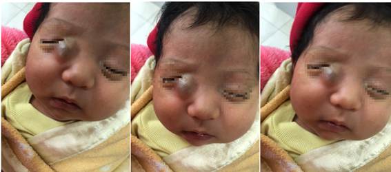
Physical examination (Fig. 1) revealed the presence of a tumor of approximately 8 mm in diameter, located in the inner canthus of the right eye, specifically in the area of the lacrimal sac. It is not painful to palpation and has a bluish color. The left eye does not present alterations.
Complementary images such as ultrasound are indicated. Encephalocele was ruled out because no pulsatile swelling was identified above the medial canthal tendon. As no imaging evidence suggesting this entity was found, and after analyzing the symptoms, the diagnosis of congenital dacryocele was made.
Non surgical treatment was decided. Warm compresses were indicated in the affected lacrimal sac area for five minutes, three times a day. It was decided to apply antibiotic therapy with tobramycin + dexamethasone in eye drops, one drop every four hours for 10 days.
At the end of the treatment, the patient showed improvement, without the need of further interventions. Follow-up by ophthalmology was indicated.
DISCUSSION
Nasolacrimal duct obstruction is a rare condition that belongs to the group of congenital anomalies that have in common the obstruction of the lacrimal duct and possible complications. Timely diagnosis and differentiation from other anomalies with similar presentation, as well as adequate postpartum management, are key to ensure a favorable outcome.
The presentation of congenital dacryocele is perinatal. The main signs described in the literature include the presence of a bluish cystic swelling at or below the medial canthal area, accompanied by epiphora. The lump tends to be turgid, fixed to deep planes, non-pulsating, violaceous or pinkish in color, usually unilateral, although bilateral presence has been described.6
The diagnosis of congenital dacryocele is clinical. Conditions that aggravate nasal congestion, such as upper respiratory tract infection, may worsen the symptoms of nasolacrimal duct occlusion. Manifestation of the lacrimal sac will result in reflux of mucus or mucous.3
A differential diagnosis should be made with choanal atresia, dacryocystitis or nasal mass such as encephalocele, gliomas, dermoid cysts and hemangiomas as one of the causes of nasolacrimal duct occlusion.
Dermoid cyst is a disease of the orbital cavity that is frequent in children from six months of age and in the first decade of life. Clinically it presents as a subcutaneous mass, hard to the touch, painless, located in the superior temporal orbit in 72 % of patients and with skeletal changes on CT scan in 85 % of patients.7)
In case of a mass with internal presence of blood flow on Doppler ultrasound, the diagnosis should be oriented towards a facial hemangioma. Anterior fossa ultrasound is usually performed during the second trimester of pregnancy and presents as a craniocervical defect with cystic contents or a solid helical mass adjacent to the brain.8
Capillary hemangioma of infancy is a benign vascular tumor involving the eyelid skin and/or orbit. It can be present from birth, however, it occurs mostly in the first months of life. Diagnosis is based on physical examination and imaging tests with Doppler ultrasound or magnetic resonance imaging.9
Obstruction of the nasolacrimal duct can be confirmed by the fluorescence disappearance test: five minutes after instillation of 1 % sodium fluorescein in the eye, a significant retention of the tear film will be observed, confirming the obstruction. Slow drainage with fluorescein is very specific for pathology of the lacrimal drainage system.9
Most congenital nasolacrimal duct obstructions resolve spontaneously or are successfully treated without the need for imaging techniques. In the presence of atypical or intractable clinical signs, its application can help to evaluate the cause of the obstruction and select the most appropriate therapy.10
Dacryocystography involves the injection of contrast material through the lacrimal puncta followed by conventional radiography, CT or MRI to visualize the filling of the system and the surrounding bony structures. Dacryoscintigraphy, on the other hand, uses artificial tears with Technetium 99m, evaluating the lacrimal system through a gamma camera.10
Most occlusions resolve on their own or with conservative medical treatment. If significant epiphora persists at one year of age or conjunctivitis recurs at an earlier age, nasolacrimal duct probing is usually treated. As a second option, mono- or bicanalicular intubation with a silicone tube, darioplasty and in special circumstances a dacryocystorhinostomy are used.11
The most important measure to unclog the nasolacrimal duct is to massage the lacrimal sac to increase hydrostatic pressure, thus breaking up any obstruction of the membrane and clearing the stagnant lacrimal sac.11
Topical antibiotherapy will be added as additional treatment in cases of secondary infection, by instilling the drops four times a day, as well as periodic cleansing of eyelids and eyelashes with warm water.12
Probing the nasolacrimal duct is the surgical procedure of choice. Some ophthalmologists perform catheterization from several months of age, early, avoiding general anesthesia and reducing cost and morbidity. Others prefer to wait up to nine -12 months and catheterize in the operating room, avoiding unnecessary interventions due to the high rate of spontaneous resolution. The only real indication for early catheterization is dacryocystocele.3
Other techniques such as dacryocystorhinostomy are also used.13) The application of diode lasers at the endocanalinular level, widely used in adults, has shown safe and effective results in children, although the experience is short.14
Prenatally, a possible respiratory complication at birth is expected due to the presence of an intranasal cyst that expands the duct into this cavity. Occasionally, this finding may be isolated and unrelated to the presence of a cyst in the orbital region. The late approach to this pathology may be complicated by bacterial proliferative dacryocystitis, as well as cellulitis if the infection spreads to nearby tissues. If left untreated, sepsis can develop rapidly and be fatal.3
CONCLUSIONS
Congenital dacryocele constitutes a congenital entity of the lacrimal ducts of low incidence. Its diagnosis is clinical; however, imaging tests are necessary to rule out other entities. Conservative medical treatment may lead to resolution of the entity, with massage and antimicrobial therapy being useful; however, surgical intervention may be required.
BIBLIOGRAPHIC REFERENCES
1. Promelle V, Demeer B, Milazzo S. Patologías congénitas en oftalmología. EMC - Pediatría [Internet]. 2020 [citado 12/10/2022]; 55(2): 1-13. Disponible en: Disponible en: https://doi.org/10.1016/S1245-1789(20)43831-4 1. . [ Links ]
2. Simón-Campos MP, Bermúdez-Azaña KL, Gonzales-Rojas AA, Vera-Abanto MC, Sevilla-Cruz TD, Celiz-Alarcón E, et al. Dacrioestenosis con dacriocistitis aguda en lactante, a propósito de un caso. Rev Med Hered [Internet]. 2021[citado 12/10/2022]; 32(1): 42-45. Disponible en: Disponible en: http://www.scielo.org.pe/scielo.php?script=sci_arttext&pid=S1018-130X2021000100042 2. [ Links ]
3. Fontoba-Poveda B, Baget-Bernaldiz M, Moll-Casamitjana D, Pineda Ortega L. Dacriocistitis aguda y crónica. Diagnóstico y tratamiento. FMC - Formación Médica Continuada en Atención Primaria [Internet]. 2022 [citado 12/10/2022]; 29(7): 358-363. Disponible en: Disponible en: https://doi.org/10.1016/j.fmc.2021.05.006 3. [ Links ]
4. Ficara A, Syngelaki A, Hammami A, Akolekar R, Nicolaides K. Value of routine ultrasound examination at 35-37 weeks' gestation in diagnosis of fetal abnormalities. Ultrasound Obstet Gynecol [Internet]. 2020 [citado 12/10/2022]; 55(1): 75-80. Disponible en: Disponible en: https://obgyn.onlinelibrary.wiley.com/doi/abs/10.1002/uog.20857 4. [ Links ]
5. Castro PT, Matos AP, Werner H, Lopez J, Ribeiro G, Araujo E, Junior E. Evaluation of fetal nasal cavity in bilateral congenital dacryocystocele: 3D reconstruction and virtual navigation by magnetic resonance imaging. Ultrasound Obstet Gynecol [Internet]. 2020 [citado 12/10/2022]; 55(1): 141-143. Disponible en: Disponible en: https://obgyn.onlinelibrary.wiley.com/doi/abs/10.1002/uog.21898 5. [ Links ]
6. Alfaro Juárez AM, Alfaro Juárez A, Sánchez Merino C. Dacriocele Congénito. Revista Atalaya Medica [Internet]. 2020 [citado 12/10/2022]; 17: 56-57. https://dialnet.unirioja.es/descarga/articulo/7889119.pdf 6. [ Links ]
7. Alfonso LS, González OF, Aranguren LV, Pereira BG. Presentación de un caso con quistes dermoides orbitarios bilaterales y múltiples. Archivos Argentinos de Oftalmología [Internet]. 2022 [citado 12/10/2022]; 21. Disponible en: Disponible en: https://www.archivosoftalmologia.com.ar/index.php/revista/article/download/198/233 7. [ Links ]
8. Llanos D, Pedraja I, Campos L, Armijo J, Ávila LF. Radiología en las tumoraciones palpables del paciente pediátrico Parte 1. Radiología [Internet]. 2022 [citado 12/10/2022]; 64(6): 552-565. Disponible en: Disponible en: https://doi.org/10.1016/j.rx.2022.08.002 8. . [ Links ]
9. Promelle V, Fortier M, Milazzo S. Aspects cliniques sensoriels et moteurs des syndromes de rétractions congénitaux : syndrome de Stilling-Duane et syndrome de Brown. Journal Français d'Ophtalmologie [Internet]. 2017 [citado 11/11/2022]; 40(5): 414-421. Disponible en: Disponible en: https://doi.org/10.1016/j.jfo.2016.10.015 9. . [ Links ]
10. Solís AL, Rojas RI, Vigoa AL, et al. Valor del ultrasonido en el diagnóstico de los tumores del saco lagrimal. Rev Cub Oftal [Internet]. 2019 [citado 12/10/2022]; 32(4):1-8. Disponible en: Disponible en: https://www.medigraphic.com/cgi-bin/new/resumen.cgi?IDARTICULO=94963 10. [ Links ]
11. Díaz-Montero C, Marqués-Fernández V, De la Herrera Flores P, Galindo-Ferreiro A. Abordaje del paciente con patología de la vía lagrimal. Indicaciones quirúrgicas. Rev ORL [Internet]. 2021 [citado 12/10/2022]; 12(2). Disponible en: Disponible en: https://scielo.isciii.es/scielo.php?script=sci_arttext&pid=S2444-79862021000200007 11. [ Links ]
12. Singh S, Nair AG, Alam MS, Mukherjee B. Outcomes of lacrimal gland injection of botulinum toxin in functional versus nonfunctional epiphora. Oman J Ophthalmol [Internet]. 2019 [citado 12/10/2022]; 12(2):104-7. Disponible en: Disponible en: https://www.ncbi.nlm.nih.gov/pmc/articles/PMC6561039/ 12. [ Links ]
13. Favier V, Crampette L. Dacriocistorrinostomía endoscópica. EMC - Cirugía Otorrinolaringológica y Cervicofacial [Internet]. 2022 [citado 12/10/2022]; 23(1):1-7. https://doi.org/10.1016/S1635-2505(22)46384-5 13. [ Links ]
14. Navarro-Hernandez E, Galindo-Ferreiro A. Dacriocistorrinostomía láser endocanalicular y sus modificaciones: revisión sistemática de técnicas y tasa de éxito. Archivos de la Sociedad Española de Oftalmología [Internet]. 2022 [citado 12/10/2022]; 97(12):692-704. https://doi.org/10.1016/j.oftal.2022.03.004 14. [ Links ]
Received: December 20, 2022; Accepted: March 08, 2023

