


 Servicios personalizados
 Inglés (pdf)
 Articulo en XML
 Referencias del artículo
 Enviar articulo por email
 Citado por SciELO
  Similares en
SciELO
Similares en
SciELO 
 Permalink
Introduction
Dental agenesis is defined as the congenital absence of a tooth bud.1 Although the aetiology of this condition is unknown, some theories have been formulated.1 Maxillary lateral incisor agenesis is the most common congenitally missing permanent tooth condition in the maxillary anterior region, representing approximately 20% of all dental anomalies.1,2 It is more prevalent in females and bilateral cases are more frequently than unilateral.3,4 Mostly, dental agenesis has been attributed to genetic factors, but they may also be caused by environmental factors such as dentoalveolar traumas, or radiation therapy.5 Functional and esthetic issues normally affect patients with congenital agenesis of upper lateral incisors at a young age, which could affect their self-esteem and social relationships.5,6
Some researchers have investigated the prevalence of congenital agenesis.7-9 Ledesma-Monteset al.7 showed that agenesis and supernumeraries were the most common developmental dental alterations in adult Mexican population. The investigators also found that hypodontia was present in 25.9% of the patients.7 In a later study, Arandiet al.8 reported a prevalence of 1.91% for missing maxillary lateral incisors in a Palestine population. Furthermore, bilateral agenesis was observed in 33.3% of the affected cases, being less common than those that displayed unilateral agenesis (66%). Despite the prevalence of missing lateral incisors was slightly higher in females (2.1%) than males (1.7%), no significant association between sex and maxillary lateral incisor agenesis was reported.8
Dallel et al.9 reported a prevalence of 7.8% in agenesis of at least one permanent tooth of the studied orthodontic population. In a decreasing order, the most frequent agenesis concerns were maxillary lateral incisors (3.6%), second mandibular premolars (2.2%), second maxillary premolars (0.9%), and mandibular incisor (0.5%).9
Treating patients with congenitally missing maxillary lateral incisors raises several important issues involving the amount of space, patient’s age, type of malocclusion, and condition of the adjacent teeth. The primary consideration when deciding which treatment option to choose is the conservation of tooth structure. Ideally, the treatment option should be the less invasive in order to satisfy the expected functional and aesthetic objectives.10
Classically, congenitally missing lateral incisors can be restored in two ways. The first treatment modality is called Canine substitution; which aim is to close the lateral incisor space by moving the canine until it is adjacent to the central incisor. Then, reshape the canine to look like to lateral incisor.10 The second option is to place the canine at its natural position within the dental arch, filling the space left by the missing lateral incisor with either a single-tooth implant or a tooth-supported restoration.5,10
Implant therapy is an established treatment modality for the rehabilitation of single or multiple missing teeth with high implant success rates in the long term.5 In addition to the high success rates of implant-supported single-tooth replacement, one of the main advantages of this type of restoration is the ability to leave the adjacent teeth untouched. This is particularly beneficial when dealing with young patients and unrestored dentitions.5,10
The aim of this case report was to determine the function and aesthetics after the therapeutic use of osseintegrated implants to replace congenitally missing upper lateral incisors in a young female at 4.5 years follow-up. This study highlights the importance of multidisciplinary diagnosis and treatment (Orthodontics, Implantology and Prosthodontics) to achieve optimal aesthetics and long-term predictability.
Case report
The patient was a 13 years old female, without systemic disease (Fig. 1A). The panoramic image showed congenitally missing upper lateral incisors (Fig. 1B). Orthodontic treatment started in April 2013 with full diagnostic procedures including articulated models, cephalometric study, TMJ evaluation, and esthetic evaluation. After the diagnosis procedures, the decision was to open the space of the congenitally missing upper lateral incisors and replace them with dental implants. This was decided due to the spaces available, the class I molar relation, the overbite and overjet present and the width of her smile. To achieve proper canine guidance, appliances such as INOVATION (Dentsply Gac International, NY, USA), Roth RX brackets, and OVATION brackets were used in the upper canines. Furthermore, archwires such as Sentalloy, Neosentalloy, Bioforce and stainless steel (Dentsply Gac International, NY, USA) were used.
Five months after the orthodontic active treatment, the space for placing the implants was achieved. Temporary crowns were placed in the position of the congenitally missing upper lateral incisors and fixed to the archwire. Upper canines erupted behind the lateral incisors temporary crowns, so they were distalized with c-chain elastomerics (Fig. 1C).
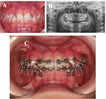
Fig. 1 A) Patient at 13 years old. B) Panoramic X- Ray image. C) Orthodontic appliance used in the patient.
After 2 years of Orthodontic treatment and the evaluation of the craniomandibular growth term by hand radioraphy, two implants were placed (Screw-Vent 3,3 ( 10, Zimmer Biomet Dental, Palm Beach Gardens, FL) with an expanded bone technique in the position of the upper lateral incisors (teeth 1.2 and 2.2) with an average surgical torque of 30 Ncm. After this, a resorbable collagen membrane (BioMend Extend, Zimmer Biomet Dental, Palm Beach Gardens, Fl) (15 mm ( 20 mm) was adapted to the bone defect. Subsequently, particulate cortical bone allograft (Puros Cortical Allograft Particulate, Zimmer Biomet Dental) (0,5 cc) was compacted between the residual buccal plate and the collagen membrane. After 4 months, osseointegration was clinically confirmed and two screw retained temporary crowns were performed using temporary plastic abutments and acrylic teeth (Fig. 2A and Fig. 2B).
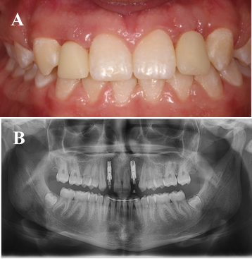
Fig. 2 A) Frontal view of screw retained temporary crowns tooth 1.2 and 2.2. B) Radiographic image of implants placed.
After several stages, two metal-ceramic screw retained crowns were obtained and installed. (Fig. 3A).
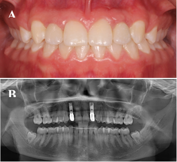
Fig. 3 A) Implant-supported single fixed prosthesis tooth 1.2 and 2.2 (screw retained). B) Radiographic control (4.5 years). Implant-supported single fixed prosthesis tooth 1.2 and 2.2.
Radiographic and clinical follow-up after 4.5 years showed a favorable evolution of the implant (Fig. 3B). No mechanical or biological complications were seen throughout this observation period. The buccal gingival margin was in a correct position.
Discussion
The aim of this case report report was to determine the function and aesthetics after the therapeutic use of osseintegrated implants to replace congenitally missing upper lateral incisors in a young female. The results showed that after multidisciplinary diagnosis and treatment, adequate aesthetics and functionality were achieved.
Treatment options available for congenitally missing upper lateral incisors are canine substitution, single-tooth implant, and tooth-supported restorations.11,12 There are specific dental and facial criteria that should be evaluated before choosing canine substitution. They include malocclusion and amount of crowding, profile, canine shape and color, and lip level. If these criteria are considered, aesthetic and functional results can be expected.12 However, many patients do not meet the criteria for canine substitution. In these patients, restorative treatment by a tooth-supported restoration or single-tooth implant should be considered.5,13
In 2011 there was a series of articles published were proponents of canine substitution leaded by Zachrisson et al,12,14 and proponents of restorative replacement leaded by Kokich et al,12 meticulously covered the topic of treatment options for this clinical challenge. Despite the advantages and disadvantages of these two treatments (e.g. indications and contraindications), there is consensus that a multidisciplinary approach is necessary to obtain an optimal outcome.11,14
The primary consideration when deciding which option to choose is the conservation of tooth structure. There is no same way to treat all patients, the diagnosis and treatment plan carried out through a multidisciplinary approach is essential and must be performed individually for each patient, considering gingival exposure, dental relation, skeleton pattern and unilateral or bilateral agenesis.10 With an adequate patient selection and diagnosis, understanding of occlusion, effective communication among operators and attention to detail, the single tooth restoration can be a predictable esthetic and long-lasting restoration.5,10,11
A systematic review performed by Andrade DC et al,13,17 concluded that there is no current evidence to support the superiority of one technique over the other for the treatment of agenesis of congenitally missing upper lateral incisors. Thus, clinicians should treat this patient with caution based on their clinical skills and experience, the clinical conditions of each patient, and patients’ expectations.
In this case report, the patient had an underdeveloped pre maxilla, wide smile, no crowding, and ample space for teeth replacement. Through diagnosis and a treatment planning with a multidisciplinary approach, it was decided to open the space of the congenitally missing upper lateral incisors and replace them with dental implants. After skeletal maturation, two implants were placed with an expanded bone technique in the position of the upper lateral incisors (teeth 1.2 and 2.2).
Implant therapy can be considered successful only when functional and esthetic requirements have been accomplished.15 The replacement of congenitally missing teeth with implants is becoming more frequent.15,16 They provide the advantages of preservation of adjacent natural tooth structure, preservation of the alveolar ridge and achievement of optimal esthetic and restorative results. However, congenitally missing teeth is a challenge because of the frequent atrophic ridges, and the therapy with dental implants require previous surgery to regenerate soft and hard tissue.16,17 Implant success rates, as well as the final esthetic outcome, have become increasingly predictable due to the hard and soft tissue grafting procedures that are available.5
In this case report, a resorbable collagen membrane (Biomend, 15 mm ( 20 mm) and particulate cortical bone allograft (0,5 cc) were adapted to the bone defect for augmenting the quantity of the available deficient alveolar bone. Guided bone regeneration has proved to be a suitable and somehow predictable technique for promoting bone regeneration. Current technique of Attia et al.17 advocates the use of a fast-resorbing material to retain the graft and promote epithelialization over the graft. The graft can be covered with a collagen material that resorbs in less than seven days.17
In the clinical follow-up (performed after 2 years) of this case report, the rehabilitation was aesthetically pleasing, the buccal and palatal gingival margin were in a correct position.
There are limitations of all case reports, however they are always a good clinical guide for cases that are common in the daily practice of the clinician. The interdisciplinary resolution of this case allows to obtain good and reliable results in the long term.
Conclusion
Multidisciplinary diagnosis and planning are essential to define the treatment option that will provide the best individual results for patients with congenitally missing upper lateral incisors. The implant approach in the anterior region is a delicate situation, which can be challenging esthetically, especially in the long term. Because of this, it is necessary to work as a team to have ideal conditions before and after implant placement.
Our results showed predictable esthetics and functional results in a patient with congenitally missing upper lateral incisors. This was possible due to a multidisciplinary approach between the diagnosis and treatment performed (Orthodontics, Implantology and Prosthodontics).

