1. Introduction
In recent years hybrid pixel detectors have shown great impact in many fields of human activity as well as experimental physics, biology, geology, medicine and imaging technology. In high energy physics these detectors are used for the registration of particles, and especially in tracking and timing application.
The Dzhelepov Laboratory of Nuclear Problems of the Joint Institute for Nuclear Researcher (JINR), in cooperation with other institutions such as Medipix international collaboration (CERN), the Institute of Experimental and Applied Physics of the Czech Technical University (Prague) and the Tomsk State University, investigates the convenience of using the GaAs:Cr as integrated sensor in hybrid pixel detectors based on the Timepix readout chip [1]. GaAs:Cr is a very promising material for the development of sensors for applications ranging from medical imaging to high energy physics due to its numerous advantages over others semiconductors [2-4]. Some of those are the possible operation at room temperature, the high resistivity to radiation damage and the high attenuation coefficient.
Several investigations about response of GaAs:Cr-based detectors to X and gamma rays have been carried out, but today the behavior of these detectors to heavy charged particles, such as alpha particles or ions is still a little studied subject.
The main objective of this work is to perform the energy calibration of a Timepix detector based on GaAs:Cr sensor with alpha particles. For this purpose, 241Am and 226Ra sources and mathematical simulation were used.
2. Materials and methods
The used detector was a Timepix device with 900 μm GaAs:Cr sensor grown by the Czochralski Liquid Encapsulation method [5] and subsequently compensated with Cr by thermal diffusion [6]. The cathode is formed by 1 μm Ni layer, whereas anode is structured in matrix of 256 x 256 pixels, each of which has dimensions of 45 x 45 μm2 and a distance between them of 10 μm. Each anode has a layer structure of Au/Ni/Cu/V type and regions between them are occupied by silicon oxide as an insulator and protection (figure 2.1 a and b).
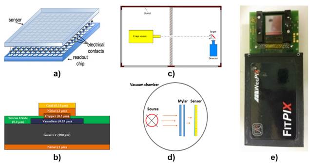
Figure 2.1 a)General scheme of the hybrid detector;b)graphical representation of the single pixel basic structure; c)schematic representation of reflection geometry used for characteristic X-rays measurements; d)schematic representation of the experimental configuration inside of the vacuum chamber; e)hybrid GaAs:Cr Timepix detector with FitPix interface.
A shielded experimental station was used for characteristic X-ray spectra measurement of different target. This workstation is equipped with a copper coated tungsten anode microfocus X-ray tube SB120 [7] employed as primary radiation source. Using a set of known materials and a reflection geometry with respect to primary X-ray beam (figure 2.1c), it is possible to obtain photon energies in a range between 7 and 57 keV as result of target fluorescence.
Table 1 shows a list of target materials employed in the experiment to obtain different X-ray energies. Only Kα1 lines were used in our case.
For measurements with alpha particles was used a vacuum chamber containing a support for radioactive source and detector alignment and a vacuum pump, high voltage source for detector bias and PC for data acquisition.
The experimental idea (see schema in figure 2.1 d) was to pass alpha particles from radioactive source through different thicknesses of Mylar films located between source and detector, in order to obtain an energy range for perform the corresponding calibration. 241Am and 226Ra were used as radiation sources, and Mylar film thicknesses were obtained from combinations of 3 μm thick pieces. From 226Ra source spectrum the line corresponding to 214Po was used for calibration.
The detector to calibrate was E08-W0153 (figure 2.1 e), and during the measurements it was kept operating with the following parameters: -300 V bias voltage, 16 MHz frequency and THL = 180 DAC.
For experimental data acquisition the PixetPro software version 1.4.4.504 [8] was used.
In order to determine the existing relationship between the energy loss of 5.5 MeV alpha particles crossing Mylar film and this material thickness, the mathematical modeling was used. For these calculations, the SRIM-2013.00 software [9] was applied.
The initial conditions were taken as follows: initial alpha particle energy E = 5.5 MeV (241Am source), the particles fall perpendicularly on the target surface, the Mylar mass density was 1.4 g/cm3. The Mylar film thicknesses were selected in the interval from 0 μm up to the value were takes place the total alpha particle absorption, taking specific points matching with the real material thicknesses.
To obtain an adequate statistic, the program was run for 1x10⁶ incident alpha particles.
3. Results and Discussion
3.1 Determining the alphas particles energies
The average energy E’ of alpha particles reaching to cross a certain Mylar film thickness d was calculated subtracting the energy loss E p inside the target, determined by mathematical modeling, to the initial energy (see table 2), taking into account only the ionization losses representing the 99.87% of the total one.
Table 2 Relationship of Mylar film thicknesses, mean energy loses within the material and the output alpha particle energies.
| d(μm) | Ep (eV) | E’ (keV) |
|---|---|---|
| 0 | 0 | 5500 |
| 5 | 573561,86 | 4930 |
| 6 | 695225,33 | 4800 |
| 10 | 1197617,39 | 4300 |
| 12 | 1468092,04 | 4030 |
| 15 | 1889793,89 | 3610 |
| 18 | 2355595,83 | 3140 |
| 20 | 2681925,97 | 2820 |
| 25 | 3635387,45 | 1860 |
| 26 | 3857148,18 | 1640 |
| 27 | 4091930,16 | 1410 |
| 28 | 4343583,83 | 1160 |
| 29 | 4614733,34 | 890 |
| 30 | 4902215,02 | 600 |
| 31 | 5181823,04 | 320 |
| 32 | 5401103,36 | 100 |
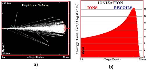
An example of the ion tracks projected in plane obtained from de SRIM simulation, is shown in figure 3.1 a. Observe that a 35 μm Mylar film thickness is sufficient to the total stopping of the 5.5 MeV alpha particles (red points represent the stopped alpha particles). The in-depth ionization profile produced by alpha particles at same Mylar film thickness can be observed in figure 3.1 b.
3.2 Energy calibration of detector with alpha particles
Energy calibration with characteristic X-rays
In order to determine the relationship between the energy of the incident alpha particle E and the cluster volume in keV units for selected DAC settings (threshold level (THL) equal to 180 DAC and clock frequency equal to 16 MHz) and bias voltage (Vbias = -300 V), it was necessary to perform a previous calibration by irradiating the detector with gamma sources. The aim is to find the relationship between the obtained in TOT mode counts and the incident particle energy.
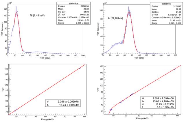
The experimental spectra measured for each X-ray energy were fitted by means of Gaussian function in order to obtain the peak position (see table 1). Only charge induction events occurring in a single pixel were taken into account. For histograms (spectrum) construction the TOT counts values obtained per pixel was placed in x axis, while in y axis the absolute frequency is indicated.
As example, some spectra obtained from two measurements made at different X-ray energies with the tube at 60 kV and 20 μA are shown in figure 3.2 (top).
From the calculated and reported in the table 1 data, the dependence of the TOT counts with the incident photon energies was performed. This curve is adjusted by means of the following function [10-11].
where a, b, c and t are fit parameters.
Keeping in mind that calibration curves present a region with lineal behavior above the 25 keV, the fitting was carried out in two steps. First, the parameters a and b were calculated using the fluorescence spectra with the higher energy (24.2, 25.27 and 57.54 keV). Second, considering the obtained lineal parameters, the general calibration was carried out to obtain the remaining parameters (c and t), that determine the behavior of the non-lineal region below 25 keV [12]. This general calibration was made fixing the obtained previously a and b values with its respective uncertainties in the lineal region. In figure 3.2 (bottom) an example of the two-step energy calibrations is presented.
Finally, to calculate the deposited in the pixel alpha particle energy in eV units, it is necessary to use the inverse of the function (3.2):
Energy calibration of detector with alpha particles and experimental verification
The calibration of the Timepix detector for alpha particles is much more complicated than calibration with characteristic X-rays because the total charge can only be revealed by making a summation of all fractional charges, i.e. by determining the cluster volume. Example of cluster volume generated by a single alpha particle is shown in figure 3.3 a.
For the visualization of the alpha cluster peaks in the obtained spectra, it was necessary to perform a simple cluster analysis. Cluster height and cluster size histograms were used to establish limits for cluster volume construction only for alpha cluster selection purposes. Only clusters with heights between 80 and 1700 keV and sizes between 10 and 10000 pixels were selected. Figures 3.3b and 3.3c show detector illumination for 5 frames and an alpha cluster for 5.5 MeV energy after alpha cluster selection respectively.
Experimental spectra after alpha cluster selection for each energy were fitted using the Gaussian, thus determining the mean points of each peak and their standard deviations, as shown in figure 3.3 d. For histograms construction, cluster volume values were placed on x axis; while in y axis the absolute frequency with which these values appear. Cluster volume values are expressed in energy unit (keV) because they were translated with equation 3.3.
Taking into account the average values of cluster volume obtained for energy, and energy values corresponding to each peak, the relationship of these magnitudes was established. Finally, the obtained curve was linearly fitted (figure 3.4 a):
where p0 and p1 are fit parameters.
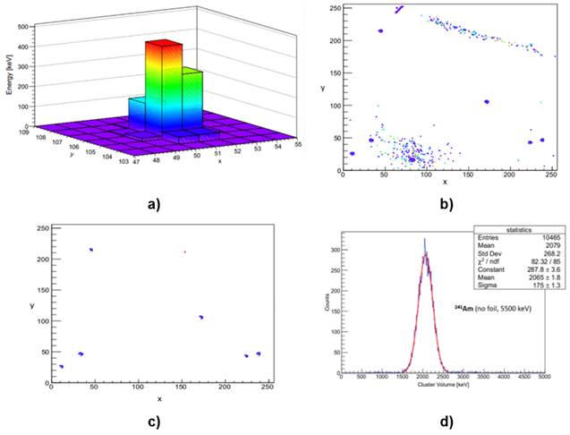
Figure 3.3 a) Pixels cluster generated by single 3140 keV alpha particle; b) detector illumination for 5 frames; and c) alpha cluster for 5.5 MeV alpha particles after cluster selection; d) example of spectra obtained in TOT mode for alpha particles from americium source. x and y axises represent the position in the pixels matrix.
In order to check the energy calibration, with same detector and under identical conditions was carried out by measuring radon in air. The measurement time was 67 hours.
The 218Po isotope is radon progeny and alpha decays to 214Pb with energy of 6115 keV [13]. This line of radon alpha spectrum was used for experimental verification of the performed calibration. Figure 3.4 b shows spectrum corresponding to this measurement after alpha cluster selection.
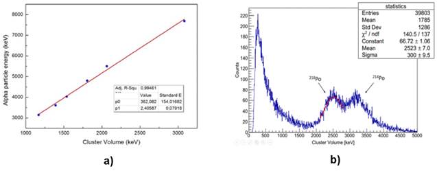
Figure 3.4 a) Energy calibration with alpha particles for selected DAC settings and bias; b) alpha cluster spectrum of radon progenies in air after cluster selection.
The mean value for 218Po peak was calibrated by the function (3.4) to translate cluster volume to incident alpha particles energy, obtaining 6432 ± 370 keV. The value calculated experimentally considering its statistical error corresponds to the reported in literature, concluding that the performed alpha particle energy calibration is acceptable.
4. Conclusions
The performed mathematical simulation of the 5.5 MeV alpha particles transport through different Mylar film thicknesses, allowed to calculate the transmitted energy making possible the experimental calibration with the use of Mylar as absorbent. It was proved that 35 μm Mylar films thickness is sufficient to the total stop of the alpha particles from a 241Am source.
By calibrating the detector with characteristic X-rays and using a two step fitting procedure, it was determined the relationship between the photon energies and the registered by the detector TOT counts.
The energy calibration with alpha particles was performed according to linear function y = 362.08 + 2.41 x, with R2 = 0.99, and was verified with the measurement of the 218Po line of radon in air.


