


 Servicios personalizados
 Inglés (pdf)
 Articulo en XML
 Referencias del artículo
 Enviar articulo por email
 Citado por SciELO
  Similares en
SciELO
Similares en
SciELO 
 Permalink
Introduction
Chest pain is a common presentation and diagnostic challenge in the emergency department (ED) setting. In the US, chest pain accounted for 11.4 million ED visits in 2005 (approximately 10% of the estimated 109 to 116 million annual ED visits). Part of the diagnostic challenge of chest pain syndrome includes variability in the patient presentation associated with age, gender, and comorbidities in addition to a wide spectrum of other potential diagnoses ranging from the benign to life-threatening. While the majority of chest pain cases presenting to the ED are eventually diagnosed as non-cardiovascular in etiology (such as gastroesophageal reflux disease or musculoskeletal problems), it is important to exclude acute life-threatening conditions, including acute myocardial infarction, pulmonary embolism, visceral perforation, and aortic dissection (AD).1
Aortic dissection implies the formation of an intimal tear with separation in the media and subsequent anterograde blood flow into the false lumen. Aortic dissection is present in approximately 90% of acute aortic syndromes. 1 Acute dissections of the ascending aorta (Stanford A) carry mortality that historically approaches 1% per hour. Although less morbid, acute dissections of the descending thoracic aorta (Stanford B) are associated with a 10% to 25% mortality at 30 days. Intramural hematoma mortality ranges from 10% to 50%, and 40% of the patients progress to over dissection. 2
According to the more popular Stanford system, dissections involving the ascending aorta are classified as type A, whereas those involving only the descending aorta are classified as type B. The older DeBakey system differentiates between dissections evolving from the ascending aorta and affecting all aortic segments (type I), less extensive ones affecting only the ascending fragment (type II), and dissections affecting only descending aorta (type III). Concerning the time from the onset of the symptoms, aortic dissections are divided into acute (presentation within 1 week), subacute (from 1 week to 1 month) and chronic (more than 1 month). 3
Aortic dissection arises from a tear in the aortic intima exposing the medial layer to the pulsatile blood flow. The intimal tear is frequently found in segments exposed to the greatest shear stress, namely the right lateral wall (opposite the main pulmonary artery) of the ascending aorta or in the proximal segment of the descending aorta. The progressive separation of the aortic wall layers results in the formation of a false lumen and its subsequent propagation can be followed either by the aortic rupture in the case of adventitial disruption or by re-entry back into the true lumen through another intimal tear. 3
The most common risk factor for aortic dissection is poorly controlled hypertension (65-75% risk with a history of hypertension). Other risk factors include age, male sex, smoking, pre-existing aortic diseases or aortic valve disease, family history of aortic diseases, history of cardiac surgery, direct blunt trauma, and the use of intravenous drugs (such as cocaine or amphetamines). 4
Sudden onset of severe chest and/or back pain is the most typical symptom. The pain may be sharp, ripping, tearing, or knife-like and is typically different from other causes of chest pain; the abruptness of its onset is the most specific characteristic. 4
Initial diagnosis is extremely important in patients with the acute aortic syndrome. When a patient has sudden abrupt chest or back pain, imaging diagnosis should be the first concern, with simultaneous laboratory tests, including a biochemical study and complete blood count, and electrocardiogram. It is especially important to note the level of D-dimers in patients with acute aortic dissection. As well as D-Dimer measures, chest X-ray, transthoracic echocardiography, transesophageal echocardiography, contrast-enhanced computed tomography, and magnetic resonance images are some of the techniques available for diagnosis. 3
Despite several cases reports and studies have been carried out in the worldwide, the present patient is the first of aortic dissection diagnosed in Cuban Hospital from the started in 2013; in addition, this condition implies a high risk of complication and death, because it is a life-threatening, based on that we decide to share this case report as a journal publication to show the clinical presentation of aortic dissection and the importance of Angio TAC in the diagnosis, with the respective reviewed and updated bibliography.
Case report
It is Indian male patient 45-years-old, arrived A&E Cuban Hospital’s department on a wheelchair, accompanied by his friend and nursing assistant complained suddenly back pain, radiated to the back between both scapula bones, as well he complained shortness of breath, at examination physician noted BP 240/140mmhg, RF 20 per minute, he looks like anxious; were ordered the lab test immediately with result showed WBC 10.52X10³/UL, CRP<5, glucose 13.0 mmol/L, Hb 16.0gm/dL, Hct 47.8%, platelet 183x103 /UL, D-Dimers >1; as well electrocardiogram, showed positive ST segment in V3-V4 no more than 2mm, no ischemic changes; a chest x-ray was performed without any positive finding.
Based on the clinical information, lab test, chest X-ray, and physical exam was suspected aortic dissection, so that on-call radiologist was called by the physician requesting for chest angiogram to confirm or rule out the diagnosis. (Fig 1)

Fig. 1. A Thorax CT with IV contrast of the patient, sagittal projection and B Thorax CT with IV contrast of the patient, coronal projection.
After order requested, inform consent obtained, vital sign and risk factor reviewed it was proceeded to perform the Angio TAC in the Radiology Department, it was administrated 60ml of Omnipaque, followed by 60ml of NaCl, it was used bolus tracking software of SIEMENS and routine Angio TAC to enhance and record the images from the full aortic artery, it was shown a separate layer at the posterior wall of the aortic artery from the emergency of the left subclavian artery to the emergency of the left renal artery, as well thrombus in false light was identified. (Fig. 2)
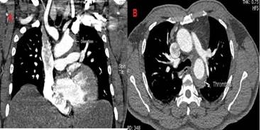
Fig. 2. A dissection at the left renal artery emergency and B thrombus attached at the posterior wall of the upper thoracic aorta.
Based on findings described above, the radiology concluded the study as a dissection of thoracic aorta grade III as per Debarked classification and type B as per Stanford classification. Based on the time of started the symptom was classified as acute aortic dissection. (Fig. 3)

Fig. 3. Volumetric reconstruction where wecan see both lights, the true and false in the lower segment of the thoracic artery.
Besides, the patient was evaluated by the on-call cardiologist, and he decided to refer him to heart Hospital from HMC to further assessment and treatment.
According to the bibliography reviewed this disease is common in older patients, but our patient is 45 years old, then, looking for any explanation why it is affecting young patient, we ask him if he has any history of trauma, drug ingestion habit, or another risk factor capable to explain this finding, and he referred none of them, posteriorly we supposed high blood pressure as a probable cause.
Discussion
The diagnosis of our case report based in tomographic finding was an acute idiopathic dissection of aortic artery grade B as per Stanford classification and grade III based in Debakey classification, the patient was on stable condition and was consulted by a cardiologist and referred by him to "Heart Hospital" from Hamad Medical Corporation as it is a tertiary institution for further assessment and final treatment. The early performed angioCT allow to do early diagnosis and proper initial treatment, it was very important because it is a life-threatening condition with a very high mortality index in the first 24-48 hours. 1-8It was possible because the clear clinical information, abrupt onset of symptoms, lab test as altered D-Dimers, oriented the physicians to properly diagnosis.
Lempel et al 2014 in their research, they analyzed incongruences in the classification of aortic dissection, because on several cases with type A and B Stanford classification, the aortic arch was also affected (7.4% for type A and 11% type B), more frequent in younger patients (mains age 50.3 years) different than the group with only affected ascending or descending aortic artery without involved aortic arch, where were are more affected aging patients (mains age 60.7 years), to resolve this incongruence they proposed a new classification, they classified type A when the dissection start in ascending aortic artery with affected or not aortic arch; type B, if it is start distal to subclavian artery and type B dissection with aortic arch involvement (B*), if there is proximal extension into the transverse arch between the innominate and left subclavian arteries and which may or may not extend distally into the descending aorta; 5 our case was classified as type B despite he is young patient, it is probable because the high blood pressure detected in the patient was the suppose cause of the dissection.
The aortic dissection is included into the aortic syndrome, as well intramural hematoma and penetrating aortic ulcer, they are uncommon causes of chest pain; DeMartino et al 2 in study done on 2018 found out stable incidence for the aortic syndrome of 4.4 per 100000 persons/years in the last years, only penetrating aortic ulcer are increased the incidence, male sex, and aging patients were the most affected (means 71.8 years), patients with an aortic syndrome had more than twice the mortality rate at 5, 10, and 20 years when compared with population-based controls evaluated in their study (5-, 10-, and 20-year mortality 39%, 57%, and 91% versus 18%, 41%, and 66%2). The patient brought by us is young and had an early diagnosis, for this reason it is probably he will have better prognostic than old patients, it is recommended further researches regarding with this entity, to do a better assessment of patients with aortic syndrome and increase the knowledge to improve the patient outcome.
Chishti MA et al reported a case of 20-years-olds nondiabetic, nonsmoker gentleman with Marfan’s syndrome admitted in Mahatma Gandhi Medical College & Hospital (MGH), Jaipur, India, with complaints of severe chest pain radiating to left shoulder since 1-day duration. He had blood pressure (BP) of 138/88 mm Hg in the right arm. There was no evidence of myocardial, cerebral, visceral, or limb ischemia. It was suspected aortic dissection and performed echocardiography showed dilated left ventricle with fair contraction and severe aortic regurgitation. The aortic root was dilated with ascending aorta measuring 58 mm and aortic annulus measuring 24 mm. Dissection flap was seen in the aorta, confirmed as well with Angio CT, the patient was surgically operated and fixed the dissection. 6 Our patient was referred to tertiary hospital to final assessment and treatment, surely surgical intervention was not suggested because it is a type B stage, for that patient BP and other risk factor control are the best option.
It was presented a case by Vululi et al in 2018, he was a fifty-four-year-old male patient admitted at Mulago Hospital in Uganda with a history of sudden onset collapse. He presented with chest and abdominal pains. He was well with no chronic diseases until two days before his admission when he suddenly collapsed with a loss of consciousness. The chest pain was piercing in nature not irradiating to any other part. Cardiac Echo done showed ascending aortic dissection with aortic regurgitation, a performed Computed tomography aortogram showed extensive aortic dissection involving the aortic root, ascending aorta, aortic arch, descending aorta and bilateral common iliac arteries, the patient was stabilized under medical treatment and discharged after eleven with planned surgical intervention to repair the dissection; unfortunately, never happened.7 Similar to our patient, the clinical history was chest pain and the diagnosis was confirmed with angio TAC.
Lei P et al in 2019 also reports the case of a 61-year-old male smoker with poorly controlled hypertension who refers a chest pain radiated to the interscapulum area, a Computed tomography angiography at admission showing Stanford type B aortic dissection, with drug-based treatment. After drug-based treatment failed, the patient was treated using endovascular techniques to place an endo-prosthesis with stenting. 8
Another patient with a similar history, and also the confirmed diagnosis was done by computed tomography angiography, as our patient the initial treatment was drug-based, but finally was necessary surgical reparation of the dissection, till moment our patient no needed surgical treatment.
As per bibliography reviewed the most frequent clinical presentation for aortic dissection is the chest pain, they are also agree with the high mortality risk for this entity, as well the importance of Angio CT in the diagnosis; the treatment is divided in medical and surgical treatment depending on Stanford classification, patient condition, and other factors.
Conclusions
As per our case report and bibliography review, the aortic dissection is a disease that should be taking in account in young patients complaining abrupt chest pain, blood hypertension, mainly if D-dimer is altered; angio CT scan is the most sensitive study for the diagnosis.
Finally, we recommend initial chest angio CT scan if it is available and it is justified by clinical history, finding, and risk factors.

