


 Serviços customizados
 Inglês (pdf)
 Artigo em XML
 Referências do artigo
 Enviar este artigo por email
 Citado por SciELO
  Similares em
SciELO
Similares em
SciELO 
 Permalink
Introduction
eHealth, understood as the application of the Internet and other related technologies in the health sector, has had a significant impact on improving access, efficiency, effectiveness and quality of clinical and business processes used by health organizations, doctors, patients and consumers, in an effort to improve the health status of patients. Radiology is one of the leading medical specialties in the use of computer systems and the Internet to streamline processes and improve patient care standards (Vialart et al., 2018). Radiology is a branch of medicine that uses images to diagnose and treat disease. Like the other branches of this science and other spheres in the current world panorama, it has benefited from the scientific and technological development that the emergence of information technology brought with it. When in 1972 the British Hounsfield presented the first computed tomography in London (Ramírez and Cano, 2012), the transition from conventional radiology to imaging was evident, since the image generated was not analog but digital. This technological development resulted in a considerable increase in the volume of information generated in health institutions (Carrillo-Zambrano et al., 2018). Its handling in a conventional way causes the loss of images, inefficient diagnoses and repeat examinations of patients; highly detrimental to their health, in addition to causing higher expenses. From this arise the PACS systems (Picture Archiving and Communication System) (Choplin et al., 1992), whose primary function is to capture, store, distribute and display medical images. For this, they have four main components: the acquisition equipment, the workstations, the medical image servers, and the network infrastructure, which establishes communication between the other components.
Due to the increasing use of computers in clinical applications and the development by dozens of manufacturers (Philips, Siemens, General Electric, Kodak and others) of a great variety of equipment, with the aim of generating digital medical images, incompatibilities arose between the different devices when storing and transferring said images and information (Aldosari, Basema and Khulud, 2018). To solve this problem, a standard method was needed, which would achieve the required interoperability regardless of the format adopted by the manufacturer. Thus, DICOM (Digital Imaging and Communications in Medicine) was created, developed in 1983 by a North American committee made up of the American College of Radiology (ACR) and the National Electrical Manufacturers Association (NEMA). It is a set of standards that establishes a unique electronic format, structured in a protocol, with which the information of the images generated by tomography, magnetic resonance and radiography equipment can be exchanged without difficulty, being integrated into the PACS systems (Bidgood and Steven, 1992).
The use of PACS systems in health institutions brings innumerable advantages. The fact that printing of radiographic films is not necessary leads to a reduction in costs. The PACS systems allow to immediately consult all the exams of a patient that the professional needs to evaluate its evolution, which makes the diagnosis a more efficient process. Its resources improve the accessibility, security and distribution of exams and reports, increasing the productivity of institutions and offering more comfort and agility to the patient (Guzmán, 2021). The Cuban state has constantly made an effort to maintain good health indicators for the population. Equipments such as Diagnostic Ultrasound (US) in real time, Computerized Axial Tomography (CT), Magnetic Resonance Imaging (MR) and Mammography (Mx) have gradually been acquired (Cidón, Torre and Cidón, 2011). In addition, PACS systems have been developed in the country, thus avoiding the need to import foreign solutions.
The first experience of this type of system in Cuba was PATRIS which was developed at the end of the 90's by the company EICISOFT. Then, in 1998, Imagis (Vázqueza, 2005) was developed by the Center for Medical Biophysics of the Universidad de Oriente. The Medical Informatics Center (CESIM), of the University of Informatics Sciences (UCI), with the purpose of carrying out the process of computerization of society, also developed a PACS solution. Initially, the system was called Cassandra PACS, then ALAS PACS and finally XAVIA PACS, a name that has changed due to the commercial and communication strategy of the UCI (Orellana and García, 2020).
The XAVIA PACS-RIS (Vega, Duque and Soler, 2020) Imaging Information Management Platform was designed to offer medical personnel working in diagnostic imaging services a range of general-purpose tools for viewing and processing digital medical images and subsequently editing of the reports issued. It is made up of several highly integrated components that are compatible with the international DICOM standard, offering a scalable and adaptable solution to the requirements of different health institutions, according to their workflow. It allows the comparison of medical images of the patient, inside and outside the institution, fidelity, reliability and reduction in the time of access to the studies from any viewing station. In Cuba, the XAVIA PACS-RIS has evolved and escalated in various hospital institutions, with an increasing presence in the national health system. This paper aims to present the impacts of the XAVIA PACS-RIS system in Cuba.
Methods
For the execution of the research, an exploratory strategy is followed and the following methods were used. Documentary analysis to obtain data and information associated with the object of study. The bibliographic documents referring to PACS solutions, radiological information systems, their integration, interoperability, as well as standards and good practices were analyzed, which allowed establishing the theoretical foundations of the present investigation. Institutions receiving the solution were contacted to obtain statistics on the operation of the system. Through the inductive-deductive method, it was possible to reach general conclusions about the processes of patient care in the diagnostic imaging services of the health institutions that use the XAVIA PACS system, based on identifying the impacts of the system on the different edges proposed in the investigation. The statistical method was applied to highlight the importance and the level of introduction of the result in Cuba.
Description of the XAVIA PACS-RIS Platform
The platform enables viewing, storage, and transmission of compressed images, supports viewing of medical images from multiple monitors, and enables simultaneous associations with the medical imaging server. It has been designed in such a way that it promotes its integration with medical equipment for imaging diagnosis (magnetic resonance imaging, computerized axial tomography, digital X-rays, among others) and with computer systems available to health institutions and organizations, due to the system standardization capability. It is made up of several independent and integrable applications that are described below:
XAVIA PACSServer (medical image server): it makes possible to manage the information of the studies that are generated in different diagnostic modalities, supports simultaneous associations, as well as guarantees the archiving of each of these studies in an orderly manner. It also makes possible to search for and retrieve studies from any workstation or imaging equipment. Additionally, the server has a group of tools for the administration of its resources and allows the creation of maintenance policies such as compression and deletion according to configuration, in addition to the execution of scheduled tasks in critical situations and the synchronization of the information that is in the databases. and the physical file.
XAVIA PACSViewer (general diagnostic station): has tools for the processing, analysis and visualization of medical images with basic tools and 3D post-processing. This component of the system allows remote connection from workstations to the hospital's image server, it receives studies directly from imaging equipment, and exchanges studies between specialist workstations. It also allows to generate imaging reports, export to common image formats, digital videos and print images on paper or radiographic films. Recorded on CD/DVD or on removable devices (USB).
XAVIA PACSReporter (imaging report editing tool): System for issuing radiological study reports that covers different flows that can occur in an imaging service. Among its newest features we can mention the work in offline mode and the configuration of new pedals for transcription through plugins. Among its main functionalities are: generating imaging reports, creation of templates for repeat diagnosis reports, printing of reports in standard document editing formats, spell checking and disease coding.
XAVIA PACSGateway (medical image acquisition and routing server): System that enables the routing of different types of imaging studies in a health institution or organization; it communicates with the image acquisition equipment, from which it receives studies of various modalities and distributes them to the diagnostic and visualization stations through a group of rules previously configured in the system.
XAVIA PACSWeb (web image viewer): System for the search, visualization and analysis of imaging studies via the web. It allows the connection via web, of devices connected to the network (computers, tablets, mobile phones) with the image server. It is compatible with images generated by state-of-the-art equipment, including multiframe. Once the images have been viewed, transformations such as: brightness, contrast, color palettes, rotations, among others, can be performed. It also gives the specialist the possibility of making measurements of distances, angles, areas and volumes on the image. The system has the Case Tray, Viewer and Configuration modules, with options similar to XAVIA PACSViewer.
XAVIA PACSServer Tools (medical image server tools): It allows synchronization of both the online image server database and the offline image server database with the images that are physically stored on the server, as well as the server that owns the images. corrupt images, to maintain a correspondence between them.
XAVIA PACSViewer Updater (update server for general diagnostic stations): It works as an update server for the medical image viewer, where updates are published and those updates are downloaded by different viewer clients.
XAVIA RIS (Radiological Information System): it allows the automation of the workflows of the imaging area, the registration of new patients to the institution, the management (planning, monitoring and control) of appointments for studies or imaging consultations, the registration of data from specialists and medical teams, the use of work lists to speed up patient care flows. It adapts to the particular conditions of health institutions, thanks to personalization through user profiles.
It allows the control of a clinical imaging history, as well as the outputs of medical statistics and charge sheets. It has a compatible DICOM work list server that communicates with the teams so that they update their work lists, and allows searches by patients, studies and medical diagnoses, facilitating the performance of morbidity studies.
Results
The platform is implemented in 27 clinical-care institutions until April 2023. Havana is the province with the highest level of introduction of the result, with presence in 19 institutions, followed by Pinar del Río and Villa Clara with three; in the case of Matanzas with two. The Covid 2019 pandemic limited the introduction of the result in new hospitals, however, during 2022 XAVIA PACS was implemented in five new institutions.

Fig. 1 - Implementations of the XAVIA PACS-RIS Platform distributed by provinces (until april 2023). Source: The authors.
Table 1 - Institutions where the XAVIA PACS-RIS solution is implemented until February 2023.
| Province | ||
|---|---|---|
| 01 | Pinar del Río | Provincial Teaching Clinical Surgical Hospital Abel Santamaria |
| 02 | Pinar del Río | Pepe Portilla Provincial Pediatric Hospital |
| 03 | Pinar del Río | Clinical Surgical Hospital Dr. León Cuervo Rubio |
| 04 | Havana | Calixto García Clinical Surgical Hospital |
| 05 | Havana | Juan Manuel Márquez Pediatric Teaching Hospital |
| 06 | Havana | Dr. Miguel Enriquez Clinical Surgical Teaching Hospital |
| 07 | Havana | Cira Garcia Central Clinic |
| 08 | Havana | Frank País International Orthopedic Scientific Center |
| 09 | Havana | Hermanos Ameijeiras Clinical Surgical Hospital |
| 10 | Havana | Dr. Salvador Allende Clinical Surgical Hospital |
| 11 | Havana | General Teaching Hospital Enrique Cabrera Cossio |
| 12 | Havana | National Center for Minimal Access Surgery |
| 13 | Havana | Julio Trigo Clinical Surgical Hospital |
| 14 | Havana | Dr. Carlos J. Finlay Central Military Hospital |
| 15 | Havana | Dr. Luis Díaz Soto Military Hospital |
| 16 | Havana | William Soler Pediatric Hospital |
| 17 | Havana | National Institute of Nephrology |
| 18 | Havana | Institute of Cardiology and Cardiovascular Surgery |
| 19 | Havana | CIMEQ Medical Surgical Research Center |
| 20 | Havana | CESAM Mental Health Center |
| 21 | Havana | MININT Clinic 43 |
| 22 | Havana | Polyclinic Gy19 |
| 23 | Matanzas | Eliseo Noel Caamaño Provincial Pediatric Hospital |
| 24 | Matanzas | Comandante Faustino Pérez Provincial Hospital |
| 25 | Villa Clara | Arnaldo Milian Castro University Clinical Surgical Provincial Hospital |
| 26 | Villa Clara | Celestino Hernández Robau University Oncology Provincial Hospital |
| 27 | Villa Clara | Provincial University Hospital Cardio Center Ernesto Guevara |
XAVIA PACSViewer and XAVIA PACSReporter have been installed in 332 diagnostic stations with 29 XAVIA PACSServers and 26 database servers. On the other hand, XAVIA PACSWeb and XAVIA RIS are present in 22 and 17institutions respectively.
Table 2 - Statistics of the resources installed by provinces until February 2023.
| Pinar del Río | 11 | 3 | 3 | 3 |
| La Habana | 291 | 22 | 15 | 12 |
| Matanzas | 13 | 2 | 2 | 1 |
| Villa Clara | 17 | 2 | 2 | 1 |
| 332 | 29 | 22 | 17 |
Economic, social and environmental impacts
To the considerable savings in time, new and improved features, greater possibilities of being attended by several specialists and remotely offered by digital radiological systems, we must add the substantial economic and environmental reductions that implementing PACS-RIS systems entails, from direct (less consumption of radiological plates, reissue of the same due to losses, deterioration) to indirect (infrastructures to store, manipulate, classify the plates, necessary inputs and various resources such as shelves, electricity, transport of the same, etc.). By going digital, and having backups and verification mechanisms, the need to carry out studies a second time due to the patient or health personnel losing or damaging the plates disappears.
Table 3 - Daily and annual average of imaging studies that do not use radiographic films.
| Military Hospital Dr. Luis Díaz Soto | 80 | 29200 |
| Central Military Hospital Dr. Carlos Juan Finlay | 80 | 29200 |
| William Soler Pediatric Hospital | 120 | 43800 |
| Clinical Surgical Hospital Dr. León Cuervo Rubio | 15 | 5580 |
| Pepe Portilla Provincial Pediatric Hospital | 9 | 2976 |
| Cira Garcia Central Clinic | 88 | 30200 |
| Provincial Teaching Clinical Surgical Hospital Abel Santamaria | 50 | 22500 |
| Julio Trigo Clinical Surgical Teaching Hospital | 18 | 6120 |
Table 3 shows an average number of daily and annual studies that stop being delivered in plates in eight (8) of the institutions that use the XAVIA PACS-RIS platform. The hospitals in the sample save 460 radiographic plates per day by introducing digital technologies and 169,576 annually, which means a high volume of contaminating materials that are not used, additionally it is not necessary to use the developing chemicals that are traditionally necessary, mainly composed of four substances:
Hydroquinone, which converts silver halide crystals exposed to X-rays into metallic black silver crystals
Sodium Sulfite, which prevents the developer from being oxidized by air
Sodium Carbonate - Sodium Hydroxide, which provides the alkaline solution needed by the developer and softens the gelatin allowing the developer to reach the silver halide crystals.
Sodium or Potassium Bromide, which limits the development of unexposed silver halide crystals.
On average, hospitals allocate 10,000 cubic meters of water per year for the development of plates, high consumption of electricity and therefore the fuel necessary for its generation. In addition, the reduction of spills and waste, consumption of paper to store and ensure the integrity of the plates, equivalent to tens of cubic meters of wood.
In terms of environmental preservation, it should be mentioned that the reactive materials for the development of radiographic films are highly polluting products, just like radiographic films, which generally do not have an adequate process when discarded. Additionally, the radiation to which patients are subjected due to repetition of studies during referral from one level of care to another and between health institutions is reduced. In addition, the consumption of paper in hospital institutions is considerable; Therefore, the use of computer tools that manage all the information without the need to print documents represent a significant contribution to the culture of savings and the consequent environmental impact.
At the social level, the use of the system improves the quality of health care received by patients, since it significantly reduces unnecessary paperwork and the wait to make appointments, perform tests, and collect results. With the deployment of the systems, both the institutions and the patients and specialists who work in them, benefit as follows:
From the patient's point of view: to be treated as a unique client, to be attended to more promptly and efficiently, to have a unique clinical imaging record that allows monitoring their health status, to reduce the radiation to which they are exposed due to the repetition of exams caused by the loss of these. Guarantee the security of all the medical information associated with your electronic medical record and obtain the results of your exams faster and with greater quality, thanks to the possibility of simultaneous viewing of the images by doctors from different areas.
From the point of view of the medical professional: guarantee the visualization of images in independent stations to the image acquisition equipment. The studies can be seen in report rooms, case discussion rooms, consultations, operating rooms, etc. This guarantees greater efficiency and effectiveness in the work of diagnostic imaging services, facilitates comparisons between imaging studies performed on a patient at different times, improves the quality of medical reports delivered to patients, puts at the disposal of the medical professional, diagnostic and clinical management tools that respond to their real needs. Have and access unique and integrated patient information to facilitate the processes of diagnosis, treatment, and other care programs, allow monitoring of patients according to the diagnoses issued in previous studies at the institution, and streamline the diagnostic process through simultaneous access. to patient studies.
From the point of view of the Health Institutions: improve the organization of the different processes that are carried out, achieve optimal use of human resources and medical equipment for image acquisition, guaranteeing the centralized generation of work lists for the specialists and teams, make the flow of information and patients viable, eliminate interference between radiologists and clinicians at the diagnostic stations incorporated into the medical teams.
From the point of view of health management: obtain statistical data in real time, making it possible to carry out scientific studies of the most common diseases, etc. And enable access and printing via the web of reports and radiological statistics.
The economic impact of digital radiology solutions must be evaluated in three fundamental dimensions (Aval and Seidman, 2007; Aval and Seidman, 2009; García. Gómez and Miranda, 2016): savings in software license costs, savings in input costs to support conventional radiology and in the preservation of the environment.
Regarding software license expenses, XAVIA PACS-RIS eliminates the importation of foreign solutions, avoiding the expenditure of foreign currency in the payment of computer tools. The usefulness of the images obtained is increased, using the technology available in the institution, since it guarantees the distribution of the images in the hospital network, regardless of the hardware characteristics of the workstations (PACSWeb). Additionally, the use of a national solution backed by technical support ensures technological sovereignty, and allows the incorporation of knowledge and needs of Cuban specialists in the field.
Although no published studies on the savings from the use of digital radiology in the country have been identified, the statistics published in (Rahoma and Chundi, 2012) were analyzed, in which the following are defined as characteristics to determine the savings: radiographic films (total cost by number of films used), developer chemical and units needed per week, silver recovery system, and chemical waste disposal. Generally, suppliers of medical equipment and supplies do not publicly disclose the prices at which these materials are marketed. However, the prices published by Rosex Medical (Rosex Medical, 2022) were used as reference prices: radiographic films ranging from 16 to 182 Euros, and developer chemicals between 12 and 100 Euros.
Discussion
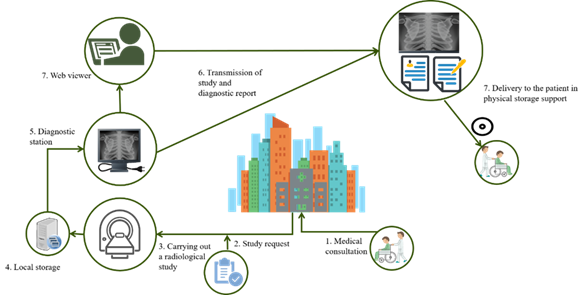
Radiology is one of the pillars of modern medicine, it is estimated that 85% of diagnoses are currently based on radiological techniques (Hunninghake et al., 2003). It is a specialty that solves problems and provides solutions. 2 shows the flow that a patient's care process follows until receiving his diagnostic report in an intrahospital model, which does not encourage exchange to and from other institutions.
The experience in the use of PACS systems and the development in health, allows us to affirm that Cuba is in a position to make a technological leap to expand the level of coverage of its services. Due to the need to reach more hospital institutions and the constant evolution of PACS platforms, new technologies are introduced to achieve their generalization. It is also important for Cuba, due to its technological limitations, to innovate on new forms of management, access to services and medical care. Medical radiology internationally is increasingly switching from on-premise PACS systems to PACS in the cloud (Muthukumaran, Umapathy and Omkumar, 2021; França, Arthur and Iano, 2022). The immediate benefits of using these platforms include cost effectiveness, scalability, and perhaps most importantly, data security. Cloud-based PACS solutions can sufficiently support a wide range of medical imaging environments, from small to medium-sized environments to large-scale installations.
There are many areas in which medical imaging is implementing technological developments with the aim of maximizing quality (better signal, faster diagnosis, better definition of the image and its attributes) and its impact on the disease to be evaluated. Images are increasingly multidimensional (they express different information that can be separated as independent channels) and multiparametric (they represent biological or physiological processes relevant to the disease). Medicine is changing from a reactive activity in the face of disease (curing) to a more proactive one in which the concepts of predicting, personalizing, participating and preventing (4P) will be the next strategic step in the most advanced centers.
As the trend in the development of artificial intelligence grows, innovation opportunities for fields such as medical imaging and radiology are imminent. However, the biggest hurdle to AI-integrated PACS solutions in radiology today lies with artificial intelligence algorithms. Currently available algorithms provide limited ability to successfully deal with the monolithic nature of many PACS systems on the market. AI vendors can be inspired to design custom PACS interfaces, and this requires a comprehensive rethink, where a PACS is built around a complete AI framework, to ensure successful widespread adoption of artificial intelligence in radiology.
Conclusiones
The XAVIA PACS-RIS platform has become a Cuban solution to contribute to the process of digital transformation in radiology. Hospitals from Pinar del Río to Villa Clara transmit and view their studies from this solution, leading to considerable savings in resources, time, and higher quality in the patient care process. The main challenges that exist to give continuity are those related to the technological infrastructure so that hospital institutions assimilate the platform, elements that lead to the search for alternatives to advance access to the entire country.

