Mi SciELO
Servicios Personalizados
Articulo
Indicadores
-
 Citado por SciELO
Links relacionados
-
 Similares en
SciELO
Compartir
Revista Cubana de Estomatología
versión On-line ISSN 1561-297X
Rev Cubana Estomatol vol.55 no.4 Ciudad de La Habana oct.-dic. 2018
ARTÍCULO ORIGINAL
Oral and maxillofacial lesions in children and adolescents
Lesiones bucales y maxilofaciales en niños y adolescentes
Renata Laís Xavier Santos1
Edmilson Zacarias da Silva Júnior1
Maria Carlla Aroucha Lyra1
Richard Alonso Ribeiro de Andrade1
Mônica Vilela Heimer1
Emanuel Sávio de Souza Andrade1
1 Faculty of Dentistry. University of Pernambuco. Recife, Brazil.
ABSTRACT
Introduction: Despite the vast literature reporting the prevalence of oral and maxillofacial diseases in the last decades, few studies have focused on lesions biopsied in the pediatric population.
Objective: To determine the prevalence of oral and maxillofacial lesions that were biopsied in children and adolescents aged 0-19 years.
Methods: Observational and descriptive study. We carried out a retrospective review of 862 reports of pathological examinations performed in an oral pathology laboratory of the northeast of Brazil, during the period from March 2001 to December 2009. The categories were neoplasms, hyperplastic/reactionary lesions, salivary gland lesions, bone lesions, healthy tissues and teeth, oral mucosal lesions, cystic lesions, odontogenic tumors, periapical inflammation, dental alteration and conclusive diagnosis.
Results: The epidemiological profile of patients was characterized by females (53.24 %), Caucasian (45.12 %), with a mean age of 13.06 years. Salivary gland lesions were the category with the largest number of cases (182), and mucocele was the most prevalent histopathological diagnosis (18.44 %), with an average size of 1.97 cm. Most cases were asymptomatic (70.88 %).
Conclusions: This study showed a predominance of lesions diagnosed as benign, the most prevalent lesions were associated with the salivary gland. Females were the most affected.
Keywords: Epidemiology; odontogenic tumors; oral medicine.
RESUMEN
Introducción: A pesar de la gran cantidad de literatura que informa sobre la prevalencia de enfermedades bucales y maxilofaciales en las últimas décadas, pocos estudios se concentraron en las lesiones biopsiadas en la población pediátrica.
Objetivo: Determinar la prevalencia de las lesiones bucales y maxilofaciales que fueron biopsiadas en los niños y adolescentes de 0 a 19 años.
Métodos: Estudio observacional y descriptivo. Se realizó una revisión retrospectiva de 862 informes de los exámenes patológicos realizados en un laboratorio de Patología Oral del nordeste de Brasil, durante el período comprendido entre marzo de 2001 y diciembre de 2009. Las categorías fueron: neoplasias, hiperplásicas / lesiones reactivas, lesiones de las glándulas salivales, lesiones óseas, tejidos y dientes sanos, lesiones de la mucosa bucal, lesiones quísticas, tumores odontogénicos, inflamación periapical, alteración dental y diagnóstico concluyente.
Resultados: El perfil epidemiológico de los pacientes se caracterizó por las mujeres (53,24 %), de raza caucásica (45,12 %) con una edad media de 13,06 años. Lesiones de las glándulas salivales fueron la categoría con el mayor número de casos (182), y el mucocele fue el diagnóstico histopatológico más frecuente (18,44 %), con un tamaño medio de 1,97 cm. La mayoría de los casos fueron asintomáticos (70,88 %).
Conclusiones: Este estudio mostró un predominio de las lesiones diagnosticadas como benignas. Las lesiones más frecuentes se relacionaron con la glándula salival. Las mujeres fueron las más afectadas.
Palabras clave: Epidemiología; tumores odontogénicos; medicina oral.
INTRODUCTION
Knowledge of oral diseases through epidemiological studies have an important role in public health, since they reveal the prevalence, incidence and progression of several diseases that affect the oral cavity, and particularize their distribution into characteristics of the environment where they are being executed.
The prevalence of oral lesions in children is demonstrated in the literature on retrospective studies that used oral biopsies performed in oral diagnostic centers in various countries1-6 or even by epidemiological studies related to specific conditions in pediatric populations, such as age, gender, systemic alterations and allergies.7
The adult and the child/adolescent populations are different in many ways, not only because of their sizes. It is known, for example, that children and adolescents are more often target by certain lesions.¹
Considering the scarcity of studies on the prevalence of oral lesions diagnosed by using histopathological exam, this study aimed to determine, in patients aged 0 to 19 years, the prevalence, clinical and histopathological characteristics of oral lesions diagnosed in a Laboratory of Oral Pathology in a Faculty of Dentistry.
METHODS
We performed a descriptive and observational study, in which pathology reports from a Laboratory of Oral Pathology files were reviewed. Out of the 4492 pathology tests evaluated, 862 reports were part of a sample of patients aged between 0 and 19 years, whose biopsies results were issued during the period from March 2001 to December 2009.
According to the criteria established by the World Health Organization (WHO) for children and adolescents, evaluations of clinical records were made, epidemiological data (age, gender, skin color) were obtained as well as the clinical characteristics of lesions (anatomical location, symptoms, size) and pathologic reports in printed and electronic media for access to the diagnosis, which were grouped into categories according to Lima et al.8 The categories were: neoplasms; hyperplastic/reactionary lesions; salivary gland lesions; bone lesions; healthy tissues and teeth; oral mucosal lesions; cystic lesions; odontogenic tumors; periapical inflammation, dental alteration and inconclusive diagnosis. The odontogenic tumors were classified according to WHO classification.9 Data were tabulated and analyzed by descriptive statistics through of absolute and percentage frequencies for the categorical variables and for the numerical variables were used mean, median and standard deviation. The Ethics Committee of the Institution approved this study.
RESULTS
In the sample analyzed, it was observed that 459 (53.24 %) patients were female, 398 (46.17 %) were male, with a mean age of 13.06 years. In five records this information was missing. Regarding race, it was found that 389 (45.12 %) patients were white, 215 (24.94 %) were black, 179 (20.97 %) were classified as other and 79 (9.16 %) were not reported.
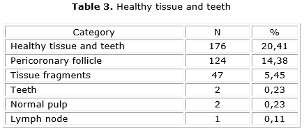
When the diagnoses were grouped into categories, we observed, respectively, that the most numerous were the salivary gland lesions (n= 182; 21.1 %) and the healthy tissue and teeth (n= 176; 20 %). The frequency of other categories were: cystic lesions (n= 117; 13.57 %), odontogenic tumors (n= 83; 9.63 %), hyperplastic/reactionary lesions (n= 79; 9.17 %), neoplasms (n= 74; 8.4 %), bone lesions (n= 67; 8 %), oral mucosal lesions (n= 57; 6.7%), inconclusive diagnosis (n=14; 2 %), periapical inflammation (n= 9; 1.6 %) and dental alteration (n= 4; 0.4 %).
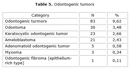
The most common lesions analyzed related to each category were fibroma (table 1), fibrous hyperplasia (table 1), mucocele (table 2), benign fibro-osseous lesion (table 2), pericoronary follicle (table 3), nonspecific chronic inflammatory process (table 4), unclassified odontogenic cysts (table 4), and odontoma (table 5). The lesions related to the categories of periapical inflammation (n= 9), dental alterations (n= 4) and inconclusive diagnosis accounted for 3.12 % of the total sample, for this reason they are not presented in the tables.
Regarding the anatomical location of the lesions, mandible, lips and tongue were the most common anatomical sites. The group of neoplasms occurred predominantly on the tongue (20.27 %), the hyperplastic / reactive lesions predominated on the gingiva (24.05 %), salivary gland lesions occurred mainly on the lower lip (71.97 %), as well as the buccal mucosal lesions (19.29 %). The mandible was the anatomical site most affected by cystic lesions (40.17 %), odontogenic tumors (55.42 %) and periapical inflammation (44.44 %). The dental alterations predominated in the maxillary (50 %).
Data relating to symptoms reported in the records showed that 186 (21.57 %) were symptomatic, 611 (70.88 %) were asymptomatic and information was missing in 65 (7.54 %). The lesion size was measured based on the length and showed an average of 1.97 cm.
DISCUSSION
In this study, the prevalence of abnormalities found in children and adolescents accounted for 19.18 % of all biopsies, higher results than those found in other studies.1,10-12
Regarding gender and race, it was observed that most of the biopsies were performed in white female patients, confirming the findings of other studies.3,4,11 It is noteworthy that some authors suggest the existence of some systematic factor inherent in females, which favors the growth of oral lesions.7 The majority of patients in this study were white, although a large number of patients were listed as unclassified or other. The miscegenation of Brazilian population raises difficulties for racial/ethnic classification.
The fact that most lesions are asymptomatic reflects the importance of professional examination of patients, as well as guidance for those responsible to often observe the oral cavity of their children searching for any deviation from normality.
The pathological diagnoses obtained were grouped into 11 categories according to the classification proposed by Lima et al.8 However, there was no standardization of diagnostic criteria used, and since there are various ways of classifying research,4,10-12 it is difficult to compare results between them.
In bone lesions, for instance, benign fibro-osseous lesion, followed by the central giant cell lesions are the most common. These findings corroborate to the results of Elarbi et al.,10 however, these authors classified these lesions as non-odontogenic benign tumors.
Regarding neoplasms, the fibroma followed by papilloma and hemangioma were the most frequent diagnosed alterations. These results were also found by Lima et al.8 Malignant neoplasms occurred in a small part of the sample, the majority of mesenchymal origin corroborating with other studies.13
The salivary gland lesions represented most of the lesions diagnosed in this study, disagreeing with the findings of Tekkesin et al.,3 who found that the cysts and pseudotumoral lesions as the most frequent oral lesions.
Among the different types of maxillofacial lesions that underwent biopsy, the most numerous in descending order were: mucocele, unclassified odontogenic cysts, nonspecific chronic inflammatory process, fibrous hyperplasia, odontoma, pyogenic granuloma, radicular cyst, keratocystic odontogenic tumor, dentigerous cyst, fibroma and ameloblastoma. Mucocele presented itself as the most frequent diagnose, with prevalence similar to that found by other authors.3,11,12,14 It is noteworthy that, in the present study, the mucocele was more prevalent on the lower lip, the same being observed by Cavalcante et al.15
Several studies in the literature focus their inquiries and research into a specific type of lesion, usually odontogenic tumors.16,17 In this study odontogenic tumors accounted for 9.62 % of 862 biopsies and these diagnoses are presented in descending order in the following sequence: odontoma, keratocystic odontogenic tumor, ameloblastoma, adenomatoid odontogenic tumor, myxoma and the most rarewas the odontogenic fibroma (epithelium-rich type). The reports of Lima et al.,8 Jaeger et al.18 and Servato et al.19 also showed odontoma as the most prevalent odontogenic tumor. It is worth noting that Elarbi et al.10 found, as the lowest prevalence tumor, the calcifying epithelial odontogenic tumor, which was not found in this study. An important change in the most recent WHO classification of odontogenic tumors published in 20059 was the inclusion of the keratocystic odontogenic tumor (previously named odontogenic keratocystic). Mosqueda-Taylor20 suggested that the inclusion of the "odontogenic keratocyst" as a tumor will be modify the relative frequency of odontogenic tumors. In this respect, some authors21-23 have shown that odontogenic tumors represent between 0,8 % and 3,7 % of all specimens sent to oral pathology laboratories and 75 % of these tumors are odontomas, ameloblastomas and myxomas. In our study odontogenic tumors were more frequently diagnosed (9.62 %) and the keratocystic odontogenic tumor was more frequent than ameloblastoma and myxoma confirming the suggestion of Mosqueda-Taylor 20 and results showed by Urs et al.24
Based on the information above, it is clear the importance of identifying the most often found lesions in children and adolescents, and thus trace the strategies adopted in accordance to the profile of the population investigated.
In this study, the epidemiological profile of patients with oral and maxillofacial lesions that underwent histopathological analysis was characterized by white females with a mean age of 13.06 years. Salivary gland lesions was the category with the largest number of cases, and histopathological diagnosis of mucocele the most prevalent. In addition, odontogenic tumors were more frequently diagnosed than in the majority of other studies. Most cases were asymptomatic, with an average size of 1.97 cm.
Conflicto de intereses
The authors declare that they have no conflict of interest.
REFERENCES
1. Mouchrek M, Gonçalves L, Bezerra-Junior J, Maia E, Silva R, Cruz M. Oral and maxillofacial biopsied lesions in Brazilian pediatric patients: A 16-year retrospective study. Rev Odonto Cienc. 2011;26(3):222-6.
2. Lei F , Chen J , Lin L, Wang W, Huang H, Chen C, Ho K, Chen Y. Retrospective study of biopsied oral and maxillofacial lesions in pediatric patients from Southern Taiwan. Journal of Dental Sciences 2014;9:351-8.
3. Tekkesin M, Tuna E, Olgac V, Aksakallı N, Alatli C. Odontogenic lesions in a pediatric population: Review of the literature and presentation of 745 cases. International Journal of Pediatric Otorhinolaryngology. 2016;86:196-9.
4. Becker M, Stefanelli S, Rougemont A, Poletti P, Merlini L. Non-odontogenic tumors of the facial bones in children and adolescents: role of multiparametric imaging. Neuroradiology. 2017;59(4):327-42.
5. Iatrou I, Theologie-Lygidakis N, Tzerbos F, Schoinohoriti OK. Oro-facial tumours and tumour-like lesions in Greek children and adolescents: An 11-year retrospective study. Journal of Cranio-Maxillo-Facial Surgery. 2013;41:437-43.
6. Krishnan R, Ramesh M, Paul G. Retrospective Evaluation of Pediatric Oral Biopsies from A Dental and Maxillofacial Surgery Centre in Salem, Tamil Nadu, India. Journal of Clinical and Diagnostic Research. 2014;8(1):221-3.
7. Hussein A, Darwazeh A, Al-Jundi S. Prevalence of oral lesions among Jordanian children. Saudi Journal of Oral Sciences. 2017;4(1):12.
8. Lima G, Fontes S, Araújo L, Etges A, Tarquino S, Gomes A. A survey of oral and maxillofacial biopsies in children: a single-center retrospective study of 20 years in Pelotas-Brazil. J Appl Oral Sci. 2008;16:397-402.
9. Barnes L, Eveson JW, Reichart P, Sidransky D (eds.) World Health Organization Classification of Tumours. Pathology and Genetics of Head and Neck Tumours. Lyon: IARC Press; 2005.
10. Elarbi M, El-Gehani R, Subhashaj K, Orafi M. Orofacial tumors in Libyan children and adolescents. A descriptive study of 213 cases. Int J Pediatr Otorhinolaryngol. 2009;73:237-42.
11. Ha W, Kelloway E, Dost F, Farah C. A retrospective analysis of oral and maxillofacial pathology in an Australian paediatric population. Australian Dental Jornal. 2014;59(2):221-5.
12. Puangwan D, Juengsomjit R, Poramaporn D, Suwimol D, Sopee D. Oral and Maxillofacial Lesions in a Thai Pediatric Population: A Retrospective Review from Two Dental Schools. J Med Assoc Thai. 2015;98(3):291-7.
13. Gultelkin S, Tokman B, Turkseven M. A review of paediatric oral biopsies in Turkey. Int Dent J. 2003;53:26-32.
14. Zuniga M, Mendez C, Kauterich R, et al. Paediatric oral pathology in a Chilean population: a 15-year review. Int j paediatr dent. 2013;23(5):346-51.
15. Cavalcante RB, Turatti E, Daniel AP, de Alencar GF, Chen Z. Retrospective review of oral and maxillofacial pathology in a Brazilian paediatric population. European Archives of Paediatric Dentistry. 2016;17(2):115-122.
16. Li N, Gao X, Xu Z, Chen Z, Zhu L., Wang J, Liu W. Prevalence of developmental odontogenic cysts in children and adolescents with emphasis on dentigerous cyst and odontogenic keratocyst (keratocystic odontogenic tumor. Acta Odontologica Scandinavica. 2014;72(8):795-800.
17. Abrahams JM, McClure SA. Pediatric odontogenic tumors. Oral and Maxillofacial Surgery Clinics of North America. 2016;28(1):45-58.
18. Jaeger F, de Noronha MS, Silva ML, Amaral MB, Grossmann SD, Horta MC, et al. Prevalence profile of odontogenic cysts and tumors on Brazilian sample after the reclassification of odontogenic keratocyst. Journal of Cranio-Maxillofacial Surgery. 2017;45(2):267-70.
19. Servato J, de Souza P, Horta M, Ribeiro DC, de Aguiar M, de Faria PR, et al. Odontogenic tumours in children and adolescents: a collaborative study of 431 cases. Int J Oral Maxillofac Surg. 2012;41:768-73.
20. Mosqueda-Taylor A. New findings and controversies in odontogenic tumors. Med Oral Patol Oral Cir Bucal. 2008;13:E555-8.
21. Mosqueda-Taylor A, Ledesma-Montes C, Caballero-Sandoval S, Portilla-Robertson J, Ruiz-Godoy Rivera LM, Meneses-Garcia A. Odontogenic tumors in Mexico: a collaborative retrospective study of 349 cases. Oral Surg Oral Med Oral Pathol Oral Radiol Endod. 1997;84:672-5.
22. Ochsenius G, Ortega A, Godoy L, Peñafiel C, Escobar E. Odontogenic tumors in Chile: a study of 362 cases. J Oral Pathol Med. 2002;31:415-20.
23. Buchner A, Merrel PW, Carpenter WM. Relative frequency of central odontogenic tumors: a study of 1,088 cases from Northern California and comparison to studies from other parts of the world. J Oral Maxillofac Surg. 2006;64:1343-52.
24. Urs A, Arora S, Singh H. Intra- Osseous Jaw Lesions in Paediatric Patients: A Retrospective Study. Journal of Clinical and Diagnostic Research. 2014;8(3):216-20.
Recibido: 09/05/2017
Aceptado: 31/07/2018
Mônica Vilela Heimer. Faculty of Dentistry. University of Pernambuco. Recife, Brazil.


