


Mi SciELO
 Servicios personalizados
Servicios Personalizados
Articulo
 Inglés (pdf)
 Articulo en XML
 Referencias del artículo
 Enviar articulo por email
Indicadores
-
 Citado por SciELO
Links relacionados
-
 Similares en
SciELO
Similares en
SciELO
Compartir

 Permalink
Revista Cubana de Estomatología
versión On-line ISSN 1561-297X
Rev Cubana Estomatol vol.52 no.2 Ciudad de La Habana abr.-jun. 2015
PRESENTACIÓN DE CASO
Basal cell adenoma in upper lip: case report
Adenoma de células basales en el labio superior: reporte de un caso
Graziele Beanes,I Liliane Lins,II Antônio Fernando Pereira FalcãoI
I Faculty of Dentistry, Federal University of Bahia, Salvador, Bahia, Brazil.
II Faculty of Medicine, Federal University of Bahia. Bahiana School of Medicine and Public Health, Salvador, Bahia.
ABSTRACT
Basal cell adenoma is a rare benign salivary gland neoplasm that accounts for 1 to 3 % of all salivary gland tumors. Most cases occur in major salivary glands and are rare in minor salivary glands. Describe a clinical case of basal cell adenoma. A 76-year-old Caucasian Brazilian woman presents with a slow-growing asymptomatic lesion of the upper lip. Physical examination revealed a nodular lesion 1.1 cm in size. The lesion was firm to palpation and the surrounding mucosa had normal color and appearance, without any ulceration. The tumor was surgically removed by excisional biopsy. Biopsy confirmed basal cell adenoma. A systematic literature review was carried out in PubMed, Lilacs and SciELO databases. The review included all articles published before 1992. Until 1991 no differentiation was made between basal cell adenoma and canalicular adenoma. Both pathologies were classified as monomorphic adenoma. This is the seventh case of upper lip basal cell adenoma reported in the literature between 1992 and 2014. Three of the seven cases reported were from Brazil.
Key words: adenoma, basal cell, pleomorphic adenoma, lip neoplasms.
RESUMEN
El adenoma de células basales es una neoplasia de la glándula salival raro benigno que representa a 1 a 3 % de todos los tumores de las glándulas salivales. La mayoría de los casos ocurren en las glándulas salivales mayores, siendo poco frecuente en las glándulas salivales menores. El objetivo de este estudio fue describir un caso clínico de adenoma de células basales. Una mujer brasileña de raza caucásica de 76 años de edad, quien se queja de un crecimiento lento y asintomático de una lesión en el labio superior. El examen físico reveló una lesión nodular de 1,1 cm de tamaño. La lesión era firme a la palpación y la mucosa circundante tenía color normal y la apariencia, sin ulceración. El tumor fue extirpado quirúrgicamente mediante una biopsia por escisión. La biopsia confirmó adenoma de células basales. Se realizó una revisión sistemática de la literatura se en las bases de datos PubMed, Lilacs y SciELO. Incluimos en esta revisión todos los artículos publicados antes de 1992. Hasta 1991, no se hizo la diferenciación entre adenoma de células basales y adenoma canalicular. Ambas patologías fueron clasificadas como adenoma monomórfico. Este es el séptimo caso de adenoma de células basales de labio superior reportado en la literatura entre 1992 y 2014. Tres de los siete casos reportados provinieron de Brasil.
Palabras clave: adenoma, basocelular, adenoma pleomorfo, neoplasias de los labios.
INTRODUCTION
Basal cell adenoma (BCA) is a rare benign salivary gland neoplasm that accounts to 1 to 3 % of all salivary gland tumours. Most cases occur in the major salivary glands (75 % parotid and 5 % submandibular glands), being rare in minor salivary glands. Tumors of the salivary glands may show morphological diversity and wide variety of biological behaviors. These characteristics, as well their rarity have hindered its diagnosis and classification.1
Basal cell adenoma, canalicular adenoma, the Warthin's tumor and oncocytoma were classified as monomorphic adenomas (MA) by the World Health Organization (WHO) in 1972. MA was considered a variant of pleomorphic adenoma, presenting few chondroid and myxomatous components. Till 1991, no differentiation was made between BCA and canalicular adenoma; both pathologies being classified as monomorphic adenoma.1 From 1992 till now, only six cases of BCA in upper lip have been described in the literature.2-6
The aim of this study is to report a clinical case of basal cell adenoma in upper lip.
CASE REPORT
A 76-year-old Caucasian woman was admitted in the Stomatology Clinic of the Faculty of Dentistry, Federal University of Bahia, Brazil, complaining of a slow-growing and asymptomatic lesion in upper lip. The patient reported that the lesion begun 3 months ago, as well the history of thyroid and breast cancers with metastasis to axillary lymph nodes.
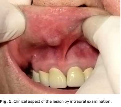
Intraoral examination has shown a delimited nodule measuring 1.1 cm in diameter, in the right side of the upper lip. The lesion was firm to palpation and the surrounding mucosa had normal color and aspect, with no ulceration (Fig. 1).
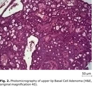
The clinical suspicion of adenoma of minor salivary glands was made. The tumour was surgically removed by excisional biopsy, under local anesthesia. The specimen was sent for histopathological examination and was stained with hematoxylin-eosin (H&E). The histopathological analysis has shown an atrophic parakeratinized stratified squamous epithelium without atypical aspects. The underlying lamina propria was infiltrated by multiple well-defined lobules composed of basaloid cells arranged in tubular-trabecular pattern. Individually, the cells showed rounded and oval nuclei, with eosinophilic cytoplasm, surrounded by a fibro-hyaline stroma. The presence of cystic areas was observed on the periphery of the lobules without calcification. The conclusion was Basal Cell Adenoma (Fig. 2). No recurrence was observed after eight years of follow-up.
This study protocols were approved by the Ethical Review Board of the Faculty of Dentistry of Federal University of Bahia, protocol number 103728, and are in accordance to the Brazilian National Health Council Resolution 466/12, as well as the Sixth Revision of the Declaration of Helsinki, 2008.
DISCUSSION
A systematic literature review was carried out in Jun 2014, using PubMed, Lilacs and Scielo databases. The following terms were used: "basal cell adenoma upper lip" and "monomorphic adenoma upper lip".
We have included in this review all papers reporting upper lip BCA cases, from 1992 to 2014, with no language restriction, and have excluded literature reviews. The search has identified a total of five papers, including six patients.2-6 For one case,5 there was no information about gender, age, and lesion recurrence. Considering the present case and the other five from literature review, all tumours were painless and had no history of recurrence after excisional biopsy. Age ranged from 51 to 76 years. There were four males and two females. Three cases came from Brazil, two from Greece, one from Spain, and one from Japan.
In the past, BCA, canalicular adenoma, the Warthin's tumour and oncocytoma were classified as monomorphic adenomas. Since 1992, the term monomorphic adenoma has been no longer used by WHO and the tumours, previously classified as MA, were individually described. BCA is a salivary gland benign tumour comprised of basaloid cells arranged in a solid, trabecular, tubular and/or membranous pattern.1,7
The parotid gland is the most frequent site of BCA occurrence, followed by the submandibular gland. BCA is extremely rare in minor salivary glands. Although a 2:1 female:male ratio is reported in literature,7 in this series, cases of upper lip BCA were more frequent among males, with a 2:1 sex ratio.
Three out the six reported cases came from Brazil, spread over three different states of the country.
The mean age (65 years, ranging from 51 to 76) of the BCA patients was according to literature.1,7 All BCA cases included in this review were painless and the time of the lesions progression ranged from 3 months to 5 years. The dimensions of the tumours ranged from 0.5 to 4.0 cm. BCA is usually described as a small and painless tumour, usually ranging from few millimeters to 3-4 cm. Secondary signs, such as the presence of ulceration or bleeding, are rare.8
In all clinical cases, included in this study, excisional biopsy was performed to remove the lesion. No recurrent cases were reported after follow-up periods varying from six to 96 months.
Histologically, BCA is characterized by the presence of basaloid cells and lack of the myxochondroid stromal component of pleomorphic adenoma. There are four patterns of histopathological BCA: solid, trabecular, tubular, and membranous. Mixed patterns may occur,7 as in the tubular-trabecular case reported in this paper.
Differential diagnosis must consider pleomorphic adenoma; mucocele, sialoadenite and Warthin’s tumour. Distinction between the BCA and malignant tumours is of great importance, particularly to avoid unnecessary aggressive therapy. Among malignancies, the basal cell adenocarcinoma requires special attention in the differential diagnosis followed by adenoid cystic carcinoma, basaloide squamous cell carcinoma, carcinoma ex pleomorphic adenoma, basal cell carcinoma, metastatic basal cell carcinoma and sialoblastoma.1,9
According to literature, BCA recurrence rate varies depending of the histological type of the lesion. For solid and trabecular variants, recurrence is almost nonexistent. The membranous type is most commonly associated with recurrence and this may be a result of multicentricity of the lesion. There are few reported cases of BCA malignant transformation and this rare condition is associated to the membranous subtype.7 Knowledge about upper lip BCA specific characteristics is limited. Therefore, caution is needed when comparing characteristics, drawn from literature, of BCAs from all sites with upper lip BCA.
This is the seventh case of upper lip BCA reported in literature from 1992 to 2014. Till 1992 there was no differentiation between cases of BCA and canalicular adenoma, both pathologies being classified as monomorphic adenoma. Although BCA affects mainly elderly women, we found a 2:1 male: female ratio concerning the cases of upper lip BCA reported. Three out of the seven reported cases came from Brazil. No recurrence was observed in 6 to 96 months follow-up after lesion excision. The specific features of the seven upper lip BCA cases reported here reinforces the importance to perform a correct diagnosis of nodule lesions in upper lip in order to establish adequate treatment.
Conflict of interests
The authors declare no conflict of interests in this paper.
BIBLIOGRAPHIC REFERENCES
1. Barnes L, Eveson JW, Reichart P, Sidransky D. Pathology and genetics of head and neck tumours. World Health Organization Classification of Tumours. 1st ed. Lyon: IARC; 2005. p. 259-60.
2. Minicucci EM, Campos EBP, Weber SAT, Domingues MAC, Ribeiro DA. Basal Cell Adenoma of the Upper Lip from Minor Salivary Gland Origin. European Journal of Dentistry. 2008;2:213-6.
3. Antoniades D, Epivatianos A, Markopoulos A, Kolokotronis A, Zaraboukas T. Coexistence of Mucous Retention Cyst and Basal Cell Adenoma Arising from the Lining Epithelium of the Cyst. Med Princ Pract. 2009;18:248-252.
4. Soares ECS, Costa FWG, Bezerra MF, Alves APNN, Sousa FB. Adenoma de células basais em lábio superior. Rev Gaúcha Odontol. 2010;58(4):533-6.
5. Vicandi B, Jiménez-Heffernan JA, López-Ferrer P, González-Peramato P, Patrón M, Viguer JM. Fine needle aspiration cytology of basal cell adenoma of the salivary gland: a cytohistological correlation study of 35 cases. Cytopathology;2012(23):315–9. DOI: 10.1111/j.1365-2303.2011.00899.x.
6. Kudok M, Harada H, Sato I, Omura K, Ishii Y. A case of basal cell adenoma of the upper lip. Case Rep Med. 2014. DOI: 10.1155/2014/795356
7. Araújo VC. Basal cell adenoma. In: Pathology and genetics of head and neck tumours.WHO Classification of Head and Neck Tumours. IARC Press; 2005. p. 261-2.
8. Pires FR, Pringle GA, Almeida OP, Chen S. Intra-oral minor salivary gland tumours: A clinicopathological study of 546 cases. Oral Oncology. 2007;43:463-70.
9. Kawata R, Yoshimur K, Lee K, Araki M, Takenaka H, Tsuji M. Basal cell adenoma of the parotid gland: a clinicopathological study of nine cases—basal cell adenoma versus pleomorphic adenoma and Warthin’s tumour. Eur Arch Otorhinolaryngol. 2010;267:779-83.
Recibido: 7 de julio de 2014.
Aprobado: 12 de agosto de 2014.
Antônio Fernando Pereira Falcão. Faculty of Dentistry, Federal University of Bahia. Rua Araújo Pinho, 62, Salvador, Bahia, Brazil. ZIP CODE: 40110-150.
Correo electrónico: afpfalcao@hotmail.com

