


Mi SciELO
 Servicios personalizados
Servicios Personalizados
Articulo
 Inglés (pdf)
 Articulo en XML
 Referencias del artículo
 Enviar articulo por email
Indicadores
-
 Citado por SciELO
Links relacionados
-
 Similares en
SciELO
Similares en
SciELO
Compartir

 Permalink
Revista Cubana de Estomatología
versión On-line ISSN 1561-297X
Rev Cubana Estomatol vol.53 no.4 Ciudad de La Habana oct.-dic. 2016
PRESENTACIÓN DE CASO
Giant lipoma on lower lip: an unusual case
Lipoma gigante en el labio inferior: Un caso inusual
Laudenice de Lucena Pereira,I Dasaiev Monteiro Dutra,II Victor Yuri Nicolau Ferreira,II Emanuene Galdino Pires,II Larissa Cavalcanti Monteiro,II Danyel Elias da Cruz Perez,III Paulo Rogério Ferreti BonanII
I Federal University of Rio Grande do Norte. Natal, Brazil.
II Federal University of Paraíba. João Pessoa, Brazil.
III Federal University of Pernambuco. João Pessoa, Brazil.
ABSTRACT
Introduction: lipomas are benign mesenchymal tumors which consist essentially of mature adipocytes, and are relatively uncommon in the oral cavity comparing with other body surfaces. Large lesions are relatively rare and lower lip is not a very usual site. This is characterized as a slow growing lesion which might reach large dimensions and are usually asymptomatic.
Objective: to show a case of giant lipoma affecting lower lip and to comment on its clinical and microscopic features.
Case report: in this study, we describe a case of giant lipoma affecting lower lip of a 55 years-old male with an asymptomatic evolution of eight years. We performed a complete excision, and the histopathological examination revealed a lipoma.
Conclusions: actually, the patient is under follow up without signs of recurrence. The clinical and microscopic characteristics were very important for the diagnosis.
Keywords: lipoma; mouth neoplasm; lip.
RESUMEN
Introducción: los lipomas son tumores mesenquimales benignos que consisten esencialmente en adipocitos maduros, poco comunes en la cavidad oral comparada con otras superficies corporales. Lesiones de gran tamaño son relativamente raras y el labio inferior no es un sitio muy habitual. Se caracteriza por ser una lesión de crecimiento lento que puede alcanzar grandes dimensiones y son generalmente asintomáticos.
Objetivo: caracterizar un caso de lipoma gigante en el labio inferior con algunas características clínicas e histopatológicas de esta lesión.
Presentación del caso: se describe un caso de lipoma gigante que afecta el labio inferior de un paciente de 55 años de edad, de sexo masculino con una evolución asintomática de ocho años. Se realizó una exéresis completa y el examen histopatológico reveló un lipoma.
Conclusiones: el paciente está bajo seguimiento y sin signos de recidiva. Las características clínicas y microscópicas fueron de gran importancia para el diagnóstico.
Palabras clave: lipoma; neoplasias de la boca; labio.
INTRODUCTION
Lipoma is defined as a benign neoplasm consisting of mature fat cells with slow growth,1 which comprises 1-5 % of all tumors that affect the oral cavity.2 Intra-orally, this tumors frequently affect the buccal mucosa and tongue and more rarely the lower lip.3 Giant lesions are also unusual and could impair important oral functions.4 The etiology of lipoma remains unclean, possible including endocrine and hereditary alterations, infection and even local trauma. Glandular cells neoplasm and fibroma are considered differential diagnosis.2 Lipoma is microscopically characterized by a proliferation of mature adipocytes arranged in lobules divided by collagen fibers.5 The treatment of the lipoma is surgical excision and currently, after monitoring the lesion, no evidence of recurrence is detected.1
Our objective is show a case of a giant lipoma affecting lower lip and comment about its clinical and microscopic features.
CASE REPORT
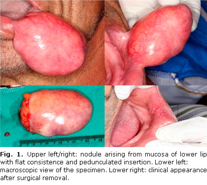
A 55 years-old non caucasian male was referred to Dental Specialties Centre of João Pessoa, in the Northeastern Brazil, due to large lesion affecting oral cavity. The patient complained of an asymptomatic lump in the mouth with a continuous growth with eight years of evolution. He also referred a rapid growth in the last year. During anamnesis, he referred good health conditions. During extraoral clinical examination, an increase in the volume of the right side of the face was detected, resulting in facial asymmetry. Besides, intraoral clinical examination unveiled a smooth well-circumscribed nodular lesion with flat consistency located in the internal mucosa of the lower lip, consisting of a slightly yellowish mass. The lesion measured approximately 6 cm in diameter. The diagnostics hipothesis of lipoma and Glandular cells neoplasm were raised (Fig. 1).
An excisional biopsy of the lesion was performed from a simple surgical excision and the chirurgic piece was placed in 10 % formalin, where a floatation was noticed. It was sent for histopathological analysis. In the microscopic examination, fat cells arranged in lobules and separated by thin bands of connective tissue were observed (Fig. 2). Thus, the diagnosis of lipoma was confirmed. The patient is under a follow-up for 2 years, showing no recurrence.

DISCUSSION
We described a case of lipoma affecting the internal mucosa of lower lip in a middle-aged man. No predilection for gender has been reported,5 while other studies highlight a higher prevalence of lipoma in male patients.6 In terms of age, the patient in our study is in accordance with a study conducted in Brazil,7 which unveiled that most patients were over 40 years old.
Large lipomas have been reported in the cheek region,8 featuring a lesion over 3 cm in diameter in a 13-year-old girl, which differed from the patient in our study, considering gender and age. However, both lesions were asymptomatic. Besides, a lipoma of large size in the tongue was reported occupying almost the entire oral cavity.9 The lipoma started to grow 3 years ago. Not accordingly, our patient reported a growth of 8 years, but both lesions impaired the chewing and speech functions. Apart from our management of the lesion, incisional biopsy was performed in this case. After the confirmation of the diagnosis, the lipoma was surgically removed with restoration of speech and masticatory function.
Regarding the diameter of the lesion, this parameter rarely reaches more than 25 mm.7 Nevertheless, we found a diameter of 6 cm, which is over the mean value related in the literature. The fact that the patient waited for eight years until the diagnosis was performed is associated with the lack of symptomatology developed in the case. Possibly, oral lipomas can interfere with chewing, speech and aesthetics.9
With regard to symptoms, researchers report that besides the foreign body sensation caused by the giant lipoma, the lesion can cause airway obstruction, depending on its magnitude.10 Despite the size of the lesion in this case, the patient had no discomfort, claiming to be accustomed to the presence of the tumor in the oral cavity.
The etiology of lipoma is very controversial, including endocrine and hereditary alterations, infection and local trauma.1,2 In this case report, however, the patient had no history of trauma.
Different histological variants of the tumor have been reported such as the angiolipoma, spindle cell lipoma, pleomorphic lipoma, lipoma of fusiform cells, intramuscular lipoma and the myxoid lipoma. However, the most common is the fibrolipoma. The lesion is microscopically characterized by a proliferation of mature adipocytes arranged in lobules that are often separated by thin bundles of collagen fibers.5 In our study, the histopathological analysis unveiled compatible characteristics with those described in the literature, excluding the hypothesis of Glandular cell neoplasm.
Considering the treatment of the lipoma, the tumor was surgically removed. After monitoring the lesion, no evidence of recurrence was detected. Oral lipomas are usually encapsulated and have a good prognosis after complete surgical resection.1
Here, we described a very rare case of giant lipoma affecting lower lip focusing the insertion of this potential diagnosis when large lesions arising from this site.
REFERENCES
1. Regezi JA, Sciubba JJ, Jordan RC. Patologia Oral: correlações clinicopatológicas. 6th ed. Rio de Janeiro: Elservier; 2012.
2. Sachdeva SK, Rout P, Dutta S, Verma P. Oral lipoma: An uncommon clinical entity. J Oral Maxillofac Radiol. 2013;1(3):118-21.
3. Naruse T, Yanamoto S, Yamada SI, Rokutanda S, Kawakita A, Takahashi H, et al. Lipomas of the oral cavity: clinicopathological and immunohistochemical study of 24 cases and review of the literature. Indian Journal of Otolaryngology and Head & Neck Surgery. 2015;67(1):67-73.
4. Raj AA, Shetty PM, Yadav SK. Lipoma of the Floor of the Mouth: Report of an Unusually Large Lesion. J Maxillofac Oral Surg. 2014;13(1):328-31.
5. Fregnani ER, Pires FR, Falzoni R, Lopes MA, Vargas PA. Lipomas of the oral cavity: clinical findings, histological classification and proliferative activity of 46 cases. Int J Oral Maxillofac Surg. 2003;32(1):49-53.
6. Park BG, Choi DJ, Park JW, Kim JS. Oral cavity lipoma: a case report. J Korean Assoc Oral Maxillofac Surg. 2015;41(4):213-6.
7. Juliasse LE, Nonaka CF, Pinto LP, Freitas RA, Miguel MCC. Lipomas of the oral cavity: clinical and histopathologic study of 41 cases in a Brazilian population. Eur Arch Otorhinolaryngol. 2010;267(3):59-465.
8. Daryani D, Gopakumar R. A large oral lipoma in a young patient: A rare combination. Contemp Clin Dent. 2014;5(2):236-9.
9. Ravi Kiran A. Purnachandrarao Naik N. Samatha Y. Vijay Kumar, A. Kalyan Kumar, D. Intraoral Lipoma: A Rare Case Report and Review of Literature. Journal of Clinical and Diagnostic Research. 2013;7(12):3090-1.
10. Koizumi T, Yane K, Yamanaka T, Kitahara T. A Method of Transoral Finger Dissection for a Giant Epiglottic Lipoma. Case Rep Otolaryngol. 2014;111(1):754-8.
Recibido: 1ro de agosto de 2015.
Aprobado: 27 de septiembre de 2016.

