


 Servicios personalizados
 Inglés (pdf)
 Articulo en XML
 Referencias del artículo
 Enviar articulo por email
 Citado por SciELO
  Similares en
SciELO
Similares en
SciELO 
 Permalink
INTRODUCTION
Posts and core are frequently used in endodontically treated teeth with excessive loss of coronal tooth structure. According to manufacturer, the luting fiber posts must be capable to adhere as it already has Bis GMA in its composition. Some factors as root canal dentin morphology, bond system, luting cement, and its cure may interfere on the hybrid layer formation along the root canal walls, affecting the post retention.1
Various luting agents have been proposed for bonding fiber reinforced composite (FRC) posts to root canal dentin. According to the adhesive strategy, the currently available resin-based cements and accompanying bonding systems can be classified as etch-and-rinse, self-etch, and self-adhesive luting agents.2
Self-adhesive cement does not require rinsing, decreasing the problem of substrate moisture control, thus simplifying the clinical procedure. No dentin pretreatment is indicated in this one-step technique. This simplification allowed by self-adhesive resin cements is attractive to clinicians.3
Although several studies have indicated that the bond-strength values of self-adhesive cements are comparable to, or even higher than, those of conventional luting strategies, their limited etching capability in the presence of the compact smear layer created within the endodontic space is a matter of concern.4
The literature showed that smear layer can be removed by different procedures, such as using chelating agents (i.e:EDTA) ,or using acids -like polyacrylic and phosphoric acids. The total or partial smear layer removal occurs according to time and concentration of these substances. Although the most self-adhesive cements manufacturers do not recommend the prior smear layer removal, this procedure may be advantageous for a better adhesion strength of these cements.5
Thus, after preparation of root canal for placement of an intraradicular retainer, there is necessity of cleaning root canal walls. Therefore, it is convenient to evaluate the optimal solution for cleaning root canals in cases of using self-adhesive cements.
METHODS
An experimental in vitro investigation was made. Freshly extracted bovine incisors with mature apices and without root curvature were obtained for this study selected at random. A digital pachymeter was employed to measure the teeth in three root regions: cervical (RC), middle (RM) and apical (RA), in mesion-distal (RMD) and buccolingual (RBL) direction, in all root length (RT). After this analysis, an average of the root dimensions was determinated and 56 teeth were selected as our sample of the study.
The bovine dentine was used in this investigation due the limited availability and the inhomogeneity of extracted human teeth. Moreover, the bioethical concerns make it difficult to collect and use human teeth for researches. In the present study, the push-out test was performed 24 h after adhesive cementation procedures because bond strength can increase during this period.
For the endodontic treatment, a step-back preparation technique was used with stainless steel K-files and Gates-Glidden (Moyco Union Broach, York, PA) (drills #2 to #4). All enlargement procedures were followed by irrigation with 1% sodium hypochlorite. Afterwards, the prepared root canals were obturated with gutta-percha cones by using the lateral condensation technique and AH Plus resin sealer (Dentsply, Germany).Subsequently, the filled roots were stored in distilled water at 37o for 48 h.
After the storage period, the root canals were prepared to ensure a standardized space for post insertion. The canal space of each root was firstly enlarged with Gates-Glidden #3, permiting access for #3 post drill with a low-speed hand piece, to a depth of 11mm.
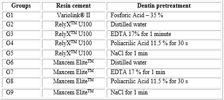
Double conicity glass fiber translucent posts (#3 White post DC, FGM) and different resins cements systems were utilized in this study, originating 9 experimental groups (n= 8).
In all groups, the posts were cleaned with 35 % phosphoric acid for 60 s followed by water rinsing and air drying. Then, a silane coupling agent (Ceramic Primer - 3MESPE) was applied in a single layer on the posts surface for 60 seconds and then, dried with air. One coat of bond (Scotch bond Multi- Purpose - 3MESPE) was also applied, when necessary (group 1). The root canal pre-treatment was performed according to the different groups.
For the etch-and-rinse resin cements (group 1), the root canal dentine was etched with 35% phosphoric acid for 15 seconds and rinsed for 30 seconds with water. After removing the water excess from the root canal with paper points, one layer of the primer (SBMP - 3M ESPE) was applied with a microbrush and gently air-dried for 5 s. Subsequently, the bond (SBMP - 3M ESPE) was applied and dried with paper points to remove the excess, and light-cured for 40 s by a LED light-curing unit Elipar Freelight II (3MESPE), with 900 mW/cm2 intensity.
For the cementation of glass fiber posts, equal amounts of resin cements agents, base and catalyst, were mixed and applied onto the posts surface and into the roots canal with a periodontal probe. Then, the post was inserted and cemented into the root canal with light finger pressure, and the luting material excess was immediately removed.
The cement was light cured for 40 s with the tip positioned parallel to the pin (at its base) and over 40 s at 45°with the long axis of the pin.
For the self-adhesive resin cements groups (2, 3, 4, 5, 6, 7, 8 and 9), the pin was cleaned with phosphoric acid followed by silane application, according to the protocol described for group G1. Adhesive application was not necessary. The root canal preparation was performed according to the different groups, with their respective dentine pretreatments, as mentioned in table 1.
Different types of dentine pre-treatment were employed in order to remove or modify the smear layer (table 1). For groups 2 and 6, the root dentine surface was irrigated with distilled water (no treatment groups). Prior to the resin cement application, for groups G3 and G7,17 % EDTA (ethylenediaminetetraacetic acid) solution was applied for a time of 1 minute andf ollowed by rinsing with water for 1 min.In the groups G4 and G8, 11.5 % polyacrylic acid was applied for 1 minute, followed by rinsing with water at the same time.Sodium hypochlorite (NaCl) was used for groups G5 and G9 during 1 minute, followed by rinsing with water.
The self-adhesive cements manipulation and pin insertion was similar to that described for group 1.
After all cementation procedures, the specimens were stored in distilled water for 24 h at 37 °C.
After the storage period, the specimens were sectioned by Isomet 1 000 cutting digital machine (Buehler UK LTD). The roots were divided in three parts, 1mm from cervical surface. Three 1mm thick precise slabs, separated by 3mm space each, were obtained per root and they were identified as cervical, middle and apical specimens. The thickness of each slab was measured by the digital machine cutting disc position along the root.
Immediately after the slabs were obtained, they were positioned on the push-out jig (1 mm diameter), which was placed on the Universal Testing Machine (MTS 810 Material Test System) with a cell load of 50 kg, at a crosshead speed of 0.5 mm/min until the post was dislodged.
The retentive strength of the post segment was expressed in MPa, by dividing the load at failure (Newtons) by the area of the post fragment (SL), by the formula SL= π (R + r) [(h2 + (R-r)2]0,5. Data were analyzed for better comprehension in a database of SPSS for Windows version 15.
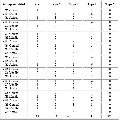
After the push-out testing, the specimens were analyzed by stereoscopic microscope to determine the failure mode: type 1, adhesive between post and resin cement (no resin cement visible around the post); type 2, mixed with resin cement covering 0-50 % of the post diameter; type 3, mixed with resin cement covering between 50 and 100 % of post surface; type 4, adhesive between resin cement and root canal (post enveloped by resin cement); type 5, cohesive in dentin.
Data were analyzed for better comprehension in a database of SPSS for Windows version 15. To evaluate the normality of the data, the Shapiro-wilk test was performed.
Al procedures were developed with high degree of seriously and medical ethic agree with the kind of study. Ethical certifications weren’t necessary cause research was in vitro.
RESULTS
According to the normality test, the data had a normal distribution, therefore, parametric statistics were performed. (ANOVA).
Push-out test: The analysis of variance showed statistically significant difference to resin cements evaluated (p< 0.05) and different thirds of root (p< 0.05). The results of the Tukey test are displayed in table 2.
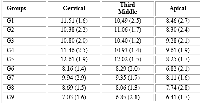
The highest values were found for the groups G3, G4 and G5, and there was no bond strength significant difference with group G2. The G6 and G9 groups showed the lowest values and had no statistical differences with G7 and G8. G1 had no significant difference with G2. G1 and G5 demonstrated statistically lower resistance values of the apical than the cervical third. G5 had lower values on the apical third when compared to the middle third. The groups G2, G3, G4, G6, G7, G8 and G9 had no significant differences along the root thirds.
The failure modes of groups and level of the root are showed in table 3. No cohesive failure in dentin (type V) was observed. Higher incidence of failure type IV (46,7 %) and type III (29,9 %) was observed in comparison to the failure type I (6,4 %). The failure type II occured in 17 % of the cases.
DISCUSSION
The self-adhesive resin cement presented a deficient hybridization of dentin along the root canal wall. The application of RelyX Unicem and Maxcem to root dentin does not result in the formation of hybrid layer or resin tags and inability to etch through the smear layer formed in the root canal. But, the self-adhesive resin cements to root canal dentin seems to be related more to the friction along the canal walls than to the adhesive bonding to root dentin. The manufacturer of RelyX U100 claims that the bonding mechanism of this self-adhesive cement is based on micromechanical retention and chemical adhesion to hydroxyapatite. A recent study showed an intense chemical interaction of RelyX U100 with hydroxyapatite.3
Rely-X U100 has limited etching potential when compared with etch-and-rinse and self-etching adhesive systems. This could possibly be explained by the methacrylated phosphoric esters present in this cement, which are not as effective as phosphoric acid in dissolving the thick smear layer in the root canal walls during post space preparation.5 The use of irrigants such as EDTA has been recommended as extremely effective in cleaning the root canal after post preparation and, as a result, improved the bond strength in each regions of the root dentin.5
Another irrigant used to clean the root dentin is NaOCl because it has the ability to remove the smear layer, which is created on the dentin surface during the post space preparation. The irrigation of root dentin with 5 % NaOCl reduce the bond strength of resin cements to dentin. This could be explained by an oxygen-enriched dentin surface after application of NaOCl, which could act as a polymerization inhibitor of resin materials. The polyacrylic acid used as a pretreatment in glass ionomer cements also has the ability to remove the smear layer and can be used as the root dentin cleaning agent.6
It is likely that phosphoric acid etching is more effective in highly tubular areas of the coronal root dentin because it removes the thick surfaces smear layer and the smear plug in dentinal tubules formed during post space preparation to allow more effective micromechanical retention of resin cements.7
In general, the root dentin should be irrigated with CHX or sterile saline solution before post cementation in order to eliminate the negative effect of NaOCl on the adhesive bond to dentine. But, the study protocol followed the manufacturers’ instructions of RelyX Unicem (3M ESPE, Seefeld, Germany), which recommend the irrigation of the root dentin with NaOCl followed by water.7
The use of ultrasonic instrumentation in association with EDTA has been suggested for a careful debridement of the post space walls improving performed prior to cementation.8
Clinical investigations have reported that the most common cause of failure is debonding of the fiber posts.9 Adhesive failure between the dentine and cement was the main failure mode. In our results, the most samples had failures located at the cement-dentin interface.
The bond strengths were significantly affected by the root canal region, but not by the self- adhesive resin cement. According to our results, the moisture tolerance is probably the factor responsible for the homogeneous bond strength values of RelyX U100 in all root dentin regions. Other aspects of the self-adhesive cements bond strengths to root dentin seem to be related more to the area of solid dentin than to the density of dentinal tubules.
The results of this study showed that RelyX U100 is the best performance among the tested cements. Variolink II showed similar results to RelyX U100 without pretreatment of the dentin. Other studies show the superiority of self-adhesive cements regarding etch-and-rinse cements. The most of these tests were performed with the self-adhesivecement (RelyXUnicem). Some authors speculated that the moisture tolerance of the self-adhesive cement may explain its favourable adhesion in root dentin.
The MaxcemElite self-adhesive resin cement showed the lowest results of bond strength,regardless of the type of pre-treatment performed on root dentin before cementation of the fiberglass post. Other studies show the low performance of Maxcem compared to RelyX Unicem. Soares et al. show in their studies bubbles in the cement Maxcem, irrespective of the location, and the cement primarily in the apical area did not appear to have polymerized.10
Conclusions
Regarding the irrigation solutions, the use of different products for partial removal of smear layer does not influence on the bond strength of the self-adhesive resin cements compared with the control distilled water. The RelyX U100 associated with pretreatment with sodium hypochlorite (recommendation of the manufacturer) showed the best results.

