


My SciELO
 Custom services
Services on Demand
Journal

Article
 English (pdf)
 Article in xml format
 Article references
 Send this article by e-mail
Indicators
-
 Cited by SciELO
Related links
-
 Similars in
SciELO
Similars in
SciELO
Share

 Permalink
Revista de Salud Animal
Print version ISSN 0253-570X
Rev Salud Anim. vol.29 no.3 La Habana Sept.-Dec. 2007
TRABAJO ORIGINAL
ANTIGENIC, BIOLOGICAL AND MOLECULAR CHARACTERIZATION OF THE CUBAN CSFV ISOLATE "MARGARITA"
CARACTERIZACIÓN ANTIGÉNICA, BIOLÓGICA Y MOLECULAR DEL AISLADO CUBANO "MARGARITA" DEL VIRUS DE
Llilianne Ganges1,2, Maritza Barrera1, Heidy Díaz de Arce1, A. Vega1, J.I. Núñez2, F. Sobrino3 y María Teresa Frías1*
1Laboratorio de Virología Animal del Centro Nacional de Sanidad Agropecuaria (CENSA), Apartado 10, San José de las Lajas,
"Margarita" CSF virus isolate (Cuba, 1958) was characterized from the antigenic, biological and molecular point of view; adapted on PK-15 and SK-6 cell lines with an infective titer of 107.5 DICT50, and biologically cloned by limit dilution keeping its pathogenicity in pigs. Genotyping of this virus was made and its phylogenetic relationship with another strain deposited at the GenBank was determined keeping located in the 1.2 sub-group very close related with the field isolates from the 1993-2006 epidemic in Cuba. Its antigenic behavior was similar to the "Alfort" strain, showing no reactivity to MAbss against vaccine strains. The complete sequence of the gene for the E2 protein deposited in the EMBL with number AJ704817 is shown, also the a.a. deduced sequence showing all the residues of cysteine highly conserved in the pestivirus genome is given being part of the different types of conformational epitopes of the glycoprotein E2. From these results, an isolate of a Cuban-native CSF virus fully characterized is available, which can be used as reference material for several purposes.
Key words: CSF; CSF reference strain; antigenic biological and molecular characterization.
RESUMEN
Con el objetivo de disponer de una cepa como material de referencia de Peste Porcina Clásica (PPC) se realizó la caracterización desde el punto de vista antigénico, biológico y molecular del aislado "Margarita" (
Palabras clave: PPC; cepa de referencia PPC; caracterización antigénica biológica y molecular.
(Recibido 15-8-2007; Aceptado 16-10-2007)
INTRODUCTION
Classical swine fiver (CSF) is a highly contagious viral disease affecting domestic and wild pigs. It is considered as one of the most severe diseases
affecting the Pork World Industry, both from the economical and sanitary point of view (25).
The etiological agent is a virus, with an icosahedric symmetry of 40 to 60 nm in diameter and with a lipid envelope (38). CSF virus (CSFV), together with the bovine viral diarrhoea virus (BVDV) and the border disease virus (BDV) conform the Genus Pestivirus, Family Flaviviridae (16).
The disease affects the swine immune system, induces immune suppression associated to important haematological changes such as leucopoenia, thrombocytopenia, coagulation disorders, thyme and bone marrow atrophy (11).
Specific viral antibodies are not detected until three weeks post infection (35) probably due to the strong leucopoenia induced by the virus. Pigs recovered from the infection generally develop neutralizing antibodies persisting during their whole life.
The severity of clinical signs mainly depends on the virulence of the viral strain, also influenced by the age and the immunological state of the animal. (25). Thus, the CSF clinical form and severity are very variable. Nevertheless, their clinical form has been classified as: i) post natal infections, including the hyper acute, acute and chronic forms, ii) trans-placentary infections, which produces foetal and neonatal affections and iii) persistent infections (8, 33).
Nowadays, CSF is enzootic in Central America and the Caribbean area, South America, Southeast Asia and
The presence of wild pigs with CSF endemic infections in some of the state members (17) is one of the problems which has been associated to the disease re-emergency, as occurred in
After some years of relative calm, some outbreaks of the disease in domestic swine farms had been notified by
All these elements make debatable the policy of no vaccination followed by the EU.
For disease control, different live vaccines, very efficient concerning protection against the disease, as well as a great quantity of new generation vaccines which have served as model for studying the immunological mechanisms related to the induced protection against the CSFV have been developed. The characteristics and uses of these vaccination strategies have been widely discussed (10).
Concerning
MATERIALS AND METHODS
Production of the virus
The viral isolate "Margarita", multiplied by successive passes in pig with a titer of 107,4 lethal dose 50 (DL50) /mL, was used.
To multiply the viral isolate "Margarita" in cell cultures, firstly a pig blood preparation with a titer of 5x10⁵ DL50 in pig was used. From this preparation, PK-15 recently grown cells were inoculated at an infection multiplicity (IM) of 0,3 DL50 /cell. The supernatant and cells were taken at 48 hours post infection (HPI), and they were submitted to three freezing-thawing cycles. The supernatant recovered after the sedimentation of cell residues was used for other two successive passes following the previous conditions. The viral suspension finally obtained was titered in PK-15 cells, detecting the presence of the virus by direct immunoperoxidase assay in plate (PLA) (39). For carrying out PLA, cells were incubated for 1 hour at 37º C with a hyperimmune serum (named a-VPPC serum) against CSFV (32). The viral titer was calculated from the last dilutions of the virus in which specifically stained infectious foci were observed using Reed and Muench's method (1938) (30). The result was expressed as the viral dose which produces infection in the 50% of the inoculated monolayers (DICT50)/mL.
This viral suspension was cloned by limit dilution, and the virus recovered from it was amplified in PK-15 cells using an IM of 0,4 which is an infective dose in tissue cultures 50 (DICT50)/ cell and named c-Margarita.
On the other hand, using the same IM, there were made 11 passes in PK-15 cells and other
The strain NADL (given by Dr. Aynaud, INRA, France) of CSFV was grown on MDBK cells at an IM of 0,2 DICT50/ cell, while strain Alfort (same origin of the previous one) was grown on PK-15 cells at a IM of 0,1.
Antigenic characterization
Direct and indirect immunofluorescence (DIF-IIF) and direct and indirect immunoperoxidase (DIP-IIP)
For carrying out DIF and IIF, the protocols described by other authors were used respectively (2, 37). For characterizing the antigenic reactivity of the virus "Margarita", 5 monoclonal antibodies (mAb), which recognized glycoprotein E2 of CSFV or BVDV, as well as others against the non-structural protein NS3 of CSFV, were used. The hyperimmune serum a-CSFV conjugated with FITC was used. The assays IIF and DIP with these mAb were carried out using laminar antigens prepared from PK-15 cells infected or not with the virus "c-Margarita", the Alfort strain of CSFV and MDBK cells infected or not with NADL strain of BVDV. The mAb used were the following:
- anti- protein E2 of CSFV: a18 at a dilution of 1:20 (36) (given by Dr. Emilia Campos,
- HC/34/3p1; anti- protein NS3 of CSFV, C 16/1/1-M; anti-E2 of BVDV: CA/3/2/22 and CA 34+1+5 (given by Dr. Greiser-Wilke, CSF World Reference Center, Hannover, Germany);
- WH211 (Central Veterinary Laboratory,
The pig hyperimmune serum a-CSFV conjugated with fluorescein isothiocyanate (FITC) was used for DIF (
For carrying out the DIP, the laminar antigens and the negative controls were incubated with the following mAb against the protein E2 of CSFV, conjugated with peroxidase: 21.2, 44.3 and 63.19 (Ceditest, Lelystad, Netherland). In the case of the IIP, cells were incubated with the hyperimmune serum a-CSFV.
Detection of the protein E2 of CSFV by electrophoresis in SDS-PAGE and immune Western blot
The proceeding described by Sambrook et al. (31) was used with some modifications briefly described here. Cells PK-15 infected and not infected with the virus "c-Margarita" were used. Such cells were washed twice with PBS, and 100 ml lysis tampon (50mM Tris-HCl pH 8; 1% NP-40;
Biological characterization. Experimental infection in pigs
In order to check if "nc-Margarita" virus multiplied in cultures and the virus bilogically cloned in PK-15 cells kept the pathogenicity characteristics in their natural target, 9 commercial domestic pigs (Landrace x Large White) from 6 to 8 weeks old (free of antibodies against CSFV) were used. They were kept in separated cages in three groups of three pigs each ones with water and commercial feedstuff ad libitum. All of them were inoculated by deep intramuscular route in the neck with their respective viral suspensions. In group 1, pigs were inoculated with 105 DL50 of "Margarita" virus adapted to the pig used as positive control to infection; the pigs from group 2 were inoculated with 105 DICT50 of "c-Margarita" virus; and those from group 3 with 104 DICT50 of "nc-Margarita" virus.
Before inoculation and during the 14 days post-infection, the rectal temperature was daily recorded and there was a pursuit of the disease clinical signs.
Necropsy and a deep anatomopathological analysis were carried out after the death or euthanasia of the pigs. Samples from tonsils, spleen, gastrohepatic and mesenteric ganglia and kidney were taken for the detection of the virus.
Molecular characterization
CSFV RNA extraction
Tripure® was used for viral RNA extraction following the manufacture´s indications (SIGMA). Samples of viral suspension, supernatant of infected cells, macerated from organs, serum and mononuclear cells of peripheric blood (MCPB) or 100 ml of "c-Margarita" virus were processed.
a) Oligonucleotides used for amplification by RT-PCR of the gene E2
The oligonucleotides couple F190 and R190 described by Lowings et al. (19) was used for amplification and latter sequencing and phylogenetic analysis of CSFV isolates (Table 1). These oligonucleotides allow amplifying a DNA fragment of 272 pb of the extreme 5´ of the gene E2.
The primers design for the complete amplification of the gene E2 was carried out using the sequences corresponding to
Three other oligonucleotides (S3a, A4b and S5) inside
b) Amplification, by RT-PCR, of the regions 5' of the gene E2 and for the complete gene E2.
The DNA fragment, corresponding to the positions 2467-2738 of the extreme 5´of the protein E2 gene of "c-Margarita" isolate, was amplified using oligonucleotides F190 and R190. The DNA amplified was purified and used for determining the sequence of 190 nucleotides comprised between the position 2508 and 2697. Amplifications were carried out following the protocol described by Díaz de Arce et al. (4), in which 2 ml of the viral DNA obtained from the suspension with the virus "Margarita" and "c-Margarita" were included. Two hundred ng of the antisense oligonucleotide (R190) and 10 U of the transcriptase reverse enzyme of the Avian Myeloblastosis virus (Seikagaku America, Inc) were also added in a final volume of 100 ml. After 40 min at
The primers design for the amplification of the protein E2 of the virus "Margarita", of unknown sequence, was carried out using the strain
c) Determination and comparison of sequences
RT-PCR products corresponding to the region 5' of the gene E2 were purified by the kit Wizard PCR Preps system (Promega) and sequenced using the kit fmol DNA Sequencing System (Promega) and the oligonucleotides F190 and R190 according to the protocol described by Díaz de Arce et al. (4). In this way, the sequence of 190 nucleotides was analyzed from the position 2508 to 2697 of the gene E2, corresponding to one of the regions used for phylogenetic comparison among different CSFV isolates (19).
Gene E2 sequence was determined using oligonucleotides SE2, S3a, A4b, S5 and AE2, and the sequencing equipment ABI automated DNA sequencer (Perkin Elmer). The results obtained were analyzed by the program Chromas (http://www.technelysium. com.au/chromas.html).
For comparison of sequences, the ones used were those corresponding to CSFV isolates of different genogroups, according to the classification of Lowing et al. (19), available in EMBL databases and the CSF World Reference Center in Hannover.
RESULTS
Growth on cell cultures
"Margarita" isolate is the one used for the CSF vaccine potency assay. This isolate (1957) was obtained by successive experimental infections from organs of a natural infected animal and never multiplied on cell cultures before (Naranjo, P. 1995, comunicación personal1).
To facilitate the virus amplification and to allow its biological cloning and characterization, the initial virus preparation was used to inoculate PK-15 cells.
As it is often seen with CSFV isolates, the inoculated cells did not show cytopatic effect.
However, the monolayer stain with a polyclonal hyperimmune serum against CSFV using the DIP technique, allowed the virus detection in the infected cells and a titer estimation of 105 DICT50/ml. for the initial "Margarita" isolate preparation; also a biological clone was obtained by limit dilution with a titer of 105,6 DICT50/mL.
A not biological cloned viral suspension was also obtained after 11 passes on PK-15 cells and two passes on SK6 cells; this suspension reached a titer of 106,9 DICT50/mL.
Antigenic characterization and molecular weight estimation of E2 protein
DIF and IIF results demonstrate that both, a-CSFV serum conjugated with FITC and mAb C 16/1/1-M directed to anti-NS3 protein which recognize different pestiviruses, had positive reaction against laminar antigens of "c-Margarita" and "nc-Margarita" viruses (Fig. 1). The reactivity observed was similar to the one with strains Alfort and NADL of BVDV.

The DIP technique was used to analyze the reactivity of "c-Margarita" virus and Alfort and NADL strains with mAb 21.2 (which detect field and vaccinal strains of CSFV), 44.3 (which differentiate CSFV vaccinal strains from field strains) and 63.19 (which differentiate vaccinal strains from field CSFV strains).
Both, Alfort strain and Maragarita virus showed positive reaction with mAb 21.2 and 44.3; NADL strain did not react with any of them. McAb 63.19 did not react with the three viruses analyzed.
The SDS-PAGE and Western blot results indicated that E2 protein appeared as a band of approximately 55 kDa, migrating parallel to the corresponding protein of Alfort strain, used as positive control (Fig. 2). The specificity of the reaction was confirmed by the absence of staining in the control lane charged with a protein extract of non infected PK-15 cells.

The targeted nucleotide sequence is variable among different virus isolates, and enough conserved to count with phylogenetic quality which makes it broadly used for CSFV sequence comparison and analysis (4, 5, 13, 18, 19).
Similarly, the corresponding sequencies from the parental "Margarita" isolate and the viral preparation grown from it were determined, during three passes in PK-15 cells, and it was used for obtaining the biological clone from which the Margarita virus came from.
Non nucleotide change was found among the three determined sequences.
With the aim to determine the phylogenetic relationship among Margarita virus and other CSFV isolates, the sequence determined was used to obtain the phylogenetic tree which is shown in Fig. 3 including the sequences corresponding to isolates representing different CSFV genogroups.
The tree topology reveals, with highly significant boostrap values, the broadly accepted genogroup classification for CSFV. Inside the tree, the Margarita sequence was located, with an adequate significant level (boostrap value 83), in the branch including the CSFV isolates belonging to the genogroup 1.2.
Study on the culture cell multiplied Margarita virus pathogenicity in swine
Pigs of control group (inoculated with "Margarita" virus adapted to pigs) developed high temperature from the 4th day post inoculation. Animals inoculated with "c-Margarita" virus showed high temperature similarly, while the ones inoculated with 104 DICT50 of "NC Margarita" virus started from the 5th day post inoculation ( Fig.4). From the 7th day all animals showed anorexia, depression and other clinical signs described for CSF, such as dehydration, conjunctivitis, constipation followed by diarrhoea, nervous disorders, skin cyanosis and prostration. All animals in the control group died between the 11th and 13th day post infection. In the other groups, one pig died in each. (Fig.4). The animals which remained alive at the 14th day were slaughtered taking into account the deterioration due to the disease.
In the necropsy, typical lesions in target organs were observed in a CSF acute case (8) as marginal infarcts in the spleen, haemorrhages in lungs, kidneys and ganglia and enteritis.
The virus was isolated in PK-15 cells from the organs obtained by necropsy in every inoculated pigs. Positive results were also obtained from all the samples when its RNAs were extracted and amplified by RT-PCR using CSFV1 and CSFV2 oligonucleotides (3) corresponding with the gene codifying the non structural protein NS5B, usually used for detecting the RNA of VPPC (data not shown).
Amplification of E2 gene by RT-PCR
The amplification strategy involved all genes described by Moorman et al. (26), including the existing RTM at the C-terminal extreme of the molecule. The amplification of purified viral RNA by RT-PCR using SE2 and AE2 oligonucleotides, allowed the obtainment of a DNA fragment of 1361 pb. The fragment contained a restriction site in the 5´ extreme to BamH I, enzyme, the Kozak consensus sequence, an AUG codon and the 1329 nucleotides corresponding to the aa 667-1109 from the E2 gene, carrying in its 3´extreme a terminal codon of the translation followed by the sequence for the restriction enzyme XbaI.
Comparison of the protein E2 sequence of the Margarita virus.
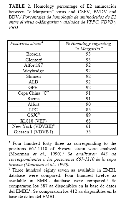
The nucleotide sequence determined allowed to deduce the aa sequence of E2 from the "c-Margarita" virus, and to do an alignment with 15 different pestivirus strains. The sequence obtained for gene E2 from "c-Margarita" virus was deposited on the EMBL database with the access number: AJ704817.
As it is shown in Table 2, the E2 sequence of Brescia and Glentorf CSFV strains were, from all analyzed, the ones showing higher homology (93 % ) with the "c-Margarita" virus sequence. These strains are on the CSFV genogroup 1.2. For Alfort strain, on the genogroup 2, the homology was 90%. With the Asiatic strains LPS and GSJC (genotype 3), the homology percents were 85 and 89 % respectively. The lowest homology percent was obtained with strain Giessen1 from BVDV type I (55%).
On the other hand, the aminoacid sequences deduced for the complete E2 from c-"Margarita" virus presented all the cysteine residues which are highly conserved in the pestivirus genomes being part of the different conformational epitopes of Glycoprotein E2.
DISCUSSION AND CONCLUSIONS
CSF is a serious problem for pig industry, in spite of the enormous efforts carried out during the last decades to eradicate the disease. Many factors make difficult the disease control, such as globalization and the increasing on trading and pig movements, the constantly growing pig populations with increased high-density areas and the increased wild pig populations acting as reservoir of the disease. Likewise, the emergency of the diseases with clinical signs related to porcine reproductive-respiratory syndrome (PRRS) and the syndrome of swine dermatitis and nephropathy complicate the diagnostic of the disease (25).
The control of the CSF in endemic areas is done by vaccination with attenuated virus strains, which not allow the differentiation between vaccinated and infected animals.
As complementary or alternative measure, depending on the severity of the outbreak and the epidemiological situation, eradication campaigns can be carried out through the policy of slaughtering animals around the points where the disease appears, together with the restriction of animal movements, as in the case of the EU countries, including
The CSF epizootic occurred in Europe in 1997 (7) was a very illustrative example of the considerable economic looses caused by the disease, mainly due to the great quantity of non infected animals slaughtered and the restriction on trading and animal movement.
The main goal approached was the growing of the "Margarita" isolate in Pk-15 cells, to facilitate its production, biological cloning and further antigenic and molecular characterization. The virus inoculation in cell monolayers did not produce detectable cytopatic effect. This situation is habitual with CSFV isolates, in which the existence of the so-called cytopatic biotype is very infrequent (21), being obtained, mainly, after several passes in cell cultures (22). Due to the lack of cytopatic effect, the virus multiplication became evident by the IIP technique using a hyperimmune serum against the virus (24). After the biological cloning by limit dilution, the recovered virus from a "c-Margarita" virus was amplified, and antigenic and genetically characterized.
The sequence amplification of CSFV by RT-PCR strategies combined with the sequencing of the products obtained allows an easy and accurate characterization of the isolates (4, 5,14, 15,18, 29). Thus, this sequence was determined for the "c-Margarita" virus amplified in cell cultures and for the parenteral virus, the "Margarita" isolate, resulting identical for both viruses.
This information allows to confirm that the "c-Margarita" virus is included in the subgroup 1.2, together with other viruses isolated in Cuba between 1993 and 1997 (4, 5).
Inside the genogroup 1.2, vaccinal strains as "C" and Pav-250 are included, and also recent isolates in
The genogroup 3 constitutes an independent branch including the viruses isolated in
As it is said, glycoprotein E2 contains the antigenic sites of CSFV recognized by neutralizing antibodies (1, 36) and it is the only protein of the virus capable of inducing the protection against the disease when it has been used in different vaccination strategies (9).
Due to the interest in using glycoprotein E2 of "c-Margarita" virus in developing a new recombinant vaccine, the antigenic characterization of this virus was carried out.
The "c- Margarita" virus was recognized by a mAb that recognizes isolates of different viruses belonging to the Genus Pestivirus (23), (37) directed against the non structural protein NS3 (more conserved among Pestiviruses). The use of mAb specifically recognizing the E2 protein from CSFV isolates, allows to confirm that "c-Margarita" virus presented equal recognizing pattern than Alfort strain, being only recognized by the specific mAb from field isolates, and not by the used specific mAb from vaccine strains.
The characterization of "c-Margarita" virus included, also, an analysis of the pathogenicity induced in pigs, after the cloning and/or amplification in PK-15 cells. The results confirmed that both, "c-Margarita" and "nc-Margarita" viruses produce acute CSF clinical signs in 6 to 8 week pigs. The virus adapted to cultures is, therefore, a highly virulent CSFV strain taking into account the classification of Mittelholzer et al. (22), due to the fact that all the pigs presented severe clinical signs of the disease, characterised by fever, progressive deterioration and death before the 15th day post infection.
To conclude, it is ratified the great interest of the results, and justify the use of the "c-Margarita" virus for the development of new vaccination strategies against the virus in
Finally, and not less important, these results have allowed to have a complete characterized native strain to be used as reference material for different purposes either in the diagnostic or for obtaining non conventional vaccine candidates .
REFERENCES
1. Andrew M, Morrissy CJ. Lenguas C, Oke P, Bruce M, Brodway M et al . Porcine Interleukin-3 enhances DNA vaccination against classical swine fever. Vaccine. 2006; 24:3241-3247.
2. Correa, P. Elaboración de un conjugado para el diagnóstico de la fiebre porcina clásica y comprobación de su especificidad y alto rendimiento. Premio Canifarma 1991 Industria Farmaceutica Veterinaria, Prem. Canif Ind Far Vet. 1991;91:47-55.
3. Díaz de Arce H, Nuñez JI, Ganges L, Barreras M, Frías MT, Sobrino F. An RT-PCR assay for the specific detection of classical swine fever virus in clinical samples. Vet Res. 1998;29:431-40.
4. Díaz de Arce H, Nuñez JI, Ganges L, Barreras M, Frías MT, Sobrino F. Molecular epidemiology of classical swine fever in Cuba. Virus Res. 1999;64:61-7.
5. Díaz de Arce H, Ganges L, Barrera M, Naranjo D, Sobrino F, Frías MT, Núñez JI. Origin and evolution of viruses causing classical swine fever in Cuba. Virus Res. 2005;112:123-131.
6. Edwards S. Survival and inactivation of classical swine fever virus. Vet Microbiol. 2000;73:175-81.
7. Edwards S, Fukusho A, Lefevre PC, Lipowski A, Pejsak Z, Roehe P, Westergaard J. Classical swine fever: the global situation. Vet Microbiol. 2000;73:103-19.
8. Frías MT, Percedo MI Reconociendo la peste porcina clásica. Manual Ilustrado, ISBN 2003;92-5-305000-4, FAO.
9. Ganges L, Barrera M, Núñez JI, Blanco I, Frias MT, Rodríguez F et al . A DNA vaccine expressing the E2 protein of CSFv elicits T cell response that can prime for rapid antibody production and confer total protection upon viral challenge. Vaccine. 2005;23: 3741-3752.
10.Ganges L, Núñez JI, Sobrino F, Borrego B, Fernández N, Frías MT, Rodríguez F. Recent advances in development of recombinant vaccines against classical swine fever virus: cellular responses also play a role in protection. Vet Journal 2007; "on line" doi:10.1016/j.tvjl.2007.01.030.
11. Gomez JC, Salguero FJ, Ruiz E, Sanchez PJ, Bautista MJ, Sierra MA. Classical Swine Fever: pathology of bone marrow. Vet Pathol 2003;40:157-63.
12.Greiser-Wilke I, Moennig V. Vaccination against classical swine fever: limitations and new strategies. Anim Health Res Rev. 2004;223-226.
13.Greiser-Wilke I, Fritzemeier J, Koenen F, Vanderhallen H, Rutili D, De Mia GM et al. Molecular epidemiology of a large classical swine fever epidemic in the European Union in 1997-1998. Vet Microbiol. 2000;77:17-27.
14.Harding M, Lutze-Wallace C, Prud'Homme I, Zhong X, Rola J. Reverse transcriptase-PCR assay for detection of hog cholera virus. J Clin Microbiol. 1994;32:2600-2.
15.Hofmann MA, Brechtbuhl K, Stauber N. Rapid characterization of new pestivirus strains by direct sequencing of PCR-amplified cDNA from the 5' noncoding region. Arch Virol. 1994;139:217-29.
16.Horzinek MC. Pestiviruses-taxonomic perspectives. Arch Virol. Suppl 1991;3:1-5.
17.Laddomada A. Incidence and control of CSF in wild boar in
18.Lowings JP, Paton DJ, Sands JJ, De Mia GM, Rutili D. Classical swine fever: genetic detection and analysis of differences between virus isolates. J Gen Virol. 1994;75:3461-8.
19.Lowings P, Ibata G, Needham J, Paton D. Classical swine fever virus diversity and evolution. J Gen Virol. 1996;77:1311-21.
20.Meyers G, Rumenapf T, Thiel HJ. Molecular cloning and nucleotide sequence of the genome of hog cholera virus. Virology. 1989;171:555-67.
21.Meyers G, Thiel HJ. Cytopathogenicity of classical swine fever virus caused by defective interfering particles. J Virol. 1995;69:3683-9.
22.Mittelholzer C, Moser C, Tratschin JD, Hofmann MA. Analysis of classical swine fever virus replication kinetics allows differentiation of highly virulent from a virulent strains. Vet Microbiol. 2000;74:293-308.
23.Moennig V, Plagemann PG. The pestiviruses. Adv Virus Res. 1992;41:53-98.
24.Moennig V. Introduction to classical swine fever: virus, disease and control policy. Vet Microbiol. 2000;73:93-102.
25.Moennig V. Floegel-Niesmann G,
26.Moormann RJ, Warmerdam PA, van der Meer B, Hulst MM Nucleotide sequence of hog cholera virus RNA: properties of the polyprotein encoded by the open reading frame spanning the viral genomic RNA. Vet Microbiol. 1990;23:185-91.
27.Parchariyanon S, Inui K, Damrongwatanapokin S, Pinyochon W, Lowings P, Paton D. Sequence analysis of E2 glycoprotein genes of classical swine fever virus: identification of a novel genogroup in Thailand. Dtsch Tierarztl Wochenschr. 2000;107:236-8.
28.Paton DJ, McGoldrick A, Belak S, Mittelholzer C, Koenen F, Vanderhallen H. et al . Classical swine fever virus: a ring test to evaluate RT-PCR detection methods. Vet Microbiol. 2000;73:159-74.
29.Paton DJ, McGoldrick A, Greiser-Wilke I, Parchariyanon S, Song JY, Liou PP. et al. Genetic typing of classical swine fever virus. Vet Microbiol. 2000;73:137-57.
30.Reed LJ, Muench H. A simple method of estimating fifty percent endpoints. Am J Hyg. 1938;27:493-7.
31.Sambrook J., Fritsch, EF., Maniatis T. Molecular cloning a Laboratory Manual. Second edition. Laboratory Press.
32.Tuero C, Díaz de Arce H, Frías MT. Elaboración de un conjugado fluorescente específico contra la peste porcina clásica y evaluación de su utilidad diagnóstica. Rev Salud Anim. 1995;17:91-93.
33.van Oirschot JT, Terpstra C. Hog cholera virus. En Virus infections of porcines. Editado por M.B. Pensaert. Elsevier 1989.
34.van Oirschot JT. Emergency vaccination against classical swine fever. Dev Biol (
35.van Oirschot JT. (2003): Vaccinology of classical swine fever: from lab to field. Vet Microbiol. 96:367-84.
36.Weiland E, Stark R, Haas B, Rumenapf T, Meyers G. y Thiel HJ. (1990): Pestivirus glycoprotein which induces neutralizing antibodies forms part of a disulfide-linked heterodimer. J Virol. 64:3563-9.
37.Weiland, F, Weiland E, Unger G, Saalmuller A y Thiel HJ. (1999): Localization of pestiviral envelope proteins E(rns) and E2 at the cell surface and on isolated particles. J Gen Virol. 80:1157-65.
38.Wengler G, Bradley DW, Collet MS, Heinz FX, Schlesinger RW, Strauss JH. (1995): Sixth report of the International Committee on taxonomy of viruses. Arch Virol Supplement. 2:223-233.
39.Wensvoort G,Terpstra C, Boonstra J, Bloemraad M y Van Zaane D. (1986): Production of monoclonal antibodies against swine fever virus and their use in laboratory diagnosis. Vet Microbiol. 12:101-8.
40.Yu X, Tu C, Li H, Hu R, Chen C, Li Z. et al. (2001): DNA-mediated protection against classical swine fever virus. Vaccine. 19:1520-5.
1 Naranjo, P. Laboratorio de control Estatal del IMV.

