


Meu SciELO
 Serviços customizados
Serviços Personalizados
Artigo
 Inglês (pdf)
 Artigo em XML
 Referências do artigo
 Enviar este artigo por email
Indicadores
-
 Citado por SciELO
Links relacionados
-
 Similares em
SciELO
Similares em
SciELO
Compartilhar

 Permalink
Revista de Salud Animal
versão impressa ISSN 0253-570X
Rev Salud Anim. v.31 n.1 La Habana jan.-abr. 2009
Short communication
POLYMERASE CHAIN REACTION DETECTION OF AVIAN LEUKOSIS VIRUS DNA IN VACCINES USED IN POULTRY
REACCIÓN EN CADENA DE LA POLIMERASA PARA LA DETECCIÓN DE VIRUS DNA DE LEUCOSIS AVIAR EN VACUNAS USADAS EN LA AVICULTURA
Ana María Acevedo, Edisleidy Rodríguez, Odalys Uffo, Damarys Relova, Julia Noda y Heidy Díaz de Arce
Departamento de Virología Animal, Dirección de Microbiología. Centro Nacional de Sanidad Agropecuaria (CENSA), Apartado 10, San José de las Lajas, La Habana, Cuba. E-mail: acevedo@censa.edu.cu
ABSTRACT
Avian leukosis viruses (ALV) provoke a variety of trasmissible bening and malign tumoral diseases affecting birds. Chickens are affected by six subgroups of ALV designs A, B, C, D, E and J of more recent world dissemination. These viruses are potential contaminants of live vaccines used in poultry. In order to research the presence of DNA from ALVs as contaminants of viral commercial vaccines to be used in poultry, different Marek´s disease vaccines were screened by a reported polymerase chain reaction (PCR) assay designed to detect all subgroups of ALVs. DNA samples extracted from seven vaccines were submitted to PCR using primers for a conserved region of env gene of HPRS-103. ALV sequences were detected in seven samples (100%). The methodology employed proved to be useful for the detection of ALVs as contaminants of imported Marek´s disease vaccines. These data suggest a high occurrence of ALVs in commercial vaccines intended for poultry disease prevention.
Key words: avian leukosis virus (ALV); polymerase chain reaction (PCR); vaccine; contamination; poultry
RESUMEN
Los virus de la leucosis aviar (ALV) provocan una variedad de enfermedades tumorales benignas y malignas transmisibles que afectan a las aves. Los pollos son afectados por seis subgrupos de ALV designados A, B, C, D, E y J de más reciente diseminación mundial. Estos virus son, además, potenciales contaminantes de vacunas vivas usadas en la avicultura. Para investigar la presencia de ADN de ALV como contaminante de vacunas virales comerciales usadas en la avicultura, monitoreamos diferentes vacunas de la enfermedad de Marek en un ensayo reportado por Reacción de la Cadena de la Polimerasa (PCR) diseñado para detectar todos los subgrupos del ALV. Las muestras de ADN extraídas de siete vacunas fueron evaluadas por PCR utilizando cebadores para una región conservada del gen de la envoltura (env) de HPRS-103. Las secuencias del ALV fueron detectadas en las siete muestras (100%). La metodología empleada resultó útil para la detección de ALV como contaminante de vacunas de Marek importadas. Nuestros datos sugieren una elevada presencia de ALV en vacunas comerciales destinadas a la prevención de enfermedad en la avicultura.
Palabras clave: virus de la leucosis aviar (ALV); reacción en cadena de la polimerasa (PCR); vacuna; contaminación; avicultura
INTRODUCTION
Avian leukosis is a disease of the birds produced by the virus of the leukosis/sarcoma group belonging to alpharetrovirus genus of the Retroviridae family (1). The ALVs that infect chickens are divided in six subgroups: A, B, C, D, E and J, which are differentiated for the antigen of the viral cover for seroneutralisation (2). The last member discovered, J subgroup, emerged at the end of the 80s (3) and it has continued in the last years with special characteristics which have caused its spread all over the world, causing great losses and it attributes a part of growth decrease in the world poultry (2,4).
These viruses are potential contaminants of live vaccines used in poultry which could produce infections in chicken populations of specific pathogens free (SPF) with the contamination of a fertile eggs proportion due to ovotransmission. The viral contamination results in the commitment of the quality of the seeds and vaccines elaborated starting from embryos of chicken or their cellular cultivations. Birds containing ALV vaccinated at very early age with biological products could develop tumors, present immunosupression and decrease humilities; thus the evaluation of the absence of contaminants from the master seed of production until the final product is of great importance.
The viral isolation in cell cultures as routine and the revealed of the viral multiplication by complement fixation test (COFAL) or ELISA to detect the presence of specific group of antigens (5,6) have been methods employed for the detection of contamination with ALV. The PCR based technology have been described for the detection of different viruses in vaccine preparations such as Newcastle disease virus (7), infectious bronchitis virus (8) and canine parvovirus (9). Fadly et al. (10,11) revealed contamination of vaccines with ALVs, specifically in two Marek´s vaccines, which confirms that these agents are potential contaminants of viral vaccines applied in poultry. This assay has meant a considerable advance due to a higher sensitivity and specificity upon differentiating the subgroups compared with ELISA. It is quicker than the viral isolation, which requires until 10 days and it needs detection by ELISA for the identification result (12).
The purpose of the present study was to research the possible presence of ALV DNA as contaminant of Marek´s disease vaccines intended to be used in poultry by ALV specific PCR assay (13).
For the PCR assay positive control, a strain of the ALV-J, the HPRS-103 donated by Dr. Venugopal of the Animal Health Institute, Compton England, was used. Seven Marek´s disease vaccines from several commercial companies were evaluated. Primary cultures of turkey´s embryo fibroblast were prepared as referred by Payne et al. (14).
To obtain cells infected with AL-J virus in order to be used as positive control in the PCR assay, cellular cultures of turkey´s embryo fibroblast of 24 hours were inoculated, as described by Fadly and Witter (5).
Such cultures were placed in incubation at 37°C and 0.5 % of CO2 and the medium changed at 24 hours, 7 days post-inoculation (PI). Observation was carried out until 12 post-inoculation, and supernant samples of the cultures at 7 days and cells and supernant were obtained at 12 days in order to evaluate viral multiplication.
The presence of ALV in infected cells was confirmed by an enzyme-linked immunosorbent assay (IDEXX, Laboratories, Inc.) (15).
DNA extraction from the cells infected with HPRS-103 strain was carried out according to the method described by Maniatis et al. (16). Extracted DNA was resuspended in 100 uL of TE buffer 1X and it was frozen to -20ºC until its evaluation.
DNA extraction of vaccine samples was carried out resuspending the vaccine bulb in 0.5 mL of saline sterile solution (500 dose).
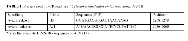
For the amplification of all ALV subgroup, the oligonucleotides (H5/AJ1) were selected (Venugopal, personnal communication) (Table 1).
The mixtures for PCR reaction in a total volume of 20 uL are as follows: 1X buffer (Promega) (50 mM KCl, 10 mM Tris-HCl pH 9.0 y 0.1% Triton® X-100), dNTP (250 uM), magnesium chloride solution (2.0 mM, Promega), 0.5 uM of each primer, 0.125 U/uL of the Taq polymerase enzyme (Promega). DNA yield and purity were determined by spectrophotometry (GenesysTM 6, USA).
As a negative control for PCR assay, DNA template was replaced by the same amount of nuclease free water (Promega, Madison, WI, USA). Amplifications were performed in MJ-ResearchTM thermocycler. The temperature profile involved a first step at 94ºC for 3 min., 30 cycles of 1 min at 94ºC, 1 min. at 54ºC and 1 min. at 74ºC. A final extension time was of 10 min. at 74ºC.
A 10 uL aliquot of each PCR product was visualized by agarose gel electrophoresis (0.8 %), containing 0.8 mg/ml ethidium bromide solution. Gels were electrophoresed for 30 min at 100 V in buffer TBE 0.5X [Tris-Borato (Tris 50mM, borate acid 50 mM] and EDTA 10 mM, pH 8.4). For the determination of the size of PCR products, molecular weight markers 1 Kb DNA ladder (Promega) with a size range from 100 to 10000 bp were included. Bands were visualized at a wave length of 312 nm and photographed.
The use of turkey's embryo fibroblast allowed to obtain virus stock of high titer according to Payne et al. (14).
Bands of expected size (1800 pb) were obtained in all Marek´s disease vaccines evaluated (Fig.1).

These results coincide with reports of Fadly et al. (10,11) and Silva (18) who revealed the presence of ALVs in vaccines, specifically ALV-A in a commercial Marek´s disease vaccine and they alerted about the need of using sensitive and specific methods for the detection of those viruses as contaminants of vaccines. Also, later Zavala and Cheng (19) carried out the identification by PCR and the characterization of ALVs in several commercial Marek´s disease vaccines and evaluated their effect in experimentally vaccinated chickens. These authors recommended the employment of these methods as complementary procedures for the detection of these viruses in the commercial vaccines destined to poultry.
Although at present, subgroups A, B (19) and E (20) of the ALVs were only identified as contaminant of vaccines, given the wide spread of the ALV-J also infecting leghorn chickens and turkeys (21,22,23), it is very important to have more sensitive and specific methods in order to reveal their presence in vaccinal products as well as in imported birds in order to avoid the dissemination of the disease in the country.
In our work, DNA extraction was carried out directly from vaccine bulb, it is very important because it reduces time to get the results. Authors like Fadly et al. (24) reveled the presence of endogenous and exogenous viruses afterwards cellular cultures from a commercial Marek´s disease by PCR.
The results obtained showed that the methodology described is a valuable tool for the detection of leukosis viruses in veterinary vaccines and could become a practical alternative for the current in vivo test for vaccine control imported or produced in our country. Our data suggest a high occurrence of ALVs in commercial viral vaccines intended for poultry disease prevention.
REFERENCES
1. Payne LN, Fadly AM. Leukosis/Sarcoma group. In: Diseases of Poultry, 10th ed., BW Calnek, HJ Barnes, CW Beard, LR McDougald and YM Saif, eds. Iowa State University Press, Ames, IA. 1997; 414-466.
2. Payne LN. HPRS-103 a retrovirus strikes back. The emergence of Subgroup J Avian Leukosis Virus. Avian Pathol. 1998;27:536-545.
3. Payne LN, Brown SR, Bumstead N, Howes K, Frazier JA, Thouless ME. A novel subgroup of exogenous Avian Leukosis Virus in chickens. J Gen Virol. 1991;72:801-807.
4. Venugopal K. Avian Leukosis Virus Subgroup J: a rapidly evolving group of oncogenic retroviruses. Review Res Vet Sci. 1999;67:113-119.
5. Fadly AM, Witter RL. Oncornaviruses: Leukosis/Sarcomas and Reticuloendotheliosis. In: SB Hitchner, CH Domermuth, HG Purchase and JE Williams (eds.), Isolation and Identification of Avian Pathogens. Am Assoc Avian Pathol. Kenneth Square, P.A.1998;185-195.
6. Anónimo. British Farmacopeia Anónimo, 1994. World Health Organization Expert Committee on Biological Standardization, requeriments for measles, mumps and rubella vaccines and combined vaccines (live). Requeriment for biological substances 47, WHO Tech Rep Ser. 1998;840:100-207.
7. Stäuber N, Brechtbühl K, Hofmann MA. Detection of Newcastle disease virus in poultry vaccines using the polymerase chain reaction and direct sequencing of amplified cDNA. Vaccine. 1995;13:360-364.
8. Falcone E, D´Amore E, Di Trani L, Puzelli S, Tollis M. Detection of avian infectious bronchitis virus in poultry vaccines by polymerase chain reaction. ATLA. 1996;24:136.
9. Senda M, Parrish CR, Harasawa R, Gamoh K, Muramatsu M, Hirayama N, Itoh O. Detection by PCR of wild-type canine parvovirus which contaminates dog vaccines. J Clin Microbiol. 1995; 33:110-13.
10.Fadly AM. Avian tumor viruses and their economic impacts. Preceedings XII Moscow International Congress of Small Animal Diseases. 2004a; 26.
11.Fadly AM, Silva RF, Hunt HD. Isolation of subgroup A avian leukosis virus from commercial Marek´s disease vaccines. In: International Marek´s Disease. Symposium Abstracts and Proceedings. 2004b; Paper No. p.8.
12.Fadly AM. Isolation and identification of avian leukosis viruses: a review. Avian Pathol. 2000; 29:529-535.
13.Venugopal K, Smith LM, Howes K, Payne LN. Antigenic variants of J subgroup avian leukosis virus: sequence analysis reveals multiple changes in the env gene. Journal of General Virology. 1998;79:757-766.
14.Payne LN, Howes K, Gillespie AM, Smith LM. Host range of Rous Sarcome virus pseudotype RSV (HPRS-103) in 12 avian species: support for a new avian retrovirus envelope subgroup, designated J. J. of Gen. Virol. 1992;73: 2995-2997.
15Payne LN, Gillespie AM, Howes K. Unsuitability of chicken sera for detection of exogenous ALV
by the group-specific antigen ELISA. Veterinary Record. 1993:555-557.
16.Maniatis TE, Fritsch EF, Sambrook J. Molecular cloning: a laboratory manual. Cold Spring Harbor, New York. 1982.
17.Bai J, Payne LN, Skinner MA. HPRS-103 Exogenous avian leukosis virus subgroup J has env gene related to those of endogenous elements EAV-0 and ES1 and B element found previously only in sarcoma viruses. J. of Virology. 1995; 69:779-784.
18.Silva RF, Fadly AM, Taylor SP. Development of a Polymerase Chain Reaction to Differentiate Avian Leukosis Virus (ALV) Subgroups: Detection of an ALV Contaminant in Commercial Marek's Disease Vaccines. Avian Dis. 2007;51(3):663-667.
19.Zavala G, Cheng S. Detection and characterization of avian leukosis virus in Marek`s vaccines. Avian Dis. 2006;50(2):209-15.
20.Fadly AM, Silva RF, Hunt HD, Pandiri A, Davis C. Isolation and characterization of an adventitious avian leukosis virus isolation from commercial Marek´s disease vaccines. Avian Dis. 2006;50:380-385.
21.Mays J, Bacon LD, Pandiri AR, Fadly AM. Response of White Leghorn Chickens of Various B Haplotypes to Infection at Hatch with Subgroup J Avian Leukosis Virus. Avian Dis. 2005;49(2):214-219.
22.Venugoplal K, Howes K, Flanner Y, Payne LN. Subgroup J avian Leukosis viruses infection in turkey: induction of rapid onset tumours by acutely transforming virus strain 966. Avian Pathol. 2000;29:319-25.
23.Venugopal K, Howes K, Payne LN. Isolation of acutely transforming subgroup J avian Leukosis viruses that induce erythroblastosis and myelocytomatosis. Avian Pathol. 2000; 29:327-332.
24.Fadly AM, Silva RF, Hunt HD. Detection of exogenous and endogenous avian leukosis virus in commercial Marek´s disease vaccine. In: Proceeding of the 170 th Annual Meeting of the United States Animal Health Association, October 9-16, San Diego, California. 2003;524-525.
(Recibido 18-11-2008; aceptado 20-1-2009)
Rev. Salud Anim. Vol. 31 No. 3 (2009)

