


Mi SciELO
 Servicios personalizados
Servicios Personalizados
Revista

Articulo
 Inglés (pdf)
 Articulo en XML
 Referencias del artículo
 Enviar articulo por email
Indicadores
-
 Citado por SciELO
Links relacionados
-
 Similares en
SciELO
Similares en
SciELO
Compartir

 Permalink
Revista de Salud Animal
versión impresa ISSN 0253-570Xversión On-line ISSN 2224-4700
Rev Salud Anim. vol.37 no.3 La Habana sep.-dic. 2015
ORIGINAL ARTICLE
Cell surface characteristics and adherence of typeable and non-typeable strains of Streptococcus suis from pig farms in Cuba
Características de la superficie celular y adherencia de cepas tipificables y no tipificables de Streptococcus suis de granjas porcinas en Cuba
Ivette Espinosa Castaño, Michel Arias Baéz, Evelyn Lobo Rivero, Siomara Martínez Marrero
Department of Microbiology, National Center for Animal and Plant Health, CENSA. Apdo. 10, San José de las Lajas, Mayabeque, Cuba. E-mail: espinosa@censa.edu.cu.
ABSTRACT
Streptococcus suis infection is considered a major problem in the swine industry. There are 35 known serotypes of S. suis based on the capsular polysaccharides (CPS) on the cell surface. Serotype 2 is the most virulent in pigs and humans. Some S. suis strains do not agglutinate with any of the typing antisera and are identified as non-typable strains, which have generally been viewed as organisms that do not cause important clinical infections. Previous studies have shown differences in the composition and properties of the cell surface among these strains. The aim of this study included the characterization of cell hydrophobicity, ability to adhere to plates, and autolysis over time of typeable and non-typeable strains of S. suis from farms of the Cuban western region. The non-typeable strains showed a hydrophobic surface and ability of adhesion to plates. In this work, a fragment of atl gene encoding for the most important autolysine in S. suis was detected in serotypes 2, 9 and non-typeable strains. Correlation between cell adherence, hydrophobicity, and autolysis was only detected in two non-typeable strains, indicating that the non-encapsulated strain was more hydrophobic than the encapsulated strain and suggesting a potential ability to form biofilms. The ability to form biofilms is not required for virulence, but it does contribute towards long-term colonization, transmission and difficulties to eradicate these infections. These results indicated that non-typable strains should be considered when implementing measures to control the pathogenesis of the infection with S. suis in Cuban farms.
Key words: Streptococcus suis, hydrophobicity, adherence, autolysine.
RESUMEN
La infección por Streptococcus suis constituye uno de los problemas de mayor importancia en la industria porcina. Existen 35 serotipos de S. suis basados en la presencia de polisacáridos capsulares (cps) sobre la superficie celular. El serotipo 2 es el más virulento para cerdos y humanos. Existen cepas que no aglutinan con algunos de los antisueros; estas se denominan cepas no tipificables y se consideran sin importancia clínica. Los estudios previos han mostrado diferencia en la composición y en las propiedades de la superficie celular de las cepas tipificables y no tipificables. El objetivo de este estudio incluyó la caracterización de la hidrofobicidad celular, la habilidad de adherirse a placa y la autolisis celular en el tiempo de cepas tipifacables y no tipificables procedentes de granjas de Cuba. Las cepas no tipificadas mostraron una superficie hidrfóbica y una capacidad de adherencia. Se detectó un fragmento del gene atl, que codifica para la más importante autolisina de S. suis, en cepas de los serotipos 2 y 9, así como en cepas no tipificables. La correlación entre hidrofobicidad, adherencia y autolisis celular fue detectada en dos cepas no tipificables, que indicaron que las mismas presentan una superficie hidrofóbica que puede contribuir a la formación de biopelículas y, de este modo, perpetuar la infección en las granjas. Aunque la producción de biopelículas no es una expresión necesaria para la virulencia, sí contribuye a la colonización a largo plazo, a la transmisión de la infección y a las dificultades para su erradicación. Estos resultados indican que los aislados no tipificables detectados en los laboratorios deben ser considerados cuando se implementan medidas para el control de la patogénesis de la infección por S. suis en granjas de Cuba, como podría ser el uso de antibiótios.
Palabras clave: Streptococcus suis, hidrofobicidad, adherencia, autolisina.
INTRODUCTION
Streptococcus suis is an important pathogen of pigs that causes high mortality and is responsible for considerable economic losses in the porcine industry. S. suis is also considered an important zoonotic pathogen causing a variety of life-threatening infections that include meningitis, arthritis and septicaemia (1, 2, 3). There are 35 known serotypes of S. suis: 1-34 and 1/2. Serotype 2 is the most virulent, and is commonly associated with disease in pigs and humans (4, 5). Although other serotypes are reported, globally, the predominant S. suis serotypes isolated from clinical cases in pigs are, in decreasing order, serotypes 2, 9, 3, 1/2 and 7. However, 15.5% of non-typeable strains by serotyping, not considered important in S. suis pathology due to the large number of non-typebeable strains, are also isolated, especially from healthy pigs (6).
Different strategies based on vaccines and antimicrobials have been used for controlling S. suis infection; however, more persistent S. suis infections are achieved in vivo (7), and hence S. suis infections may be difficult to treat (8). Non-typeable S. suis strains have generally been considered as organisms that do not cause important clinical infections; however, previous studies have shown the unencapsulated serotype 2 and non-typeable strains to be more adhesive than the encapsulated strains (9).
Attachment of microbial cells to biotic or abiotic surfaces depends on several factors such as Brownian movement, van der Waals attraction, gravitational forces and surface electrostatic charges. Another important factor is the cell hydrophobicity. Hydrophobic cells play a key role in the formation of biofilms on tissues; the biofilms are an important problem because of the strong resistance of these microbial structures to drugs (10).
The non-typeable strains of S. suis are being taken into account more each time in the last years (6): a similar fact happens with nontypeable strains of other species of the genus Streptococcus recognized important for human health like S. pneumonaie (11).
The aim of this study was to characterize typeable and nontypeable isolates of Streptococcus suis from pig farms in the Cuban western region, in relation to those characters contributing to persistence such as cell hydrophobicity, adherence ability, and autolysin activity.
MATERIALS AND METHODS
Bacterial strains and culture conditions: The S. suis strains used in the present study are shown in Table 1. All strains were clinical isolates from lung samples of pneumonia diseased pigs previously characterized at the Bacteriology Laboratory of the National Centre for Animal and Plant Health over the period 2002-2014. The samples were cultured on Columbia Blood Agar (Oxoid) containing 5% (v/v) sheep blood and incubated aerobically at 37oC for 48 h. All the isolates were biochemically typed using the API 20 STREP test kit (Bio Mérieux, France). Serotyping was carried out by the coagglutination test using rabbit hyperimmune sera against reference strains of all serotypes of S. suis, as previously described by Higgins and Gottschalk 1990 (12).

All S. suis-like strains were confirmed by PCR with the amplification of a 294bp fragment of 16S rDNA gene using S. suis species-specific primers (13). The colony of each isolate from blood agar plates was transferred to 50 µl of nuclease-free water and boiled in a heating block at 100oC for 5 min. After centrifugation at 5000 g for 5 min, the supernatant was collected and stored at -20oC until use. The PCR assays were carried out on a final reaction volume of 25 µl and using PCR Master Mix (Invitrogen) according to the manufacturer`s instructions; 5 µl of DNA sample was used in each reaction. The primers were synthesized by the Center of Genetic Engineer and Biotechnology (CIGB). They were used at a concentration of 0.2 µM .Amplification was done in the PCR system (Mastercycler); each isolate was tested twice under the same conditions. PCR amplicons were electrophoresed on 2% agarose gels and visualized by UV transillumination after ethidium bromide staining (0.5 µg/ml). The strains were maintained as stock cultures in Todd-Hewitt broth (THB, Oxoid) containing 20% glycerol at 20oC.
Detecting gene atl fragment
The gene atl encoding for the autolysine protein was identified by PCR as previously described by Cun-Xiang et al. (14).
Autolysis assay
The autolysis assay was carried out as previously described (14). Cells were grown to stationary phase (1×10⁸ CFUml-1) in THB at 37°C and pelleted by centrifugation. The cells were washed once and resuspended in 50 mM Tris-HCl (pH 7.0) containing 0.05% Triton X-100 to an absorbance 600 of 0.6. The cell suspensions were incubated at 37°C with gentle shaking. The decrease in absorbance was monitored
Surface hydrophobicity assay
Surface hydrophobicity was assessed using the modified salting aggregation test (SAT) assay (15). S. suis cultures in THB incubated at 37°C to late-log phase (1×10⁸ CFUml-1) were harvested, washed twice with PBS, resuspended in PBS (pH 7,2), and `salted out' (aggregated) by combining 25 µl volumes with 25 µl volumes of ammonium sulphate (NH4)2SO4 solutions at different concentrations (0,2, 0,5, 1, 1,5, 2, 2,5, 3 and 4 mol l-1) on microscope slides followed by agitation for 4 min at room temperature. The lowest final concentration of (NH4)2SO4 causing aggregation was recorded as the SAT value and classified as follows: <0.1 mol l-1= highly hydrophobic; 0.1-1.0 mol l-1=hydrophobic and >1.0 mol l-1= hydrophilic. The assays were performed in duplicate at two separate occasions.
Microtitre plate adherence assay
The cultures of each strain in THB containing 2.108 bacteria was diluted into wells of polystyrene plates containing the minimal medium (MM) described by Grenier et al.(2009) (15). After 24 hours, the plates were washed three times with sterile double-distilled water. They were allowed to air-dry for 1 hour at 42°C and then stained with 1% crystal violet (Sigma). They were quantified by adding 30% acetic acid (Sigma) and measuring the absorbance at 492 nm using a microtiter plate reader (SUMA, PR-621, Cuba). Wells with sterile broth medium served as controls. The isolates were classified as described by Christensen et al. 1985 (16).
Statistics and Reproducibility of results
The microtiter plate assays were performed in duplicate wells. All experiments were repeated independently three times. One-way ANOVA was used to compare groups followed by Bonferroni's multiple comparison post-test by using Info Stat Ver. 1.1 (2002). The significance level was p< 0.05.
RESULTS AND DISCUSSION
Serotyping, a procedure that relies on the composition of capsular material, is an important step in the identification of S. suis (6, 12). Serotype 2 is most frequently associated with pathology, although other serotypes are also the source of many infections (12). The non-typeable isolates are increasingly more reported associated with pneumonia cases in pigs. More specifically, Wei et al. (17) characterized 407 strains of S. suis isolated from diseased pigs in China and recovered 5.4% of nontypeable isolates. In Canada, between 12% and 20% of strains recovered from diseased pigs were untypeable (18). In a previous study in Cuba, non-typeable isolates were also recovered from pneumonic pigs (19). To gain clarity on the characteristics of Cuban isolates recovered from non-invasive disease sources and presumptively identified as typeable and non-typeable, one genotypic and two phenotypic assays were performed.
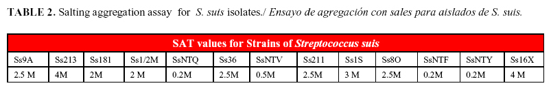
There are various methods of recognition of hydrophobic properties of microorganisms. In this study, the SAT values were expressed as the minimal molar concentration of (NH4)2SO4 necessary to cause agglutination of the bacterial cells (Table 1). SAT indices ranged from 0.2 to 4 M (Table 2). The non-typeable isolates (SsNTF, SsNTQ and SsNTY) agglutinated in the presence of the most low concentration of (NH4)2SO4, indicating their hydrophobic surface, while the typeable isolates corresponding to serotypes1, ½, 2, 3, 8, and 16 only agglutinated showing a hydrophilic surface in the presence of (NH4)2SO4 concentrations higher than 2 M. Bonifait et al. (20) described the lack of a capsule in the non-typeable isolates to correlate with a greater cell-surface hydrophobicity when compared with that of capsulated serotype 2 isolates, thereby favoring the cell adherence and biofilm formation.
The average absorbance at 492nm obtained for isolates of S. suis in the assay for adherence to plates are shown in Figure 1. Significant differences (p < 0.05) were observed. Only the non-typeable isolates showed some ability to adhere to plates. One isolate (SsNTF) was classified as strongly adherent (SsNTF), two isolates (SsNTQ and SsNTV) as moderately adherent, while the rest were weakly or non-adherent.
According to our results, only few adherence-producing isolates could be detected, which was in agreement with the observations of other researchers who found non-typeable isolates producing adherence. The polystyrene microtitre plate assay measures the amount of biological material sticking to the surface of a container after the bacteria have been cultured in it. It is not clear if this assay is an estimator of an increase in biofilm biomass or if it detects an increased ability of the biofilm material to attach to the sides of the plastic wells (21). By the other way, Bonifait et al. (22) showed that supplementing the culture medium with fibrinogen induced biofilm formation by different serotypes of S. suis in a dose-dependent manner.
Nowadays, it is well accepted that, in most environments, microorganisms can switch from a free-living state to a sessile mode of life to form biofilms displaying specific properties. Among these specific properties is an enhanced tolerance to all sort of adverse conditions including desiccation and high concentrations of antimicrobial agents such as biocides, antibiotics, and antifungal compounds. The ability to form biofilms is not required for virulence, but it does contribute towards long-term colonization, transmission and difficulties to eradicate these infections (23, 24, 25).
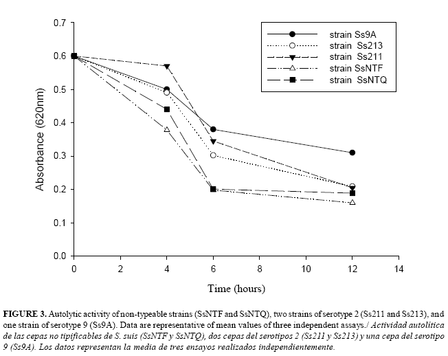
The results of this study confirmed the presence of a fragment atl corresponding to start codon of the autolysine in serotype 2 strains and in one isolate of serotype 9, but they also revealed the presence of this fragment in non-typeable isolates (Figure 2). Autolysins are bacterial cell wall hydrolytic enzymes that mediate an important role in cell wall metabolism during the antibiotic-induced lysis and may function as important virulence factors for bacterial pathogens (11).
The autolytic activity of strains of S. suis was determined, and the absorbance of strains where it was possible to detect atl gene is shown in the Figure 3. Results of the autolysis assay showed an absorbance decrease over time for the whole-cell suspensions in buffer in all the strains evaluated. However, the non-typeable S. suis strains reached the lowest values of absorbance over time, while S. suis serotype 9 showed the highest value. The non-typeable strains were also the most adherent in the plate assay. Probably, the autolysine also contributes to the biofilm formation because the developmental process requires the release of extracellular polymeric substances (EPS) by the biofilm forming community (8).
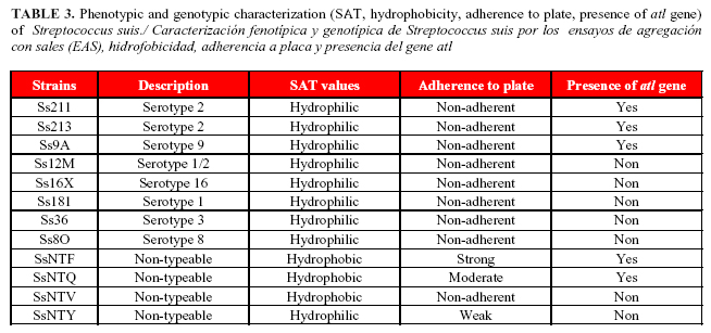
It was interesting that the correlation between hydrophobicity, adherence and presence of atl gene was only detected in the non-typeable strains (Table 3). There was no correlation between adherence and hydrophobicity in typeable strains.
The non-typeable isolates showed a hydrophobic surface in the SAT test, ability to adhere to the plate, presence of atl and autolysis activity; all these properties favor the cell persistence through the biofilm formation by these strains.
For years, the attempts for controlling infections by S. suis have been focused only on typeable strains because non-typeable strains have generally been regarded as organisms which do not cause important clinical infections. It is also difficult to be certain that these strains were already non-encapsulated when causing disease, or if they had lost their CPS during isolation and culture. It has been reported that 34% of isolates belonging to serotype 1/2 or 2 recovered from cases of endocarditis in Japan were non-encapsulated due to deletions and insertions in the genes of the CPS locus (6). These aspects suggest to consider the non-typeable isolates when implementing measures to control infections by S. suis in Cuban farms.
REFERENCES
1. Baums CG, Verku¨hlen GJ, Rehm T, Beyerbach M, Pohlmeyer K, Weigand PV. Prevalence of Streptococcus suis Genotypes in Wild Boars of Northwestern Germany. Appl Environ Microbiol. 2007;73:711-717.
2. Chanter N, Jones PW, Alexander TJ. Meningitis in pigs caused by Streptococcus suis-a speculative review. Vet Microbiol. 1999;36:39-55.
3. Christensen G, Sorensen V, Mousing J. Diseases of swine. In: Straw BE, D'Allaire S, Mengeling WL, Taylor DJ, eds. Diseases in Swine, 8th Edn. Oxford: Blackwell Science 1999; pp. 913-940.
4. Gottschalk M, Higgins R, Boudreau M. Use of polyvalent coagglutination reagents for serotyping of Streptococcus suis. J Clin Microbiol. 1993;31:2192-2194.
5. Gottschalk M, Segura M, Xu J. Streptococcus suis infections in humans: the Chinese experience and the situation in North America. Anim Health Res Rev. 2007;8:29-45.
6. Guillaume GD, Jean-Philippe A, Jianguo X, Segura M, Gottschalk M. Streptococcus suis, an important pig pathogen and emerging zoonotic agent-an update on the worldwide distribution based on serotyping and sequence typing. Emerging Microbes and Infections 3, e45; doi:10.1038/emi. 2014.
7. Brown MR, Allison DG, Gilbert P. Resistance of bacterial biofilms to antibiotics: a growth-rate related effect? J Antimicrob Chemother. 1988;22:777-780.
8. Brady RA, Leid JG, Calhoun JH, Costerton JW, Shirtliff ME. Osteomyelitis and the role of biofilms in chronic infection. FEMS Immunol Med Microbiol. 2008;52:13-22.
9. Benga L, Goethe R, Rohde M, Valentin-Weigand P. Non-encapsulated strains reveal novel insights in invasion and survival of Streptococcus suis in epithelial cells. Cell Microbiol. 2004;6:867-881.
10.Krasowska A, Sigler K. How microorganisms use hydrophobicity and what does this mean for human needs? Frontiers in Cellular and Infection Microbiology. 2014. http://www.frontiersin.org.
11.Sa´-Lea~o R, Simo~es AS, Nunes S, Sousa NG, Fraza~o N, et al. Identification, prevalence and population structure of non-typable Streptococcus pneumoniae in carriage samples isolated from preschoolers attending day-care centres. Microbiol. 2006;152:367-376.
12.Higgins R, Gottschalk M. An update on Streptococcus suis identification. J Vet Diagn Invest. 1990;2:249-252.
13.Marois C, Le Devendec L, Gottschalk M, Kobisch M. Detection and molecular typing of Streptococcus suis in tonsils from live pigs in France. Can J Vet Res. 2007;71:14-22.
14.Cun-Xiang J, Hong-Wei G, Cheng-Ping L. Characterization and Functional Analysis of atl, a Novel Gene Encoding Autolysin in Streptococcus suis. J Bacteriol. 2011;1464-1473.
15.Basson A, Flemming LA, Chenia HY. Evaluation of adherence, hydrophobicity, aggregation characteristics and biofilm development of Flavobacterium johnsoniae-like isolates from South African aquaculture systems. Microb Ecol. 2008;55:1-14.
16.Grenier D, Grignon L, Gottschalk M. Characterization of biofilm formation by a Streptococcus suis meningitis isolate. Vet J. 2009;179:292-295.
17.Wei Z, Li R, Zhang A, Hongkui He, Yafeng Hua, et al. Characterization of Streptococcus suis isolates from the diseased pigs in China between 2003 and 2007. Vet Microbiol. 2009;137:196-201.
18.Gottschalk M, Lacouture S, Bonifait L, David R, Nahuel F, Daniel G. Characterization of Streptococcus suis isolates recovered between 2008 and 2011 from diseased pigs in Qu´ebec, Canada. Vet Microbiol. 2013;162(2-4):819-825.
19.Espinosa I, Báez M, Corona B, Chong D, Lobo E, Martínez S. Molecular typing of Streptococcus suis from pigs in Cuba. Biotecnol Aplic. 2013;30(1):39-34.
20.Bonifait L, Gottschalk M, Grenier D. Cell surface characteristics of nontypeable isolates of Streptococcus suis. FEMS Microbiol Lett. 2010;311(2):160-166.
21.Siva W, Xiaojin L, Gunawardana M, Maguire K, Guerrero-Given D, Schaudinn C, et al. Beta- Lactam Antibiotics Stimulate Biofilm Formation in Non-Typeable Haemophilus influenzae by Up-Regulating Carbohydrate Metabolism. PLOS ONE. 2014;9(7). http://www.plosone.org.
22.Bonifait L, Louis G, Daniel G. Fibrinogen Induces Biofilm Formation by Streptococcus suis and Enhances Its Antibiotic Resistance. Applied Environ Microb. 2008;4969-4972.
23.Parsek MR, Singh PK. Bacterial biofilms: an emerging link to disease pathogenesis. Annu Rev Microbiol. 2003;57:677-701.
24.Yifan X, Kreth J. Role of LytF and AtlS in eDNA Release by Streptococcus gordonii. PLOS ONE. 2013. http://www.plosone.org. 8(4).
25. Lebeaux D, Ashwini C, Olaya R, Christophe B. From in vitro to in vivo Models of Bacterial Biofilm-Related Infection. Pathogens. 2013;2:288-356.
Recibido: 20-6-2015.
Aceptado: 25-10-2015.

