


Mi SciELO
 Servicios personalizados
Servicios Personalizados
Revista

Articulo
 Inglés (pdf)
 Articulo en XML
 Referencias del artículo
 Enviar articulo por email
Indicadores
-
 Citado por SciELO
Links relacionados
-
 Similares en
SciELO
Similares en
SciELO
Compartir

 Permalink
Revista de Salud Animal
versión impresa ISSN 0253-570Xversión On-line ISSN 2224-4700
Rev Salud Anim. vol.39 no.2 La Habana may.-ago. 2017
ORIGINAL ARTICLE
Occurrence of bovine dermatophilosis in Huambo province, Angola
Ocurrencia de dermatofilosis bovina en la provincia Huambo, Angola
Aires Walter1, Armindo Paixão1, Siomara Martínez2, Ivette Espinosa2*
1 Faculty of Veterinary Medicine, Jose Eduardo Dos Santos University, Angola.
2 Departament of Microbiology-Epidemiology, National Center for Animal and Plant Health (CENSA), Apartado 10, San José de las Lajas, Mayabeque, Cuba.
ABSTRACT
Dermatophilosis is an exudative pustular dermatitis that affects cattle, sheep, horses, goats, dogs, cats, reptiles, and occasionally humans. This disease is caused by Dermatophilus congolensis, a pleomorphic Gram-positive actinomycete. According to the Veterinary Services, the highest incidence of the disease in Angola was identified in the provinces Huambo, Kwanza Sul and Malang during May 2013 and April 2015, but only supported on clinical sings. In the present work ninety crust samples were taken from animals with lesions on the skin in Huambo province, in the period May 2013-April 2015. The bacteria were isolated following Haalstra´s method. Presumptive colonies of the Dermathophilus genus were subjected to Gram stain, biochemical identification, and molecular detection by PCR. Eight Dermathophilus congolensis isolates were tested for their sensitivity to eleven different antibiotics. A total of 30 samples showed typical colonies of the Dermatophilus genus. The amplicon of 817 bp corresponding to the 16S rRNA region was obtained in the 30 isolates suspecting of being D. congolensis. Drug sensitivity test revealed that Amoxicillin/clavulanate, Clindamycin, Erythromycin, Lincomycin, Azithromycin, Rifampicin, and Enrofloxacin were highly effective against the eight D. congolensis isolates. Observation of lesions and microbiologic diagnosis using biochemical and molecular techniques allowed confirming bovine dermatophilosis in the region of Huambo, Angola.
Key words: Dermatophilus congolensis, bovine, diagnosis, antimicrobial resistance.
RESUMEN
Dermatofilosis es una dermatitis pustular exudativa que afecta al ganado bovino, ovejas, caballos, cabras, perros, gatos, reptiles y, ocasionalmente, a humanos. Esta enfermedad es causada por Dermatophilus congolensis, un actinomiceto pleomórfico Gram-positivo. Según los Servicios Veterinarios, la mayor incidencia de la enfermedad en Angola se identificó en las provincias Huambo, Huila, Kwanza Sul y Malang durante mayo de 2013 y abril de 2015, pero solo se basó en lesiones clínicas. En el presente trabajo se obtuvieron 90 muestras de costra de animales con lesiones en la piel en Huambo, Angola, en mayo de 2013 y abril de 2015. Las bacterias se aislaron siguiendo el método de Haalstra. A las colonias presuntivas del género Dermathophilus se les realizó tinción de Gram, identificación bioquímica y detección molecular por PCR. Se seleccionaron ocho aislamientos de Dermathophilus congolensis para su sensibilidad a 11 antibióticos diferentes. Un total de 30 muestras mostraron colonias típicas del género Dermatophilus. El amplicón de 817 pb correspondiente a la región ARNr 16S se obtuvo en treinta cepas sospechosas de D. congolensis. La prueba de sensibilidad a las drogas reveló que Amoxicilina/clavulanato, Clindamicina, Eritromicina, Lincomicina, Azitromicina, Rifampicina y Enrofloxacina fueron altamente eficaces contra los ocho aislados de D. congolensis. La observación de lesiones y la identificación por técnicas bioquímicas y moleculares confirmaron la dermatophilosis bovina en la región de Huambo, Angola.
Palabras clave: Dermathophilus congolensis, bovino, diagnóstico, resistencia antimicrobiana.
INTRODUCTION
Dermatophilosis is a contagious skin disease caused by Dermatophilus congolensis. Such disease is characterized by exudative, proliferative or hyper keratotic dermatitis, accompanied by the production of crusts and foliculitis (1,2,3). The causative organism is an aerobic Gram-positive bacterium that produces motile zoospores. This bacterium invades the skin and causes an acute, subacute or chronic skin disease. This disease affects cattle, sheep, horses, goats, wild animals, and man, being considered a zoonosis (4,5,6).
This disease, which has a worldwide distribution, prevails in tropical areas and it is associated to humid environments and other factors. The first case was reported in Belgian Congo, the current Democratic Congo, by Van Saceghem in 1915, and since then, the bacterium has been isolated from animal infections chiefly in Africa (Kenya, Ethiopia, Tanzania, Nigeria, and South Africa), Asia (Turkey, India and China), and Central and South America (Argentina, Uruguay and Brazil). The disease has also been isolated in Australia, the United States (New York, Kentucky, Florida, and Texas), Canada, and Europe (France, Spain, and Germany), but as a chronic endemic disease or, more rarely, as an acute and epidemic infection (7,8,9).
In Angola, the dermatophilosis is considered as a re-emerging disease. The costs are calculated in terms of productivity losses, reproduction problems in cows with severe vulva infection, and also chemotherapy and chemoprophylaxis costs, as well as losses due to culling and death of the infected animals. The disease has occurred in Angola with the highest incidence in the provinces of Huambo, Kwanza Sul and Malang, as it was reported by the General Directory of the Veterinary Services of Angola, (10). However, this information has been supported only on clinical examination, but there are also other diseases affecting cattle and producing similar injuries to the bovine skin. For these reasons, the application of a polyphasic diagnosis combining phenotypic and molecular assays has been necessary.
This work was aimed at confirming the presence of D. congolensis in animals with clinical lesions, similar to the dermatophilosis in the bovine farms in Huambo province, Angola, by using microbiological, phenotypic and molecular methods.
MATERIALS AND METHODS
A randomized, cross-sectional and prospective study was conducted on farms and communities with traditional systems and extensive breeding located in five municipalities in Huambo province, Angola. The total cattle population consisted of 752 animals, which included the Nelore race and indigenous cattle. The samples were taken in two stages in the period May 2013–April 2015. Animals with skin lesions were selected specifically for the production of crust, purulent discharge and alopecia, similar to those described by Topa and Iseensee (11) and Tavanaeimanesh et al. (12).
A prevalence of 30 % based on previous studies was considered (13), and the minimum sample size was calculated with 95 % confidence and 5 % estimated error. The following equation was used: n=Nz2pq/dz(N-1)+z2pq (14). Based on this equation, the minimum sample size was 73 bovines for obtaining the ratio of animals positive for D. congolensis.
Isolation of Dermatophilus congolensis from the infected scabs
The samples were collected in sterile plastic tubes by scraping the lesions located on different parts of the animal body and conserved in refrigeration, maintaining the biosecurity in the whole process of specimen collection. Then they were transported to the Veterinary Medical Microbiology Laboratory, Faculty of Agriculture and Veterinary Medicine of the University of Brasilia, Brazil. The containers with specimen were properly closed and decontaminated in each farm.
Dermatophilus species isolation was carried out by the Haalstra’s method with minor modifications (15). The samples were fragmented with a scalpel, moistened with sterile saline solution, and stained by the Gram stain. Small pieces of scab were placed in a vial with 1 ml of sterile distilled water and stored at room temperature for 3-4 hours. The opened vial was placed for 15 minutes in a candle jar. The samples of the liquid surface were extracted with a bacteriological loop and cultured on Blood Agar Base (Oxoid) supplemented with 5 % sheep blood and incubated at 37°C under 5 % CO2 tension in an incubator for 72 hours.
The colonies with the presumptive characteristics of the Dermatophilus genus (tiny, smooth, creamy white-colored, beta-hemolytic and adherent) were sub-cultured to obtain pure cultures. Subsequently, identification was done by using Gram stain and two biochemical tests, catalase with 3 % hydrogen peroxide and the oxidase test (1 % dihydrochloridetetramethyl-p-phenylenediamine) (Probac, Brasil). Ten strains were selected for their identification by the biochemical tests. The fallowing test were used: indole production, nitrate reduction, gelatin hydrolysis, urea production, and the use of glucose, fructose, maltose, sucrose, salicin, sorbitol, xylose, mannitol, and lactose. The strain cultures were incubated at 37°C and observed for five days for any activity by the organisms.
Antibiotic susceptibility test
The cultures of the eight strains were performed on Muller Hinton Agar medium (Oxoid) supplemented with 5 % blood. The antibiotics used were Amoxicillin + clavulanate (10 ug, Oxoid), Clindamycin (2 ug, Oxoid), Streptomycin (10 ug, Oxoid), Sulfazotrim (30 ug, Oxoid) Erythromycin (15 ug, Oxoid), Lincomycin (2 ug, Oxoid), Azithromycin (15 ug, Oxoid), Rifampicin (5 ug, Oxoid), Enrofloxacin (10 ug, Oxoid), and Sulfonamides (250 ug). The inhibition zone diameter was measured after 48 h incubation at 37°C. The results were analyzed according to the Clinical and Laboratory Standards Institute (16), specifically for Gram positive microorganisms.
D. congolensis detection by PCR
Bacterial DNA was extracted from colonies which had been resuspended in 200 µl of nuclease-free water. Proteinase K (20 mg/ml) was added and incubated at 37oC for 90 min. Then, phenol-chloroform extractions and ethanol precipitation were made. The concentration and purity of the DNA from all samples were determined by Colibri Microvolume Spectrophotometer (Titertek-Berthold, Pforzheim, Germany). DNA was stored at -20°C until use. The molecular confirmation of D. congolensis was performed by PCR according to the conditions designed by Amor (17) for the amplification of an 817 bp fragment of the 16S rRNA gene, using the primers 5´CGTGCAGTGGGTACGGGCAG3´ and 5´TGTTACTTGATCCCCAATCGCCAGT3´. Amplification was performed in a final volume of 25 µl containing 1.5 µl of each primer (25 pmol), 1µl of the DNA sample, 12.5 µl of Green Master Mix 2X (Promega, Madison, WI), and 8.5 µl of water. The reaction mixture was subjected to a program: 10 min at 94°C, 40 cycles of 30 s at 94°C, 30 s at 60°C, and 1 min and 15 s at 72°C, and a final extension for 1 min at 72°C.
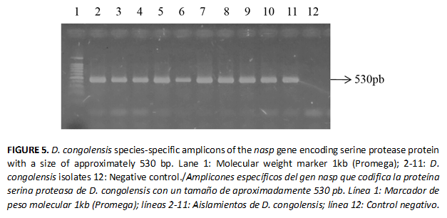
A second PCR assay was carried out to detect the presence of the nasp gene (530 pb) fragment encoding for the D. congolensis serine protease protein, which constituted a factor for D. congolensis virulence , and the protocol described by Garcia-Sanchez et al. (18) was followed. Primer sequences were 5´GATGGAAAATGCAAGGAGCAG3´ and 5´GTCTTCGGGGTCCATGAACAT3´. The PCR program carried out consisted of one denaturation step at 940C for 5 min, and then 35 cycles of 1 min at 94°C, 1 min at 55°C, and 1, 5 min at 72°C.
The amplification reactions were performed in a Thermocycler Eppendorf Mastercycler Personal. The products of all amplification reactions were analyzed by gel electrophoresis on 0.8 % agarose and 0.5X TBE buffer staining with ethidium bromide 0.5 ug/ml. The run was performed in 0.5X TBE buffer at a constant voltage of 100 V for 40 min and using a 1kb molecular weight marker (Promega). The bands were visualized in a UV transilluminator.
DNA sequencing
The genomic DNA corresponding to 2015 Angola strain was sequenced at Macrogene Advancing through Genomics. The 16S rRNA gene of 2015 Angola strain was amplified with an expected size of 1465 bp by using the oligonucleotide primers 5´AGAGTTTGATCATGGCTCAG3´ and 5´GTGTGACGGGCGGTGTGTAC3´ (Korea, http://dna.macrogen.com/kor/), which corresponded to bases 8 to 27 and to bases 1391 to 1410 of the 16S rRNA. Sequences were aligned and assembled using the Vector NTI® software (Invitrogen, USA) to obtain a sequence of 1380 bp, which was deposited in GenBank under accession numbers KX884879.1. They were aligned using MEGA 7 software (19) for phylogenetic inference using the Neighbor-Joined method (20).
Ethical considerations
Samples were collected by clinical veterinarians as part of the usual screening scheme on farms, and the ethical guidelines and animal welfare regulations (OIE, 2010) were strictly respected. All herd owners had given an informed consent prior to the study.
RESULTS
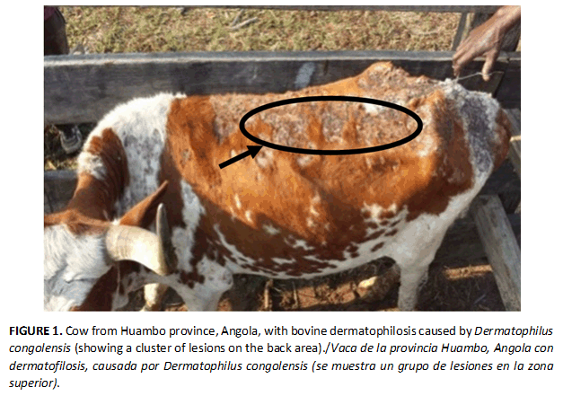
Ninety animals were found to have typical lesions of bovine dermatophilosis. Scabs with exudates were observed on those parts of the body with alopecia or matted hairs (Figure 1). Dermatophylosis diagnosis is often confused with others cutaneous infections (dermatophitosis) due to the close resemblance of its clinical presentations; therefore, it is necessary to have a microbiological confirmation.
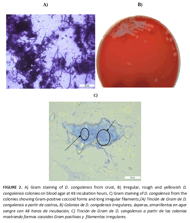
Thirty suspected isolates of D. congolensis were obtained from the 90 clinical samples with an overall prevalence of 33% (30/90). The frequency of D. congolensis isolates and their origin of isolation are listed in Table 1. Based on the morphological characterization and biochemical performance, the isolates were identified as possible D. congolensis; the colonies 0.5 to 1 mm had a rough surface, a yellow-golden pigmentation, and produced β-hemolysis. The Gram stained smears from both scabs and colonies revealed Gram positive branching rods which were thick and filamentous, suggesting the presence of this bacterium in the lesions (Figure 2).
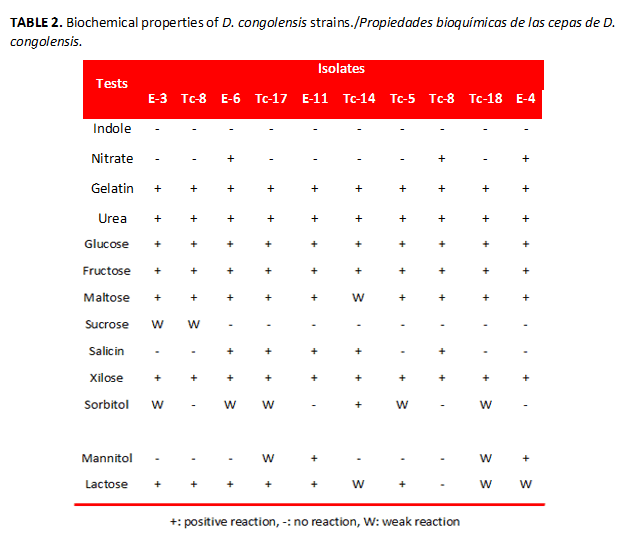
The phenotypic characterization showed the isolates producing catalase, while the oxidase test was negative. All isolates produced urease and digested gelatin. Only three isolates were able to reduce nitrate to nitrite, the rest (n=10) were unable. The indole test was negative. In the sugar fermentation test, all D. congolensis isolates fermented glucose, fructose, maltose, and xilose, while only two isolates fermented manitol (Table 2).
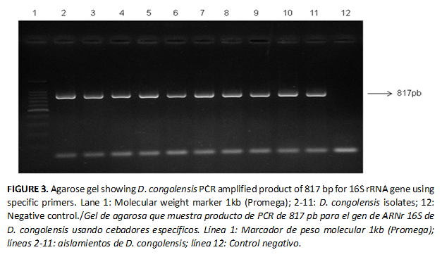
From the cultures (n=30) with presumptive characteristics of the genus Dermatophilus, a fragment of approximately 817 bp was amplified (Figure 3). PCR detection limit with purified genomic DNA as the template was > 0,014 ng DNA/PCR.
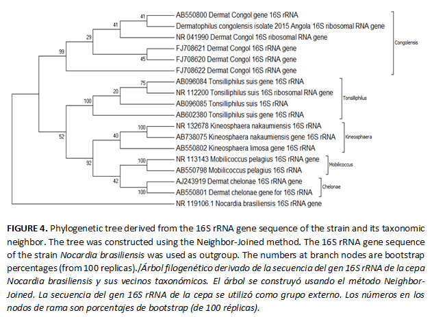
The phylogenetic analysis based on the 16S rRNA gene sequence revealed that the 2015 Angola strain was related to the type strain of D. congolensis (Figure 4). The 2015 Angola strain joined the phylogenetic lineage of D. congolensis species with a bootstrap resampling value of 99 %. This result supported the affiliation of 2015 Angola strain to D. congolensis species. The highest sequence identity value of the 2015 Angola strain was observed in D. congolensis strain: NBRC 105199.
An expected product of 530 bp in size, using the specific primers for nasp gene, was amplified from all isolates subjected to a second PCR assay (Figure 5).
D. congolensis isolates were sensitive in vitro to Amoxicillin/clavulanate, Clindamycin, Streptomycin, Erythromycin, Lincomycin, Azithromycin, Rifampicin, and Enrofloxacin. They were resistant only to Sulfonamides and Sulfazotrim.
DISCUSSION
Dermatophilosis infection has been reported in cattle and other mammal species in different countries of the African continent. The Veterinary Services have demonstrated the presence of the disease based only on clinical sings in the northern, central and southern provinces of Angola (21). The confirmation of D. congolensis was established based on bovine clinical signs and lesions, as well as through its isolation and identification in the laboratory using phenotypic and genotypic assays.
The dark brown scabs observed on the shoulders were very similar to those described by Prasad et al., (22) and Reddy et al. (23). Comparable to previous findings, the colonies were 0.5 to 1 mm and produced ß-haemolysis (24,25,26).
Glucose and maltose fermentation agreed with previous results (13). Only two isolates fermented sucrose, differing from the results of most authors, who have reported a negative response to this sugar (27). Six isolates were lactose positive; however many of the published works have shown a negative response to this sugar, except the study made by Shaibu et al. (28), in which only two isolates from goat and castle were reported to be lactose positive. Otherwise, the reaction to sorbitol and salicin was similar to that found in other studies which declared a pattern in a negative to positive range (9,27). Differences in the biochemical profile of strains with the results of other authors can be attributed to intra-species diversity for D. congolensis, an aspect that has been little discussed in the literature, which is probably because different biotypes exist. Many of the results based on biochemical tests have been carried out from the D. congolensis strains that had different origins such as goats, cattle and horses.
The isolation of D. congolensis was not possible from all the animals showing typical lesions. Some samples produced mixed cultures in which the bacterial growth, typical of other genera like Staphylococcus and Bacillus, was observed. Culturing this bacterium from lesions is difficult; it may be masked by the overgrowth of other opportunistic microorganisms that are part of the skin microbiota (15). Difficulties to isolate and identify D. congolensis have consequently been the reasons for the lack of reports of more dermatophilosis cases (18). The use of special techniques like filtration and chemotaxis, including culture media with antibiotics, are sometimes recommended for the bacteriological isolation (3,15).
The isolates were identified as D. congolensis by species-specific PCR assay. For the detection of D. congolensis by PCR, different targets have been referred: the alkaline ceramidase gene (18) and 16S rRNA (9,17,28). The latter has been more frequently used in different formats such as a real-time assay (4). Besides, the use of this molecule has allowed identifying D. congolensis isolates of different species of animals like goats and sheep (28).
The nasp gene virulence gene was detected in all the isolates previously identified as D. congolensis. According to Garcia-Sanchez et al. (18), the nasp gene encodes for the serine protease protein which constitutes an important virulence factor for the development of D. congolensis infection. The product of this gene (serine protease) contributes to break the main protective barriers like keratin and cutaneous lipids, allowing the bacterial penetration and survival in deeper epidermal layers and the completion of its life cycle. This protein can be considered as a candidate antigen for vaccines and a diagnostic tool for dermatophilosis (18).
The susceptibility of most of the antibiotics tested was in agreement with the results of others authors (9,29). The dermatophilosis treatment in animals has been attempted with a wide variety of topical and parenteral antibiotics, but it has been largely ineffective, probably because of the inability of the topical antibiotics to reach organisms in the deep epidermis of parenteral antibiotics to obtain the avascular upper epidermal layers (9). All antibiotics used in this study are of parenteral application.
In the farms visited, the most frequently antibiotics used have been compounds derived from penicillin (Penfort® PPU, Pen & Strep, and Oxitetraciclin), but they have not been effective for the control and eradication of this infection. Therefore, it would be convenient to evaluate other strategies such as the use of medicinal plants or the combination of drugs (30). The crust detachment was observed from the skin and growth of new hair after the application of a cream ointment prepared from Tephrosia vogelii leaves.
Data on antimicrobial susceptibility in D. congolensis strains are scarce. And similar to what occurs for many pathogens of veterinary interest for this species, regional or global standardized data do not exist yet. This aspect constitutes a difficulty in the antimicrobial susceptibility studies and the comparison of reports among laboratories. The results obtained in this work can contribute to the D. congolensis databases and its susceptibility patterns for the African continent.
The detection of the animals affected by dermatophilosis is often difficult and the disease can be confused with clinically similar diseases. For this reason, specific methodologies allowing the confirmation of the microorganism are required. Prior to this work, phenotypic and genotypic assays had not been used to confirm the presence of D. congolensis in Huambo, Angola. The present study is the first confirmation about the presence of this disease in bovine farms with clinical signs in Huambo.
- Yeruham I, Elad D, Perl S. Dermatophilosis in goats in the Judean foothills. Revue Medicine Veterinary. 2003;154(12):785-786.
- Dalis SJ, Kazzem HM, Makinde AA, Fatihu MY. Bacteria associated with bovine dermatophilosis in Zaria, Nigeria. Afr J Microbiol Res. 2010;4:1475-1476.
- García A, Martínez R, Medina JM. Development of a real-time SYBR Green PCR assay for the rapid detection of Dermatophilus congolensis. J Vet Sci. 2013;14(4):491-494.
- Shoorijeh SJ, Badiee K, Behzadi MA, Tamadon A. First report of Dermatophilus congolensis dermatitis in dairy cows in Shiraz, southern Iran. Iranian J Vet Res. 2008;9(3):281-283.
- Agüero P, Briosso M. Descripción de un Caso de Dermatofilosis en Tambo. Boletín electrónico Agosto 2010. Laboratorio de Diagnóstico de Centro Diagnóstico Veterinario (CDV). [en línea] 2010 Agosto (Consultado: 12 de Julio de 2016). Disponible en: http://www.cdvsa.com.ar/pdf/boletin-electronico1.pdf.
- Dickson C, DeElías-Costa MRI. Dermatofilosis humana y animal. Presentación de un caso atípico y revisión de la literatura. [en línea] 2010;(Consultado 2 de Julio de 2016);16(5):349-353.Disponible en: http://www.dermatolarg.org.ar/index.php/dermatnolarg/article/viewFile/526/309.
- Sharma DR, Kwatra MS, Saini SS, Dhillon SS, Gill BS. Epidemiological studies on dermatophilosis outbreaks in Punjab, Indian. J Comp MicrobiolImmunol and Infect Dis. 1992;(13):5-9.
- CIE. Dermatofiliasis. CIE-10 A48.8. Otras enfermedades bacterianas especificadas. [en línea] 2006. (Consultado 2 de julio de 2016) Disponible en: http://bvs1.panaftosa.org.br/local/file/textoc/Achav1dermatofiliasis.pdf.
- Burd EM, Juzych LA, Rudrik JT, Habib F. Pustular dermatitis caused by Dermatophilus congolensis. J Clin Microbiol. 2007;45(5):1655-1658.
- Institute of Veterinary Services. (ISV). General Direction. Report on Activities Developed during the year Ministry of Agriculture, Rural Development, and Fisheries. Luanda Angola. 2010.
- Topa MC, Iseensee K, Thompson G. Um caso de dermatofilose em bovino. Rev Port Cienc Vet. 2001;96(583):89-93.
- Tavanaeimanesh H, Sasani F, Atyabi N, Rasekh M, Eftekhari Z, Hashemian M .An outbreak of atypical dermatophilosis mixed by Pseudomonas aeruginosa in a sheep herd after dipping. Iranian J Vet Med. 2015;9(4):303-306.
- Zacarias T, Eliseu A. Prevalence and control of bovine dermatophilosis in Angola. AO/IAEA International Symposium on Sustainable Improvement of Animal Production and Health, SYNOPSES Vienna, Austria:8–11 June, CN- 2008;174-62.
- Aguilar B. Fórmulas para el cálculo de la muestra en investigaciones de salud. Salud en Tabasco. 2005;11(1-2):333-338.
- World Organisation for Animal Health (OIE). Dermatophilosis: A Manual of Diagnostic tests for Terrestrial Animals, 5th ed., Office of International des Epizootics. 2008;725-727.
- Clinical and Laboratory Standards Institute (CLSI). Performance Standards for Antimicrobial Susceptibility Testing 26th ed, M100S, Wayne, PA. 2016.
- Amor A, Enriquenz A, Corcuera MT, Toro C, Herroro D, Baquero M. Is infection by Dermatophilus congolensis under diagnosed? J Clin Microbiol. 2011;49(1):449-451.
- García SA, Cerrato C, Larrasa AJ. Identification of analkaline ceramidase gene from Dermatophilus congolensis. Vet Microbiol. 2004;99:67-74.
- Tamura K, Stecher G, Kumar S. 2016. Mega7: Molecular Evolutionary Genetics Analysis Version 7.0 for Bigger Datasets. Mol Biol Evol:33:1870-1874.
- Saitou N, Nei M. The Neighhbor-Joining method: a new method for reconstructing phylogenetic trees. Mol Biol Evol. 1987;4(4):406-25.
- Institute of Veterinary Services (ISV). General Direction. Report on Activities Developed during the year Ministry of Agriculture, Rural Development, and Fisheries. Luanda Angola. 2012.
- Prasad BS, Prameela DR, Sreenivasulu D, Vijayalaxmi S. Prevalence of bovine dermatophilosis in Andhra Pradesh. Inter J VetSci. 2016;5(1):41-43.
- Reddy BS, Rani PD, Sivajothi S, Venkatasivakumar R, Solmon KGR. Dermatohilus in Cross-Bred Cattle in Y.S.R. District of Andhara Pradesh. International J Sci Env Technol. 2014;3(4):1371-1374.
- Ellis TM, Masters AM, Sutherland SS, Carson JM, Gregory AR. Variation in cultural, morphological, biochemical properties and infectivity of Australian isolates of Dermatophilus congolensis. Vet Microbiol. 1993;38(1-2):81-102.
- Masters AM, Ellis MT, Grein SB. Dermatophilus congolensis. Strain difference in expression of phospholiphase activities, Elsevier Veterinary Microbiology. 1997;57:199-213.
- Tarazi YH, Al-Ani FK. An Outbreak of dermatophilosis and case ous lymphadenistis mixed infection in camels in Jordan. J Inf Dev Ctries. 2016;10(5):506-511.
- Shaibu SJ, Kazeem HM, Abdullahi US, Fatihu MY. Phenotypic and Genotypic characterization of isolates of Dermatophilus congolensis from cattle, sheep and goats in Jos, Nigeria., African J Microbiol Res. 2011;5(5):467-474.
- Shaibu SJ, Kazeem HM, Abdullahi US, Fatihu MY. The use of polymerase chain reaction in the diagnosis of dermatophilosis from cattle, sheep and goats in Nigeria. J Anim Vet Advan. 2010;9(6):1034-1036.
- Towersey LE, Takiya CM, Londero AT. Dermatophilus congolensis human infection. J Am Acad Dermatol. 1993;29:351-354.
- Makoshi MS, Arowolo ROA. Therapeutic effects of Tephrosia vogelii ointment in the treatment of bovine dermatophilosis. J Vet Med Anim Health. 2011;3(4):51-55.
Recibido: 6/2/2017
Aceptado: 9/6/2017
*Autor para correspondencia: Ivette Espinosa. E-mail: espinosa@censa.edu.cu

