


Mi SciELO
 Servicios personalizados
Servicios Personalizados
Revista

Articulo
 Inglés (pdf)
 Articulo en XML
 Referencias del artículo
 Enviar articulo por email
Indicadores
-
 Citado por SciELO
Links relacionados
-
 Similares en
SciELO
Similares en
SciELO
Compartir

 Permalink
Revista Cubana de Medicina Tropical
versión impresa ISSN 0375-0760versión On-line ISSN 1561-3054
Rev Cubana Med Trop vol.70 no.1 Ciudad de la Habana ene.-abr. 2018
COMUNICACIÓN BREVE
Procedure optimization for concentration and detection of protozoa and helminths in great volumes of water
Optimización del procedimiento para la concentración y detección de protozoos y helmintos en grandes volúmenes de agua
Sandra Ríos Tobón,I,II Juliana López Jiménez,I,II Laura Francisca Campo Polanco,I Ruth Marina Agudelo Cadavid,I,II Lina Andrea Gutiérrez BuilesI
I Grupo Biología de Sistemas, Escuela de Ciencias de la Salud, Facultad de Medicina, Universidad Pontificia Bolivariana. Medellín, Antioquia, Colombia.
II Grupo de Investigación Salud y Ambiente, Facultad Nacional de Salud Pública, Universidad de Antioquia. Medellín, Antioquia, Colombia.
ABSTRACT
Feces-contaminated water is a vehicle of transmission of potentially pathogenic microorganisms responsible for illnesses that represent main causes of death worldwide. A protocol for detection of intestinal parasites in great volumes of water was optimized. It includes: membrane filtration, mechanical agitation with detergent, centrifugation, chemical concentration with Mini Parasep® and microscopic examination. From samples of feces-contaminated water containing parasitic forms, a total recovery percentage of 85.7 % of parasites was achieved after tests. This procedure provides a useful alternative method that could be subjected to validation as a routine methodology in the diagnosis of microbiological water quality.
Keywords: parasitology; waterborne diseases; water quality.
RESUMEN
El agua contaminada con heces es un vehículo de transmisión de microorganismos potencialmente patógenos causantes de enfermedades que constituyen causas principales de muerte a nivel mundial. Se optimizó un protocolo para la detección de parásitos intestinales en grandes volúmenes de agua. Este incluye: filtración por membrana, agitación mecánica con detergente, centrifugación, concentración química con Mini Parasep® y examen microscópico. En muestras de agua contaminada con heces que contenían formas parasitarias, se obtuvo un porcentaje de recuperación total del 85.7 % de estas formas después de aplicar el protocolo. El procedimiento constituye un método alternativo que podría someterse a validación como metodología habitual para el diagnóstico de la calidad microbiológica del agua.
Palabras clave: parasitología; enfermedades transmitidas por el agua; calidad del agua.
Human or animal feces-contaminated water is a vehicle of transmission of potentially pathogenic microorganisms, responsible for illnesses that represent one of the main causes of death worldwide.1-3 Drinking water safety is regulated by both national and international standards that aim for a reduction of the risks associated to its consumption,4 being microorganisms, the most important aspect to be evaluated.1
At least 99 water-transmitted disease outbreaks reported worldwide as of 2010, were caused by parasites.2,3 Cryptosporidium spp., the main responsible organism, has caused the death of thousands of people, both in countries with deficiencies in drinking water quality surveillance programs, as well as countries with great quality standards.3,4 During the drinking water treatments, the parasitic forms through water transmission have a great survival capacity to different environmental elements and physicochemical treatments and disinfection, which allow them to persist and maintain their pathogenic capacity.4,5
The microbiological quality of water has been evaluated mainly through parasitic and bacterial-origin bioindicators,6 determining their presence is necessary for identifying the pathogens that put entire communities at risk.7 Research of water-transmitted parasites has become more important in the last years, and the World Health Organization has suggested the creation of a protocol that includes their study in microbiologic water assessments.5
In order to detect parasites in great water volumes, it is necessary to concentrate them8 by using traditional reactants, instruments and specialized protocols.2,6,8-10 Several different density gradient based methods have been described, which allow phase separation and the consequent shape recovery;4 however, comparative studies have determined that these techniques have low specificity and propose finding more efficient alternatives in detection of the parasitic charge in drinking water.11 A protocol for such optimization was developed in this work, which includes: membrane filtration, mechanic agitation with detergent, centrifugation and chemical concentration through Mini Parasep® for detection of parasites in great water volumes.
The protocol evaluated in the present work (Fig.) and described below, was based on the instructions by Pérez et al., Alarcon et al., Jara et al., and Saens et al. 4,6,11,12 using a water sample contaminated with parasitic forms of different nature (table).
Step 1. Sample preparation: Each liter of water Type I (ultra-pure water 18.2 MΩ.cm at 25 ºC Synergy® Millipore system) was contaminated with 500 μL of the water contaminated sample (table). This procedure was performed three times followed by magnetic agitation for 10 min.
Step 2. Membrane filtration: The membrane filtration method described by Pérez et al. 4 was implemented with the following modifications: the water sample volume was passed through membranes (0.45 µm, Millipore®), until reaching its saturation point; the filtered liquid was then refiltered using 0.2 µm (Millipore®) membranes until reaching its saturation point, thus guaranteeing filtration of all the water.
Step 3. Mechanic agitation with detergent: Each 0.2 and 0.45 µm membrane used in the step above was cut with sterile scissors into 1 cm-wide segments. The segments corresponding to the three membranes were submerged in conical cubes containing 50 mL of Tween 80 at 0.1 % and agitated mechanically with vortex at 45 times gravity (×g). This step was followed by agitation with a glass stirring rod until the saturation material detached from the membranes, which were then discarded using sterile tongs.
Step 4. Centrifugation and microscopic evaluation: The tubes were centrifuged for 10 min at 3 000 rpm and the supernatant was discarded. The sediment was diluted in 500 µL NaCl at 0.85 %, submitted to vortex agitation and divided into two fractions: one for the wet- mount method, direct sediment reading and modified Ziehl Neelsen, (Merck KGaA, Darmstadt, Germany) and Field colorings (Químicos Albor, Bogotá, Colombia), and another one for the chemical concentration method.
Step 5. Chemical concentration with Mini Parasep® Solvent Free and microscopic evaluation: The density-based concentration process proposed by Pérez et al., was replaced by the commercial Mini Parasep® Faecal Parasite Concentrator method, following the manufacturer's instructions of dry samples, using 500 µL of sediment in each tube. Microscopic evaluations of both the fresh sediment and through modified Ziehl Neelsen coloring as well as Field colorings were performed.
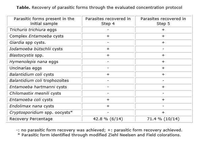
The concentration protocol used in the present work allowed for recovery of 85.7 % of parasitic forms in the initial contaminated sample, performing microscopic evaluation in two different moments (step 4 and step 5) (Fig.). This finding suggests that with the suggested optimization process, a high recovery percentage is achieved, notably increasing the possibility of parasites detection in great water volumes. The parasites recovered in Step 4 (table), corresponded to 42.8 % (6/14) of the total contained in the initial sample. In Step 5, a 71.4 % (10/14) recovery was achieved. However, the parasites recovered in both cases corresponded to 28.57 % (4/14) of the total parasites, which shows the importance of performing readings in both steps. Chilomastix mesnili cysts were not observed after the treatment. Nevertheless, since equally small or even smaller forms such as Entamoeba hartmanni y Endolimax nana were detected (6-8 μm), failure to recover small parasitic forms cannot be attributed to procedure limitations.
The lack of recovery of Iodamoeba bütschlii and Endolimax nana cysts in Step 5, evident also in Step 4, could be related to a low parasitic charge of these species in the initial sample, or to the sensitivity of the microscopic evaluation ─for Iodamoeba bütschlii, is 50 % and for Endolimax nana, 89 %.13
On the other hand, recovery of Balantidium coli trophozoites was not observed, as expected, given the fact that the mobile forms of these parasites are susceptible to degradation during concentration processes.14
The requirements of high cost technologies used for recovery of parasites from water samples has motivated the search of equally efficient and less costly methodologies. In this regard, the method integration attempted in the present work ─membrane filtration, mechanic agitation, centrifugation, chemical concentration, direct and by coloring microscopical exam─ allowed the recovery of the greatest amount of parasitic forms present in water, with methodologies that do not require major investments.
The chemical concentration process was fast and favored the optimization of laboratory resources. It also increased the probability of detection of parasitic forms, achieving separation of big particles that otherwise could sediment in the conical tube during centrifugation. The sensitivity of this kit has been previously described, for eggs and helminths larvae, protozoa cysts and oocysts such as: Hypmenolepis nana,Schistosoma mansoni, Ancylostoma spp., Strongyloides stercoralis, Ascaris lumbricoides, Entamoeba coli, Giardia lamblia, complex Entamoeba, among others.12 The use of modified Ziehl Neelsen and Field colorings allowed the visualization of acid-alcohol resistant parasitic forms (Cryptosporidium spp oocysts) and the staining of nucleic acids, increasing the morphologic definition and the valuation of diagnostic characteristics of specie.15
The procedure evaluated in the present work suggests an alternative and reliable tool for concentrating and detecting parasitic forms in the monitoring of drinking and non-drinking water. It is particularly optimal to be applied in developing countries where resources are scarce, in areas without access to specialized laboratories, where monitoring of drinking water is necessary to avoid spreading of waterborne diseases.
Conflict of interest
This document was prepared and revised with the participation of all the authors above mentioned, who hereby declare no conflict of interests that puts at risk the validity of the information here presented.
Financial support
Centro de Investigaciones para el Desarrollo y la Innovación -CIDI, Universidad Pontificia Bolivariana (Project number 251B08/14-65).
Fondo de Apoyo Docente de la Facultad Nacional de Salud Pública, Universidad de Antioquia (record number INV 444-13).
BIBLIOGRAPHIC REFERENCES
1. Fawell J, Nieuwenhuijsen MJ. Contaminants in drinking water. Br Med Bull. 2003;68(1):199-208.
2. UNICEF - Agua, saneamiento e higiene - Common water and sanitation-related diseases. 2013 [cited 2016 Oct 10]. Available from: http://www.unicef.org/spanish/wash/index_wes_related.html
3. Baldursson S, Karanis P. Waterborne transmission of protozoan parasites: review of worldwide outbreaks - an update 2004-2010. Water Res. 2011;45(20):6603-14.
4. Pérez-Cordón G, Rosales MJ, Valdez RA, Vargas-Vásquez F, Córdova O. Detección de parásitos intestinales en agua y alimentos de Trujillo, Perú. Rev Peru Med Exp Salud Pública. Instituto Nacional de Salud. 2008;25(1):144-8.
5. Organización Mundial de la Salud. Guías para la calidad del agua potable 3ra. ed. Ginebra, Suiza: OMS; 2006 [citado 15 Jul 2016]. Available from: http://www.apps.who.int/water_sanitation_health/dwq/gdwq3_es_fulll_lowsres.pdf?ua=1
6. Alarcón MA, Beltrán M, Cárdenas ML, Campos MC. Recuento y determinación de viabilidad de Giardia spp. y Cryptosporidium spp. en aguas potables y residuales en la cuenca alta del río Bogotá. Biomédica. 2005;25(3):353-65.
7. Vásquez G, Castro G, González I, Pérez R, Castro T. Bioindicadores como herramientas para determinar la calidad del agua. ContactoS. 2006;(60):41-8.
8. Lora-Suarez F, Rivera R, Triviño Valencia J, Gómez Marín JE. Detection of protozoa in water samples by formalin/ether concentration method. Water Res. 2016;100(1):377-81.
9. Menocal L, Caraballo Y. Importancia de la vigilancia sanitaria de los parásitos en la calidad del agua, según su uso. Rev Cubana Hig Epidemiol. 2014;52(2):196-209.
10. Betancourt WQ, Rose JB. Drinking water treatment processes for removal of Cryptosporidium and Giardia. Vet Parasitol. 2004;126(1-2):219-34.
11. Jara CA, Minchón Medina CA, Zárate Asmat C. Comparación de las técnicas de Willis y de Sheather para el diagnóstico coproparasitoscópico. Rev Rebiol. 2007;1(1):1-6.
12. Saez AC, Manser MM, Andrews N, Chiodini PL. Comparison between the Midi Parasep and Midi Parasep Solvent Free (SF) faecal parasite concentrators. J Clin Pathol. 2011;64(10):901-4.
13. Campo-Polanco L, Botero LE, Gutiérrez LA, Cardona Arias JA. Reproducibilidad del examen directo de heces y de la concentración formol-éter y validez del examen directo de heces para el diagnóstico de parásitos intestinales. Arch Med. 2015;11(4:4):1-9.
14. Plutzer J, Karanis P. Neglected waterborne parasitic protozoa and their detection in water. Water Res. 2016;101:318-32.
15. Tahvildar-Biderouni F, Salehi N. Detection of Cryptosporidium infection by modified Ziehl-Neelsen and PCR methods in children with diarrheal samples in pediatric hospitals in Tehran. Gastroenterol Hepatol Bed Bench. 2014;7(2):125-30.
Recibido: 29 de diciembre de 2016.
Aprobado: 19 de octubre de 2017.
Sandra Ríos Tobón. Grupo Biología de Sistemas, Escuela de Ciencias de la Salud, Facultad de Medicina, Universidad Pontificia Bolivariana. Grupo de Investigación Salud y Ambiente, Facultad Nacional de Salud Pública, Universidad de Antioquia. Medellín, Antioquia, Colombia. E-mail: sandra.riost@udea.edu.co

