


 Servicios personalizados
 Inglés (pdf)
 Articulo en XML
 Referencias del artículo
 Enviar articulo por email
 Citado por SciELO
  Similares en
SciELO
Similares en
SciELO 
 Permalink
Introduction
The presence of metal artifacts has been a major problem in x-ray computed tomography (CT). Metal parts in the field of view attenuate most x-ray photons and generate dark and bright streaks after reconstruction. Metal induced streaking artifacts deteriorate the diagnostic quality and quantitative value of CT images. They obscure the anatomical structures surrounding the metallic objects and preclude confident diagnosis of the disease [1]-[3].
Different approaches towards metal artifact reduction (MAR) have been published, which can be divided mainly into five classes: Acquisition Improvement, Physics-based Pre-processing, Projection Completion, Iterative Reconstruction and Image based Approaches [4]. A common approach is to directly correct or replace by interpolation the corrupt projection data in the raw projection’s data [5]-[9]. A less study MAR method is to perform post-processing, which aims to correct in the image domain. Post-processing algorithms reduce artifacts after the image has been reconstructed, and do not depend on access to raw projection data [10].
In this paper a novel MAR technique is presented in the image domain that uses image smoothing via L0 Gradient Minimization method [11], which is assessed in presence of dental fillings and metal hip prosthesis. Finally, the results using a real CT dataset are shown. They demonstrate that the present MAR method can produce better or similar results than LI [12], NMAR [8] and FSMAR [7], without the use of the raw CT data.
Materials and methods
MAR based on edge-preserving smoothing via L0 Gradient Minimization filter
The ISMAR method (figure 1) is composed by four steps: metal trace’s segmentation (IMetal), prior image generation (IPrior), a projection completion, followed by image reconstruction (IRec) and finally, a smoothing algorithm is applied to obtain the final result as will be explained subsequently.
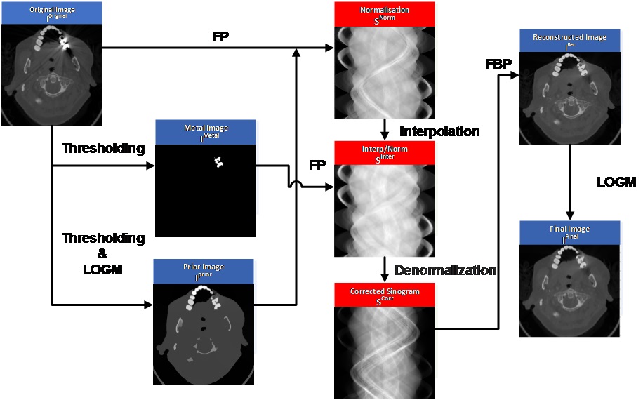
Figure 1 Flow diagram showing the steps of the new MAR based on edge-preserving smoothing via L0 Gradient Minimization filter algorithm.
A. Metal traces segmentation
In the original image (IOriginal), metals are segmented out based on thresholding. A simple thresholding was applied to find the image metal (IMetal). In this work, 3000 HU is selected as the threshold, which is proper to extract metal according to the literature [13]. Then the forward projection (FP) of the obtained IMetal is performed to get metal traces, which specifies the projections affected by metals.
B. Prior image generation
The main step of the present approach is to generate a good prior image (IPrior), which should contain the most possible soft tissue and bone regions. The MAR methods, which complete sinogram by using forward projection of a prior image, can achieve good results as long as the prior image is close enough to the ground truth. However, most of these MAR methods generate the prior image in a simple way, and the prior image is usually not perfect enough because in some occasions contains also artifacts.
To overcome the above problem, in this work, the IPrior isgotten using a global thresholding fixed between 5HU and 10 HU and a LO Gradient Minimization (L0GM) smoothing algorithm, which are applied to the IOriginal. L0GM method was selected due to the mechanism of discretely counting spatial changes, because it can remove low-amplitude structures and globally preserve and enhance outgoing edges, even if they are boundaries of very narrow objects. In this method λ is the smoothing parameter, which controls the degree of smooth, and κ controls the iterations rate. Five iterations are generally performed in the algorithm to get a good IPrior, with κ = 1.8 and λ = 9 x 10⁻⁶. More iterations could yield an over-smoothing, losing the image information.
C. Projection completion and image reconstruction
In this step, sinogram from IOriginal, IPrior and IMetal were obtained using forward projection. Following the well-known method developed by [8], the original sinogram is normalized (SNorm) by dividing it by the sinogram of the IPrior pixelwise. Moreover, all values from the INorm that lie within the metal trace (IMetal) are replaced in each row by linear interpolation (See Kalender et al. [12] for further details). Afterwards, the corrected sinogram (SCorr) is gotten by denormalization of the interpolated (SInter). Lastly, the reconstructed image (IRec) is obtained using filtered back projection (FBP) with linear interpolation and Ram-Lak filter.
Patients
The study was performed by using the DICOM images from CT data sets obtained from 15patientsranging in age from 21-82 years; and body weight ranged from 39.2-98.9 kg. The eligible criterion was the presence of a metallic implant in the examination area. Only the image data of patients was used in the study, keeping all other patient’s information anonymous.
Image acquisition
The CT datasets were obtained for this research from two different CT scanners: a kV on-board imaging (OBI) system integrated in a TrueBeamTMmedical linear accelerator (Varian Medical System, Palo Alto, CA) and a Siemens SOMATOM Sensation 16 scanner CT using helical scanning geometry (Santa Clara, Cuba). In both CT scanners the tube voltage was set to 120 kVp. The x-ray tube current was set at 25 mA and 22.6 mA respectively. The matrix of reconstructed image was 512 x 512 pixels, corresponding pixel sizes were 1 mm X 1 mm for OBI and 0.776 mm X 0.776 mm for Siemens SOMATOM Sensation.
Image analysis
Qualitative image analyses
Clinical images obtained by using LI, NMAR, FSMAR and the new algorithm were evaluated by two board certified radiologists independently, each with more than 10 years of clinical experience, blinded to all patient data and image parameters. The images were displayed in random order, and reviewed in the soft tissue window (window level 20 HU, window width 400 HU) and bone window (window level 300 HU, window width 2,500 HU).
The artifacts and the diagnostic interpretability were graded on a five point rating scale by observers. The degree of metal artifacts on images was scored on a scale from 1 to 5 (1, very severe streaks; 2, severe streaks; 3, moderate streaks; 4, minimal streaks; 5, no streak artifacts). The diagnostic image quality was similarly scored on CT images on a scale from 1 to 5 (1, severely reduced image quality, non-diagnostic; 2, markedly reduced image quality, with impaired diagnostic interpretability; 3, acceptable image quality and diagnostic interpretability; 4, good image quality, with high diagnostic confidence; 5, excellent image quality, with full diagnostic interpretability).
Statistical analysis
All numeric values were reported as the mean ± SD. To compare image quality scores of the data sets, non-parametric Friedman, Wilcoxon signed-rank (paired samples) and Mann-Whitney (unpaired samples) tests were employed. To assess inter-observer agreement was used Cohen’s kappa. The κ values of 0.01-0.20 were considered to indicate slight agreement, 0.21-0.40 fair agreement, 0.41-0.60 moderate agreement, 0.61-0.80 substantial agreement and 0.81-1.00 almost perfect agreement.
Statistical analyses were performed with statistical software (SPSS, version 22.0; IBM, Chicago, IL, USA). For all statistical analyses, p-values less than 0.05 were considered to represent statistically significant differences.
Results
Artifacts from metallic devices were significantly lower in all algorithms compared with the original image (figure 2), obtained using FBP from raw data. ISMAR led a significant artifact reduction and tissue preservation compared with LI and NMAR. FSMAR and ISMAR had similar results in some cases, but in presence of severe artifacts in the dental fillings, ISMAR had better results. Two examples of images from our study are shown in figure 3.
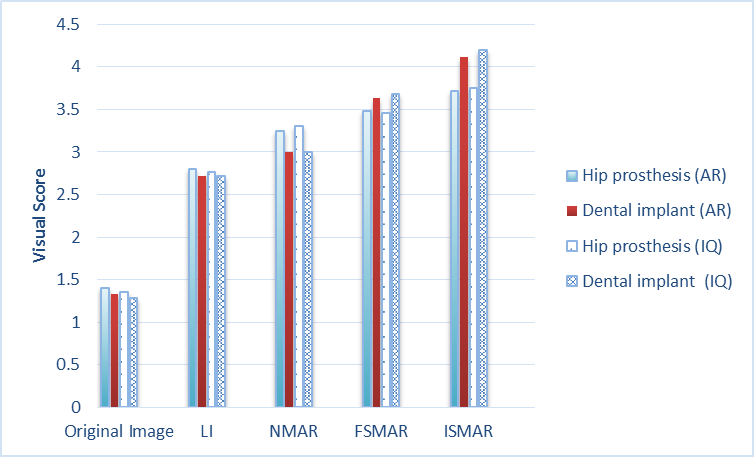
Figure 2 Comparison of metal artifact reduction and images quality scores between MAR algorithms obtained from non-parametric Friedman test, existing significant difference among methods both hip prosthesis as well as dental fillings with p<0.001. IQ an AR represent image quality and artifacts reduction respectively.
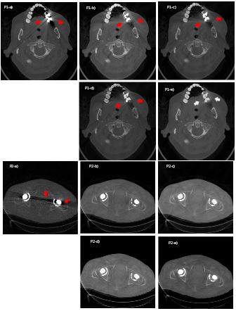
Figure 3 Patient 1 (P1) with two big dental fillings distributed in therightside of the lower jaw and patient 2 (P2) with bilateral hip prosthesis (a) The original artifact CT image (FBP) and the corrected images using the various MAR approaches: (b) LI, (c) NMAR, (d) FSMAR and (e) ISMAR method. The window width and window level was (WW =2500 HU, WL=300 HU).
From table 1 the results on image quality obtained, shown that in presence of dental implants there was significant difference among methods. The Wilcoxon test did not show statistically significant among NMAR and FSMAR (p=0.271), as well as among FSMAR and ISMAR (p=0.058).
Table 1 Differences in image quality among the algorithms obtained from subjective analysis using Wilcoxon signed-rank (paired samples) and Mann-Whitney (unpaired samples) tests.
| Dental implants | ||||
|---|---|---|---|---|
| LI | NMAR | FSMAR | ISMAR | |
| FBP | < 0.001 | < 0.001 | < 0.001 | < 0.001 |
| LI | 0.003 | 0.002 | < 0.001 | |
| NMAR | 0.005 | < 0.001 | ||
| FSMAR | 0.009 | |||
| Hip implants | ||||
| LI | NMAR | FSMAR | ISMAR | |
| FBP | < 0.001 | 0.001 | < 0.001 | < 0.001 |
| LI | 0.008 | 0.002 | 0.001 | |
| NMAR | 0.271 | 0.015 | ||
| FSMAR | 0.058 | |||
Interobserver agreement for all assessments of image quality is shown in table 2. The κ values for the two observers ranged from 0.681 to 0.779 for image quality of the dental fillings (P < 0.01) and from 0.703 to 0.831 for image quality of the hip prosthesis (P < 0.01); indicating that substantial to almost perfect interobserver agreements were obtained.
Table 2 Interobserver agreement for all image quality assessments and all visualization capability assessments.
| Original Image | LI | NMAR | FSMAR | ISMAR | ||||||
|---|---|---|---|---|---|---|---|---|---|---|
| κ | P | κ | P | κ | P | κ | P | κ | P | |
| Hip prosthesis | 0.831 | < 0.01 | 0.737 | < 0.01 | 0.703 | <0.01 | 0.722 | <0.01 | 0.818 | < 0.01 |
| Dental fillings | 0.758 | < 0.01 | 0.681 | < 0.01 | 0.762 | < 0.01 | 0.779 | <0.01 | 0.748 | <0.01 |
Discussion
Artifacts related to metal hardware are a frequent problem in CT. In this paper, a new method for the suppression of metal artifacts was proposed. The main goal of ISMAR was the creation of a prior image using a L0GM smoothing algorithm, because a good prior image can avoid wrong tissue classification and consequently a better final image.
On the other side, the major metal streak artifacts are corrected in the output image of the proposed algorithm. A prior image ensures that traces of high-contrast structures are continued through the metal trace. Therefore, the typical artifacts tangent to the metal implants, and connecting metal implants and high-contrast structures, are reduced to a minimum or even removed completely.
The combination of linear interpolation and the normalization approach led to a quick and efficient reconstruction performance. ISMAR also has advantages for clinical work flow. It has the ability to operate directly on DICOM images, reducing the artifacts and improving image quality. Many MAR algorithms require access to raw projection data, but these often cannot be implemented on commercial CT scanners due to restrictions. From this algorithm could develop a dedicated software for use in workstations by radiologists, even for personal laptops.
The main limitation of this contribution is the number of patients studied. It would be important increase the sample for evaluating its performance, as well as investigate other common sources of streaking artifacts, such as seed implants, spinal screws and surgical clips.
Conclusion
In this work, we presented a novel method for metal artifact reduction in CT images based on edge-preserving smoothing via L0 Gradient Minimization filter. In clinical case studies, the algorithm developed is able to improve image quality. Results obtained using real datasets show that ISMAR is effective reducing metal artifacts and also producing better segmentation, without the use of the raw CT data.

