


 Serviços customizados
 Inglês (pdf)
 Artigo em XML
 Referências do artigo
 Enviar este artigo por email
 Citado por SciELO
  Similares em
SciELO
Similares em
SciELO 
 Permalink
Introduction
Developmental Dysplasia of the Hip (DDH) encompasses a wide range of anatomical abnormalities of the coxofemoral joint including abnormalities in the size, shape, orientation, or organization of the femoral head, acetabulum, or both. It can result in subluxation or dislocation of the femoral head affecting hip development.
DDH is the cause of 2.6-9.1% of all total hip replacements (THR) at any age and it is the most frequent cause of THR in young patients (21-29%). Coxarthrosis occurs in patients with hip subluxation. Neonatal hip instability has a 2.6-fold increased risk of hip replacements in young adulthood compared to stable hips at birth.1
Total hip replacements at young ages are associated with a higher long-term risk of revision surgeries. Revision total hip replacement (RTHR) is a longer, more complex procedure and it is associated with extensive loss of bone support, both in the femur and in the acetabulum. Revision surgeries are related to vascular lesions in 0.2% and the external iliac artery is the most frequently involved.2
Aseptic loosening in THR is related to a painful hip and the presence of more than 2 mm radiolucent lines at the bone-cement or bone-prosthesis interface, component migration, osteolysis, osteopenia, or an obvious component malalignment evidenced in plain radiographies.3 Multiple factors can cause aseptic loosening. Genetical, surgical factors, a chronical process resulting from a cell-mediated response to wear debris, or a chronic impact of the prosthesis in demanding activities.4
In RTHR, acetabular defects interfere with prosthetic components compromising their stability. It requires a good classification of the acetabular defects and good presurgical planning. Several classifications of acetabular defects were established in the late eighties and early nineties. Paprosky Classification identifies acetabular supporting structures’ capacities for biologic augments and synthetic components placement. Based on this classification there are different treatment options like autologous bone grafting, reconstruction plating, and acetabular augmentations.5 We report a case of a 26-year-old patient with DDH treated initially with a THR, who presented aseptic loosening with a Paprosky 3A acetabular defect and underwent revision surgery through a modified Stoppa approach. We wanted to determine the advantages of this approach and the risks associated to reconstruct acetabular defects. Stoppa approach is widely used for acetabulum fractures,6 a review of the literature identified one case-report using this approach for acetabular defects reconstruction.7 No other reports of surgical procedures were identified making quantification of risks associated.
The Hospital Local Committee for Research and Research Ethics granted ethical approval for the study. The authors requested verbal informed consent from the patient for the presentation of this case, always maintaining anonymity and confidentiality.
Case report
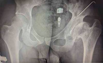
Our patient has developmental dysplasia of the left hip (DDH) and was treated with a pelvic osteotomy at 1.5 years old (fig. 1). At the age of 23, he received a THR but presented prosthesis hip dislocation two months later. The liner was changed, and we put a femoral cerclage wire. However, three years later, the patient complained of increasing left hip pain. Radiographic examination demonstrated aseptic loosening and protrusion of the acetabular component. CT scan showed an acetabular defect Paprosky 3A (fig. 2) (No acetabular rim, severely compromised acetabular walls, non-supportive columns, supero-lateral migration of the component, moderate destruction of the teardrop, and moderate lysis of the ischium).5

Removal of all prosthetic material was initially performed. A second time, acetabuloplasty was accomplished with an autologous bone graft of the contralateral iliac crest. Bone graft was attached endopelvic with two reconstruction plates of 6 and 7 orifices through a modified Stoppa approach. In a third and last surgical time, having a good previous bone graft integration, RTHR was performed placing a Gription® TF acetabular system [Acetabular augment 58 mm] (DePuy Synthes, Warsaw, USA) and a Corail® Hip System, [PINNACLE™ Acetabular Cup System 46, liner 46; Revision stem STD offset 12; ARTICUL/EZE BIOLOX® delta Head 28+1.5], (DePuy Synthes, Warsaw, USA) (fig. 3).

Our standard post-operative regime was followed, including routine antibiotic prophylaxis. The patient made uneventful postoperative recoveries and was discharged from the hospital. Two years after the third operation our patient has a cane-aided painless bipedal gait and no limitations on his daily living activities, 85° of hip flexion, 30° of hip internal rotation, 40° of hip external rotation, 35° of hip abduction. Our patient is very satisfied with the results.
Discussion
Hip revision surgeries are always a complex challenge and require good preoperative planning. Planning helps to visualize the operation after a review of the clinical and radiographic findings. We decided to perform a modified Stoppa approach, dissecting over the suprapubic area obtaining a wide exposure of the quadrilateral surface and the posterior column. Iliac crest grafts attached to reconstruction plates were placed to repair the endopelvic defect. Once an adequate integration of the bone autograft was obtained, we decided to place a porous acetabular augment of titanium that enhances osseointegration and has an elastic modulus like bone.8
Del Gaizo et al9 report up to 21% of complications in 37 patients operated with acetabular defect Paprosky type 3A using revision acetabular augmentations, but they only reported one aseptic loosening. Jenkins et al,10 n 2017, reported a study of 85 hips operated using porous acetabular augmentations in Paprosky 3A and 3B acetabular defects and concluded that the use of these devices provides lasting fixation with favorable clinical outcomes. Although the Stoppa approach is not the usual surgical approach for this type of surgery and its technique is complex with possibilities of vascular damage that can be fatal (such as damage to the "Corona Mortis"), the exposure of the quadrilateral surface and the posterior column is excellent to solve acetabular defects, which could not be carried out from other less extensive approaches. For this reason, we emphasize the usefulness of the approach for the placement of a new endopelvic acetabular roof to subsequently treat the joint through acetabular augmentation and revision of prosthetic components. The result of this case opens the possibility for prospective studies to examine severe acetabular defects that require endopelvic damage control and assessment of associated risks.

