


Mi SciELO
 Servicios personalizados
Servicios Personalizados
Articulo
 Inglés (pdf)
 Articulo en XML
 Referencias del artículo
 Enviar articulo por email
Indicadores
-
 Citado por SciELO
Links relacionados
-
 Similares en
SciELO
Similares en
SciELO
Compartir

 Permalink
Revista de Protección Vegetal
versión impresa ISSN 1010-2752
Rev. Protección Veg. vol.28 no.2 La Habana mayo-ago. 2013
SHORT COMMUNICATION
Quantification of phenols in lesions caused by Mycosphaerella fijiensis Morelet in `Cavendish naine'
Cuantificación de fenoles en lesiones causadas por Mycosphaerella fijiensis Morelet en `Cavendish naine'
Cynthia Sanchez-García, Yelenys Alvarado-Capó, Mayra Acosta-Suárez, Michel Leiva-Mora, Mileidy Cruz-Martín, Berkys Roque
Instituto de Biotecnología de las Plantas (IBP). Universidad Central Marta Abreude Las Villas. Carretera a Camajuaní km 5.5, Santa Clara. Cuba. Email: cyn@ibp.co.cu.
ABSTRACT
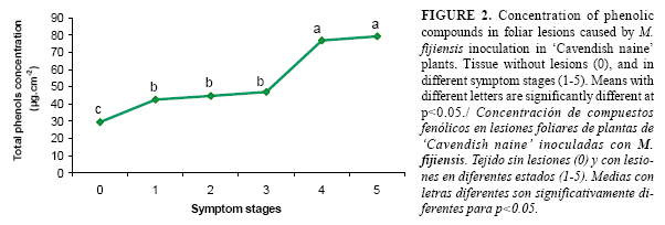
The presence of phenols was demonstrated in leaf lesions caused by Mycosphaerella fijiensis Morelet in inoculated plants of `Cavendish naine'. Phenolic compounds were microscopically detected in plant tissue with symptoms in different development stages by using staining cathecol derivatives, and they were quantified by spectrophotometry. An accumulation of phenols was observed since the first lesions appeared and gradually increased as symptoms progressed. It varied from 29µg.cm2 in the early lesion stage to 79µg.cm2 in the last stage of the disease. Although phenols were not effective in preventing further disease development in this compatible interaction, these results constituted an evidence of the presence of these biochemical compounds as part of the plant defense response against pathogen infection.
Key words: Black Sigatoka, cathecol derivatives, histochemical detection.
RESUMEN
Se demostró la acumulación de fenoles en lesiones foliares causadas por Mycosphaerella fijiensis Morelet en plantas inoculadas de `Cavendish naine'. Los compuestos fenólicos fueron detectados microscópicamente, en el tejido vegetal correspondiente a síntomas en diferentes estados de desarrollo, mediante la tinción de derivados del catecol y cuantificados por espectrofotometría. Se observó que la acumulación de fenoles ocurrió desde la aparición de las primeras lesiones e incrementó a medida que evolucionaron los síntomas, desde un valor de concentración de 29µg.cm2 en el estado más temprano de la enfermedad hasta 79µg.cm2 en el estado más avanzado. Aunque la inducción de fenoles no fue efectiva en el control del progreso de la enfermedad en esta interacción compatible, estos resultados constituyen una evidencia de la presencia de estos compuestos como parte de la respuesta de la planta ante la infección del patógeno.
Palabras clave: Sigatoka negra, derivados del catecol, detección histoquímica.
Histochemical techniques have been used in many kinds of studies of plant structures because of their simplicity and rapidness, which made them quite useful for a first approach to the phenomenon to be studied. They have been used in many plants to evaluate their morphological changes during growth (1), the pathogen spread in plant tissues, and also for observing the biochemical response of plant cells to pathogen infection in some pathosystems (2,3).
Accumulation of phenolic compounds in the host as a response to fungal attack to strengthen the cell wall is well known l (4), and it has been correlated with disease resistance in a number of plant pathogen interactions.
Musa sp.-Mycosphaerella fijiensis Morelet pathosystem has been studied from many perspectives with the aim of increasing the knowledge of this plant-pathogen interaction. In Cuba, some investigations have been carried out in the field of the molecular biology related to this pathosystem (6) and the use of epidemiological variables and components of resistance to differentiate genotypes of Musa spp. (7). However, the use of biochemical techniques to detect and quantify molecular compounds involved in banana plant defence, such as phenols, has been little successful.
The symptoms caused by M. fijiensis, causal agent of Black Sigatoka, in susceptible Musa spp. plants are characterized by the presence of pronounced lesions with chlorotic halos since the stage 3 of the disease that suggests a photo-oxidative damage, the release of pathogen toxins and the triggering of the plant defense response (5). In this sense, obtaining of evidences of local accumulation of biochemical compounds, specifically phenols, in lesions of banana plants inoculated with M. fijiensisis is very important in studying this plant-pathogen interaction.
The objective of this work was the histochemical detection and quantification of phenolic compounds in the lesions caused by M. fijiensis in inoculated plants of `Cavendish naine'.
`Cavendish naine' plants (Musa AAA, susceptible to Black Sigatoka) was used as the plant material. They were obtained from ¨La Cuba¨ enterprise (Ciego de Ávila, Cuba), propagated by tissue culture according to the protocol described by Orellana (8) and acclimatized during three months in a greenhouse until they reached 20cm of height and with more than three developed leaves. The pathogenic strain CCIBP-Pf83 of M. fijiensis from the microbial culture collection of Plant Biotechnology Institute (IBP, Cuba) was used.
Preparation of M. fijiensis mycelia suspension, artificial inoculation and evaluation of the symptom development and evolution were carried out following the protocol described by Alvarado-Capó et al. (9). The inoculum concentration was adjusted to approximately 105 mycelia fragments.mL-1. The first three leaves of 20 plants were inoculated with the fungal suspension and other 20 plants were inoculated only with 1% (w/v) gelatin and included as controls. Plants were kept under greenhouse conditions with 80% of humidity and with a sun light intensity of 3 841 µmol.m2.s (measured with Extech Light Meter 401025, USA).
For the histochemical detection and quantification of phenolic compounds, 20 leaf segments (2cm2) with one lesion per symptom stage (from 1 to 5), according to the scale proposed by Alvarado et al. (9) and without lesions were randomly sampled from inoculated `Cavendish naine' plants and from control plants, respectively.
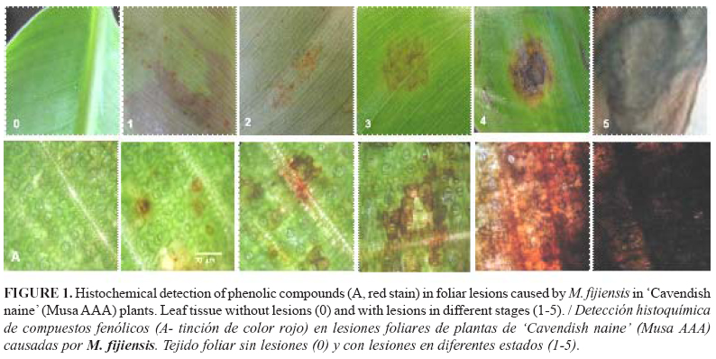
Leaf segments (samples) were placed on clean glass microscope slides before staining. The accumulation of phenolic compounds was visualized by the protocol described by Reeve (10) for cathecol derivatives. One drop of 10% (v/v) aqueous sodium nitrate, one drop of 10% (v/v) aqueous acetic acid and one drop of 20% (w/v) aqueous urea were added. After 3 min, two drops of 2N NaOH were finally added. Samples were examined with a compound light microscope (Olympus, 200X) and photographed immediately with a digital camera (Canon PC1201). A deep cherry red colouration was observed in the presence of phenolics.
The total phenol concentration was determined colorimetrically according to Bray and Thorpe (11). The samples were grounded in liquid nitrogen and extracted in 10 ml of 80% (v/v) ethanol, boiled at 50ºC for 30 min, and then centrifuged at 8 000 g for 10 min. Reaction volume included 1 ml supernatant, 3 ml distilled water, 1 ml Folin Ciocalteau reagent and 2 ml 20% (w/v) sodium carbonate. The ethanolic extract was heated for 1 min in a boiling water bath and cooled in tap water. The solution was diluted to 10 ml with distilled water and the intensity of the blue colour was measured at 750 nm in a spectrophotometer UV-visible (Genesys 6, Thermo Electron Corporation, USA) using a blank (the blank was obtained with 3 ml distilled water instead of the extract and the colour was developed as described above). A standard curve was prepared from known concentrations of gallic acid to calculate the concentration of phenolic compounds in the samples. The concentration of phenolic compounds was expressed as total phenol concentration (µg. cm-2).
The data were analyzed by using the Statistic Package for Social Science (SPSS) version 19.0 for Windows. The values obtained were analyzed statistically by nonparametric tests of Mann-Whitney after verifying the assumptions of normality and heterogeneity of variance.
The accumulation of phenols was detected in the foliar tissue as deep red coloured zone corresponding to lesions in all the symptom stages, which increased gradually while lesions progressed (Fig. 1). Similarly, it was observed in the total phenol concentrations (Fig. 2), where the highest values were obtained in the stages 4 and 5.
It has been demonstrated that, after inoculation, many structural and physiological changes occurs in the cells surrounding the infection site for restricting pathogen spread and for reducing fluid loss (12).
Phenol accumulation in foliar lesions is an evidence of the triggering of the defensive response in plants to pathogen attack, which has been described as one of the most important molecular events (4). In addition, it has been demonstrated that phenol accumulation after infection are directly toxic to pathogens (12) and their polymerization makes the cell wall thicker and stronger (11). This phenomenon has been observed in many pathosystem such as Sorghum vulgare Pers.-Sclerotium rolfsii Sacc. (15), Solanum lycopersicum L.-Botrytis cinerea Pers. (2), Eucalyptus sp.-Mycosphaerella sp. (3) and Lactuca sativa L.- Bremia lactucae Regel (16).
In Musa sp.- M. fijiensis interaction, those evidences are very limited; however, Portal (6) mentioned the possible relationship of the phenol metabolic pathway in Musa defense response to M. fijiensis in the susceptible genotype `Grande naine' by using a subtractive gen library. Accordingly, the results of this work provide a new direct evidence of a local accumulation of phenolic compounds in foliar lesions of `Cavendish naine' plants after M. fijiensis inoculation as part of the defence response of banana plants in this compatible interaction. At the same time, they validate the use of this histochemical tool to detect biochemical compounds as a simple and rapid alternative for testing different resistance phenotypes under controlled conditions, which can be applied in Musa breeding programs. In general, they may also contribute to a better understanding of this important plant-pathogen interaction.
ACKNOWLEDGMENT
This work was developed in the frame of the Program of Institutional University Cooperation among the Universidad Central ¨Marta Abreu¨ de Las Villas and the Flemish Interuniversity Council of Belgium (IUC UCLV/VLIR).
REFERENCES
1. Valerio R, Lindorf H, de García E. Relationship between leaf anatomy of some varieties of Musa sp. and its behaviour towards Sigatoka (yellow and black) disease. Agronomía Tropical. 2002;52(4):507-521.
2. Asselbergh B, Curvens K, França SC, Audenaert K, Vuylsteke van BF, Höfte M. Resistance to Botrytis cinerea in sitiens, an abscisic acid-deficient tomato mutant, involves timely production of hydrogen peroxide and cell wall modifications in the epidermis. Plant Physiology. 2007;144:1863-4877.
3. Smith AH, Gill WM, Pinkard EA, Mohammed CL. Anatomical and histochemical defence responses induced in juvenile leaves of Eucalyptus globulus and Eucalyptus nitens by Mycosphaerella infection. For Path. 2007;37:361-373.
4. Walters D, Newton A, Lyon G. Induced resistance: Helping plants to help themselves. Biologist. 2005;52:28-33.
5. Carlier J, Fouré E, Gauhl F, Jones D, Lepoivre P, Mourichon X, et al. Black Leaf Streak. In: Jones DR (ed.). Fungal Disease of the Foliage. 2000; 37-79.
6. Portal O. Development of molecular tools to study the interaction between banana and Mycosphaerella fijiensis, the causal agent of Black Leaf Streak Disease. 2008. PhD thesis. Ghent University, Belgium.
7. Leiva-Mora M, Alvarado-Capó Y, Acosta-Suárez M, Cruz-Martín M, Sánchez-García C, Roque B. Protocolo para la inoculación artificial de plantas de Musa spp. con Mycosphaerella fijiensis y evaluación de su respuesta mediante variables epifitiológicas y componentes de la resistencia. Biotecnología Vegetal. 2010;10(2):79-88.
8. Orellana P. Tecnología para la micropropagación in vitro de clones de Musa spp. 1994. Tesis de Doctor en Ciencias. Universidad Central ¨Marta Abreu¨ de Las Villas-Instituto de Biotecnología de las Plantas, Santa Clara, Cuba.
9. Alvarado Y, Leiva M, Rodríguez MA, Acosta M, Cruz M, Portal O, et al. Early evaluation of Black leaf streak resistance by using mycelial suspension of Mycosphaerella fijiensis. In: Jacome L, Leproive P, Martin D, Ortiz R, Romero R, Escalant JV (eds). Mycosphaerella leaf spot diseases of bananas: present status and outlook. 2003; 169-175. INIBAP, Montpellier. ISBN 2-910810-57-7.
10.Reeve RM. Histochemical tests for polyphenols in plant tissues. Stain Tech. 1951;26:91-96.
11.Bray W, Thorpe V. Analysis of phenolic compounds of interest in metabolism. Meth Biochem Analysis. 1954;1:27-52.
12.Ferreira RB, Monteiro S, Freitas R, Santos CN, Chen Z, Batista LM, et al. The role of plant defence proteins in fungal pathogenesis. Molecular Plant Pathology. 2007;5:677-700.
13.Hernández Y, Portillo F, Portillo MP, Navarro C, Rodríguez M, Velazco J. Densidad estomática en materiales de plátano (Musa AAB, AAAB y ABB) susceptibles y resistentes a Sigatoka Negra (Mycosphaerella fijiensis, Morelet). Rev Fac Agron. 2006;23(3):114-121.
14.Basha SA, Sarma BK, Singh DP, Singh UP. Differential methods of inoculation of plant growth-promoting rhizobacteria induce synthesis of phenylalanine ammonia-lyase and phenolic compounds differentially in chickpea. Folia Microbiol. 2006;51:463-468.
15.Maurya S, Singh R, Singh DP, Singh HB, Srivastav JS, Singh U. Phenolic compounds of Sorghum vulgare in response to Sclerotium rolfsii infection S. Journal of Plant Interactions. 2007;2(1):25-29.
16.Lebeda A, Sedlarova M, Petrivalsky M, Prokopova J. Diversity of defence mechanisms in plant-Oomycete interactions: a case study of Lactuca spp. and Bremia lactucae. European Journal of Plant Pathology. 2008;122:71-89.
Recibido: 8-5-2012.
Aceptado: 26-9-2012.

