


Mi SciELO
 Servicios personalizados
Servicios Personalizados
Revista

Articulo
 Inglés (pdf)
 Articulo en XML
 Referencias del artículo
 Enviar articulo por email
Indicadores
-
 Citado por SciELO
Links relacionados
-
 Similares en
SciELO
Similares en
SciELO
Compartir

 Permalink
Vaccimonitor
versión impresa ISSN 1025-028X
Vaccimonitor vol.20 no.2 Ciudad de la Habana Mayo.-ago. 2011
ARTICULOS ORIGINALES
Development and characterization of murine monoclonal antibody specific for the P1.4 PorA proteins from strain B:4:P1.(7b).4. of Neisseria meningitidis
Desarrollo y caracterización de un anticuerpo monoclonal murino específico a la proteína PorA P1.4 de la cepa B:4:P1.(7b).4 de Neisseria meningitidis
María Elena Pérez,* Ramón Barberá, Francisco Domínguez, Oscar Otero, Mercedes Gutiérrez, Gustavo Falero, Grethel Reyes, Franklin Sotolongo, Gustavo Sierra
Instituto Finlay. Centro de Investigación y Producción de Vacunas. Ave. 27 No. 19805, La Lisa, A.P. 16017 Cod. 11600. La Habana, Cuba.
*Máster en Ciencias e Investigadora Auxiliar. Instituto Finlay, Centro de Investigación-Producción de Vacunas. email:meperez@finlay.edu.cu
ABSTRACT
Neisseria meningitidis isolates are conventionally classified by serosubtyping that characterizes the reactivities of the PorA outer membrane protein variable-region epitopes with monoclonal antibodies. Porins are outer membrane proteins (OMPs) of N. meningitidis serogroup B and have attracted study principally for two reasons: their use in the classification of meningococcal isolates into serotype and subtype and as potential components of vaccines against this important pathogen. New murine hybridomas, secreting specific monoclonal antibodies against PorA serotype P1.4 of N. meningitidis serogroup B, were generated using conventional hybridoma procedures. The monoclonal antibodies obtained were characterized by Western blot and whole cell ELISA, using reference strains from different N. meningitidis serotypes and subtypes. All monoclonal antibodies belong to isotype IgG1. Others hybridomas producing MAbs against PorB and FrpB were also obtained.
Keywords: Monoclonal Antibodies, PorA, Neisseria meningitidis.
RESUMEN
Los aislamientos de Neisseria meningitidis se clasifican convencionalmente por serosubtipos. Su reactividad se realiza entre el epítope de la región variable de la proteína de membrana externa PorA con anticuerpos monoclonales. Las porinas, proteínas de membrana externa de N. meningitidis del serogrupo B, son atractivas para su estudio principalmente por la clasificación en serotipo y subtipo de los aislamientos del meningococo y como posibles componentes de vacunas contra este importante agente patógeno. Se generaron nuevos hibridomas murinos secretores de anticuerpos monoclonales específicos contra la proteína PorA subtipo P1.4 de N. meningitidis del serogrupo B, mediante los procedimientos convencionales de hibridomas. Los anticuerpos monoclonales, pertenecientes al isotipo IgG1, fueron caracterizados mediante Western blot y ELISA de células enteras. Se utilizaron cepas de referencia de diferentes serotipos y subtipos de N. meningitidis y se obtuvieron hibridomas productores de anticuerpos monoclonales contra otras proteínas como PorB y FrpB.
Palabras clave: Anticuerpos monoclonales, PorA, Neisseria meningitidis.
INTRODUCTION
The obligate human pathogen Neisseria meningitidis is a Gram-negative bacterium that variably colonizes the nasopharynx of healthy individual (1,2). Invasive meningococcal disease causes a significant public health burden throughout the world, with estimates of 500 000 cases and more that 50 000 deaths reported annually (3).
The inability to use a polysaccharide vaccine to protect against group B meningococci has limited the ability to control group B meningococcal disease by vaccination. The weak immunogenicity of the group B polysaccharide means that subcapsular antigens, and in particular the PorA and PorB outer membrane proteins, have become a focus as vaccine components.
The class 1 protein is PorA. Expression of the class 2 and class 3 proteins (PorB) is mutually exclusive, and they are products of the porB locus. PorA and PorB are important epidemiological markers that are the targets of serosubtyping and serotyping antibodies, respectively and are the targets of bactericidal antibodies (4).
Since mid-1991, New Zealand has experienced an epidemic of meningococcal disease. The epidemic has been caused by serogroup B meningococci expressing PorA type P1.7-2, 4, belonging to the ST-41/ST-44 complex, lineage III. In various western countries, subtype P1.4 of Neisseria meningitidis serogroup B causes the greatest incidence of meningococcal disease too (5).
We report the generation of hybridomas producing monoclonal antibodies (MAbs) that recognized PorA outer membrane protein only from N. meningitidis strains subtype P1.4.
MATERIALS AND METHODS
N. meningitidis outer membrane vesicles
Outer membrane vesicles (OMV) were manufactured by a detergent extraction method (6). Briefly:OMVs were obtained from live bacteria by gentle extraction with 10% deoxycholate (Merck). Bacterial debris was removed by centrifugation and nucleic acids were eliminated by enzymatic treatment with nucleases (Merck). OMVs were purified by gel filtration chromatography on Sephacryl S-300 (Pharmacia Fine Chemicals) followed by precipitation with 96% ethanol. The outer membrane vesicles from B:4:P1.(7b).4 strain were used for immunizing the BALB/c mice and ELISA.
Bacterial strains, growth condition and monoclonal antibodies
Prototype strains used in this work were stored at -700C in 10% skim milk (Oxoid, England) containing 20% (v/v) glycerol; before use they were cultured on GC (Difco) agar plate overnight in a humid atmosphere containing 5% CO2. The bacteria were then scraped from the plate with sterile cotton swabs and suspended in phosphate-buffered saline (PBS).
After inactivation of bacteria at 560C in a water bath for a minimum of 30 minutes, the suspension was adjusted to an absorbance of 0.09 or less, and then stored at 40C until used.
Specific P1.4 subtype MAb (MN20B9.34, IgG2a) was used as control, and it was supplied by Pierre Voet from GSK.
Enzyme immunoassay for detection of specific antibodies
Polystyrene micro titer ELISA plates (Costar, USA) were coated overnight at 40C with 100 mL per well of OMV a concentration of 5 mg/mL in PBS. The wells were subsequently filled with 2% skim milk solution in PBS and incubated for 1 h at room temperature (RT) to reduce non-specific binding. Samples, consisting in serial dilution of sera or culture supernatants, were added and incubated for 2 h at RT. Plates were washed with 0.05% tween-20 in PBS (PBS-T) and incubated with peroxidase-conjugated anti-mouse IgG (whole molecule) (Sigma Chemical Co., USA). Color was developed with o-phenylenediamine (0.4 mg/mL) and 0.4% H2O2 in 0.1 M sodium citrate buffer, pH 5.0. The reaction was stopped by adding 2.5 N H2SO4 and the O.D492nm read with a plus-multiscan microplate reader (Labsystem, UK).
Whole-cell enzyme immunoassay
Several suspensions of N. meningitidis strains were used for coating the well of polystyrene microtiter ELISA plates (Costar, USA) overnight at 37 0C. Samples, consisting in purified monoclonal antibodies or culture supernatants were added and incubated for 2 h at RT. Plates were washed with PBS-T and incubated with peroxidase-conjugated anti-mouse IgG (whole molecule) (Sigma Chemical Co., USA). Color was developed with o-phenylenediamine (0.4 mg/mL) and 0.4% H2O2 in 0.1 M sodium citrate buffer, pH 5.0. The reaction was stopped by adding 2.5 N H2SO4 and the O.D492nm read with a plus-multiscan microplate reader (Labsystem, UK).
Production of MAbs
Female BALB/c mice (6-8 week old) were immunized by SC injection of OMV (20 µg) emulsified in Freund's adjuvant (Sigma Chemical Co., USA). Each animal received four injections administrated at 2-week intervals. Mice were bled by their tail veins 7 to 10 days after the final injection and their serum tested for anti-OMV antibodies in the ELISA described above. Three to four days before cell fusion, the appropriate mouse received a final injection of antigen (10 mg) in PBS. Splenocytes were fused with the P3X63-Ag 8.653 mouse myeloma cell line using polyethylene glycol 1300 Hybri-Max (Sigma Chemical Co., USA) as described by Campbell (7). The hybrid cells were screened for their ability to secrete antibodies binding OMV in the direct ELISA (350 clones assay) and 200 clones for Western blot.
A second round of screening was made by Western blot analysis using as whole-cell and a reference MAb specific to subtype P1.4. Hybrids-secreting reactive antibodies were subcloned by limiting dilution and stabilized. The selected hybridoma cells were grown as ascites in the peritoneal cavity of pristane-primed BALB/c mice. Ascites fluid was tapped from the peritoneal cavity and rendered cell-free by centrifugation at 1000 g for 15 min at 4 0C. The MAbs were purified from ascites fluid using protein A affinity chromatography (8).
Isotyping
Classes and subclasses of MAbs secreted by hybridoma were determined with an ImmunoType Kit (Sigma Chemical Co., USA) following manufacturer's instructions.
Immunoblot analysis of MAbs
OMV or whole-cell lysates were fractionated in 12.5% SDS-PAGE according to Laemmli (9) and transferred to nitrocellulose paper as described by Towbin (10). After blocking with 5% skim milk in PBS, the blot was reacted with a suitable dilution of MAbs. The papers were washed with PBS-T and incubated with peroxidase-conjugated anti-mouse IgG (whole molecule) (Sigma Chemical Co., USA). Color was developed with diaminobenzidine (0.2 mg/mL) (Sigma Chemical Co., USA) and 0.4% H2O2 in tris-buffered saline, pH 8.0.
RESULTS
Hybrids producing anti-OMV antibodies were screening by a direct ELISA and Immunoblotting. Table 1 shows the results of specific MAb-secreting clones obtained against different antigens (PorA, PorB and FrpB).
Immunoblot analysis and ELISA of MAb anti PorA
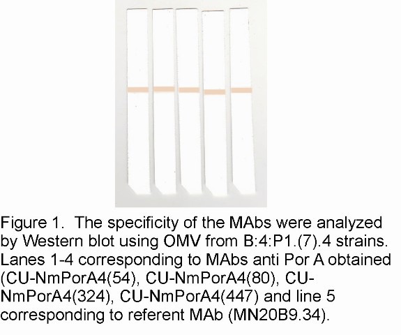
The specificity of the obtained MAbs anti PorA were studied by Immunoblot analysis using a strain of N. meningitidis separated by SDS-PAGE. Figure 1 (lane 1-4) shows that all MAbs recognized a 46-kDa antigen, corresponding to PorA protein from N. meningitidis serogroup B strain B:4:P1.(7b).4. This finding was corroborated by using reference MAbs anti PorA protein against subtype P1.4. The four MAbs obtained were coded as follow: CU-NmPorA4 (54); CU-NmPorA4(80); CU-NmPorA4(324) and CU-NmPorA4(447). The isotype of all MAbs were IgG1.
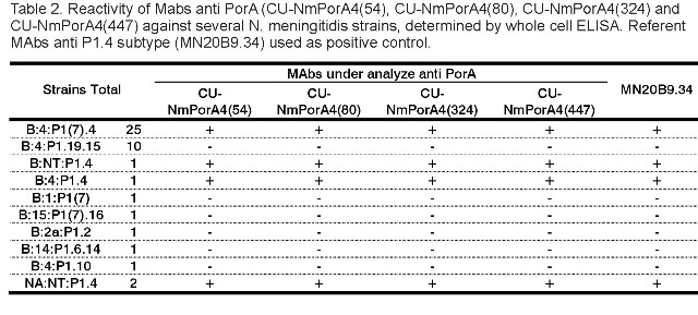
We also evaluated the specificity for subtypes P1 (7) and P1.4, corresponding to RV1 and RV2, respectively. To do this we tested the MAbs anti PorA in whole-cell enzyme immunoassay and Immunoblot using a panel of different serotype and subtype N. meningitidis strains. The MAbs only recognized the strains that included in their antigenic structure P1.4 protein as shown in Table 2 and Figure 2 (A, B, C, D). The results were endorsed by using anti P1.4 specific MAb in both techniques.
DISCUSSION
Methods, which use a 96-well configuration (ELISA), are clearly the most practical tool for screening large number of wells required for monoclonal antibody production. Here a double-screening method (Immunoblot and ELISA) was used in the primary screening in order to identify positive clones.
The decision to use Immunoblotting as screening strategy in hybridoma production against OMV proteins was based on several factors: the antigen preparation used for both immunization and screening was OMV; the inaccessible P1.7 subtype epitope from PorA protein only is available after protein denaturalization (11); previous findings showed that when OMV from CU-385-83 strains was used for MAbs generation only was obtained MAbs against PorA and PorB3 proteins (12); and finally sera collected from mice immunized with OMV protein and examined by Immunoblotting recognized several different antigens, including PorA, PorB3 and FrpB, among others.
As it is shown in the Table 1, MAbs against to FrpB only were detected when using Immunobloting assay as the selection methods for primary screening, whereas MAbs against PorA and PorB were detected using both Immunobloting assay and ELISA. MAbs against PorA were analyzed in more detail later and all of them reacted in whole cell ELISA and Immunoblotting only against subtype P1.4 strains.
The use of this screening strategies contributed to obtain MAbs against FrpB, PorA subtype P1.4 and PorB3 proteins from outer membrane of Neisseria meningitidis. The reason for the inaccessibility of the epitope from FprB in ELISA assay using OMV as coating antigen, are now being studied.
Characterization of meningococcal strains has been based on antigenic differences in the capsule (serogroup), the four variable regions (VRs) of the PorB OMP (serotype), and the two variable regions (VRs) of the PorA OMP (serosubtype) (13). This last one is being considered a promising vaccine candidate (14).
Monomeric PorA is a transmembrane protein with eight outer loops, among which the first and the fourth loop contain the hypervariable regions, called VR1 and VR2, respectively (15). These two regions define the dual PorA subtype, designated P1. x, y, where "P1" stands for the class 1 protein and "x" and "y" stand for numbers denoting the VR1 and VR2 domains, respectively. The two subtype regions from PorA are generally determined by an whole cells ELISA or a blotting assay (16) using referent MAbs directed against epitopes in VR1 and VR2; consequently, each PorA can bind two different subtype specific MAbs.
We have produced and characterized four MAbs against PorA outer membrane protein only from N. meningitidis strains subtype P1.4. Western blot analysis (Figure 1) showed that MAbs anti PorA recognized an approximately 46 kDa protein presented in N. meningitidis B:4:P1.(7b).4. strain, that closely corresponds to the molecular mass predicted for the product of the Por A gene (16).
The antibodies only reacted with subtype P1.4 strains when it was evaluated by Immunoblot analysis and whole cell ELISA (see Figure 2 A, B, C, D and Table 2). These results suggested that our MAbs were specific for VR2 of PorA protein.
The most appropriate screening methods and subtyping analysis are required for an adequate investigation. A panel of well-characterized subtype-specific MAbs should be selected, in order to classify more strains.
The production of new MAbs will improve and facilitate the study of the structure and function of different N. meningitidis antigens in great detail, permitting to carry out other characterization studies from clinical isolates, necessary to increase the epidemiological knowledge.
REFERENCES
1. TrotterCL, Gay NJ, Edmunds WJ. The natural history of meningococcal carriage and disease. Epidemiol Infect 2006;134:556-66.
2. Stephens DS, Greenwood B, Brandtzaeg P. Epidemic meningitis, meningococcemia and Neisseria meningitidis. Lancet 2007;369:2196-210.
3. Harrison LH, Trotter CL, Ramsay ME. Global epidemiology of meningococcal disease.Vaccine 2009;27S:B51-63.
4. Frasch CE, Borrow R, Donnelly J. Bactericidal antibody is the immunologic surrogate of protection against meningococcal disease. Vaccine 2009;27S:B112-6.
5. Oomen CJ, Hoogerhout P, Kuipers B, Vidarsson G, van Alphen L, Gros P. Crystal Structure of an Anti-meningococcal Subtype P1.4 PorA Antibody Provides Basis for Peptide-Vaccine Design. Journal of Molecular Biology 2005;351:1070-80.
6. Huergo CC, Sierra GV, Gutiérrez MM, Biset G, García L, Puentes G, et al. Centro Nacional de Biopreparados, assignee. Method of producing Neisseria meningitidis B vaccine. US Patent 5,597,572. 1997, Jan 28.
7. Gavilondo JV. Anticuerpos Monoclonales. La Habana: Elfos Scientiae 1995. p. 46-9.
8. Tu YY, James PF, Goldenberg DM: Temperature affects binding of murine monoclonal antibodies to proteins. A.J Immunol Methods 1998;109:43-7.
9. Laemmli UK. Cleavage of structural proteins during the assembly of bacteriophage T4. Nature 1990;227:680-5.
10. Towbin H, Staehlin T, Gordon J. Electrophoretic transfer of proteins from poliacrylamide gels to nitrocellulose sheets procedure and some applications. Proc Natl Acad Sci. USA. 1979;76:4350-4.
11. Wedege E, Dalseg R, Caugant DA, Poolman JT, Frholm LO. Expression of an inaccessible P1.7 subtype epitope on meningococcal class 1 proteins. J Med Microbiol 1993;38:23-8.
12. Pérez ME, Barberá R, Domínguez F, Otero O, Gutiérrez M, Falero G, et al. Development and characterization of murine monoclonal antibody specific for the P1.15 PorA proteins from a Cuban vaccine strain B:4,7:P1.19,15 of Neisseria meningitidis. Hibridoma 2006;25:243-7.
13. Stephens DS. Conquering the meningococcus. FEMS Microbiol Rev 2007;31:3-14.
14. Sadarangani M, Pollard A. Serogroup B meningococcal vaccines an unfinished story. Lancet Infect Dis 2010;10:112-24.
15. Climent Y, Urwin R, Yero D, Martínez I, Martín A, Sotolongo F, et al. The genetic structure of Neisseria meningitidis populations in Cuba before and after the introduction of a serogroup BC vaccine. Infection, Genetics and Evolution 2010;10:546-54.
16. Climent Y, Yero D, Martínez I, Martín A. Jolley KA, Sotolongo F, et al. Clonal distribution of disease-associated and healthy carrier isolates of Neisseria meningitidis between 1983 and 2005 in Cuba. Journal of Clinical Microbiology 2010;48:802-10.
Recibido: Noviembre de 2010
Aceptado: Febrero de 2011

