


Mi SciELO
 Servicios personalizados
Servicios Personalizados
Revista

Articulo
 Inglés (pdf)
 Articulo en XML
 Referencias del artículo
 Enviar articulo por email
Indicadores
-
 Citado por SciELO
Links relacionados
-
 Similares en
SciELO
Similares en
SciELO
Compartir

 Permalink
Biotecnología Aplicada
versión On-line ISSN 1027-2852
Biotecnol Apl vol.33 no.4 La Habana oct.-dic. 2016
TECHNIQUE
A simple method for determining protein-bound homocysteine and cysteine in human plasma by capillary electrophoresis
Método simple para la determinación de homocisteína y cisteína unida a proteínas en muestras de plasma humano mediante electroforesis capilar
Virus E Danielevich, Ivanov A Vladimirovich, Kuchukova M Iusupovna, Luzyanin B Petrovich, Kubatiev A Amirkhanovich
FSBSI Institute of General Pathology and Pathophysiology. Moscow, 125315, Baltiyskaya str.,8, Moscow, Russia.
ABSTRACT
A capillary electrophoresis method with UV detection has been developed for the determination of protein-bound cysteine and homocysteine in human plasma based on the combination of derivatization step with pH-mediated base stacking. Centrifugal ultrafiltration was carried out to clarify plasma proteins from salts and low-molecular weight compounds. Thereafter, the sample was incubated with dithiothreitol to reduce the disulfides and release protein-bound aminothiols. The released thiols were derivatized with thiocarbonyldiimidazole and injected into the capillary electrophoresis by voltage. Due to the stacking effects it is possible to perform a considerable on-line pre-concentration of the analytes. The proposed approach allows to reach a detection limit of 1 μmol/L in blood plasma using 48.5 cm total length 50-μm i.d. uncoated capillary. The coefficient of variation of the assay was within ± 5 % for both homocysteine and cysteine.
Keywords: protein-bound homocysteine, protein-bound cysteine, thiocarbonyldiimidazole, capillary electrophoresis, human plasma, UV detection.
RESUMEN
Se desarrolló un método basado en electroforesis capilar para la detección mediante luz ultravioleta de residuos de cisteína y homocisteína unidos a proteínas en plasma humano, mediante la combinación de un paso de derivatización con apilamiento mediado por pH. Las sales y los compuestos de bajo peso molecular se eliminaron de la mezcla de proteínas mediante ultracentrifugación. A continuación, la muestra se incubó con ditiotreitol para reducir los puentes disulfuro. Los compuestos derivatizados se inyectaron a la electroforesis capilar mediante su aplicación por voltaje. El efecto de apilamiento permitió una considerable preconcentración de los analitos antes de su separación. El enfoque propuesto permite alcanzar un límite de detección de 1 μmol/L en muestras de plasma sanguíneo con el empleo de un capilar no recubierto de 48.5 cm de largo y un diámetro interno de 50 μm. El coeficiente de variación del ensayo para la homocisteína y la cisteína fue de ± 5 %.
Palabras clave: cisteína unida a proteínas, homocisteína unida a proteínas, tiocarbonildiimidazol, electroforesis capilar, plasma humano, detección UV.
INTRODUCTION
About of 70 % of total Hcy and 50 % of Cys are bound to proteins in blood plasma [1]. Elevated level of Hcy (hyperhomocysteinemia) is independent marker of various cardiovascular diseases [2]. The determination of protein-bound Hcy is attractive because some plasma proteins are discussed as carriers of this compound into the cells given that their presence causes cell dysfunction, especially endothelial dysfunction [3, 4]. Furthermore, the homocysteinilation may be associated with oxidative stress and/or with dysfunction of plasma proteins [5-7].
Plasma cysteine is an end product of Hcy transsulfuration pathway and major competitor for binding sites on proteins [8]. Close metabolic relations of this amino acids cause an interest to using of Hcy/Cys ratio as alternative total Hcy. This ratio may better reflect abnormalities of transsulfuration enzymes, affects bioavailability of Hcy or reveal of preanalytical errors [8]. Also it was shown that high plasma homocysteine concentration is associated with increased risk of colorectal cancer among postmenopausal women, whereas high cysteine is associated with decreased risk [9].
Although immunoassay analysis [10] is a widespread routine method for determination of total (oxidized + reduced) Hcy but it cannot been applied to the determination of Cys. To solve this problem many analytical methods based on HPLC or CE with laser induced fluorescence (LIF) [1, 11, 12], electrochemical [13] or mass spectrometry (MS) detectors [14, 15] have been proposed. Most methods are relevant to the analysis of total aminothiols, but they may also be applied to their bound forms. Unfortunately, the poor throughput of these approaches makes it difficult their implementation in clinical laboratories. In our opinion, available HPLC and CE with ultraviolet light (UV)-detectors are more accessible for clinical laboratories. However, only several HPLC-UV methods have been proposed for the quantitative determination of aminothiols in blood plasma [16-18]. Some CE-UV methods with a derivatization step were proposed [19-22], but their sensitivity was insufficient to detect plasma Hcy, in contrast to CE-LIF and HPLC-UV.
Several years ago, Zinellu et al. have shown that CE-UV sensitivity is sufficient for the direct determination of Hcy and Cys at 190 nm in the micromolar range in model mixtures [23]. Obviously, high salts concentration and potential interferences will hamper to the sensitive CE-UV detection of protein-bound homocysteine and cysteine in human plasma. However, noteworthy works of Kubalzcyk et al. where a good LOD (1 μmol/L) was reached for total Hcy in human plasma samples [24, 25]. They applied Hcy derivatization with 2-chloro-1-methylquinolinium tetrafluoroborate. Unfortunately, this reagent is not commercially available. High sensitivity was also achieved by Chang and Tseng [26]. They used gold nanoparticles to achieve a great concentration effect with achieving of LOD 10-65 nM. Despite the attractiveness of this approach, it is time-consuming because of multistage sample preparation and long analysis time. Moreover, protein-bound thiols were calculated as the difference in concentration between total and free their forms. Therefore from our point of view, indirect methods in combination with in-capillary concentration would be preferable.
In this case, reagents should meet the following requirements. First, its derivatives must be suitable for pre-concentration. Second, excess of reagent must not create interferences. Early used reagents (5,5’-(2-dithiobisnitrobenzoic) acid, monobromobimane, 4-aminosulfonyl-7-fluoro-2,1,3-benzoxadiazole and 2,2’-dipyridyl disulfide) were not are not completely satisfied with these requirements. On the other hand, Amarnath et al. used thiocarbonyldiimidazole (TCDI) for derivatization of Cys, Hcy and HPLC for separation of corresponding derivatives which can be able to absorb in UV region [27]. TCDI binds NH2- and SH-groups of analytes via thiocarbonyl moiety. Thus, the reaction products acquire acidic character as their carboxyl groups of aminothiols are preserved.
TCDI and products of its hydrolysis are also weak electrolytes and they do not have a significant impact on in-capillary concentration. This allows us to use pH-mediated base stacking [28]. This approach includes electrokinetic injection (EI) of sample and subsequent postinjection of alkaline solution (base stacking). Sample anions (include analytes) migrate in capillary and formed an injection zone at EI stage. The key parameter of EI effectiveness (i.e. amount of injected analytes) is ratio BGE/sample conductivity or ionic strength. BGE ionic strength is limited by heating during CE therefore it is necessary to minimize sample ionic strength. A possibility subsequent removing of salts and plasma proteins, low-ionizable properties of TCDI are appropriate conditions for EI. If injection time is a sustained then length of injection zone may be too long for good separation. In that case an alkaline postinjection is used for titration and narrowing of injection zone [28].
Thereby the aim of this study was to develop a sensitive, fast, simple and available electrophoretic method with UV-detection for determination of protein-bound Hcy, Cys and corresponding Cys/ Hcy ratio in human blood plasma based on the combination of derivatization step with in-capillary concentration.
MATERIALS AND METHODS
Reagents
Formic acid for LC-MS (Fluka, Germany), TCDI purum (Sigma-Aldrich, Switzerland), ammonium acetate pure (Reahim, Russia), acetonitrile HPLC grade (Himmed, Russia), NaCl purum p.a. (Fluka, Switzerland), D-penicillamine (PA; Sigma-Aldrich, Germany), DL-dithiotreitol (DTT, > 99.5 %; Fluka, Germany), Cys 97 % (Aldrich, USA), cysteinylgly-cine (CysGly) > 85 % (Sigma, Germany), Hcy > 95 % (Sigma, Germany), hexadecyltrimethylammonium bromide (CTAB; Sigma, India), NaOH purum (Dia- M, Germany).
Deionized water was prepared by using a deionizer Millipore Simplicity 185 with Simpakor 1 cartridge. Stock solutions of Cys (100 mmol/L), Hcy (50 mmol/L) and PA (50 mmol/L) were prepared by dissolving of appropriate amounts of analytes 0.1 % (v/v) formic acid and kept at –80 ºC. For plasma proteins reduction prior to derivatization a fresh 0.1 mol/L solution of DTT was prepared. TCDI solution was prepared by dissolving of appropriate amount of the reagent in one volume acetonitrile and dilution by 9 volumes of water.
Instrumentation
Agilent CE 3D system (Germany) with diode-array detector and 50 μm i.d. capillary 48.5 cm length (40 cm eff.) was used. Absorption was detected at 254 ± 20 nm with frequency 5 s-1. Centrifuge filters Amicon Ultra-3K (Millipore, Ireland) were used for ultrafiltration.
Sample preparation
Venous blood was voluntarily donated by women who consented to provide the samples required for the study (N = 19, 35-47 years old, mean 41 years). Samples were collected in vacuum tubes with 3.8 % sodium citrate (Greiner Bio-One GmbH, Austria) and processed at 3000 g for 3 min. Plasma was separated and stored at –20 ºC. Blood plasma (50 μL) was mixed with deionized water (450 μL) and was filtrated through Amicon Ultra 3K filter at 14 000 g for 15 min. Then residual (upper) solution (~100 μL) was diluted in 400 μL water and was filtrated again. Then 50 μL of 100 μmol/L PA (internal standard) with 20 mmol/L DTT was added to residual solution. This mixture was incubated 10 min under 37 ºC and 100 μL of 50 mmol/L TCDI was added. Then mixture was centrifuged at 5000 g for 5 min and supernatant was ultrafiltrated 15 min at 14 000 g. Prepared samples were stored at 4 ºC up to 3 days before analysis.
Validation
Calibration solutions were performed using water and mixture of filtrated plasma samples as matrixes. For calibration in water 7 standard solutions were prepared (corresponding of Cys concentrations 0, 15.6, 31.3, 62.5, 125, 250 and 500 μmol/L and Hcy concentrations 0, 3.1, 6.3, 12.5, 25, 50 and 100 μmol/L). For calibration of bound thiols in plasma a solution of plasma proteins were prepared according to sample preparation procedure using ultrafiltration of eight plasma samples mixture. 5 portions of this filtrate were spiked with thiols (0, 62.5, 125, 250, 500 μmol/L Cys and 0, 12.5, 25, 50, 100 μmol/L Hcy) and incubated 10 min at 25 ºC. Reduction and derivatization were carried out according to the sample preparation procedure. Peak areas vs. the spiked concentrations were used to generate the calibration curves, which were fitted by least squares linear regression analysis.
Estimation of sensitivity (LOQ) was performed only for bound Hcy since its concentration in plasma lower (~4-8 μmol/L) than bound Cys (60-207 μmol/L) [1, 8, 29]. To take account “matrix effect” a “blank” plasma (i.e., without reduction but with derivatization) was prepared.
CE procedure
Before acquisition the capillary was rinsed 1 mol/L NaOH, 10 mmol/L CTAB, water and BGE (0.2 mol/L ammonium acetate with 25 μmol/L CTAB) for 2 min each one. CE was performed at –15kV 15 min. Flushing was 1 min water and 2 min BGE. Sample injection was at –15kV for 30 s the sample and 60 s for 0.2 mol/L NaOH.
RESULTS AND DISCUSSION
Advantageously, the concentration of sample ions in the injection zone in EI tends to the concentration of BGE ions, the sample being concentrated against it. Since our analytes are anions, a reverse polarity mode is required (cathode on inlet) for EI and separation. CTAB was used to invert electroosmotic flow (EOF) in the same direction. A typical electropherogram of human plasma with pH-mediated base stacking is shown in Figure 1. Noteworthy, it is also possible to determinate thiol-containing dipeptide CysGly, a product of plasma glutathione cleavage. Analytes were cleared from salts and proteins by ultrafiltration.
Widespread methods such as acid or alcohol precipitation and solid-phase extraction are difficult to combine with pH-mediated base stacking because they have drawbacks, due to the sample’s high ionic strength and/or sample dilution. At the same time, desalting leads to overestimations, while the addition of small amounts of NaCl (10 mmol/L) to prepared samples results in 10-fold reduced signal intensity (data not shown). Relative retention times of Cys and Hcy were 0.8 ± 0.02 and 0.887 ± 0.014, respectively. RRT was been calculated as: retention time of the analyte/retention time of internal standard (IS).
A calibration was carried out by adding internal standard (PA) and analytes in the concentration range 0-500 and 0-100 μmol/L for Cys and Hcy, respectively, to plasma protein solution and water. Calibration curves demonstrated a linear relationship with correlation coefficients above 0.995 (p < 0.0001) in all the cases. Linear regression equations were: y = 0.0135x + 1.4 and y = 0.0086x + 0.029 for Cys and Hcy, respectively. The close slope was taken from analytes calibration in water solution (y = 0.0136x + 0.124 and y = 0.0083x + 0.0095). Reproducibility was measured using six continuous injections and CV of Hcy/PA and Cys/PA were within the ± 5 % range. The absolute intensity varied slightly higher (8-12 %).
Two probes selected for sensitivity estimation: plasma with reduction and derivatization, and the other “blank” with plasma without reduction but with TCDI also. The first probe (plasma sample with reduction and derivatization) was diluted by second probe (blank sample without reduction but with TCDI also) in 2.4 and 8 times. In the latter case the S/N ratio for Hcy was about of 7 that corresponding its concentration 0.8 μM in the sample.
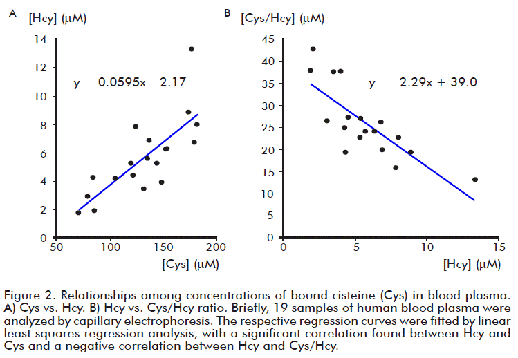
We analyzed 19 samples of human blood plasma. Concentrations of bound Cys and Hcy were 132 ± 35 (70-182) and 5.7 ± 2.7 (1.9-13.3) μmol/L, respectively. These levels are between those previously reported for control groups of healthy people [1, 8, 29]. The Cys/Hcy ratio was 26 ± 8 (13-43). It should be noted that CV for Hcy (47 %) was slightly higher than the CV for Cys/Hcy (31 %). There was a significant correlation between Hcy and Cys (Figure 2A, r = 0.77, p < 0.001), and a negative correlation between Hcy and Cys/Hcy (Figure 2B, r = –0.79, p < 0.001). These findings are similar for total Hcy and Cys as previously reported [8]. Additionally, a strongly positive correlation was observed between Hcy and Hcy/Cys (r = 0.86, p < 0.001, data not shown). Also, there was no positive correlation between Cys and Cys/Hcy (r = –0.41, data not shown).
Thus, CE-UV approach for the determination of bound plasma Hcy and Cys has been developed. Plasma proteins are separated from free thiols and salts by ultrafiltration, but it is possible to use gel-filtration chromatography as alternative. Since bound thiols exist in an oxidize state, there is necessary to convert them to the reduce form by DTT before the derivatization step. The specific reagent TCDI was used to obtain derivatives able to be detected by UV absorption. Because of low salts concentration in samples and titratable acidic character of them, it was possible in-capillary concentration using pH-mediated base stacking [28]. Weak base character of TCDI and imidazole also favored EI of analytes too. Due to the high resolution power of CE, it was possible to resolve peaks of analytes from interfering compounds, and the theoretical plate number of this approach was about 65 000. It should be noted that this CE analysis, contrary to HPLC [27], SPE is not require, hence, the method proposed follows a simple sample preparation step. Additionally, the sensitivity of CE-UV analysis was enough to determine bound Hcy and at a level similar to that of an early reported HPLC-UV method [27].
CONCLUSIONS
The developed approach provides an opportunity to determine bound aminothiols with enough sensitivity (less than 1 μmol/L) by CE-UV, without using nanoparticles and with a commercially available derivatization reagent. It was shown a positive correlation between protein-bound Hcy and Cys.
ACKNOWLEDGEMENTS
This study was supported by grant from the Russian Science Foundation (project No. 16-15-10340).
REFERENCES
1. Williams RH, Maggiore JA, Reynolds RD, Helgason CM. Novel approach for the determination of the redox status of homocysteine and other aminothiols in plasma from healthy subjects and patients with ischemic stroke. Clin Chem. 2001;47(6):1031-9.
2. Newton LA, Sandhu K, Livingstone C, Leslie R, Davis J. Clinical diagnostics for homocysteine: a rogue amino acid? Expert Rev Mol Diagn. 2010;10(4):489-500.
3. Sengupta S, Wehbe C, Majors AK, Ketterer ME, DiBello PM, Jacobsen DW. Relative roles of albumin and ceruloplasmin in the formation of homocystine, homocysteine-cysteine-mixed disulfide, and cystine in circulation. J Biol Chem. 2001;276(50):46896-904.
4. Zhloba AA, Subbotina TF. Homocysteinylation score of high-molecular weight plasma proteins. Amino Acids. 2014;46(4):893-9.
5. Majors AK, Sengupta S, Willard B, Kinter MT, Pyeritz RE, Jacobsen DW. Homocysteine binds to human plasma fibronectin and inhibits its interaction with fibrin. Arterioscler Thromb Vasc Biol. 2002;22(8):1354-9.
6. McCully KS. Chemical pathology of homocysteine. IV. Excitotoxicity, oxidative stress, endothelial dysfunction, and inflammation. Ann Clin Lab Sci. 2009;39(3):219-32.
7. Zinellu A, Sotgia S, Deiana L, Carru C. Quantification of thiol-containing amino acids linked by disulfides to LDL. Clin Chem. 2005;51(3):658-60.
8. Hortin GL, Sullivan P, Csako G. Relationships among plasma homocysteine, cysteine, and albumin concentrations: potential utility of assessing the cysteine/homocysteine ratio. Clin Chem. 2001;47(6):1121-4.
9. Miller JW, Beresford SA, Neuhouser ML, Cheng TY, Song X, Brown EC, et al. Homocysteine, cysteine, and risk of incident colorectal cancer in the Women’s Health Initiative observational cohort. Am J Clin Nutr. 2013;97(4):827-34.
10. Pernet P, Lasnier E, Vaubourdolle M. Evaluation of the AxSYM homocysteine assay and comparison with the IMx homocysteine assay. Clin Chem. 2000;46(9):1440-1.
11. Wada M, Kuroki M, Minami Y, Ikeda R, Sekitani Y, Takamura N, et al. Quantitation of sulfur-containing amino acids, homocysteine, methionine and cysteine in dried blood spot from newborn baby by HPLC-fluorescence detection. Biomed Chromatogr. 2014;28(6):810-4.
12. Zinellu A, Lepedda A, Sotgia S, Zinellu E, Marongiu G, Usai MF, et al. Albumin-bound low molecular weight thiols analysis in plasma and carotid plaques by CE. J Sep Sci. 2010;33(1):126-31.
13. Baron M, Sochor J. Estimation of thiol compounds cysteine and homocysteine in sources of protein by means of electrochemical techniques. Int J Electrochem Sci. 2013;8:11072-86.
14. Jiang Z, Liang Q, Luo G, Hu P, Li P, Wang Y. HPLC-electrospray tandem mass spectrometry for simultaneous quantitation of eight plasma aminothiols: application to studies of diabetic nephropathy. Talanta. 2009;77(4):1279-84.
15. Ivanov AV, Luzyanin BP, Kubatiev AA. The use of N-ethylmaleimide for mass spectrometric detection of homocysteine fractions in blood plasma. Bull Exp Biol Med. 2012;152(3):289-92.
16. Andersson A, Isaksson A, Brattstrom L, Hultberg B. Homocysteine and other thiols determined in plasma by HPLC and thiol-specific postcolumn derivatization. Clin Chem. 1993;39(8):1590-7.
17. Katrusiak AE, Paterson PG, Kamencic H, Shoker A, Lyon AW. Pre-column derivatization high-performance liquid chromatographic method for determination of cysteine, cysteinyl-glycine, homocysteine and glutathione in plasma and cell extracts. J Chromatogr B Biomed Sci Appl. 2001;758(2):207-12.
18. Kusmierek K, Bald E. Reversed-phase liquid chromatography method for the determination of total plasma thiols after derivatization with 1-benzyl-2-chloropyridinium bromide. Biomed Chromatogr. 2009;23(7):770-5.
19. Russell J, Rabenstein DL. Speciation and quantitation of underivatized and Ellman’s derivatized biological thiols and disulfides by capillary electrophoresis. Anal Biochem. 1996;242(1):136-44.
20. Ivanov AR, Nazimov IV, Baratova LA. Determination of biologically active low-molecular-mass thiols in human blood. II. High-performance capillary electrophoresis with photometric detection. J Chromatogr A. 2000;895(1-2):167-71.
21. Kang SH, Wei W, Yeung ES. On-column derivatization for the analysis of homocysteine and other thiols by capillary electrophoresis with laser-induced fluorescence detection. J Chromatogr B Biomed Sci Appl. 2000;744(1):149-56.
22. Sevcikova P, Glatz Z, Tomandl J. Determination of homocysteine in human plasma by micellar electrokinetic chromatography and in-capillary detection reaction with 2,2’-dipyridyl disulfide. J Chromatogr A. 2003;990(1-2):197-204.
23. Zinellu A, Sotgia S, Scanu B, Pisanu E, Sanna M, Sati S, et al. Determination of homocysteine thiolactone, reduced homocysteine, homocystine, homocysteine-cysteine mixed disulfide, cysteine and cystine in a reaction mixture by overimposed pressure/ voltage capillary electrophoresis. Talanta. 2010;82(4):1281-5.
24. Kubalczyk P, Bald E. Transient pseudo-isotachophoretic stacking in analysis of plasma for homocysteine by capillary zone electrophoresis. Anal Bioanal Chem. 2006;384(5):1181-5.
25. Kubalczyk P, Bald E, Furmaniaka P, Glowacki R. Simultaneous determination of total homocysteine and cysteine in human plasma by capillary zone electrophoresis with pH-mediated sample stacking. Anal Methods. 2014;6(12):4138-43.
26. Chang CW, Tseng WL. Gold nanoparticle extraction followed by capillary electrophoresis to determine the total, free, and protein-bound aminothiols in plasma. Anal Chem. 2010;82(7):2696-702.
27. Amarnath K, Amarnath V, Amarnath K, Valentine HL, Valentine WM. A specific HPLC-UV method for the determination of cysteine and related aminothiols in biological samples. Talanta. 2003;60(6):1229-38.
28. Hoque ME, Arnett SD, Lunte CE. On-column preconcentration of glutathione and glutathione disulfide using pH-mediated base stacking for the analysis of microdialysis samples by capillary electrophoresis. J Chromatogr B Analyt Technol Biomed Life Sci. 2005;827(1):51-7.
29. Andersson A, Lindgren A, Hultberg B. Effect of thiol oxidation and thiol export from erythrocytes on determination of redox status of homocysteine and other thiols in plasma from healthy subjects and patients with cerebral infarction. Clin Chem. 1995;41(3):361-6.
Received in October, 2016.
Accepted in December, 2016.
Virus E Danielevich. FSBSI Institute of General Pathology and PathophysiologyMoscow, 125315, Baltiyskaya str.,8, Moscow, Russia. E-mail: edwardvirus@yandex.ru.

