SciELO - Scientific Electronic Library Online

 
vol.21 issue4Protective effect of NeuroEPO on the reproduction of diabetic ratsFamily inherited, prenatal and perinatal risk factors in Cuban children with primary autism author indexsubject indexarticles search
Home Pagealphabetic serial listing  

My SciELO

Services on Demand

Journal

Article

Indicators

  • Have no cited articlesCited by SciELO

Related links

  • Have no similar articlesSimilars in SciELO

Share


Revista Habanera de Ciencias Médicas

On-line version ISSN 1729-519X

Rev haban cienc méd vol.21 no.4 La Habana July.-Aug. 2022  Epub Aug 10, 2022


Biomedical basic sciences

Intranasal administration of NeuroEPO does not affect the structure of respiratory mucosa in Wistar rats

La administración de NeuroEPO intranasal no afecta la estructura de la mucosa respiratoria en ratas Wistar

Ketty Suárez Borrás1  * 
http://orcid.org/0000-0002-3998-1657

Gisselle Fernández Peña1 
http://orcid.org/0000-0002-5339-8118

Yamila Rodríguez Cruz1 
http://orcid.org/0000-0001-8492-9797

Giselle Puldón Seguí1 
http://orcid.org/0000-0002-1980-9016

1Universidad de Ciencias Médicas de La Habana, Instituto de Ciencias Básicas y Preclínicas “Victoria de Girón”. La Habana, Cuba.

ABSTRACT

Introduction:

Diseases associated with the Central Nervous System represent a group of conditions with important social and economic repercussions. New treatment strategies with NeuroEPO offer new opportunities to prevent the onset and progression of these disorders. NeuroEPO administered intranasally can reach the Central Nervous System through various mechanisms involving the olfactory and trigeminal nerves, respiratory and olfactory mucosa, nasal vasculature, cerebrospinal fluid, and the lymphatic system.

Objective:

To determine the effect of intranasal administration of NeuroEPO on the histologic structure of the respiratory mucosa and its associated lymphatic tissue in Wistar rats.

Material and Methods:

An experimental, descriptive, longitudinal, prospective study was conducted, using the Wistar rat as a biological model. Ten healthy animals were randomly distributed in two groups of five animals each. One of the groups received intranasal NeuroEPO for 28 days at doses of 300 μg/kg. The other group was given a vehicle at a rate of 0,3μl/g. The histological characteristics of the respiratory mucosa were studied. The Mann-Whitney test was used to compare the means.

Results:

No alterations in the histological characteristics of the respiratory mucosa and the lymphatic tissue associated with the nasal mucosa were observed in Wistar rats after the administration of NeuroEPO.

Conclusion:

Intranasal administration of NeuroEPO does not cause pathological changes in the histological structure of the respiratory mucosa or the lymphatic tissue associated with the nasal mucosa of Wistar rats in our experimental conditions.

Keywords: NeuroEPO; respiratory mucosa; neurodegenerative diseases; neuroprotection

RESUMEN

Introducción:

Las enfermedades asociadas al Sistema Nervioso Central representan un grupo de padecimientos con importante repercusión social y económica. Las nuevas estrategias de tratamiento con NeuroEPO ofrecen nuevas oportunidades para prevenir la aparición y la progresión de estos desórdenes. La NeuroEPO administrada por vía intranasal puede alcanzar al Sistema Nervioso Central a través de varios mecanismos que involucran los nervios olfatorios y trigéminos, mucosa respiratoria y olfatoria, vasculatura nasal, fluido cerebroespinal y el sistema linfático.

Objetivo:

Determinar el efecto de la administración intranasal de NeuroEPO sobre la estructura histológica de la mucosa respiratoria y de su tejido linfático asociado en ratas Wistar.

Material y métodos:

Se realizó un estudio experimental, descriptivo, longitudinal, prospectivo, utilizando la rata Wistar como modelo biológico. Se utilizaron diez animales sanos distribuidos aleatoriamente en dos grupos de cinco cada uno. Uno de los grupos recibió NeuroEPO intranasal durante 28 días a dosis de 300 µg/ kg. Al otro grupo se le administró vehículo a una razón de 0,3µl /g. Se estudiaron las características histológicas de la mucosa respiratoria. Las medianas de los grupos fueron comparadas mediante la prueba U de Mann-Whitney.

Resultados:

No se observaron alteraciones en las características histológicas de la mucosa respiratoria, ni del tejido linfático asociado a la mucosa nasal en ratas Wistar, tras la administración de la NeuroEPO.

Conclusión:

La administración intranasal de NeuroEPO no provoca cambios patológicos sobre la estructura histológica de la mucosa respiratoria ni del tejido linfático asociado a la mucosa nasal de ratas Wistar en nuestras condiciones experimentales.

Palabras clave: NeuroEPO; mucosa respiratoria; enfermedades neurodegenerativas; neuroprotección

Introduction

Diseases associated with the Central Nervous System (CNS) represent a group of conditions with significant social and economic repercussions. They are characterized by a high prevalence in both developed and developing countries. Cerebrovascular diseases (CVD) and those caused by neuronal degeneration attract the attention of major research centers. Nowadays, there is no effective treatment on the market that achieves a cure for these diseases.1,2

Currently, there is a lot of research on the neurobiological mechanisms related to brain aging and associated diseases. Scientists continue to search for methods to improve their treatment, both clinical and psychosocial.3,4

Despite the great effort undertaken in the analysis of the pathophysiology of neurological conditions and the development of new drugs to treat these conditions, the success obtained is limited.1,2,4 A growing line of research in neuroscience is the use of human recombinant molecules. Recombinant human erythropoietin with low sialic acid content (NeuroEPO) is a relevant example of the use of these molecules.1,2,3,4 NeuroEPO has cytoprotective, hypoglycemic and neuroprotective effect.4,5,6,7)

The systemic administration of NeuroEPO has the disadvantage that it is quickly degradable at the hepatic level due to the lower amount of sialic acid.1,2,3 That is why different routes of administration have been explored. Intranasal administration would prevent NeuroEPO liver clearance.

Recent studies have shown that intranasal administration of NeuroEPO has a neuroprotective effect on different biomodels of neurological diseases.1,2,3,4,8 This neuroprotective effect has also been demonstrated in controlled clinical trials in Parkinson's and Alzheimer's disease.9,10

However, it is not known whether its intranasal application at maintained doses, for a longer period, could cause alterations in the histological structure of the respiratory mucosa in rats.

The objective of this research is to determine the effect of intranasal administration of NeuroEPO on the histological structure of the respiratory mucosa and its associated lymphatic tissue in Wistar rats.

Materials and Methods

An experimental, longitudinal, and prospective study was conducted. The experiment was conducted at the National Center for the Production of Laboratory Animals (CENPALAB) and the Institute of Basic and Pre-Clinical Sciences Victoria de Girón (ICBP-UCMH) between 2016 and 2017.

Wistar rats were used as a biological model, approximately 21 days old, weighing between 150-175 g, nulliparous and non-gravid, from the CENPALAB. The animals were kept in the animal testing area of the Bioterium, with controlled conditions of temperature (19-25 oC) and relative humidity (40-70 %), light-dark cycles 12/12h, and access to water and food ad libitum according to national and international regulations.11 The experiment was carried out with the approval of the Institutional Committee for the Care and Use of Laboratory Animals (CICUAL) of CENPALAB.

NeuroEPO Formulation

The nasal formulation of NeuroEPO (PATENTS PCT / cu2006 / 000001 and 20050138 to CIDEM, Havana, Cuba)12 was prepared by the Center for Drug Research and Development (CIDEM) and supplied by the Center for Molecular Immunology (CIM) through the marketer of biopharmaceutical products of this center (Havana, Cuba).

The IFA (Active Pharmaceutical Ingredient) of this formulation is rHu-EPO with low sialic acid content; it also contains the bioadhesive polymer D-hydroxy-propylcellulose to increase the residence time in the nasal cavity and decrease its elimination by ciliary movement. This was diluted in phosphate-buffered saline (pH 7,0) at 0,15 mM. The nasal formulation also contains other excipients for the stability of the formulation: benzalkonium chloride as a preservative to avoid microbiological contamination to regulate osmotic pressure.12 The vehicle was supplied by the Center for Drug Research and Development (CIDEM) and contained all substances (excipients) except NeuroEPO.

A total of ten healthy animals were randomly distributed in two groups of five animals each, as follows:

  1. Control group, which were treated with vehicle solution intranasally.

  2. Treated group, which was administered NeuroEPO 300 μg / Kg, volume (10 μL) intranasally.

Intranasal application (IN)

Rats in both groups underwent intranasal (IN) treatment. The animal was restrained, placed in supine position and the formulation was administered in each nostril slowly using an automatic pipette; the approximate application time was one or two minutes. The treatment was performed for 28 days, at that day euthanasia was performed.

The control group was given a vehicle at a rate of 0,3μl/g. The treated group received 300 μg/kg/day from NeuroEPO. This dose was obtained from the human dose of recombinant EPO of 48 μg/kg/day or 1000 IU/kg/day, which is twice the dose used to stimulate erythropoiesis with rHu-EPO.9,13

Processing and staining of nasal structures for histological study

The nasal structures were removed, washed with 0,9 % sodium chloride solution and fixed in neutral 4 % formalin. They were then decalcified with formic acid for 14 days. Tissue samples for histopathological examination were obtained from the third level (T3) of the nasal cavity; the palatine structures (through the second palatine ridge and includes the first upper molar) were taken as a reference.14,15 The samples were processed by the conventional method of inclusion in paraffin.16,17

Four slides were obtained per animal, with three cuts of five μm thickness each, obtained in a Histo-Line Laboratories MR 300 microtome with steel blades. The staining techniques used were hematoxylin and eosin (three slides), and PAS / Alcián Blue (one slide).

Qualitative histological study

The slides were observed under a Motic BA 210 Digital optical microscope at 100x, 400x, and 1000x in search for morphological changes in both the epithelium and the lamina propria of the respiratory mucosa. In each cut, ten fields per animal randomly chosen were observed. A general assessment of the entire cut was made, aspects related to the shape, size, coloration, and arrangement of the structures were analyzed; in addition, the presence of possible inflammatory or degenerative changes was considered.18

Morphometric study of the respiratory mucosa

The quantitative study of the respiratory mucosa was carried out. Epithelium height was determined from the basement membrane to the apical surface not including cilia. In the same way, the thickness of the lamina propria was determined.

The morphometric study of the respiratory mucosa was analyzed at the level of the maxillary sinuses, nasopharyngeal duct and ventral portion of the middle septum. For the digitization of the images, a Motic BA 210 Digital microscope was used, with 10x, 40x and 100x lens, Widelfield binocular 300 (F.N.20) Light distribution 100 projection tube and a high-resolution Moticam model digital camera attached to the microscope, equipped with data analysis software with USB 2.0 input to a computer. For the morphometric analysis, the Image Tool Software 3.0 for Windows19 was used.

Statistical analysis

The values of the variables were collected in the database; the Microsoft Excel program was used. The results of the variables were processed using the statistical software Grafpad Prism version 5.01 for Windows.20 The mean and standard deviation were determined as descriptive statisticians. Normality tests were performed on all data (Kolmogorov-Smirnov test). As these did not show normal distribution, the Mann-Whitney test was used to compare the means. In all cases the differences were considered significant for a p< 0,05.

The datasets generated and/or analyzed during the current study are available in the Mendeley Data repository.21

Results

Histological features of the respiratory mucosa

In the respiratory mucosa of the group treated with NeuroEPO, the presence of inflammatory infiltration and signs of fibrosis were not evident. The epithelium of cylindrical ciliated pseudostratified lining with goblet cells in all its extension showed a normal morphological pattern. Mucus-secreting goblet cells were observed with normal histological characteristics. In the underlying lamina propria, abundant blood vessels and seromucosal glands with well-constituted acines of normal morphological appearance were evident. These results correspond to what was shown in the control group. (Figure 1).

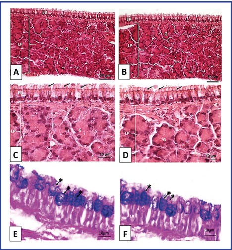
Fig. 1 Optical photomicrography of the respiratory mucosa of Wistar rats. T3 Maxillary sinus. A, C and E: Control group. B, D and F: Group treated with NeuroEPO. ER: respiratory epithelium, LP: self-contained payroll, Arrows: cilia, G: glands. Staining with H/E. A and B magnification 400X. C and D magnification 1000X. PAS/Blue Coloration of Alcián. E and F Magnification 1000X. Arrows with asterisks: goblet cells.  

Morphometric Study

Height of the respiratory epithelium and thickness of the lamina propria

The height of the respiratory epithelium, and the thickness of the lamina propria of the respiratory mucosa of animals treated with NeuroEPO were similar to the control. (Figure 2).

Fig 2 Height of the epithelium and thickness of the lamina propria of the respiratory mucosa of Wistar rats. Groups control and treated with NeuroEPO. Significance level p >0.05. 

Histological features of lymphoepithelium and nasal mucosal associated lymphatic tissue (NALT)

The histological features observed in the lymphoepithelium and NALT of rats in this group were normal. The lymphoepithelium lining the aggregates of lymphatic follicles showed typical morphological features. The lymphatic follicles showed shape and size similar to those of the control group. Lymphocytes of normal morphological aspect were observed. (Figure 3).

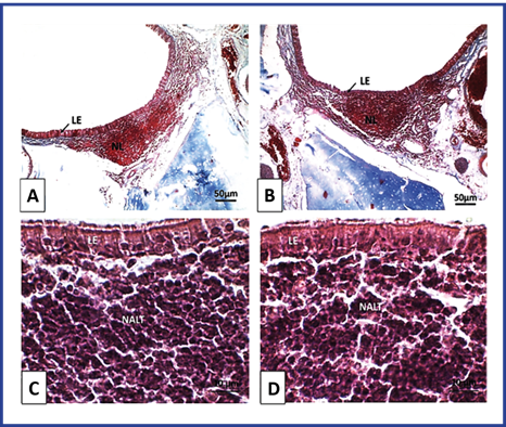
Fig. 3 Optical photomicrography of lymphoepithelium and NALT of Wistar rats. T3. Mucosa of the lateral wall of the anterior orifice of the nasopharyngeal duct). A and C: Control group. B and D: Group treated with NeuroEPO. LE: lymphoepithelium, NL: lymph nodes. NALT: lymphatic tissue associated with the nasal mucosa. A and B Coloration with Mallory's Trichromics. Magnification 100X. C and D Staining with H/E. Magnification 1000X. 

Discussion

In the present research, no changes were observed in the histological characteristics of the epithelium or the lamina propria of the respiratory mucosa. Likewise, no signs indicative of acute or chronic inflammatory response were evidenced with intranasal administration of NeuroEPO to Wistar rats. The control and treated groups exhibited similar morphological characteristics.

There are no previous studies describing the effect of intranasal administration of NeuroEPO in the structure of respiratory mucosa. In studies with other molecules as chlorine gas, ozone, formaldehyde, and xylene, exposure causes cilia loss, caliciform cell metaplasia, squamous cell hyperplasia, degeneration, epithelial necrosis, and inflammatory infiltration. Regenerative epithelial proliferation with or without squamous metaplasia and associated inflammatory response have also been described.22,23,24,25,26

The results of the current study differ from some reports suggesting that prolonged use of some drugs nasally triggers drug rhinitis, characterized by squamous metaplasia of the respiratory epithelium and/or ulceration, increased goblet cells, and thickened and hyalinized basement membrane. The lamina propria of the respiratory mucosa in these cases may exhibit edema or fibrosis with a diffuse infiltrate of lymphocytes, histiocytes and eosinophils. Such is the case with some nasal decongestants and anti-inflammatories, such as oxymetazoline, phenylephrine, mometasone and fluticasone.27,28

Recent studies have evaluated the effect of intranasal administration of NeuroEPO on the nasal and olfactory mucosa in rats (after 14 and 28 days respectively).29,30 Both studies showed that the administration of NeuroEPO did not affect the morphology of the structures explored.

In the morphometric study of the respiratory mucosa, no significant differences were observed in the height of the respiratory epithelium or the thickness of the lamina propria in the study groups. There are no reports in the literature describing the effects of the administration of NeuroEPO on the morphometry of the respiratory mucosa. The results differ from some studies carried out in experimental animals in which a decrease in respiratory epithelium and thickness of the lamina propria corroborated by morphometry has been reported in rats exposed to ethanol and cathelicidin.31,32

It is likely that the results obtained in the present study are related to the fact that the distribution of lesions in the nasal cavity depends on the local dose used and the susceptibility of the tissues to damage. The respiratory epithelium is less susceptible to damage by chemical agents than others of the epithelia of the nasal cavity; in addition, the dose used and the time of exposure to it may have influenced the current results.33

In the present research, the control and treated groups showed a normal morphological pattern of the epithelium and the lamina propria of the respiratory mucosa corroborated with the morphometric study. No signs of acute or chronic inflammation were observed in the study groups; this suggests that NeuroEPO administered nasally at doses of 300 μg/kg/day for 28 days does not produce alterations on the respiratory mucosa in Wistar rats.

In our study, no changes were observed in the histological structure of the lymphoepithelium of rats exposed to high doses of nasal NeuroEPO, behaving similarly to the control group. When observing the histological characteristics of the NALT, it was possible to appreciate that its shape and size were similar in both groups.

No reports were found in the reviewed literature about the effects of NeuroEPO on lymphoepithelium and NALT in rats, which could be compared with those of current research. However, the current results differ from those obtained in other studies, in which it has been reported that animals exposed to toxins nasally triggered alterations in the immune system shortly after their exposure, among these are: decrease in the number of intraepithelial lymphocytes, and lymphocytes present from the lymphatic follicles. These changes had an impact on the shape and size of the NALT of the animals under study.34,35

Rats exposed to high concentrations of inhaled formaldehyde have shown lymphoepithelial hyperplasia. The NALT in these cases showed decreased cellularity of T and B lymphocytes.36

One of the mechanisms through which a toxic agent causes alteration in the immune system is related to the stress it generates on the cells. The sympathetic nervous system is essential to restore homeostasis, altered by stress and to promote the survival of the organism. For each type of aggressor agent, a certain type of specific response is generated.37,38 Stress causes an increase in the secretion of glucocorticoid hormones and catecholamines by activation of the hypothalamic-pituitary-adrenal axis. These hormones interact with receptors on immune cells and cause changes in their response that include modifications in cell traffic and proliferation, changes in cytokine secretion, antibodies, and cytolytic activity.39

Glucocorticoids inhibit myelopoiesis and production of pro-inflammatory cytokines (IL-1, IL-6, TNF). They also induce expression of MHC class II molecules and suppress the function of Th1 lymphocytes.36,37,38 Cortisol is the main glucocorticoid involved in immunomodulation, triggering immunosuppressive and anti-inflammatory effects.37,38,39

One of the factors involved in immunogenicity is the concentration of the drug. A high dose was used in the present study, as reported in the literature.13 However, no effect on lymphoepithelium or NALT was observed. This could be related to a tolerogenic response, and this favored an immunological tolerance.18 This may be related to the fact that the structure of NeuroEPO is very similar to that of endogenous EPO (due to its human recombinant origin).

As a result, NeuroEPO administered nasally at doses of 300 μg/kg/day for 28 days does not produce alterations in lymphoepithelium and NALT in Wistar rats.

This research was limited to the evaluation of the nasal administration of NeruroEPO in the histological structure of the respiratory mucosa and NALT, but further research could be addressed to evaluate the effect in functional aspects.

Conclusions

The administration of NeuroEPO nasally at doses of 300 μg/kg/day for 28 days does not cause damage to the histological structure in the respiratory mucosa or the lymphatic tissue associated with the nasal mucosa of Wistar rats.

References

1. Garzón F, Rodríguez Y, García JC, Rama R. Neuroprotective Effects of neuroEPO Using an In Vitro Model of Stroke. Behav Sci.2018;8(2):26. [ Links ]

2. Rama R, Garzón F, Rodríguez Cruz Y, Maurice T, García Rodríguez JC. Neuroprotective effect of Neuro-EPO in neurodegenerative diseases: “Aleajacta est.” Neural Regen Res [Online]. 2019 Sep [Cited 04/02/2022];14(9):1519. Available from: Available from: https://www.ncbi.nlm.nih.gov/pmc/articles/PMC6557108/Links ]

3. Rey F, Balsari A, Giallongo T. Erythropoietin as a Neuroprotective Molecule: An Overview of Its Therapeutic Potential in Neurodegenerative Diseases. ASN Neuro [Online]. 2019 [Cited 04/02/2022];11:1759091419871420. Available from: Available from: https://www.ncbi.nlm.nih.gov/pmc/articles/PMC6712762/Links ]

4. Zucchella C, Sinforiani E, Tamburin S, Federico A, Mantovani E, Bernini S, et al. The Multidisciplinary Approach to Alzheimer'sDisease and Dementia. A Narrative Review of Non-Pharmacological Treatment. Front Neurol [Online]. 2018 Dec [Cited 04/02/2022];9:1058. Available from: Available from: https://www.ncbi.nlm.nih.gov/pmc/articles/PMC6300511/Links ]

5. Fernández Romero T, Clapés Hernández S, Pérez Hernández CL, Barreto López JJ, Fernández Peña G. Efecto hipoglicemiante de la NeuroEPO en ratas con y sin diabetes mellitus. Rev haban cienc méd [Online]. 2022 [Cited 17/04/2022];21(1):e4617. Disponible en: Disponible en: http://www.revhabanera.sld.cu/index.php/rhab/article/view/4617Links ]

6. Ul Islam S, Shehzad A, Bilal Ahmed M, Lee YS. Intranasal Delivery of Nanoformulations: A Potential Way of Treatment for Neurological Disorders. Molecules [Online]. 2020 Apr [Cited 04/02/2022];25(8):[Aprox. 2 p.]. Available from: Available from: https://pubmed.ncbi.nlm.nih.gov/32326318/Links ]

7. Fernández Romero T, Clapés Hernández S, Pérez CL, Núñez López N, Suárez Román G, Fernández G. Protective effect of NeuroEPO in the reproduction of diabetic rats. Rev haban cienc méd [Online]. 2022 [Cited 08/08/2022];21(4):[Aprox. 6 p.]. Available from: Available from: http://www.revhabanera.sld.cu/index.php/rhab/article/view/4797Links ]

8. Rodríguez Y, Strehaiano M, Rodríguez T, García JC, Maurice T. An intranasal formulation of erythropoietin (Neuro-EPO) prevents memory deficits and amyloid toxicity in the APPSwe transgenic mouse model of Alzheimer’s disease. J Alzheimer´s Dis [Online]. 2017;55(1):231-48. Available from: https://www.doi.org/10.3233/JAD-160500 [ Links ]

9. Vega MLB, Shengnan L, Leon YR, Rodríguez AM, Fernández EC, Llano MG, et al. NeuroEPO improves cognition in Parkinson’s disease. Preliminary report. medRxiv [Online]. New York: medRxiv; 2022 [Cited 17/04/2022]. Available from: Available from: https://www.medrxiv.org/content/10.1101/2022.02.24.22271444v2Links ]

10. Pérez L, Sosa S, Valenzuela C, Bringas G, Urrutia N, Peñalver AI, et al. Clinical trial of neuroEPO in mild-to-moderate Alzheimer’s disease. Alzheimer's Dement [Online]. 2021;17:e050212. Available from: https://doi.org/10.1002/alz.050212 [ Links ]

11. McCormick-Ell J, Connell N. Laboratory safety, biosecurity, and responsible animal use. ILAR journal [Online]. 2019;60(1):24-33. Available from: https://doi.org/10.1093/ilar/ilz012 [ Links ]

12. Muñoz Cernada A, García Rodríguez JC, Núñez Figueredo Y, Pardo Ruiz Z, García Selman JD, Sosa Testé I, et al. Formulaciones nasales de EPOrh con bajo contenido de ácido siálico para el tratamiento de enfermedades del sistema nervioso central [Online]. La Habana: PATENTSCOPE WO2007009404, 2007 [Cited 17/04/2022]. Available from: Available from: https://patentscope.wipo.int/search/es/detail.jsf?docId=WO2007009404Links ]

13. Tveden Nyborg P, Lykkesfeldt J. Animal Models in Pharmacology and Toxicology. In: Handbook of Laboratory Animal Science [Online]. Florida: CRC Press; 2021 [Cited ]. 25/07/2022. p. 665-94Available from: Available from: https://www.taylorfrancis.com/chapters/edit/10.1201/9780429439964-30/animal-models-pharmacology-toxicology-pernille-tveden-nyborg-jens-lykkesfeldtLinks ]

14. Uraih LC, Maronpot RR. Normal histology of the nasal cavity and application of special techniques. Environ Health Perspect [Online]. 1990;85:187-208. Available from: https://www.doi.org/10.1016/s0272-0590(81)80037-1 [ Links ]

15. Young JT. Histopathologic examination of the rat nasal cavity. Fundam Appl Toxicol [Online]. 1981;1(4):309-12. [ Links ]

16. Grada A, Mervis J, Falanga V. Research techniques made simple: animal models of wound healing. Journal of Investigative Dermatology [Online]. 2018;138(10):2095-105 [ Links ]

17. Verma A, Verma M, Singh A. Animal tissue culture principles and applications. Animal Biotechnology [Online]. 2020:269-93. Available from: http://www.doi.org/10.1016/B978-0-12-811710-1.00012-4 [ Links ]

18. Abbas KA, Lichtman HA, Pillai S. Células y tejidos del sistema inmunitario. In: Inmunología celular y molecular. 7 ed. Barcelona: Elsevier Saunders; 2012.p.15-32. [ Links ]

19. Dan Wilcox, Brent Dove. UTHSCSA image tool (3.0)[Online]. USA: Informer Technologies, Inc; 2022 [Cited 25/07/2022]. Available from: Available from: https://imagetool.software.informer.com/Descargar-gratis/Links ]

20. Motulsky H, Chistopoulos A, Miller JR. Graphpad Prism Version 5.0 Software Inc, USA [Online]. San Diego: GraphPad; 2022 [Cited 25/07/2022]. Available from: Available from: http://www.graphpad.comLinks ]

21. Suarez K, Fernández G, Rodriguez Y, Puldon G (2022), “Administration of intranasal NeuroEPO does not affect the structure of the respiratory mucosa in Wistar rats” [Online]. Amsterdam: Mendeley Data; 2022. Available from: http://doi.org/10.17632/c5zhgv532g.1 [ Links ]

22. Jiang XZ, Buckley LA, Morgan KT. Pathology of toxic responses to the RD50 concentration of chlorine gas in the nasal passages of rats and mice. Toxicol Appl Pharmacol [Online]. 1983;71(2):225-36. Available from: http://www.doi.org/10.1016/0041-008x(83)90339-3 [ Links ]

23. Harkema JR, Wagner JG. Innate Lymphoid Cell-Dependent Airway Epithelial and Inflammatory Responses to Inhaled Ozone: A New Paradigm in Pathogenesis. Toxicol Pathol [Online]. 2019;47(8):993-1003. Available from: http://www.doi.org/10.1177/0192623319873872 [ Links ]

24. Edrissi B, Taghizadeh K, Moeller BC, Yu R, Kracko D, Doyle Eisele M, et al. N6-Formyllysine as a Biomarker of Formaldehyde Exposure: Formation and Loss of N6-Formyllysine in Nasal Epithelium in Long-Term, Low-Dose Inhalation Studies in Rats. Chem Res Toxicol [Online]. 2017;30(8):1572-6. Available from: http://www.doi.org/10.1021/acs.chemrestox.7b00075 [ Links ]

25. Tesfaye S, Hamba N, Gerbi A, Negeri Z. Oxidative Stress and Carcinogenic Effect of Formaldehyde Exposure: Systematic Review & Analysis. Endocrinol Metab Syndr [Online]. 2020 Sep [Cited 04/02/2022];9(6):1-11. Available from: Available from: https://www.longdom.org/open-access/oxidative-stress-and-carcinogenic-effect-of-formaldehyde-exposure-systematic-review-analysis-58212.htmlLinks ]

26. Eda A, Baver S, Seyla SB, Hüseyin Ö, Deveci E. Effects of Xylene on Respiratory Mucosa in Rats. Int J Morphol [Online]. 2016 Sep [Cited 04/02/2022];34(3):934-8. Available from: Available from: http://www.scielo.cl/scielo.php?script=sci_arttext&pid=S0717-95022016000300019&lng=en&nrm=iso&tlng=enLinks ]

27. Horak F, Zieglmayer UP, Zieglmayer R, Kavina A, Marschall K, Munzel U, et al. Azelastine nasal spray and desloratadine tablets in pollen-induced seasonal allergic rhinitis: a pharmacodynamic study of onset of action and efficacy. Curr Med Res Opin [Online]. 2006;22(1):151-7. Available from: http://www.doi.org/10.1185/030079906X80305 [ Links ]

28. Small P, Keith PK, Kim H. Allergic rhinitis. Allergy Asthma Clin Immunol [Online]. 2018 [Cited 04/02/2022];14(2):1-11. Available from: Available from: https://aacijournal.biomedcentral.com/articles/10.1186/s13223-018-0280-7Links ]

29. Lagarto A, Bueno V, Guerra I, Valdés O, Couret M, López R, et al. Absence of hematological side effects in acute and subacute nasal dosing of erythropoietin with a low content of sialic acid. Exp Toxicol Pathol [Online]. 2011 [Cited 04/02/2022];63(6):563-7. Available from: Available from: https://pubmed.ncbi.nlm.nih.gov/20488687/Links ]

30. Rodríguez Cruz Y, Suárez Borrás K, Fernández Peña G, Puldón Seguí G. Efectos de la NeuroEPO sobre las características histológicas de la mucosa olfatoria en ratas wistar. En: Congreso MorfoVirtual 2020 [Online]. La Habana: Sociedad Cuabana de Ciencias Morfológicas; 2020 [Cited 25/07/2022]. Available from: Available from: http://www.morfovirtual2020.sld.cu/index.php/morfovirtual/morfovirtual2020/paper/view/25/Links ]

31. Marcondes de Godoy M, Lopes RA, Sala MA, Vinha D, Regalo SCH. Acción del Etanol sobre el Epitelio Nasal y Glándulas Septales de Ratas, Durante la Lactancia. Int J Morphol [Online]. 2005 [Cited 04/02/2022];23(4):293-300. Available from: Available from: http://www.scielo.cl/scielo.php?script=sci_arttext&pid=S0717-95022005000400002&lng=es&nrm=iso&tlng=esLinks ]

32. Alt JA, Qin X, Pulsipher A, Orb Q, Orlandi RR, Zhang J, et al. Topical cathelicidin (LL-37) an innate immune peptide induces acute olfactory epithelium inflammation in a mouse model. Int Forum Allergy Rhinol [Online]. 2015 Dec;5(12):1141-50. Available from: http://www.doi.org/10.1002/alr.21634 [ Links ]

33. Chamanza R, Wright J. A review of the comparative anatomy, histology, physiology and pathology of the nasal cavity of rats, mice, dogs and non-human primates. Relevance to inhalation toxicology and human health risk assessment. Journal of Comparative Pathology [Online]. 2015;153(4):287-314. Available from: https://doi.org/10.1016/j.jcpa.2015.08.009 [ Links ]

34. Elmore SA. Enhanced Histopathology Evaluation of Lymphoid Organs. Methods Mol Biol [Online]. 2018 [Cited 30/03/2022];1803:147-68. Available from: Available from: https://link.springer.com/protocol/10.1007/978-1-4939-8549-4_10Links ]

35. Elmore SA. Enhanced Histopathology of Mucosa-Associated Lymphoid Tissue. Toxicol Pathol [Online]. 2006 Jun [Cited 30/03/2022];34(5):687-96. Available from: Available from: https://journals.sagepub.com/doi/full/10.1080/01926230600939989Links ]

36. Frieke Kuper C, van Oostrum L, Ma-Hock L, Durrer S, Woutersen RA. Hyperplasia of the lymphoepithelium of NALT in rats but not in mice upon 28-day exposure to 15ppm formaldehyde vapor. Exp Toxicol Pathol [Online]. 2011;63(1):25-32. Available from: https://www.sciencedirect.com/science/article/pii/S0940299309002437Links ]

37. Marino JH, Teague TK. The Immune System as a Sensor and Regulator of Stress: Implications in Human Development and Disease. In: Biobehavioral Markers in Risk and Resilience Research [Online]. Switzerland: Springer; 2019. p. 1-11. Available from: https://doi.org/10.1007/978-3-030-05952-1_1 [ Links ]

38. Kuper CF, Wijnands MVW, Zander SAL. Mucosa-Associated Lymphoid Tissues BT - Immunopathology in Toxicology and Drug Development. In: Parker GA, ed. Organ Systems [Online]. Switzerland: Springer International Publishing; 2017. p. 81-121. Available from: https://doi.org/10.1007/978-3-319-47385-7_4 [ Links ]

39. Pacák K, Palkovits M. Stressor specificity of central neuroendocrine responses: implications for stress-related disorders. Endocr Rev [Online]. 2001 Aug;22(4):502-48. Available from: http://www.doi.org/10.1210/edrv.22.4.0436 [ Links ]

Authors´ contributions

2Ketty Suarez Borras: Conceptualization, data curation, research, methodology, formal analysis, original draft writing, writing-review, and editing.

3Gisselle Fernández Peña: Conceptualization, data curation, research, methodology, formal analysis, original draft writing, writing-review, and editing.

4Yamila Rodriguez Cruz: Acquisition of funds and administration, data curation, formal analysis, original draft writing, writing-revision, and editing.

5Giselle Puldón Seguí: Data curation, formal analysis, original draft writing, writing-revision, and editing.

Received: April 19, 2022; Accepted: June 08, 2022

*Corresponding authorsuarezkety42@gmail.com

The authors declare that there are no conflicts of interest.

All authors participated in the discussion of the results and have read, reviewed, and approved the final version of the article.

Creative Commons License This is an open-access article distributed under the terms of the Creative Commons Attribution License