<?xml version="1.0" encoding="ISO-8859-1"?><article xmlns:mml="http://www.w3.org/1998/Math/MathML" xmlns:xlink="http://www.w3.org/1999/xlink" xmlns:xsi="http://www.w3.org/2001/XMLSchema-instance">
<front>
<journal-meta>
<journal-id>0034-7507</journal-id>
<journal-title><![CDATA[Revista Cubana de Estomatología]]></journal-title>
<abbrev-journal-title><![CDATA[Rev Cubana Estomatol]]></abbrev-journal-title>
<issn>0034-7507</issn>
<publisher>
<publisher-name><![CDATA[Editorial Ciencias Médicas]]></publisher-name>
</publisher>
</journal-meta>
<article-meta>
<article-id>S0034-75072012000100005</article-id>
<title-group>
<article-title xml:lang="en"><![CDATA[Mucoepidermoid carcinoma of the salivary glands in Brazil: clinicopathological outcomes]]></article-title>
<article-title xml:lang="es"><![CDATA[Carcinoma mucoepidermoide de glándulas salivales en Brasil: relación clinicopatológica]]></article-title>
</title-group>
<contrib-group>
<contrib contrib-type="author">
<name>
<surname><![CDATA[Oliveira]]></surname>
<given-names><![CDATA[Lucinei Roberto]]></given-names>
</name>
<xref ref-type="aff" rid="A01"/>
</contrib>
<contrib contrib-type="author">
<name>
<surname><![CDATA[Figueiredo Soave]]></surname>
<given-names><![CDATA[Danilo]]></given-names>
</name>
<xref ref-type="aff" rid="A02"/>
</contrib>
<contrib contrib-type="author">
<name>
<surname><![CDATA[Oliveira-Costa]]></surname>
<given-names><![CDATA[João Paulo]]></given-names>
</name>
<xref ref-type="aff" rid="A02"/>
</contrib>
<contrib contrib-type="author">
<name>
<surname><![CDATA[Sala Di Matteo]]></surname>
<given-names><![CDATA[Miguel Angel]]></given-names>
</name>
<xref ref-type="aff" rid="A02"/>
</contrib>
<contrib contrib-type="author">
<name>
<surname><![CDATA[Ribeiro-Silva]]></surname>
<given-names><![CDATA[Alfredo]]></given-names>
</name>
<xref ref-type="aff" rid="A02"/>
</contrib>
</contrib-group>
<aff id="A01">
<institution><![CDATA[,Vale do Rio Verde University  ]]></institution>
<addr-line><![CDATA[ ]]></addr-line>
<country>Brazil</country>
</aff>
<aff id="A02">
<institution><![CDATA[,University of Sao Paulo  ]]></institution>
<addr-line><![CDATA[ ]]></addr-line>
<country>Brazil</country>
</aff>
<pub-date pub-type="pub">
<day>00</day>
<month>03</month>
<year>2012</year>
</pub-date>
<pub-date pub-type="epub">
<day>00</day>
<month>03</month>
<year>2012</year>
</pub-date>
<volume>49</volume>
<numero>1</numero>
<fpage>0</fpage>
<lpage>0</lpage>
<copyright-statement/>
<copyright-year/>
<self-uri xlink:href="http://scielo.sld.cu/scielo.php?script=sci_arttext&amp;pid=S0034-75072012000100005&amp;lng=en&amp;nrm=iso"></self-uri><self-uri xlink:href="http://scielo.sld.cu/scielo.php?script=sci_abstract&amp;pid=S0034-75072012000100005&amp;lng=en&amp;nrm=iso"></self-uri><self-uri xlink:href="http://scielo.sld.cu/scielo.php?script=sci_pdf&amp;pid=S0034-75072012000100005&amp;lng=en&amp;nrm=iso"></self-uri><abstract abstract-type="short" xml:lang="en"><p><![CDATA[The biological features and clinical behavior of mucoepidermoid carcinomas are widely variable and poorly understood. This study aimed to investigate prognostic factors that may affect survival in patients with a primary diagnosis of head and neck mucoepidermoid carcinomas. The effects of age, gender, anatomic localization, tumor size, clinical stage, histological grade, recurrence, metastasis, compromised surgical margins and treatment on clinicopathological outcomes were investigated. Survival curves were generated using the Kaplan-Meier method and analyses were performed using the log rank test. A total of 16 cases were analyzed over a period of 18 years; males were 68.7 %, with ages ranging from 13 to 83 years. The 75 % of the tumors developed in the major salivary glands, 56.3 % in the parotid gland and they were predominantly classified as stage II 37.5 % and low-grade lesions 37.5 % at diagnosis. Surgical resection was performed in all patients. The follow-up period in this study ranged from 6 to 217 months. The 5 and 10-year overall survival rates were both 85.6 %. Disease-free survival rates were 81.8 % (5 years) and 68.2 % (10 years). There were statistically significant effects of tumor size (p= 0.05), metastasis (p= 0.04) and primary anatomic localization (p= 0.04) on disease-free survival rates. Through a long follow-up period in present study we could highlight the relevance of primary anatomical site, tumor size and metastasis as useful prognostic factors that may affect survival in patients with a primary diagnosis of head and neck mucoepidermoid carcinomas.]]></p></abstract>
<abstract abstract-type="short" xml:lang="es"><p><![CDATA[Las características biológicas y el comportamiento clínico del carcinoma mucoepidermoide son muy variados y aún poco conocidos. El propósito de este estudio fue investigar los factores pronósticos que puedan afectar la supervivencia de los pacientes con diagnóstico primario de carcinoma mucoepidermoide de cabeza y cuello. Se estudiaron la edad, el sexo, la localización anatómica, el tamaño del tumor, el estadio clínico, el grado histológico, la recidiva, la metástasis, los bordes quirúrgicos comprometidos y el tratamiento, sobre los resultados clínico-patológicos. Las curvas de supervivencia fueron construidas con el método de Kaplan-Meier y el análisis estadístico fue realizado mediante la prueba del log-rank. Fueron analizados 16 casos durante un periodo de 18 años. Se constató un 68,7 % de pacientes del sexo masculino y de edades comprendidas entre los 13 y los 83 años. El 75 % de los tumores se localizó en las glándulas salivales mayores, el 56,3 % en parótida y las clasificaciones predominantes en el momento del diagnóstico fueron lesiones de bajo grado y estadio II con un 37,5 %. La resección quirúrgica fue realizada en todos los pacientes. El periodo de seguimiento en este estudio varió entre 6 y 217 meses. La tasa general de supervivencia, tanto a los 5 como a los 10 años fue de 85,6 %, mientras que las tasas de supervivencia libre de enfermedad fueron de 81,8 % a los 5 años y de 68,2 % a los 10 años. Se demostró la influencia estadísticamente significativa del tamaño del tumor (p= 0,05), la presencia de metástasis (p= 0,04) y de la localización anatómica primaria (p= 0,04) sobre las tasas de supervivencia libre de enfermedad. Los resultados obtenidos demostraron la importancia de la localización anatómica primaria del tumor, de su tamaño y de la presencia de metástasis, en la supervivencia de los pacientes con diagnóstico primario de carcinoma mucoepidermoide.]]></p></abstract>
<kwd-group>
<kwd lng="en"><![CDATA[Salivary gland neoplasms]]></kwd>
<kwd lng="en"><![CDATA[mucoepidermoid carcinoma]]></kwd>
<kwd lng="en"><![CDATA[disease-free survival]]></kwd>
<kwd lng="es"><![CDATA[neoplasias de las glándulas salivales]]></kwd>
<kwd lng="es"><![CDATA[carcinoma mucoepidermoide]]></kwd>
<kwd lng="es"><![CDATA[supervivencia libre de enfermedad]]></kwd>
</kwd-group>
</article-meta>
</front><body><![CDATA[ <p align="right"><font face="Verdana, Arial, Helvetica, sans-serif" size="2">    <B>ART&Iacute;CULO ORIGINAL</B></font></p>     <p>&nbsp;</p>     <p><font face="Verdana, Arial, Helvetica, sans-serif" size="4"><b>Mucoepidermoid    carcinoma of the salivary glands in Brazil: clinicopathological outcomes</b></font><font face="Verdana, Arial, Helvetica, sans-serif" size="2">    </font></p> <B>    <P>      <P>&nbsp;     <P><font face="Verdana, Arial, Helvetica, sans-serif" size="3">Carcinoma mucoepidermoide    de gl&aacute;ndulas salivales en Brasil: relaci&oacute;n clinicopatol&oacute;gica    </font>     <P>&nbsp;     <P>&nbsp;     <P>      <P>  </B>     ]]></body>
<body><![CDATA[<P><b><font face="Verdana, Arial, Helvetica, sans-serif" size="2">Lucinei Roberto    Oliveira,<SUP>I</SUP> Danilo Figueiredo Soave,<SUP>II</SUP> Jo&atilde;o Paulo    Oliveira-Costa,<SUP>II</SUP> Miguel Angel Sala Di Matteo,<SUP>II</SUP> Alfredo    Ribeiro-Silva<SUP>II</SUP></font> </b>      <P>      <P><font face="Verdana, Arial, Helvetica, sans-serif" size="2"><SUP>I </SUP> Vale    do Rio Verde University, Brazil.    <br>   </font><font face="Verdana, Arial, Helvetica, sans-serif" size="2"><SUP>II </SUP>    University of Sao Paulo, Brazil.    <br>   </font>     <P>&nbsp;     <P>&nbsp;     <P><HR size="1" noshade>     <p><font face="Verdana, Arial, Helvetica, sans-serif" size="2"><B>ABSTRACT</B>    </font> </p>     <P>      ]]></body>
<body><![CDATA[<P><font face="Verdana, Arial, Helvetica, sans-serif" size="2">The biological    features and clinical behavior of mucoepidermoid carcinomas are widely variable    and poorly understood. This study aimed to investigate prognostic factors that    may affect survival in patients with a primary diagnosis of head and neck mucoepidermoid    carcinomas. The effects of age, gender, anatomic localization, tumor size, clinical    stage, histological grade, recurrence, metastasis, compromised surgical margins    and treatment on clinicopathological outcomes were investigated. Survival curves    were generated using the Kaplan-Meier method and analyses were performed using    the log rank test. A total of 16 cases were analyzed over a period of 18 years;    males were 68.7 %, with ages ranging from 13 to 83 years. The 75 % of the tumors    developed in the major salivary glands, 56.3 % in the parotid gland and they    were predominantly classified as stage II 37.5 % and low-grade lesions 37.5    % at diagnosis. Surgical resection was performed in all patients. The follow-up    period in this study ranged from 6 to 217 months. The 5 and 10-year overall    survival rates were both 85.6 %. Disease-free survival rates were 81.8 % (5    years) and 68.2 % (10 years). There were statistically significant effects of    tumor size (p= 0.05), metastasis (p= 0.04) and primary anatomic localization    (p= 0.04) on disease-free survival rates. Through a long follow-up period in    present study we could highlight the relevance of primary anatomical site, tumor    size and metastasis as useful prognostic factors that may affect survival in    patients with a primary diagnosis of head and neck mucoepidermoid carcinomas.    </font>     <P>      <P>  <b><font face="Verdana, Arial, Helvetica, sans-serif" size="2"><I>Key words</I>:</font></b><font face="Verdana, Arial, Helvetica, sans-serif" size="2">  Salivary gland neoplasms, mucoepidermoid carcinoma, <font color="#FF0000"><font color="#000000">disease-free  survival.</font></font>  <HR size="1" noshade></font>     <P>      <P><font face="Verdana, Arial, Helvetica, sans-serif" size="2"><B>RESUMEN</B>    </font>     <P>      <P><font face="Verdana, Arial, Helvetica, sans-serif" size="2">Las caracter&iacute;sticas    biol&oacute;gicas y el comportamiento cl&iacute;nico del carcinoma mucoepidermoide    son muy variados y a&uacute;n poco conocidos. El prop&oacute;sito de este estudio    fue investigar los factores pron&oacute;sticos que puedan afectar la supervivencia    de los pacientes con diagn&oacute;stico primario de carcinoma mucoepidermoide    de cabeza y cuello. Se estudiaron la edad, el sexo, la localizaci&oacute;n anat&oacute;mica,    el tama&ntilde;o del tumor, el estadio cl&iacute;nico, el grado histol&oacute;gico,    la recidiva, la met&aacute;stasis, los bordes quir&uacute;rgicos comprometidos    y el tratamiento, sobre los resultados cl&iacute;nico-patol&oacute;gicos. Las    curvas de supervivencia fueron construidas con el m&eacute;todo de Kaplan-Meier    y el an&aacute;lisis estad&iacute;stico fue realizado mediante la prueba del    log-rank. Fueron analizados 16 casos durante un periodo de 18 a&ntilde;os. Se    constat&oacute; un 68,7 % de pacientes del sexo masculino y de edades comprendidas    entre los 13 y los 83 a&ntilde;os. El 75 % de los tumores se localiz&oacute;    en las gl&aacute;ndulas salivales mayores, el 56,3 % en par&oacute;tida y las    clasificaciones predominantes en el momento del diagn&oacute;stico fueron lesiones    de bajo grado y estadio II con un 37,5 %. La resecci&oacute;n quir&uacute;rgica    fue realizada en todos los pacientes. El periodo de seguimiento en este estudio    vari&oacute; entre 6 y 217 meses. La tasa general de supervivencia, tanto a    los 5 como a los 10 a&ntilde;os fue de 85,6 %, mientras que las tasas de supervivencia    libre de enfermedad fueron de 81,8 % a los 5 a&ntilde;os y de 68,2 % a los 10    a&ntilde;os. Se demostr&oacute; la influencia estad&iacute;sticamente significativa    del tama&ntilde;o del tumor (p= 0,05), la presencia de met&aacute;stasis (p=    0,04) y de la localizaci&oacute;n anat&oacute;mica primaria (p= 0,04) sobre    las tasas de supervivencia libre de enfermedad. Los resultados obtenidos demostraron    la importancia de la localizaci&oacute;n anat&oacute;mica primaria del tumor,    de su tama&ntilde;o y de la presencia de met&aacute;stasis, en la supervivencia    de los pacientes con diagn&oacute;stico primario de carcinoma mucoepidermoide.</font>      <P>      <P> <font face="Verdana, Arial, Helvetica, sans-serif" size="2"><I><b>Palabras clave</b></I><b>:</b>  neoplasias de las gl&aacute;ndulas salivales, carcinoma mucoepidermoide, <b></b><font face="Verdana, Arial, Helvetica, sans-serif" size="2">supervivencia  libre de enfermedad.</font>  <HR size="1" noshade> </font>      <p>&nbsp;</p>     ]]></body>
<body><![CDATA[<p>&nbsp;</p>     <P>      <P>      <P><font face="Verdana, Arial, Helvetica, sans-serif" size="2"><B><font size="3">INTRODUCTION</font></B>    </font>      <P>      <P><font face="Verdana, Arial, Helvetica, sans-serif" size="2">Primary malignant    salivary gland tumors represent less than 3 % of head and neck cancers and 10-15    % of all glandular tumors. There is a wide variation in these malignant neoplasms    over different geographic areas and ethnic groups. Due to the singularity and    diverse histology of these tumors, prognostic factors have been difficult to    elucidate.<SUP>1</SUP> </font>     <P>      <P><font face="Verdana, Arial, Helvetica, sans-serif" size="2">The establishment    and description of mucoepidermoid carcinoma (MEC) as a distinct salivary gland    tumor was originally credited to <I>Stewart and others</I><SUP>2</SUP> (1945).    Since then MEC has been described as the most common malignant salivary gland    tumor, accounting for close to 10 % of all salivary gland neoplasms.<SUP>3,4    </SUP>The clinical behavior of MEC is widely variable, but it seems to correlate    with tumor stage and grade. </font>     <P>      <P><font face="Verdana, Arial, Helvetica, sans-serif" size="2">The mucoepidermoid    carcinoma generally shows an extremely aggressive pattern for high-grade tumors,    whereas its respectivee low-grade counterpart is often indolent and slow-growing.<SUP>5,6</SUP>    However metastases have been described in some cases of low-grade MEC.<SUP>7</SUP>    These discrepancies exist likely because there are several histological graduation    systems that have been recommended for MECs,<SUP>7-10</SUP> none of which is    universally accepted, which makes the retrospective investigations of clinical    outcomes difficult.<SUP>3</SUP> In the same way, although complete resection    with free surgical margins is the main treatment for MEC, there is still a lack    of consensus on the postoperative use of radio and chemotherapy in some cases.<SUP>3,4</SUP>    </font>     ]]></body>
<body><![CDATA[<P>      <P><font face="Verdana, Arial, Helvetica, sans-serif" size="2">The mechanisms    of pathogenesis and progression of salivary gland tumors are still poorly understood    and studies of prognostic factors that evaluate only one type of salivary gland    tumor are scarce for several countries in the world. This study aimed to investigate    prognostic factors that may affect survival in patients with a primary diagnosis    of head and neck mucoepidermoid carcinomas.</font>     <P>&nbsp;     <P><font face="Verdana, Arial, Helvetica, sans-serif" size="2"><b><font size="3">METHODS</font></b></font>    <B></B>      <P>      <P><font face="Verdana, Arial, Helvetica, sans-serif" size="2">A retrospective    study was carried out of patients with a primary diagnosis of head and neck    MEC treated between 1990 and 2008 at the General Hospital of Ribeirao Preto    School of Medicine-USP, Brazil. The medical and surgical records of all cases    were reviewed for clinicopathological factors, such as age, gender, primary    tumor location, tumor size, clinical stage, histological grade, treatment, compromised    surgical margins, tumor recurrence, metastasis, disease-free survival (DFS)    and overall survival (OS). The study protocol was performed with prior approval    of the local Human Research Ethics Committee (approval number HC/FMRP-USP 10142/2010).    </font>     <P>      <P><font face="Verdana, Arial, Helvetica, sans-serif" size="2">All cases met the    criteria proposed by the World Health Organization for the diagnosis of salivary    gland MEC.<SUP>11</SUP> Additional inclusion criteria were documented treatment    of primary MEC at our institution and a minimum of six months of follow-up information.    MECs were staged according to the TNM classification of malignant tumors,<SUP>12</SUP>    and minor salivary gland tumors were staged according to their site of origin    in a similar fashion to squamous cell carcinomas. Two oral pathologists reviewed    all cases to histopathologically classify these tumors according to the protocol    published by <I>Brandwein and others</I><SUP>10</SUP> (2001), which classifies    tumors into low (Grade I), intermediate (Grade II) or high (Grade III) grades.    Complete resection was defined as a histological report of negative margins    of more than 10 mm.<SUP>13</SUP> </font>      <P>      <P><font face="Verdana, Arial, Helvetica, sans-serif" size="2">Patients' features    were summarized through descriptive statistics (mean, range for continuous variables    and frequency and percentage for categorical variables). The DFS was calculated    as the time interval between the date of first treatment and the date of local    disease recurrence or last information for censored observations when the patient    was known to be disease-free. The OS was defined as the interval between the    beginning of the treatment and the date of death or last information for censored    observations. Data concerning survival recurrence and metastasis were evaluated.    The Kaplan-Meier method was used to plot survival curves with the log rank test    for analysis of cumulative survival rates. Statistical significance was defined    as a 2-tailed p value of d </font><font face="Symbol" size="2">&pound; </font><font face="Verdana, Arial, Helvetica, sans-serif" size="2">0.05.    </font>      ]]></body>
<body><![CDATA[<P>      <P>&nbsp;     <P><font face="Verdana, Arial, Helvetica, sans-serif" size="2"><B><font size="3">RESULTS</font></B>    </font>      <P>      <P><font face="Verdana, Arial, Helvetica, sans-serif" size="2">
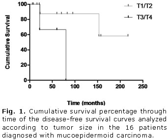
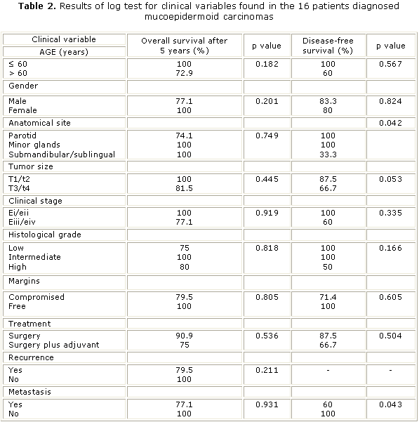
A total of 16 surgically-treated    cases of MEC met the inclusion criteria for this survey during the specified    period. The clinicopathologic features and results of log rank tests for clinical    variables are shown in <a href="/img/revistas/est/v49n1/t0105112.gif">tables 1</a> and <a href="/img/revistas/est/v49n1/t0205112.gif">2</a>,    respectively. The follow-up period in this study ranged from 6 to 217 months    (median 65 months), and the 5 and 10-year OS rates were both 85.6 %. The disease-free    interval for recurrences and metastases ranged from 22 to 153 months and 22    to 157 months, respectively. Rates of 81.8 % and 68.2 % were found for DFS over    a period of 5 and 10 years, respectively.</font>      
<P><font face="Verdana, Arial, Helvetica, sans-serif" size="2">
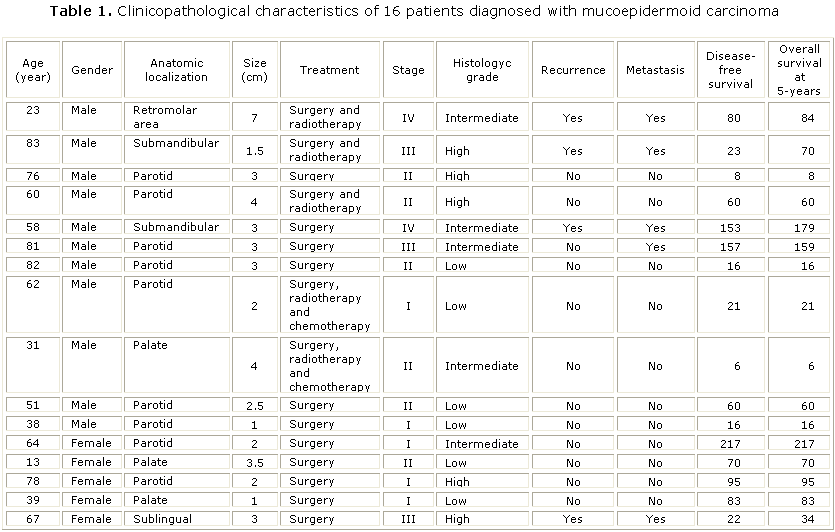
There were no statistically    significant differences for any analyzed variable affecting OS curves. However    as illustrated in <a href="#fig1_05">figures 1</a> and <a href="#fig2_05">2</a>,    the DFS curves showed significant differences for tumor size (p= 0.05) and metastasis    (p= 0.04), respectively. Additionally, a significant influence on prognosis    was observed in DFS curves depending on the primary anatomical site. Tumors    were stratified into three groups: parotid, minor glands and submandibular/sublingual    glands. All tumors diagnosed in the submandibular and sublingual glands had    recurrences and metastases (<a href="/img/revistas/est/v49n1/t0105112.gif">table 1</a>) and consequently,    a worse clinical outcome (p= 0.04) (<a href="#fig3_05">Fig. 3</a>).</font>      
<P align="center"><a name="fig1_05"></a><img src="/img/revistas/est/v49n1/f0105112.jpg" width="342" height="285">      
<P align="center"><a name="fig2_05"></a><img src="/img/revistas/est/v49n1/f0205112.jpg" width="344" height="292">      
<P align="center"><a name="fig3_05"></a><img src="/img/revistas/est/v49n1/f0305112.jpg" width="381" height="287">      
<P align="left">&nbsp;     ]]></body>
<body><![CDATA[<P>      <P><font face="Verdana, Arial, Helvetica, sans-serif" size="2"><B><font size="3">DISCUSSION</font></B>    </font>      <P>      <P><font face="Verdana, Arial, Helvetica, sans-serif" size="2">The MEC is the    most common malignant neoplasm observed in the major and minor salivary glands,    comprising about one-third of all patients affected by salivary malignancies.<SUP>14</SUP>    Diagnosis can occur over a wide age range but occurs predominantly between the    third and sixth decades and affects slightly more women than men (3:2). Almost    half of these tumors occur in the major salivary glands, appearing predominantly    in the parotid (45 %).<SUP>10</SUP> However, due to its infrequency and histopathological    diversity, the epidemiological distribution pattern of primary MECs observed    in one country can differ from others and there are still few published studies    investigating the behavior of these tumors through long follow-up periods. In    the present study, was set out to examine MEC behavior and patient outcomes    to provide additional information on potential factors that could significantly    affect the prognosis of these tumors. </font>     <P>      <P><font face="Verdana, Arial, Helvetica, sans-serif" size="2">There were 16 cases    of MEC diagnosed over an 18-year period at our institution, which is similar    to results reported by <I>Triantafillidou and others</I><SUP>14</SUP> (2006)    who found 16 cases over a 15-year interval. The broad age range observed in    our series supports that reported in literature.<SUP>4,13</SUP> Although there    are a few studies corroborating our results,<SUP>13,15</SUP> the masculine gender    prevalence found differed from commonly reported data that patients diagnosed    with MEC are predominantly female.<SUP>4,16</SUP> </font>     <P><font face="Verdana, Arial, Helvetica, sans-serif" size="2">In agreement with    most other studies,<SUP>4,5,13,15</SUP> was found a prevalence of primary tumors    diagnosed in the major salivary glands, especially in the parotid gland, followed    by intraoral MECs identified in minor salivary glands sited in the hard palate.<SUP>16,17</SUP>    In contrast with other investigations,<SUP>4,13,15</SUP> there was an unusual    case of a MEC found in the sublingual gland. As in most other studies,<SUP>4,6,13</SUP>    Was found a predominance of MECs diagnosed in early clinical stages as T1/T2    and our findings showed a significant influence of tumor size on DFS.<SUP>4,15</SUP>    The low rates of recurrence and metastasis found in this study are in agreement    with previous reports.<SUP>4,6 </SUP>A significant influence of primary anatomical    site was observed in DFS curves when there was stratification into three groups    because all tumors diagnosed in the submandibular and sublingual glands had    recurrences and metastases, which negatively affected the prognosis, this finding    confirms previous observations.<SUP>7,18</SUP></font>      <P>      <P><font face="Verdana, Arial, Helvetica, sans-serif" size="2">Despite several    attempts, an established grading system for MEC does not yet exist. The three-level    grading system commonly used by pathologists for MEC classification mainly considers    the relative proportion of cell types (epidermoid, intermediate and mucinous    cells), their respective degrees of atypia and growth patterns (cystic, solid,    or infiltrative), together with neural and vascular invasion.<SUP>7-10</SUP>    The grading system proposed by <I>Brandwein and others</I><SUP>10</SUP> (2001)    was adopted in this study because it is objective and easy to use and reproduce,<SUP>3,4</SUP>    which will help future standardization. Unlike other studies that used this    same grading system,<SUP>4,13</SUP> the results demonstrate a balance in distribution    among the three tumor grades subtypes. Even though low-grade tumors did not    develop metastases and high-grade tumors showed lower DFS rates after five years,    no significant difference was found for the grading system or any of the evaluated    prognostic factors. </font>     <P>      ]]></body>
<body><![CDATA[<P><font face="Verdana, Arial, Helvetica, sans-serif" size="2">In contrast to    results reported by <I>Nance and </I>others<SUP>4</SUP> (2008) in present study    was not observed any association between positive surgical margins and decreased    DFS. Although MEC has been described as a radioresistant tumor, postoperative    radiotherapy has been associated with decreased recurrence in some reports.<SUP>13</SUP>    Conversely, in the present investigation, a trend toward better survival was    demonstrated in the group who underwent surgical treatment alone, although this    finding was not statistically significant. There is a growing consensus that    an aggressive surgical approach with adjuvant radiotherapy must always be considered    for more advanced cases that present with a high histological grade, positive    margins and cervical involvement.<SUP>3,4,15,19</SUP> </font>     <P>      <P><font face="Verdana, Arial, Helvetica, sans-serif" size="2">The clinical progression    of MEC is usually slow and therefore, requires long-term follow-up to establish    prognostic factors that could influence clinical outcome. Published works usually    lose relevant survival information through time. Although this current investigation    was limited by a relatively small sample size, there was a long follow-up period    in which we could verify and confirm the influence of some prognostic factors.    Further investigation of potential factors that may influence the survival of    these patients should be encouraged through longer follow-up periods and larger    samples. </font>     <P>      <P>      <P><font face="Verdana, Arial, Helvetica, sans-serif" size="2">Through a long    follow-up period in present study was highlight the relevance of primary anatomical    site, tumor size and metastasis as useful prognostic factors that may affect    survival in patients with a primary diagnosis of head and neck mucoepidermoid    carcinomas. Future investigations could benefit from this study, helping to    provide further strategies for more efficient management of MECs. </font>      <P>&nbsp;     <P>      <P><font face="Verdana, Arial, Helvetica, sans-serif" size="2"><B><font size="3">BIBLIOGRAPHIC    REFERENCES</font></B> </font>      <P>      ]]></body>
<body><![CDATA[<!-- ref --><P><font face="Verdana, Arial, Helvetica, sans-serif" size="2">1. Drivas EI, Skoulakis    CE, Symvoulakism EK, Bizaki AG, Lachanas VA, Bizakis JG. Pattern of parotid    gland tumors on Crete, Greece: a retrospective study of 131 cases. Med Sci Monit.    2007;13(3):136-40.     </font>     <P><font face="Verdana, Arial, Helvetica, sans-serif" size="2">2. Stewart FW,    Foote FW Jr, Becker WF. Mucoepidermoid tumors of the salivary glands. Ann Surg.    1945;122:820-44. </font>     <!-- ref --><P><font face="Verdana, Arial, Helvetica, sans-serif" size="2">3. Luna MA. Salivary    mucoepidermoid carcinoma: revisited. Adv Anat Pathol. 2006;13(6):293-307.     </font>     <!-- ref --><P><font face="Verdana, Arial, Helvetica, sans-serif" size="2">4. Nance MA, Seethala    RR, Wang Y, Chiosea SI, Myers EN, Johnson JT, et al. Treatment and survival    outcomes based on histologic grading in patients with head and neck mucoepidermoid    carcinoma. Cancer. 2008;113(8):2082-9.     </font>     <!-- ref --><P><font face="Verdana, Arial, Helvetica, sans-serif" size="2">5. Guzzo M, Andreola    S, Sirizzotti G, Cantu G. Mucoepidermoid carcinoma of the salivary glands: clinicopathologic    review of 108 patients treated at the National Cancer Institute of Milan. Ann    Surg Oncol. 2002;9(7):688-95.     </font>     <!-- ref --><P><font face="Verdana, Arial, Helvetica, sans-serif" size="2">6. Kokemueller    H, Brueggemann N, Swennen G, Eckardt A. Mucoepidermoid carcinoma of the salivary    glands clinical review of 42 cases. Oral Oncol. 2005;41(1):3-10.     </font>     <!-- ref --><P><font face="Verdana, Arial, Helvetica, sans-serif" size="2">7. Goode RK, Auclair    PL, Ellis GL. Mucoepidermoid carcinoma of the major salivary glands: clinical    and histopathologic analysis of 234 cases with evaluation of grading criteria.    Cancer. 1998;82(7):1217-24.     </font>     <!-- ref --><P><font face="Verdana, Arial, Helvetica, sans-serif" size="2">8. Batsakis JG,    Luna MA. Histopathologic grading of salivary gland neoplasms. Mucoepidermoid    carcinomas. Ann Otol Rhinol Laryngol. 1990;99(10 Pt 1):835-8.    </font>      <!-- ref --><P><font face="Verdana, Arial, Helvetica, sans-serif" size="2">9. Auclair PL,    Goode RK, Ellis GL. Mucoepidermoid carcinoma of intraoral salivary glands. Evaluation    and application of grading criteria in 143 cases. Cancer. 1992;69(8):2021-30.        </font>     <!-- ref --><P><font face="Verdana, Arial, Helvetica, sans-serif" size="2">10. Brandwein MS,    Ivanov K, Wallace DI, Hille JJ, Wang B, Fahmy A, et al. Mucoepidermoid carcinoma:    a clinicopathologic study of 80 patients with special reference to histological    grading. Am J Surg Pathol. 2001;25(7):835-45.     </font>     <!-- ref --><P><font face="Verdana, Arial, Helvetica, sans-serif" size="2">11. Seifert G,    Sobin LH. Histological classification of salivary gland tumours. En: World Health    Organization. International histological classification of tumours. Berlin:    Springer-Verlag; 1991.     </font>     <!-- ref --><P><font face="Verdana, Arial, Helvetica, sans-serif" size="2">12. Wittekind C,    Greene FL, Hutter RVP, Klimpfinger M, Sobin LH. TNM Atlas: Illustrated guide    to the TNM classification of malignant tumours (UICC). Heidelberg: Springer;    2004.     </font>     <!-- ref --><P><font face="Verdana, Arial, Helvetica, sans-serif" size="2">13. Rapidis AD,    Givalos N, Gakiopoulou H, Stavrianos SD, Faratzis G, Lagogiannis GA, et al.    Mucoepidermoid carcinoma of the salivary glands. Review of the literature and    clinicopathological analysis of 18 patients. Oral Oncol. 2007;43(2):130-6.     </font>     <!-- ref --><P><font face="Verdana, Arial, Helvetica, sans-serif" size="2">14. Triantafillidou    K, Dimitrakopoulos J, Iordanidis F, Koufogiannis D. Mucoepidermoid carcinoma    of minor salivary glands: a clinical study of 16 cases and review of the literature.    Oral Dis. 2006;12(4):364-70.     </font>     <!-- ref --><P><font face="Verdana, Arial, Helvetica, sans-serif" size="2">15. Ozawa H, Tomita    T, Sakamoto K, Tagawa T, Fujii R, Kanzaki S, et al. Mucoepidermoid carcinoma    of the head and neck: clinical analysis of 43 patients. Jpn J Clin Oncol. 2008;38(6):414-8.        </font>     <!-- ref --><P><font face="Verdana, Arial, Helvetica, sans-serif" size="2">16. Bernardes VF,    Ramos-Jorge ML, Vieira Carmo MA, Cardoso SV, Alves Mesquita R, Ferreira Aguiar    MC. Intraoral mucoepidermoid carcinoma of salivary glands. Lack of Association    Among Clinicopathological Features and Immunoexpression of c-erbB-2 in 29 Cases.    Int J Morphol. 2008;26(4):1005-11.     </font>     <!-- ref --><P><font face="Verdana, Arial, Helvetica, sans-serif" size="2">17. Lopes MA, da    Cruz Perez DE, de Abreu Alves F, de Almeida OP, Kowalski LP. Clinicopathologic    and immunohistochemical study of intraoral mucoepidermoid carcinoma. Otolaryngol    Head Neck Surg. 2006;134(4):622-6.     </font>     <!-- ref --><P><font face="Verdana, Arial, Helvetica, sans-serif" size="2">18. Wahlberg P,    Anderson H, Bi&ouml;rklund A, M&ouml;ller T, Perfekt R. Carcinoma of the parotid    and submandibular glands a study of survival in 2465 patients. Oral Oncol. 2002;38(7):706-13.        </font>     <!-- ref --><P><font face="Verdana, Arial, Helvetica, sans-serif" size="2">19. Garc&iacute;a-Roco    P&eacute;rez O. Tumores de gl&aacute;ndulas salivales: su comportamiento en    10 a&ntilde;os de trabajo (1993-2002). Rev Cubana Estomatol. 2003;40(3). Disponible    en: <a href="http://scielo.sld.cu/scielo.php?script=sci_arttext&pid=S0034-75072003000300001&lng=es&nrm=iso&tlng=es" target="_blank">http://scielo.sld.cu/scielo.php?script=sci_arttext&amp;pid=S0034-75072003000300001&amp;lng=es&amp;nrm=iso&amp;tlng=es</a></font>      <P>&nbsp;     <P>&nbsp;      <P>      <P><font face="Verdana, Arial, Helvetica, sans-serif" size="2">Recibido: 6 de    diciembre de 2011.    ]]></body>
<body><![CDATA[<br>   </font><font face="Verdana, Arial, Helvetica, sans-serif" size="2">Aprobado:    22 de diciembre de 2011. </font>      <P>&nbsp;     <P>&nbsp;     <P>      <P><font face="Verdana, Arial, Helvetica, sans-serif" size="2"><I>Dr. Alfredo    Ribeiro-Silva</I>. University of Sao Paulo, Brazil. Correo electr&oacute;nico:    <U><FONT  COLOR="#0000ff"><a href="mailto:arsilva@fmrp.usp.br">arsilva@fmrp.usp.br</a></FONT></U>    </font>       ]]></body><back>
<ref-list>
<ref id="B1">
<label>1</label><nlm-citation citation-type="journal">
<person-group person-group-type="author">
<name>
<surname><![CDATA[Drivas]]></surname>
<given-names><![CDATA[EI]]></given-names>
</name>
<name>
<surname><![CDATA[Skoulakis]]></surname>
<given-names><![CDATA[CE]]></given-names>
</name>
<name>
<surname><![CDATA[Symvoulakism]]></surname>
<given-names><![CDATA[EK]]></given-names>
</name>
<name>
<surname><![CDATA[Bizaki]]></surname>
<given-names><![CDATA[AG]]></given-names>
</name>
<name>
<surname><![CDATA[Lachanas]]></surname>
<given-names><![CDATA[VA]]></given-names>
</name>
<name>
<surname><![CDATA[Bizakis]]></surname>
<given-names><![CDATA[JG]]></given-names>
</name>
</person-group>
<article-title xml:lang="en"><![CDATA[Pattern of parotid gland tumors on Crete, Greece: a retrospective study of 131 cases]]></article-title>
<source><![CDATA[Med Sci Monit.]]></source>
<year>2007</year>
<volume>13</volume>
<numero>3</numero>
<issue>3</issue>
<page-range>136-40</page-range></nlm-citation>
</ref>
<ref id="B2">
<label>2</label><nlm-citation citation-type="journal">
<person-group person-group-type="author">
<name>
<surname><![CDATA[Stewart]]></surname>
<given-names><![CDATA[FW]]></given-names>
</name>
<name>
<surname><![CDATA[Foote]]></surname>
<given-names><![CDATA[FW]]></given-names>
</name>
<name>
<surname><![CDATA[Becker]]></surname>
<given-names><![CDATA[WF]]></given-names>
</name>
</person-group>
<article-title xml:lang="en"><![CDATA[Mucoepidermoid tumors of the salivary glands]]></article-title>
<source><![CDATA[Ann Surg.]]></source>
<year>1945</year>
<volume>122</volume>
<page-range>820-44</page-range></nlm-citation>
</ref>
<ref id="B3">
<label>3</label><nlm-citation citation-type="journal">
<person-group person-group-type="author">
<name>
<surname><![CDATA[Luna]]></surname>
<given-names><![CDATA[MA]]></given-names>
</name>
</person-group>
<article-title xml:lang="en"><![CDATA[Salivary mucoepidermoid carcinoma: revisited]]></article-title>
<source><![CDATA[Adv Anat Pathol.]]></source>
<year>2006</year>
<volume>13</volume>
<numero>6</numero>
<issue>6</issue>
<page-range>293-307</page-range></nlm-citation>
</ref>
<ref id="B4">
<label>4</label><nlm-citation citation-type="journal">
<person-group person-group-type="author">
<name>
<surname><![CDATA[Nance]]></surname>
<given-names><![CDATA[MA]]></given-names>
</name>
<name>
<surname><![CDATA[Seethala]]></surname>
<given-names><![CDATA[RR]]></given-names>
</name>
<name>
<surname><![CDATA[Wang]]></surname>
<given-names><![CDATA[Y]]></given-names>
</name>
<name>
<surname><![CDATA[Chiosea]]></surname>
<given-names><![CDATA[SI]]></given-names>
</name>
<name>
<surname><![CDATA[Myers]]></surname>
<given-names><![CDATA[EN]]></given-names>
</name>
<name>
<surname><![CDATA[Johnson]]></surname>
<given-names><![CDATA[JT]]></given-names>
</name>
</person-group>
<article-title xml:lang="en"><![CDATA[Treatment and survival outcomes based on histologic grading in patients with head and neck mucoepidermoid carcinoma]]></article-title>
<source><![CDATA[Cancer.]]></source>
<year>2008</year>
<volume>113</volume>
<numero>8</numero>
<issue>8</issue>
<page-range>2082-9</page-range></nlm-citation>
</ref>
<ref id="B5">
<label>5</label><nlm-citation citation-type="journal">
<person-group person-group-type="author">
<name>
<surname><![CDATA[Guzzo]]></surname>
<given-names><![CDATA[M]]></given-names>
</name>
<name>
<surname><![CDATA[Andreola]]></surname>
<given-names><![CDATA[S]]></given-names>
</name>
<name>
<surname><![CDATA[Sirizzotti]]></surname>
<given-names><![CDATA[G]]></given-names>
</name>
<name>
<surname><![CDATA[Cantu]]></surname>
<given-names><![CDATA[G]]></given-names>
</name>
</person-group>
<article-title xml:lang="en"><![CDATA[Mucoepidermoid carcinoma of the salivary glands: clinicopathologic review of 108 patients treated at the National Cancer Institute of Milan]]></article-title>
<source><![CDATA[Ann Surg Oncol.]]></source>
<year>2002</year>
<volume>9</volume>
<numero>7</numero>
<issue>7</issue>
<page-range>688-95</page-range></nlm-citation>
</ref>
<ref id="B6">
<label>6</label><nlm-citation citation-type="journal">
<person-group person-group-type="author">
<name>
<surname><![CDATA[Kokemueller]]></surname>
<given-names><![CDATA[H]]></given-names>
</name>
<name>
<surname><![CDATA[Brueggemann]]></surname>
<given-names><![CDATA[N]]></given-names>
</name>
<name>
<surname><![CDATA[Swennen]]></surname>
<given-names><![CDATA[G]]></given-names>
</name>
<name>
<surname><![CDATA[Eckardt]]></surname>
<given-names><![CDATA[A]]></given-names>
</name>
</person-group>
<article-title xml:lang="en"><![CDATA[Mucoepidermoid carcinoma of the salivary glands clinical review of 42 cases]]></article-title>
<source><![CDATA[Oral Oncol.]]></source>
<year>2005</year>
<volume>41</volume>
<numero>1</numero>
<issue>1</issue>
<page-range>3-10</page-range></nlm-citation>
</ref>
<ref id="B7">
<label>7</label><nlm-citation citation-type="journal">
<person-group person-group-type="author">
<name>
<surname><![CDATA[Goode]]></surname>
<given-names><![CDATA[RK]]></given-names>
</name>
<name>
<surname><![CDATA[Auclair]]></surname>
<given-names><![CDATA[PL]]></given-names>
</name>
<name>
<surname><![CDATA[Ellis]]></surname>
<given-names><![CDATA[GL]]></given-names>
</name>
</person-group>
<article-title xml:lang="en"><![CDATA[Mucoepidermoid carcinoma of the major salivary glands: clinical and histopathologic analysis of 234 cases with evaluation of grading criteria]]></article-title>
<source><![CDATA[Cancer.]]></source>
<year>1998</year>
<volume>82</volume>
<numero>7</numero>
<issue>7</issue>
<page-range>1217-24</page-range></nlm-citation>
</ref>
<ref id="B8">
<label>8</label><nlm-citation citation-type="journal">
<person-group person-group-type="author">
<name>
<surname><![CDATA[Batsakis]]></surname>
<given-names><![CDATA[JG]]></given-names>
</name>
<name>
<surname><![CDATA[Luna]]></surname>
<given-names><![CDATA[MA]]></given-names>
</name>
</person-group>
<article-title xml:lang="en"><![CDATA[Histopathologic grading of salivary gland neoplasms: Mucoepidermoid carcinomas]]></article-title>
<source><![CDATA[Ann Otol Rhinol Laryngol.]]></source>
<year>1990</year>
<volume>99</volume>
<numero>10</numero>
<issue>10</issue>
<page-range>835-8</page-range></nlm-citation>
</ref>
<ref id="B9">
<label>9</label><nlm-citation citation-type="journal">
<person-group person-group-type="author">
<name>
<surname><![CDATA[Auclair]]></surname>
<given-names><![CDATA[PL]]></given-names>
</name>
<name>
<surname><![CDATA[Goode]]></surname>
<given-names><![CDATA[RK]]></given-names>
</name>
<name>
<surname><![CDATA[Ellis]]></surname>
<given-names><![CDATA[GL]]></given-names>
</name>
</person-group>
<article-title xml:lang="en"><![CDATA[Mucoepidermoid carcinoma of intraoral salivary glands: Evaluation and application of grading criteria in 143 cases]]></article-title>
<source><![CDATA[Cancer.]]></source>
<year>1992</year>
<volume>69</volume>
<numero>8</numero>
<issue>8</issue>
<page-range>2021-30</page-range></nlm-citation>
</ref>
<ref id="B10">
<label>10</label><nlm-citation citation-type="journal">
<person-group person-group-type="author">
<name>
<surname><![CDATA[Brandwein]]></surname>
<given-names><![CDATA[MS]]></given-names>
</name>
<name>
<surname><![CDATA[Ivanov]]></surname>
<given-names><![CDATA[K]]></given-names>
</name>
<name>
<surname><![CDATA[Wallace]]></surname>
<given-names><![CDATA[DI]]></given-names>
</name>
<name>
<surname><![CDATA[Hille]]></surname>
<given-names><![CDATA[JJ]]></given-names>
</name>
<name>
<surname><![CDATA[Wang]]></surname>
<given-names><![CDATA[B]]></given-names>
</name>
<name>
<surname><![CDATA[Fahmy]]></surname>
<given-names><![CDATA[A]]></given-names>
</name>
</person-group>
<article-title xml:lang="en"><![CDATA[Mucoepidermoid carcinoma: a clinicopathologic study of 80 patients with special reference to histological grading]]></article-title>
<source><![CDATA[Am J Surg Pathol.]]></source>
<year>2001</year>
<volume>25</volume>
<numero>7</numero>
<issue>7</issue>
<page-range>835-45</page-range></nlm-citation>
</ref>
<ref id="B11">
<label>11</label><nlm-citation citation-type="book">
<person-group person-group-type="author">
<name>
<surname><![CDATA[Seifert]]></surname>
<given-names><![CDATA[G]]></given-names>
</name>
<name>
<surname><![CDATA[Sobin]]></surname>
<given-names><![CDATA[LH]]></given-names>
</name>
</person-group>
<article-title xml:lang="en"><![CDATA[Histological classification of salivary gland tumours]]></article-title>
<collab>World Health Organization</collab>
<source><![CDATA[International histological classification of tumours]]></source>
<year>1991</year>
<publisher-name><![CDATA[Springer-Verlag]]></publisher-name>
</nlm-citation>
</ref>
<ref id="B12">
<label>12</label><nlm-citation citation-type="book">
<person-group person-group-type="author">
<name>
<surname><![CDATA[Wittekind]]></surname>
<given-names><![CDATA[C]]></given-names>
</name>
<name>
<surname><![CDATA[Greene]]></surname>
<given-names><![CDATA[FL]]></given-names>
</name>
<name>
<surname><![CDATA[Hutter]]></surname>
<given-names><![CDATA[RVP]]></given-names>
</name>
<name>
<surname><![CDATA[Klimpfinger]]></surname>
<given-names><![CDATA[M]]></given-names>
</name>
<name>
<surname><![CDATA[Sobin]]></surname>
<given-names><![CDATA[LH]]></given-names>
</name>
</person-group>
<source><![CDATA[TNM Atlas: Illustrated guide to the TNM classification of malignant tumours (UICC)]]></source>
<year>2004</year>
<publisher-loc><![CDATA[Heidelberg ]]></publisher-loc>
<publisher-name><![CDATA[Springer]]></publisher-name>
</nlm-citation>
</ref>
<ref id="B13">
<label>13</label><nlm-citation citation-type="journal">
<person-group person-group-type="author">
<name>
<surname><![CDATA[Rapidis]]></surname>
<given-names><![CDATA[AD]]></given-names>
</name>
<name>
<surname><![CDATA[Givalos]]></surname>
<given-names><![CDATA[N]]></given-names>
</name>
<name>
<surname><![CDATA[Gakiopoulou]]></surname>
<given-names><![CDATA[H]]></given-names>
</name>
<name>
<surname><![CDATA[Stavrianos]]></surname>
<given-names><![CDATA[SD]]></given-names>
</name>
<name>
<surname><![CDATA[Faratzis]]></surname>
<given-names><![CDATA[G]]></given-names>
</name>
<name>
<surname><![CDATA[Lagogiannis]]></surname>
<given-names><![CDATA[GA]]></given-names>
</name>
</person-group>
<article-title xml:lang="en"><![CDATA[Mucoepidermoid carcinoma of the salivary glands: Review of the literature and clinicopathological analysis of 18 patients]]></article-title>
<source><![CDATA[Oral Oncol.]]></source>
<year>2007</year>
<volume>43</volume>
<numero>2</numero>
<issue>2</issue>
<page-range>130-6</page-range></nlm-citation>
</ref>
<ref id="B14">
<label>14</label><nlm-citation citation-type="journal">
<person-group person-group-type="author">
<name>
<surname><![CDATA[Triantafillidou]]></surname>
<given-names><![CDATA[K]]></given-names>
</name>
<name>
<surname><![CDATA[Dimitrakopoulos]]></surname>
<given-names><![CDATA[J]]></given-names>
</name>
<name>
<surname><![CDATA[Iordanidis]]></surname>
<given-names><![CDATA[F]]></given-names>
</name>
<name>
<surname><![CDATA[Koufogiannis]]></surname>
<given-names><![CDATA[D]]></given-names>
</name>
</person-group>
<article-title xml:lang="en"><![CDATA[Mucoepidermoid carcinoma of minor salivary glands: a clinical study of 16 cases and review of the literature]]></article-title>
<source><![CDATA[Oral Dis.]]></source>
<year>2006</year>
<volume>12</volume>
<numero>4</numero>
<issue>4</issue>
<page-range>364-70</page-range></nlm-citation>
</ref>
<ref id="B15">
<label>15</label><nlm-citation citation-type="journal">
<person-group person-group-type="author">
<name>
<surname><![CDATA[Ozawa]]></surname>
<given-names><![CDATA[H]]></given-names>
</name>
<name>
<surname><![CDATA[Tomita]]></surname>
<given-names><![CDATA[T]]></given-names>
</name>
<name>
<surname><![CDATA[Sakamoto]]></surname>
<given-names><![CDATA[K]]></given-names>
</name>
<name>
<surname><![CDATA[Tagawa]]></surname>
<given-names><![CDATA[T]]></given-names>
</name>
<name>
<surname><![CDATA[Fujii]]></surname>
<given-names><![CDATA[R]]></given-names>
</name>
<name>
<surname><![CDATA[Kanzaki]]></surname>
<given-names><![CDATA[S]]></given-names>
</name>
</person-group>
<article-title xml:lang="en"><![CDATA[Mucoepidermoid carcinoma of the head and neck: clinical analysis of 43 patients]]></article-title>
<source><![CDATA[Jpn J Clin Oncol.]]></source>
<year>2008</year>
<volume>38</volume>
<numero>6</numero>
<issue>6</issue>
<page-range>414-8</page-range></nlm-citation>
</ref>
<ref id="B16">
<label>16</label><nlm-citation citation-type="journal">
<person-group person-group-type="author">
<name>
<surname><![CDATA[Bernardes]]></surname>
<given-names><![CDATA[VF]]></given-names>
</name>
<name>
<surname><![CDATA[Ramos-Jorge]]></surname>
<given-names><![CDATA[ML]]></given-names>
</name>
<name>
<surname><![CDATA[Vieira Carmo]]></surname>
<given-names><![CDATA[MA]]></given-names>
</name>
<name>
<surname><![CDATA[Cardoso]]></surname>
<given-names><![CDATA[SV]]></given-names>
</name>
<name>
<surname><![CDATA[Alves Mesquita]]></surname>
<given-names><![CDATA[R]]></given-names>
</name>
<name>
<surname><![CDATA[Ferreira Aguiar]]></surname>
<given-names><![CDATA[MC]]></given-names>
</name>
</person-group>
<article-title xml:lang="en"><![CDATA[Intraoral mucoepidermoid carcinoma of salivary glands: Lack of Association Among Clinicopathological Features and Immunoexpression of c-erbB-2 in 29 Cases]]></article-title>
<source><![CDATA[Int J Morphol.]]></source>
<year>2008</year>
<volume>26</volume>
<numero>4</numero>
<issue>4</issue>
<page-range>1005-11</page-range></nlm-citation>
</ref>
<ref id="B17">
<label>17</label><nlm-citation citation-type="journal">
<person-group person-group-type="author">
<name>
<surname><![CDATA[Lopes]]></surname>
<given-names><![CDATA[MA]]></given-names>
</name>
<name>
<surname><![CDATA[da Cruz Perez]]></surname>
<given-names><![CDATA[DE]]></given-names>
</name>
<name>
<surname><![CDATA[de Abreu Alves]]></surname>
<given-names><![CDATA[F]]></given-names>
</name>
<name>
<surname><![CDATA[de Almeida]]></surname>
<given-names><![CDATA[OP]]></given-names>
</name>
<name>
<surname><![CDATA[Kowalski]]></surname>
<given-names><![CDATA[LP]]></given-names>
</name>
</person-group>
<article-title xml:lang="en"><![CDATA[Clinicopathologic and immunohistochemical study of intraoral mucoepidermoid carcinoma]]></article-title>
<source><![CDATA[Otolaryngol Head Neck Surg.]]></source>
<year>2006</year>
<volume>134</volume>
<numero>4</numero>
<issue>4</issue>
<page-range>622-6</page-range></nlm-citation>
</ref>
<ref id="B18">
<label>18</label><nlm-citation citation-type="journal">
<person-group person-group-type="author">
<name>
<surname><![CDATA[Wahlberg]]></surname>
<given-names><![CDATA[P]]></given-names>
</name>
<name>
<surname><![CDATA[Anderson]]></surname>
<given-names><![CDATA[H]]></given-names>
</name>
<name>
<surname><![CDATA[Biörklund]]></surname>
<given-names><![CDATA[A]]></given-names>
</name>
<name>
<surname><![CDATA[Möller]]></surname>
<given-names><![CDATA[T]]></given-names>
</name>
<name>
<surname><![CDATA[Perfekt]]></surname>
<given-names><![CDATA[R]]></given-names>
</name>
</person-group>
<article-title xml:lang="en"><![CDATA[Carcinoma of the parotid and submandibular glands a study of survival in 2465 patients]]></article-title>
<source><![CDATA[Oral Oncol.]]></source>
<year>2002</year>
<volume>38</volume>
<numero>7</numero>
<issue>7</issue>
<page-range>706-13</page-range></nlm-citation>
</ref>
<ref id="B19">
<label>19</label><nlm-citation citation-type="journal">
<person-group person-group-type="author">
<name>
<surname><![CDATA[García-Roco Pérez]]></surname>
<given-names><![CDATA[O]]></given-names>
</name>
</person-group>
<article-title xml:lang="es"><![CDATA[Tumores de glándulas salivales: su comportamiento en 10 años de trabajo (1993-2002)]]></article-title>
<source><![CDATA[Rev Cubana Estomatol]]></source>
<year>2003</year>
<volume>40</volume>
<numero>3</numero>
<issue>3</issue>
</nlm-citation>
</ref>
</ref-list>
</back>
</article>
