<?xml version="1.0" encoding="ISO-8859-1"?><article xmlns:mml="http://www.w3.org/1998/Math/MathML" xmlns:xlink="http://www.w3.org/1999/xlink" xmlns:xsi="http://www.w3.org/2001/XMLSchema-instance">
<front>
<journal-meta>
<journal-id>0864-215X</journal-id>
<journal-title><![CDATA[Revista Cubana de Ortopedia y Traumatología]]></journal-title>
<abbrev-journal-title><![CDATA[Rev Cubana Ortop Traumatol]]></abbrev-journal-title>
<issn>0864-215X</issn>
<publisher>
<publisher-name><![CDATA[Editorial Ciencias Médicas]]></publisher-name>
</publisher>
</journal-meta>
<article-meta>
<article-id>S0864-215X2016000100007</article-id>
<title-group>
<article-title xml:lang="en"><![CDATA[Results of the Electrolysis Percutaneous Intratissue in the shoulder pain: infraspinatus, A Randomized Controlled Trial]]></article-title>
<article-title xml:lang="es"><![CDATA[Resultados de la electrólisis percutánea intratisular en el dolor en el hombro: infraespinoso, un ensayo controlado aleatorio]]></article-title>
<article-title xml:lang="fr"><![CDATA[Résultats de l'Électrolyse Percutanée Intra-tissulaire contre la douleur de l'épaule: Muscle sous-épineux. Une étude contrôlée et randomisée]]></article-title>
</title-group>
<contrib-group>
<contrib contrib-type="author">
<name>
<surname><![CDATA[R-Moreno]]></surname>
<given-names><![CDATA[María Dolores]]></given-names>
</name>
<xref ref-type="aff" rid="A01"/>
</contrib>
</contrib-group>
<aff id="A01">
<institution><![CDATA[,Universidad de Alcalá  ]]></institution>
<addr-line><![CDATA[Madrid ]]></addr-line>
<country>España</country>
</aff>
<pub-date pub-type="pub">
<day>00</day>
<month>06</month>
<year>2016</year>
</pub-date>
<pub-date pub-type="epub">
<day>00</day>
<month>06</month>
<year>2016</year>
</pub-date>
<volume>30</volume>
<numero>1</numero>
<fpage>76</fpage>
<lpage>87</lpage>
<copyright-statement/>
<copyright-year/>
<self-uri xlink:href="http://scielo.sld.cu/scielo.php?script=sci_arttext&amp;pid=S0864-215X2016000100007&amp;lng=en&amp;nrm=iso"></self-uri><self-uri xlink:href="http://scielo.sld.cu/scielo.php?script=sci_abstract&amp;pid=S0864-215X2016000100007&amp;lng=en&amp;nrm=iso"></self-uri><self-uri xlink:href="http://scielo.sld.cu/scielo.php?script=sci_pdf&amp;pid=S0864-215X2016000100007&amp;lng=en&amp;nrm=iso"></self-uri><kwd-group>
<kwd lng="en"><![CDATA[EPI®]]></kwd>
<kwd lng="en"><![CDATA[muscle-tendon complex infraspinatus]]></kwd>
<kwd lng="en"><![CDATA[trigger points]]></kwd>
<kwd lng="en"><![CDATA[tendon pain]]></kwd>
<kwd lng="en"><![CDATA[mobility restrictions]]></kwd>
<kwd lng="en"><![CDATA[randomized trial]]></kwd>
<kwd lng="es"><![CDATA[EPI®]]></kwd>
<kwd lng="es"><![CDATA[músculo-tendón infraespinoso complejo]]></kwd>
<kwd lng="es"><![CDATA[puntos de activación]]></kwd>
<kwd lng="es"><![CDATA[dolor en los tendones]]></kwd>
<kwd lng="es"><![CDATA[restricciones a la movilidad]]></kwd>
<kwd lng="es"><![CDATA[estudio aleatorio]]></kwd>
<kwd lng="fr"><![CDATA[EPI®]]></kwd>
<kwd lng="fr"><![CDATA[complexe musculo-tendineux sous-épineux]]></kwd>
<kwd lng="fr"><![CDATA[points de stimulation]]></kwd>
<kwd lng="fr"><![CDATA[douleur tendineuse]]></kwd>
<kwd lng="fr"><![CDATA[limitations de la mobilit]]></kwd>
<kwd lng="fr"><![CDATA[étude randomisée]]></kwd>
</kwd-group>
</article-meta>
</front><body><![CDATA[ <p align="right"><font face="Verdana, Arial, Helvetica, sans-serif" size="2">    <b><font face="Verdana">ORIGINAL ARTICLE </font></b></font></p>     <p align="left">&nbsp;</p>     <p align="left"><font face="Verdana, Arial, Helvetica, sans-serif" size="2"><b><font size="4">Results    of the Electrolysis Percutaneous Intratissue in the shoulder pain: infraspinatus,    A Randomized Controlled Trial </font></b></font></p>     <p align="left">&nbsp;</p>     <p align="left"><b><font face="Verdana" size="3">Resultados de la electr&#243;lisis    percut&#225;nea intratisular en el dolor en el hombro: infraespinoso, un ensayo    controlado aleatorio</font><font face="Verdana, Arial, Helvetica, sans-serif" size="4">    </font></b></p>     <p align="left">&nbsp;</p>     <p align="left"><font face="Verdana" size="3"><b>R&eacute;sultats de l'&Eacute;lectrolyse    Percutan&eacute;e Intra-tissulaire contre la douleur de l'&eacute;paule : Muscle    sous-&eacute;pineux. Une &eacute;tude contr&ocirc;l&eacute;e et randomis&eacute;e</b></font></p>     <p align="left">&nbsp;</p>     <p align="left">&nbsp;</p>     <p align="left"><font face="Verdana, Arial, Helvetica, sans-serif" size="2"><b>Mar&#237;a    Dolores R-Moreno </b></font></p>     ]]></body>
<body><![CDATA[<p><font face="Verdana, Arial, Helvetica, sans-serif" size="2"> Universidad de    Alcal&#225;. Madrid, Espa&#241;a.</font></p>     <p>&nbsp;</p>     <p>&nbsp;</p> <hr noshade size="1">     <p><font face="Verdana, Arial, Helvetica, sans-serif" size="2"><b>ABSTRACT</b>    </font></p>     <p align="left"> <font face="Verdana, Arial, Helvetica, sans-serif" size="2"><b>Background:    </b> The Electrolysis Percutaneous Intratissue (EPI&#174;) is a novel technique    that provokes a local inflammatory process, allowing the phagocytises and affected    tissue to repair. <br/>   <b>Objectives: </b> The work is aimed to: a) verify the effectiveness of the    EPI&#174; when there is shoulder pain, b) locate where the EPI&#174; should    be applied, c) and find the possible interaction between the trigger points    and the tendon pain.     <br>   <b>Design: </b> Randomized controlled trial. <br/>   <b>Setting: </b> Institute of Physiotherapy and Sports.    <br>   </font><font face="Verdana, Arial, Helvetica, sans-serif" size="2"><b>Method:    </b> A double randomized experimental longitudinal study was conducted on four    groups of 10 people aged 34-47 years with pain in the shoulder. In the first    study there were three intervention groups and a control group. In the second    study, the group with the best results in the first study served as a control    group. <br/>   <b>Measurements: </b> The variables measured were the perceived pain and the    restriction for abduction, internal and external rotation. <br/>   <b>Results: </b> Although the three intervention groups improved respect to    the control group when the EPI&#174; was applied, the results show that the    EPI&#174; is more effective when it is applied in all detected trigger points    and to tendon pain. <br/>   <b>Conclusions: </b> The EPI&#174; is more effective if applied in the infraspinatus    muscle and the tendon than applied only to one of the two structures, when both    structures have pain. <br/>   <b>Limitations: </b> The study could have tested the involvement of different    structures and its related biomechanical implications. It could have also considered    more variables. </font></p>     <p align="left"> <font face="Verdana, Arial, Helvetica, sans-serif" size="2"><b>Keywords:</b>    EPI&#174;; muscle-tendon complex infraspinatus; trigger points; tendon pain;    mobility restrictions; randomized trial. </font></p> <hr noshade size="1">     <p align="left"><font face="Verdana, Arial, Helvetica, sans-serif" size="2"><b>RESUMEN</b>    </font></p>     <p align="left"><font face="Verdana, Arial, Helvetica, sans-serif" size="2"> <b>Introducci&#243;n:</b>    la electr&#243;lisis percut&#225;nea intratisular (EPI&#174;) es una novedosa    t&#233;cnica que provoca un proceso inflamatorio local, que permite la fagocitosis    y la reparaci&#243;n del tejido afectado.    ]]></body>
<body><![CDATA[<br>   <b>Objetivos:</b><font size="4"> </font>verificar la efectividad de la EPI&reg;    cuando hay dolor del hombro, b) localizar d&oacute;nde deber&iacute;a ser aplicada    la EPI&reg; y C) y determinar la interacci&oacute;n entre los posibles puntos    de activaci&oacute;n y el dolor del tend&oacute;n.    <br>   <b>Dise&#241;o:</b> estudio controlado aleatorio.    <br>   <b>Ubicaci&#243;n: </b> Instituto de Fisioterapia y el Deporte. <br/>   <b>M&#233;todo:</b> se realiz&#243; un estudio longitudinal experimental aleatorizado    doble en cuatro grupos de 10 personas con edades entre 34-47 a&#241;os que sufr&#237;an    dolor en el hombro. En el primer estudio hubo tres grupos de intervenci&#243;n    y un grupo de control. En el segundo estudio, el grupo que tuvo mejores resultados    en el primer estudio sirvi&#243; como grupo de control.    <br>   <b>Mediciones</b><b>: </b>las variables que se midieron fueron dolor percibido    y la restricci&#243;n de la abducci&#243;n, rotaci&#243;n interna y rotaci&oacute;n    externa.    <br>   <b>Resultados</b><b>:</b> aunque los tres grupos de intervenci&#243;n mejoraron    respecto al grupo de control cuando se aplic&#243; la EPI&#174;, los resultados    muestran que la EPI&#174; es m&#225;s eficaz cuando se aplica en todos los puntos    de activaci&#243;n detectados y<b><font color="#FF0000"> </font></b><font color="#FF0000"><font color="#000000">donde    hay dolor en los tendones</font></font><font color="#000000">.</font>    <br>   <b>Conclusiones:</b> la EPI&#174; es m&#225;s eficaz si se aplica en el m&#250;sculo    infraespinoso y el tend&#243;n que si se aplica solo a una de las dos estructuras,    cuando <font color="#FF0000"> <font color="#000000">ambas presentan dolor</font></font><font color="#000000">.</font>    <br>   </font><font face="Verdana, Arial, Helvetica, sans-serif" size="2"><b>Limitaciones:    </b>el estudio podr&#237;a haber probado la participaci&#243;n de diferentes    estructuras y sus implicaciones biomec&#225;nicas relacionadas. Podr&#237;a    tambi&#233;n haber tenido en cuenta m&#225;s variables<b>.</b> </font></p>     <p align="left"> <font face="Verdana, Arial, Helvetica, sans-serif" size="2"><b>Palabras    clave: </b> EPI&#174;; m&#250;sculo-tend&#243;n infraespinoso complejo; puntos    de activaci&#243;n; dolor en los tendones; restricciones a la movilidad; estudio    aleatorio. </font></p> <hr noshade size="1">     <p align="left"><font face="Verdana" size="2"><b>R&Eacute;SUM&Eacute;</b></font>    <br> </p>     ]]></body>
<body><![CDATA[<p align="left"><font size="2" face="Verdana"><b>Introduction:</b> L'&Eacute;lectrolyse    Percutan&eacute;e Intra-tissulaire (EPI&reg;) est une nouvelle technique qui    produit une r&eacute;action inflammatoire locale permettant la r&eacute;g&eacute;n&eacute;ration    tissulaire du tendon, ligament, muscle, etc.    <br>   <b>Objectifs: </b>Le but de ce travail est de, a) confirmer l'effectivit&eacute;    de l'EPI&reg; lorsqu'il y a une douleur au niveau de l'&eacute;paule, b) localiser    la r&eacute;gion sur laquelle l'EPI&reg; doit &ecirc;tre appliqu&eacute;, et    c) trouver la possible interaction entre les points de stimulation et la douleur    tendineuse.    <br>   <b>Dessin:</b> Une &eacute;tude contr&ocirc;l&eacute;e et randomis&eacute;e.    <br>   <b>Lieu:</b> Institut de physioth&eacute;rapie et de sports.    <br>   <b>M&eacute;thode:</b> Une &eacute;tude randomis&eacute;e, exp&eacute;rimentale    et longitudinale de quatre groupes de 10 personnes, &acirc;g&eacute;es de 34    - 47 ans et atteintes d'une douleur au niveau de l'&eacute;paule, a &eacute;t&eacute;    r&eacute;alis&eacute;e. Dans la premi&egrave;re &eacute;tude, il y a eu trois    groupes exp&eacute;rimentaux et un groupe t&eacute;moin. Dans la deuxi&egrave;me    &eacute;tude, le groupe ayant les meilleurs r&eacute;sultats dans la premi&egrave;re    &eacute;tude a servi de groupe t&eacute;moin.    <br>   <b>&Eacute;valuations:</b> Parmi les variables analys&eacute;es, on peut trouver    la perception de la douleur et la limitation de l'adduction et de la rotation    interne et externe.    <br>   <b>R&eacute;sultats:</b> Quoique les trois groupes exp&eacute;rimentaux ont    &eacute;prouv&eacute; une am&eacute;lioration vis-&agrave;-vis le groupe t&eacute;moin    apr&egrave;s l'application de l'EPI&reg;, les r&eacute;sultats ont montr&eacute;    que cette technique est plus effective si elle est appliqu&eacute;e sur tous    les points de stimulation d&eacute;tect&eacute;s et contre la douleur tendineuse.    <br>   <b>Conclusions:</b> L'EPI&reg; est plus effective si elle est appliqu&eacute;e    sur le muscle sous-&eacute;pineux et les tendons que sur une seule de ces deux    structures, quand toutes les deux sont douloureuses.    <br>   <b>Limitations:</b> L'&eacute;tude pouvait avoir examin&eacute; les diff&eacute;rentes    structures compromises et leurs implications biom&eacute;caniques associ&eacute;es.    Elle pouvait avoir aussi consid&eacute;r&eacute; beaucoup plus de variables.</font></p>     <p><font size="2" face="Verdana"><b>Mots cl&eacute;s:</b> EPI&reg;; complexe musculo-tendineux    sous-&eacute;pineux; points de stimulation; douleur tendineuse; limitations    de la mobilit; &eacute;tude randomis&eacute;e.</font></p> <hr noshade size="1">     ]]></body>
<body><![CDATA[<p align="left">&nbsp;</p>     <p align="left">&nbsp;</p>     <p align="left"><font face="Verdana" size="3"><b>INTRODUCTION</b></font><b><font face="Verdana, Arial, Helvetica, sans-serif" size="2">    </font></b></p>     <p><font face="Verdana, Arial, Helvetica, sans-serif" size="2"> Shoulder pain    is one of the most frequent pains in humans. Generally, the patient points to    a wide area that can go from the cervical spine, and the scapular-thoracic region    reaching the upper limb. In general, muscle and tendon structures contribute    to the maintenance of bone segments.<sup>1</sup> Then, the initial anamnesis    is very important to clarify what the patient means when s/he speaks of shoulder    pain and, fundamentally, what is the range of possible pathologies that would    define the diagnostic, therapeutic and preventive procedures. </font></p>     <p><font face="Verdana, Arial, Helvetica, sans-serif" size="2"> In general, studying    and analyzing all possible causes of the onset of shoulder pain and the different    procedures for therapeutic-palliative approach is quasi-infinite. </font></p>     <p><font face="Verdana, Arial, Helvetica, sans-serif" size="2"> The first author    of the article has been the Physiotherapist for the National handball team for    several years. His experience and his concern in shoulder injuries has shown    him that ailments in the infraspinatus muscle was one of the main causes that    prevented the athletes from practicing sports. </font></p>     <p><font face="Verdana, Arial, Helvetica, sans-serif" size="2"> Also, outside    the sport arena, we have observed that the muscle-tendon complex infraspinatus    is frequently the origin of pain, and there is little published work despite    its incidence and prevalence in clinical practice. These facts have aroused    the curiosity of testing the muscle-tendon complex infraspinatus, and we have    tried to discover the level of involvement and possible interaction in the muscle-tendon    pain when the patient lies on his/her shoulder and/or back. </font></p>     <p><font face="Verdana, Arial, Helvetica, sans-serif" size="2"> The infraspinatus    muscle-tendon complex is determined by the position of the scapula, which in    turn influences the direction of the lever arm in execution of its functions.    The tendon is a fundamental element of the musculoskeletal system, and once    the injury is produced, the tendon enters an initial healing phase-repair process    that if prolonged enters a progressive degradation process called tendinosis.<sup>2-4    </sup>The muscle tissue is another key part of the locomotor system and a metabolic    reservoir formed by contractile cells that specializes in carrying out the work    of the interactions of contractile proteins actin and myosin from chemical energy.    </font></p>     <p><font face="Verdana, Arial, Helvetica, sans-serif" size="2"> It is common to    listen to the muscle-contract designation, but in recent years we are also getting    used to the trigger point term. By examining various related segments, we note    that there are a number of points called trigger points in the infraspinatus    muscle that sometimes produce referred pain in the examination and sometimes    not. </font></p>     <p><font face="Verdana, Arial, Helvetica, sans-serif" size="2"> The trigger point    is defined as an irritable area located in a tense band of muscle fibers. When    we press it, the pain perception increases, which may or may not produce referred    pain. </font></p>     ]]></body>
<body><![CDATA[<p><font face="Verdana, Arial, Helvetica, sans-serif" size="2"> The work done    by <i>Shah</i><sup>5 </sup>shows that in the trigger points there is a high    level of concentration of substances such as P substance, interleucina1-&#946;,    serotonin, bradykinin, glutamate, among others, and that the dry needle puncture    of these trigger points decreases the concentration level of these substances.    </font></p>     <p><font face="Verdana, Arial, Helvetica, sans-serif" size="2"> In general, professionals    in the field assign the cause of pain to the tendon due to a tendinopathy, or    to the muscle due to the trigger points, and we proceed to address it in isolation    or together when we know that the muscle and tendon structures contribute in    a relationship between origin and insertion.<sup>1</sup> </font></p>     <p><font face="Verdana, Arial, Helvetica, sans-serif" size="2"> Recently, it has    been proved that the Electrolysis Percutaneous Intratissue (EPI&#174;) can stimulate    the tendon biology, initiate an inflammatory response, and promote wound healing    in tendons of rats.<sup>6</sup> Some studies show the efficacy in chronic lateral    elbow epicondylitis.<sup>7</sup> It is emerging as the great therapeutic alternative    to the lack of evidence with other therapies. In particular the lack of evidence    reflected in conservative therapies.<sup>8-10</sup> </font></p>     <p><font face="Verdana, Arial, Helvetica, sans-serif" size="2"> The EPI&#174;    consists of applying a galvanic current through puncture needles that acts as    a negative electrode (cathode), causing an electrochemical reaction in the tissue.    This reaction provokes a local inflammatory process, allowing the phagocytises    and affected tissue to be repaired. </font></p>     <p><font face="Verdana, Arial, Helvetica, sans-serif" size="2"> It mobilizes and    attracts cells involved in regeneration-healing, opening calcium channels<sup>11,12</sup>    DNA synthesis, and collagen produced.<sup>13,14</sup> </font></p>     <p><font face="Verdana, Arial, Helvetica, sans-serif" size="2"> Then, in this    study we want to test the EPI&#174; in patients with shoulder pain and analyse    where it should be applied. </font></p>     <p align="left"> <font face="Verdana, Arial, Helvetica, sans-serif" size="2"><b>Objective</b>    </font></p>     <p><font face="Verdana, Arial, Helvetica, sans-serif" size="2"> The purpose of    the study was threefold. First, it aimed to verify the effectiveness of the    EPI&#174; when there is shoulder pain. Second, it located where the EPI&#174;    should be applied. Third, it identified the possible interaction between the    trigger points and the tendon pain, and its bidirectionality. </font></p>     <p align="left">&nbsp;</p>     <p align="left"><font face="Verdana, Arial, Helvetica, sans-serif" size="2"><font size="3"><b>METHOD</b></font>    </font></p>     ]]></body>
<body><![CDATA[<p align="left"> <font face="Verdana, Arial, Helvetica, sans-serif" size="2"><b>Design    Overview </b> </font></p>     <p><font face="Verdana, Arial, Helvetica, sans-serif" size="2"> We have performed    a longitudinal parallel study consisting of 40 subjects randomly divided into    4 groups of 10, each group with a balanced distribution by gender (5 males and    5 females). The sample size was selected following the criteria: 1) a homogeneous    balance between the groups 2) a size that guarantied the statistical significance    and that could be collected by the institute within a reasonable time. </font></p>     <p><font face="Verdana, Arial, Helvetica, sans-serif" size="2"> The trial was    divided into two parts: in the first part there was three intervention groups    and a control group. In the second one, the group with the best results in the    first study served as a control group. </font></p>     <p align="left"> <font face="Verdana, Arial, Helvetica, sans-serif" size="2"><b>Participants</b>    </font></p>     <p><font face="Verdana, Arial, Helvetica, sans-serif" size="2"> The age limit    was set in a decade that would have allowed the development of the disease between    34 to 47 years. Some authors<sup>7</sup> consider that the inflammatory process    is what sets the acute phase of the injury, and it lasts approximately 3 weeks.    </font></p>     <p><font face="Verdana, Arial, Helvetica, sans-serif" size="2"> The exclusion    criterion was being operated and/or suffering systemic diseases. We have discarded    7 patients under this criterion (2 with systemic diseases and 5 for being operated).    </font></p>     <p><font face="Verdana, Arial, Helvetica, sans-serif" size="2"> The inclusion    criterion in the study was the presence of trigger points in the infraspinatus    muscle and pain in the tendon of at least a 3 month duration. The mobility of    the shoulder in abduction, internal rotation and external rotation, was limited    by the appearance of pain while performing the movement. The level of pain could    not be less than 65 in the Visual Analogue Scale (VAS). </font></p>     <p align="left"> <font face="Verdana, Arial, Helvetica, sans-serif" size="2"><b>Settings</b>    </font></p>     <p><font face="Verdana, Arial, Helvetica, sans-serif" size="2"> We have used the    following to carry out the experimental work: </font></p>     <blockquote>       ]]></body>
<body><![CDATA[<p><font face="Verdana, Arial, Helvetica, sans-serif" size="2"> - The facilities      of the Institute of Physiotherapy and Sports. </font></p>       <p><font face="Verdana, Arial, Helvetica, sans-serif" size="2"> - Electric current      by the apparatus with marked EPI&#174; EEC Cesmar manufactured by serial number      114, power supply voltage 12v. DC; maximum current output of 330 mA and maximum      output voltage of 30V. Electrical protection type BF Class IIa. </font></p>       <p><font face="Verdana, Arial, Helvetica, sans-serif" size="2"> - The application      of EPI&#174; was 6 mA intensity, 4 seconds duration and 3 impacts. </font></p>       <p><font face="Verdana, Arial, Helvetica, sans-serif" size="2"> - Physiotherapy      invasive needles CE 0197. The dimensions of the needles depended on the estimation      of anthropometry of the subject and the estimation of the depth we wanted      to access. </font></p>       <p><font face="Verdana, Arial, Helvetica, sans-serif" size="2"> - The Visual      Analogue Scale (VAS) brand Uniphy (www.uniphy.nl) for measuring the pain variable      perceived. The range of values is from 0 to 100, being 100 the highest pain      value. </font></p>       <p><font face="Verdana, Arial, Helvetica, sans-serif" size="2"> - Goniometer      provided by Gidotti-Farma (Menarini Group) for measuring the range of movement      and establishing the joint limitation for onset of pain during abduction,      internal rotation and external rotation of the shoulder. The values were measured      in degrees. </font></p> </blockquote>     <p align="left"> <font face="Verdana, Arial, Helvetica, sans-serif" size="2"><b>Randomization    and Interventions</b> </font></p>     <p><font face="Verdana, Arial, Helvetica, sans-serif" size="2"> In order to assess    the effects of the treatment, we have compared the results with patients who    did not receive it.<sup>15</sup> In previous published studies<sup>7</sup> a    control group was not included. </font></p>     <p><font face="Verdana, Arial, Helvetica, sans-serif" size="2"> The features of    each group are: </font></p>     <blockquote>       ]]></body>
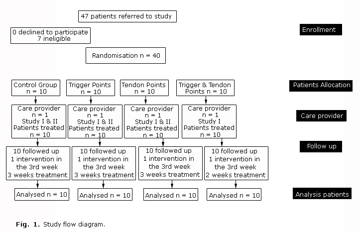
<body><![CDATA[<p><font face="Verdana, Arial, Helvetica, sans-serif" size="2"> 1. Control group      that continued with their daily activities (CT). </font></p>       <p><font face="Verdana, Arial, Helvetica, sans-serif" size="2"> 2. Intervention      group where the EPI&#174; was applied only in all detected trigger points      (TP). </font></p>       <p><font face="Verdana, Arial, Helvetica, sans-serif" size="2"> 3. Intervention      group where the EPI&#174; was applied only in the tendon of the infraspinatus      (T). </font></p>       <p><font face="Verdana, Arial, Helvetica, sans-serif" size="2"> 4. Intervention      group where the EPI&#174; was applied in all detected trigger points and the      tendon (TTP). </font></p> </blockquote>     <p><font face="Verdana, Arial, Helvetica, sans-serif" size="2"> An Ethic Committee    at the <i>Institute of Physiotherapy and Sports </i>was formed on 15<sup>th</sup>    January 2013 to approve the trial. It verified the results on 2<sup>nd</sup>    July 2013. The randomized controlled trial started at the end of March 2013    and concluded during the second week of June 2013. Each patient that arrived    to the facilities and met the inclusion criteria was randomly allocated to a    group depending on sex. That is, the first man and the first woman to the first    group, and the second man and the second woman to the next group, and so on    until the first's participants of the 4 groups were completed. Then, we repeated    the same process for the second participants and so on, until we collected ten    persons in each group. All the patients knew that they were enrolled in a trial,    but they never knew what group they belonged to nor the existence of any other    group. All the patients signed a consent document. </font></p>     <p align="left"> <font face="Verdana, Arial, Helvetica, sans-serif" size="2"><b>Outcomes    and Follow-up</b> </font></p>     <p><font face="Verdana, Arial, Helvetica, sans-serif" size="2"> In total we performed    four measurements: an initial one before initiating the treatment and three    subsequent ones performed after application of a weekly treatment.<a href="#_edn1" name="_ednref1" title="">[a]</a>    We measured the level of pain, the limit of abduction, internal rotation and    external rotation. We divided the study into two parts according to the evolution    of the variables. The first study corresponds to the first three measurements,    and the second one to the fourth measurement. None of the enrolled participants    gave up the trial, and there were no changes in the trial after it commenced.    Finally there was no harm or unintended effects on any of the intervention groups.    <a href="/img/revistas/ort/v30n1/f0107116.jpg">Figure 1</a> summarizes the participant flow diagram.    </font></p>     <p><font face="Verdana, Arial, Helvetica, sans-serif" size="2"> </font></p>     <p align="left"><font face="Verdana, Arial, Helvetica, sans-serif" size="2"><font size="3"><b>RESULTS    </b></font></font></p>     <p align="left"><font face="Verdana, Arial, Helvetica, sans-serif" size="2"> <a href="/img/revistas/ort/v30n1/t0107116.gif">Tables    1</a> and <a href="/img/revistas/ort/v30n1/t0207116.gif">2</a> show the values for the different variables    measured for the 4 groups under study. The rows represent the mean and the standard    deviation of each group, while the columns represent first the age, and then    the initial pain value (VAS<sub>i</sub>) followed by the pain value measured    after one week after the treatment, and the same sequence for the abduction    limits, internal and external rotation limits. <a href="#fig2">Figure 2</a>    represents graphically the VAS values for the 4 measurements for each group.    </font></p>     ]]></body>
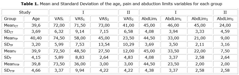
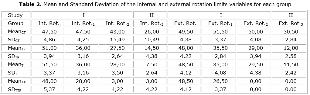
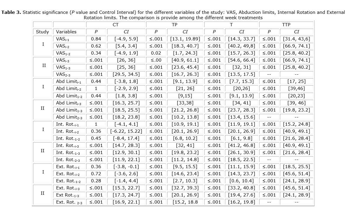
<body><![CDATA[<p align="center"><font face="Verdana, Arial, Helvetica, sans-serif" size="2">    <img src="/img/revistas/ort/v30n1/f0207116.jpg" width="420" height="434"><a name="fig2"></a></font></p>     <p align="left"><font face="Verdana, Arial, Helvetica, sans-serif" size="2"> <a href="/img/revistas/ort/v30n1/t0307116.gif">Table    3</a> represents the statistic significance for all the variables under study    (p-values and confidence intervals). </font></p>     <p align="left"><font face="Verdana, Arial, Helvetica, sans-serif" size="2">From    <a href="/img/revistas/ort/v30n1/t0307116.gif">table 3</a>, we can see that there was changes between    VAS<sub>i</sub> with VAS<sub>1 </sub>in the three intervention groups (p&lt;0.001)    and between VAS <sub>i </sub>&amp; VAS<sub>2</sub> and VAS<sub>1 </sub>&amp;    VAS<sub>2</sub>(p&lt;0.001) in T and TTP but in TP(p&lt;0.05). There were no    significant differences in the level of perceived pain in relation to age (p&gt;0.05).    There were also no significance differences in the level of perceived pain in    relation to sex (p&gt;0.05). </font></p>     <p align="left"><font face="Verdana, Arial, Helvetica, sans-serif" size="2"> In    the comparison between the VAS<sub>i</sub> with VAS<sub>1</sub> and VAS<sub>2</sub>,    the CT group did not decrease the perception of pain at any time (p&gt; 0.05),    and there were significance differences with the rest of the groups (p&lt;0.001).    The best results were for the TTP group followed by the T group and in last    place, the TP group. The comparison between the VAS<sub>1</sub> and VAS<sub>2</sub>    is similar to the results between VAS<sub>i</sub> and VAS<sub>2</sub>. </font></p>     <p align="left"><font face="Verdana, Arial, Helvetica, sans-serif" size="2"> In    the descriptive analysis of the restriction of mobility for abduction (Abd.Limit),    we observed changes (p&lt;0.001). In the Abduction limits comparison between    Abd.Limit<sub>i </sub>&amp; Abd.Limit<sub>1</sub>, Abd.Limit<sub>i </sub>&amp;    Abd.Limit<sub>2</sub>, and Abd.Limit<sub>1</sub> with Abd.Limit <sub>2</sub>,    there was interaction of our interventions and variations were obtained for    the 3 groups (p&lt;0.001). There were no significant differences in the level    of restriction of mobility for abduction in relation to age (p&gt;0.05) or in    relation to sex (p&gt;0.05). </font></p>     <p align="left"><font face="Verdana, Arial, Helvetica, sans-serif" size="2"> In    the comparison between the Abd.Limit<sub>i </sub>&amp; Abd.Limit<sub>1</sub>,    Abd.Limit<sub>i </sub>&amp; Abd.Limit<sub>2</sub>, and Abd.Limit <sub>1 </sub>&amp;    Abd.Limit<sub>2 </sub>the CT group did not improve its level of mobility restriction    at any time (p&gt;0.05). But all intervention groups improved (p&lt;0.001) with    significant differences respect to the CT group, except for the comparison between    the TP and T groups (p&gt;0.05). The TTP group obtained the best result compared    to the other groups (p&lt;0.001). </font></p>     <p align="left"><font face="Verdana, Arial, Helvetica, sans-serif" size="2"> Related    to the Internal Rotation limits comparison between Int.Rot<sub>i </sub>&amp;    Int.Rot<sub>1</sub>, Int.Rot<sub>i </sub>&amp; Int.Rot<sub>2</sub>, and Int.Rot<sub>1    </sub>&amp; Int.Rot<sub>2</sub> there was interaction of our interventions and    variations that were obtained for the 3 groups (p&lt;0.001). There were no significant    differences in the level of restriction of internal rotation limits in relation    to age (p&gt;0.05), nor in relation to sex (p&gt;0.05). </font></p>     <p align="left"><font face="Verdana, Arial, Helvetica, sans-serif" size="2"> In    the comparison between the Int.Rot<sub>i </sub>&amp; Int.Rot<sub>1</sub>, Int.Rot<sub>i    </sub>&amp; Int.Rot<sub>2</sub> and Int.Rot<sub>1 </sub>&amp; Int.Rot<sub>2</sub>the    CT group did not improve its level of mobility restriction at any time (p&gt;0.05);    however, all intervention groups improved (p&lt;0.001) with significant differences    with respect to the CT group, but the T and TP groups did not present differences    between them (p&gt;0.05). </font></p>     <p><font face="Verdana, Arial, Helvetica, sans-serif" size="2"> Finally, we present    the results for the External Rotation Limits. In the comparison between Ext.Rot<sub>i    </sub>&amp; Ext.Rot<sub>1</sub> and Ext.Rot <sub>i </sub>&amp; Ext.Rot<sub>2</sub>,    and Ext.Rot<sub>1</sub>&amp; Ext.Rot<sub>2</sub> there was interaction of our    interventions and variations that were obtained for the 3 groups (p&lt;0.001).    There were no statistic differences in the level of restriction of mobility    for external rotation in relation to age (p&gt;0.05) and not also in relation    to sex (p&gt;0.05). </font></p>     <p align="left"><font face="Verdana, Arial, Helvetica, sans-serif" size="2"> In    the comparison between the Ext.Rot<sub>i</sub> and the Ext.Rot<sub>1</sub> the    CT group did not improve its level of mobility restriction at any time (p&gt;0.05);    however, all intervention groups improved (p&lt;0.001) with significant differences    with respect to the CT group, except for the T and TP groups without differences    between them (p&gt;0.05). But there were differences of the TTP group in relation    to the T and TP groups. </font></p>     ]]></body>
<body><![CDATA[<p align="left"><font face="Verdana, Arial, Helvetica, sans-serif" size="2"> For    the second study, we started from the 3 measures already collected and performed    a final measurement. As mentioned before, the TTP group served as a control    group since it was the group with the best results: the pain perception was    almost null and the mobility was recovered (then, any treatment was applied    for the TTP group). In the fourth measurement, we observed that there were changes    (p&lt;0.001) in all the variables measured. The Post Hoc shows significant differences    between all groups (p&lt;0.001) after the EPI&#174; application in all the trigger    points and in the tendon. </font></p>     <p align="left">&nbsp;</p>     <p align="left"><font face="Verdana, Arial, Helvetica, sans-serif" size="2"><b><font size="3">CONCLUSIONS</font></b>    </font></p>     <p><font face="Verdana, Arial, Helvetica, sans-serif" size="2"> According to the    results, we can infer that our intervention could introduce variations in the    pain perception in the three groups under study and no improvement in the control    group. Furthermore, we observed that the degree of improvement was increasing    in our successive interventions. Besides, during our interventions, we did not    reach significant differences to lower the pain perception between the TP and    T groups, despite the large decreased in pain perception between the two groups.    </font></p>     <p><font face="Verdana, Arial, Helvetica, sans-serif" size="2"> In summary, we    found that our interventions decreased the scale pain perception from our first    intervention for the TTP and T groups, and in the second intervention for the    3 intervention groups. The TTP group presented the best results, and the positive    response of the T group was earlier than the TP group. Then, we can say that:    </font></p>     <blockquote>       <p><font face="Verdana, Arial, Helvetica, sans-serif" size="2"> 1. The application      of the EPI&#174; in the trigger points and the tendon of the infraspinatus      muscle (together or alone) is effective in reducing the pain and the mobility      restriction in abduction, internal rotation and external rotation limits when      there is pain in the tendon and trigger points below the infraspinatus muscle.      </font></p>       <p><font face="Verdana, Arial, Helvetica, sans-serif" size="2"> 2. The EPI&#174;      is more effective if applied in the infraspinatus muscle and the tendon than      applied only in one of the two structures, when both structures have pain.      </font></p>       <p><font face="Verdana, Arial, Helvetica, sans-serif" size="2"> 3. In the case      of comparing the application of the EPI&#174; in the muscle or the tendon,      there were no differences after two applications, but the perception of the      pain diminishes in the first application in the tendon, and its application      in the muscle required two applications to arise the same effect. </font></p>       <p><font face="Verdana, Arial, Helvetica, sans-serif" size="2"> 4. The application      of two sessions of EPI&#174; together in the tendon and the muscle with an      interval of one week was enough for a quasi-full recovery, while the isolated      application of EPI&#174; on two sessions in the tendon or muscle did not produce      the similar recovery, leaving pain and partial limitations. </font></p>       ]]></body>
<body><![CDATA[<p><font face="Verdana, Arial, Helvetica, sans-serif" size="2"> 5. The significant      improvement in the CT group and the TP and T groups with the third EPI&#174;      when applied together in the tendon and the muscle, once again confirmed the      efficiency of the treatment in both structures. </font></p>       <p><font face="Verdana, Arial, Helvetica, sans-serif" size="2"> 6. The tendon      and muscle can interact in mutual damage. </font></p> </blockquote>     <p align="left"><font face="Verdana, Arial, Helvetica, sans-serif" size="2"> <b>Limitations    of the Study </b></font></p>     <p align="left"><font face="Verdana, Arial, Helvetica, sans-serif" size="2"> The    study could have been more ambitious by testing the involvement of different    structures and its related biomechanical implications, and it could have considered    more variables. </font></p>     <p align="left"><font face="Verdana, Arial, Helvetica, sans-serif" size="2"> <b>Conflict    of Interest </b></font></p>     <p><font face="Verdana, Arial, Helvetica, sans-serif" size="2"> None declared.    </font></p>     <div>        <p> <font face="Verdana, Arial, Helvetica, sans-serif" size="2"><b>What this      study adds?</b> </font></p>       <p><font face="Verdana, Arial, Helvetica, sans-serif" size="2"> The study has      shown that when there are trigger points in the muscle and pain in the tendon      of the infraspinatus: </font></p> </div>     <blockquote>       ]]></body>
<body><![CDATA[<div>         <p><font face="Verdana, Arial, Helvetica, sans-serif" size="2"> 1) The EPI&#174;        is an effective treatment that helps to lower the pain and mobility limits        values. </font></p>         <p><font face="Verdana, Arial, Helvetica, sans-serif" size="2">2) The EPI&#174;        is more effective when it is applied together in the trigger points and        the tendon, although improvements are appreciated when it is applied in        isolation. These findings suggest the existence of interaction on both structures.        </font></p>   </div>   </blockquote>     <div>        <p> <font face="Verdana, Arial, Helvetica, sans-serif" size="2"><b>What is already      known on this topic?</b> </font></p>       <p><font face="Verdana, Arial, Helvetica, sans-serif" size="2"> This novel treatment      obtains excellent results in a short period of time compared to existing treatments.      We believe is going to have a big impact, in the short term, of the clinical      practice. </font></p> </div>     <p align="left">&nbsp;</p>     <p align="left"><font face="Verdana, Arial, Helvetica, sans-serif" size="2"><b><font size="3">BIBLIOGRAPHIC    REFERENCES </font></b></font></p>     <!-- ref --><p><font face="Verdana, Arial, Helvetica, sans-serif" size="2"> 1. Stoller DW.    MRI, Arthroscopy, and Surgical Anatomy of the Joints. Philadelphia: Lippincott-Raven;    1999. ISBN-13: 978-0781715805.     </font></p>     ]]></body>
<body><![CDATA[<!-- ref --><p><font face="Verdana, Arial, Helvetica, sans-serif" size="2"> 2. Cook JL, Purdam    CR. Is tendon pathology a continuum? A pathology model to explain the clinical    presentation of load-induced tendinopathy. Br J Sport Med. 2009;43:409-16.     </font></p>     <!-- ref --><p><font face="Verdana, Arial, Helvetica, sans-serif" size="2"> 3. Khan KM, Cook    JL, Kannus P, Maffulli N, Bonar SF. Time to abandon the "tendinitis" myth. BMJ.    2002;324:626-7.     </font></p>     <!-- ref --><p><font face="Verdana, Arial, Helvetica, sans-serif" size="2"> 4. Fu SC, Rolf    C, Cheuk YC, Lui PP, Chan KM. Deciphering the pathogenesis of tendinopathy:    a three-stages process. Sports Med Arthros Rehabil Ther Technol. 2010 Dec 13;2:30.    doi: 10.1186/1758-2555-2-30.     </font></p>     <!-- ref --><p><font face="Verdana, Arial, Helvetica, sans-serif" size="2"> 5. Shah JP, Phillips    TM, Danoff JV, Gerber LH. An in vivo microanalytical technique for measuring    the local biochemical milieu of human skeletal muscle. J Appl Physiol. 2005;99:1977-84.        </font></p>     <!-- ref --><p><font face="Verdana, Arial, Helvetica, sans-serif" size="2"> 6. Valera-Garrido    F, Minaya-Mu&#241;oz F, S&#225;nchez-Ib&#225;&#241;ez JM, Garc&#237;a-Palencia    P, Valderrama-Canales F, Medina-Mirapeix F, et al. Comparison of the acute inflammatory    response and proliferation of dry needling and Electrolysis Percutaneous Intratissue    (EPI) in healthy rat Achilles tendons. Br J Sports Med. 2013;47:32. doi: 10.1136/bjsports-2013-092459.56    </font><!-- ref --><p><font face="Verdana, Arial, Helvetica, sans-serif" size="2"> 7. Valera-Garrido    F, Minaya-Mu&#241;oz F, S&#225;nchez-Ib&#225;&#241;ez JM, Medina-Mirapeix F,    Polidori F. Short- and long-term outcomes of electrolysis percutaneous intratissue    (EPI) in chronic lateral elbow epicondylitis. Br J Sports Med. 2013;47:3. DOI:10.1136/bjsports-2013-092459.38    </font><!-- ref --><p><font face="Verdana, Arial, Helvetica, sans-serif" size="2"> 8. Maffulli N,    Longo UG. Conservative management for tendinopathy; is there enough scientific    evidence? Rheumatology. 2008;47:390-1.     </font></p>     <!-- ref --><p><font face="Verdana, Arial, Helvetica, sans-serif" size="2"> 9. Fredberg U,    Bolvig L. Jumper&#180;s knee. Review of the literature. Scand J Med Sci Sports.    1999;9:66-73.     </font></p>     <!-- ref --><p><font face="Verdana, Arial, Helvetica, sans-serif" size="2"> 10. Peers KH<sup>1</sup>,    Lysens RJ. Pattelar tendinopathy in athletes; current diagnostic and therapeutic    recommendations. Sports Med. 2005;35:71-87.     </font></p>     <!-- ref --><p><font face="Verdana, Arial, Helvetica, sans-serif" size="2"> 11. Huttenlocher    A, Horwitz AR. Wound healing with healing with electrical potential. N Engl    J Med. 2007;356:303-4.     </font></p>     <!-- ref --><p><font face="Verdana, Arial, Helvetica, sans-serif" size="2"> 12. Jaffe LF,    Vanable JW Jr. Electric fields and wound healing. Clin Dermatol. 1984;2:34-44.        </font></p>     ]]></body>
<body><![CDATA[<!-- ref --><p><font face="Verdana, Arial, Helvetica, sans-serif" size="2"> 13. Cheng N, Van    Hoof H, Bockx E, Hoogmartens MJ, Mulier JC, De Dijcker FJ, et al. The effects    of electric currents on ATP generation, protein synthesis and membrane transport    of rat skin. Clin Orthop Relat Res. 1982;(171):264-72.     </font></p>     <!-- ref --><p><font face="Verdana, Arial, Helvetica, sans-serif" size="2"> 14. Bourguignon    GJ, Jy W, Bourguignon LYW. Electric stimulation of human fibroblasts causes    an increase in Ca<sup>2+</sup> influx and the exposure of additional insulin    in receptors. J Cell Physiol. 1989;379-85.     </font></p>     <!-- ref --><p><font face="Verdana, Arial, Helvetica, sans-serif" size="2"> 15. Hancock M,    Herbert RD, Maher CG. A guide to interpretation of studies investigating subgroups    of responders to physical therapy interventions. Phys Ther. 2009;89(7):698-704.    doi: 10.2522/ptj.20080351.     </font></p>     <p>&nbsp;</p>     <p>&nbsp;</p>     <p><font face="Verdana, Arial, Helvetica, sans-serif" size="2">Recibido: 16 de    noviembre de 2015.     <br>   <a>Aprobado:</a> 18 de enero de 2016.</font></p>     ]]></body>
<body><![CDATA[<p>&nbsp; </p>     <p>&nbsp;</p>     <p><font face="Verdana, Arial, Helvetica, sans-serif" size="2"><i>Mar&#237;a Dolores    R-Moreno</i>. Departamento de Autom&#225;tica, E-318. Escuela Polit&#233;cnica    Superior. Carretera Madrid-Barcelona, Km. 33,6. Universidad de Alcal&#225; 28805,    Alcal&#225; de Henares. Madrid, Espa&#241;a.    <br>   </font><font face="Verdana, Arial, Helvetica, sans-serif" size="2">Phone: +34    91 885 6607 Fax: +34 91 885 6641 </font><font face="Verdana, Arial, Helvetica, sans-serif" size="2">E-mail:    <a href="mailto:mdolores@aut.uah.es">mdolores@aut.uah.es</a> <a href="mailto:malola.rmoreno@uah.es">malola.rmoreno@uah.es</a>    </font><font face="Verdana, Arial, Helvetica, sans-serif" size="2">URL: <a href="http://atc1.aut.uah.es/~mdolores/">http://atc1.aut.uah.es/~mdolores/</a>    </font></p> <hr align="left" size="1" width="33%"/>        <div id="edn1">        <p> <font face="Verdana, Arial, Helvetica, sans-serif" size="2"><a href="#_ednref1" name="_edn1" title="">a)</a>      The CT group did not receive any treatment until the second study, that is,      the third scheduled week of the study.</font></p>   </div>      ]]></body><back>
<ref-list>
<ref id="B1">
<label>1</label><nlm-citation citation-type="book">
<person-group person-group-type="author">
<name>
<surname><![CDATA[Stoller]]></surname>
<given-names><![CDATA[DW]]></given-names>
</name>
</person-group>
<source><![CDATA[MRI, Arthroscopy, and Surgical Anatomy of the Joints]]></source>
<year>1999</year>
<publisher-loc><![CDATA[Philadelphia ]]></publisher-loc>
<publisher-name><![CDATA[Lippincott-Raven]]></publisher-name>
</nlm-citation>
</ref>
<ref id="B2">
<label>2</label><nlm-citation citation-type="journal">
<person-group person-group-type="author">
<name>
<surname><![CDATA[Cook]]></surname>
<given-names><![CDATA[JL]]></given-names>
</name>
<name>
<surname><![CDATA[Purdam]]></surname>
<given-names><![CDATA[CR]]></given-names>
</name>
</person-group>
<article-title xml:lang="en"><![CDATA[Is tendon pathology a continuum: A pathology model to explain the clinical presentation of load-induced tendinopathy]]></article-title>
<source><![CDATA[Br J Sport Med]]></source>
<year>2009</year>
<volume>43</volume>
<page-range>409-16</page-range></nlm-citation>
</ref>
<ref id="B3">
<label>3</label><nlm-citation citation-type="journal">
<person-group person-group-type="author">
<name>
<surname><![CDATA[Khan]]></surname>
<given-names><![CDATA[KM]]></given-names>
</name>
<name>
<surname><![CDATA[Cook]]></surname>
<given-names><![CDATA[JL]]></given-names>
</name>
<name>
<surname><![CDATA[Kannus]]></surname>
<given-names><![CDATA[P]]></given-names>
</name>
<name>
<surname><![CDATA[Maffulli]]></surname>
<given-names><![CDATA[N]]></given-names>
</name>
<name>
<surname><![CDATA[Bonar]]></surname>
<given-names><![CDATA[SF]]></given-names>
</name>
</person-group>
<article-title xml:lang="en"><![CDATA[Time to abandon the "tendinitis" myth]]></article-title>
<source><![CDATA[BMJ]]></source>
<year>2002</year>
<volume>324</volume>
<page-range>626-7</page-range></nlm-citation>
</ref>
<ref id="B4">
<label>4</label><nlm-citation citation-type="journal">
<person-group person-group-type="author">
<name>
<surname><![CDATA[Fu]]></surname>
<given-names><![CDATA[SC]]></given-names>
</name>
<name>
<surname><![CDATA[Rolf]]></surname>
<given-names><![CDATA[C]]></given-names>
</name>
<name>
<surname><![CDATA[Cheuk]]></surname>
<given-names><![CDATA[YC]]></given-names>
</name>
<name>
<surname><![CDATA[Lui]]></surname>
<given-names><![CDATA[PP]]></given-names>
</name>
<name>
<surname><![CDATA[Chan]]></surname>
<given-names><![CDATA[KM]]></given-names>
</name>
</person-group>
<article-title xml:lang="en"><![CDATA[Deciphering the pathogenesis of tendinopathy: a three-stages process]]></article-title>
<source><![CDATA[Sports Med Arthros Rehabil Ther Technol]]></source>
<year>2010</year>
<month>12</month>
<day>13</day>
<volume>2</volume>
<page-range>30</page-range></nlm-citation>
</ref>
<ref id="B5">
<label>5</label><nlm-citation citation-type="journal">
<person-group person-group-type="author">
<name>
<surname><![CDATA[Shah]]></surname>
<given-names><![CDATA[JP]]></given-names>
</name>
<name>
<surname><![CDATA[Phillips]]></surname>
<given-names><![CDATA[TM]]></given-names>
</name>
<name>
<surname><![CDATA[Danoff]]></surname>
<given-names><![CDATA[JV]]></given-names>
</name>
<name>
<surname><![CDATA[Gerber]]></surname>
<given-names><![CDATA[LH]]></given-names>
</name>
</person-group>
<article-title xml:lang="en"><![CDATA[An in vivo microanalytical technique for measuring the local biochemical milieu of human skeletal muscle]]></article-title>
<source><![CDATA[J Appl Physiol]]></source>
<year>2005</year>
<volume>99</volume>
<page-range>1977-84</page-range></nlm-citation>
</ref>
<ref id="B6">
<label>6</label><nlm-citation citation-type="journal">
<person-group person-group-type="author">
<name>
<surname><![CDATA[Valera-Garrido]]></surname>
<given-names><![CDATA[F]]></given-names>
</name>
<name>
<surname><![CDATA[Minaya-Muñoz]]></surname>
<given-names><![CDATA[F]]></given-names>
</name>
<name>
<surname><![CDATA[Sánchez-Ibáñez]]></surname>
<given-names><![CDATA[JM]]></given-names>
</name>
<name>
<surname><![CDATA[García-Palencia]]></surname>
<given-names><![CDATA[P]]></given-names>
</name>
<name>
<surname><![CDATA[Valderrama-Canales]]></surname>
<given-names><![CDATA[F]]></given-names>
</name>
<name>
<surname><![CDATA[Medina-Mirapeix]]></surname>
<given-names><![CDATA[F]]></given-names>
</name>
</person-group>
<article-title xml:lang="en"><![CDATA[Comparison of the acute inflammatory response and proliferation of dry needling and Electrolysis Percutaneous Intratissue (EPI) in healthy rat Achilles tendons]]></article-title>
<source><![CDATA[Br J Sports Med]]></source>
<year>2013</year>
<volume>47</volume>
<page-range>32</page-range></nlm-citation>
</ref>
<ref id="B7">
<label>7</label><nlm-citation citation-type="journal">
<person-group person-group-type="author">
<name>
<surname><![CDATA[Valera-Garrido]]></surname>
<given-names><![CDATA[F]]></given-names>
</name>
<name>
<surname><![CDATA[Minaya-Muñoz]]></surname>
<given-names><![CDATA[F]]></given-names>
</name>
<name>
<surname><![CDATA[Sánchez-Ibáñez]]></surname>
<given-names><![CDATA[JM]]></given-names>
</name>
<name>
<surname><![CDATA[Medina-Mirapeix]]></surname>
<given-names><![CDATA[F]]></given-names>
</name>
<name>
<surname><![CDATA[Polidori]]></surname>
<given-names><![CDATA[F]]></given-names>
</name>
</person-group>
<article-title xml:lang="en"><![CDATA[Short- and long-term outcomes of electrolysis percutaneous intratissue (EPI) in chronic lateral elbow epicondylitis]]></article-title>
<source><![CDATA[Br J Sports Med]]></source>
<year>2013</year>
<volume>47</volume>
<page-range>3</page-range></nlm-citation>
</ref>
<ref id="B8">
<label>8</label><nlm-citation citation-type="journal">
<person-group person-group-type="author">
<name>
<surname><![CDATA[Maffulli]]></surname>
<given-names><![CDATA[N]]></given-names>
</name>
<name>
<surname><![CDATA[Longo]]></surname>
<given-names><![CDATA[UG]]></given-names>
</name>
</person-group>
<article-title xml:lang="en"><![CDATA[Conservative management for tendinopathy; is there enough scientific evidence]]></article-title>
<source><![CDATA[Rheumatology]]></source>
<year>2008</year>
<volume>47</volume>
<page-range>390-1</page-range></nlm-citation>
</ref>
<ref id="B9">
<label>9</label><nlm-citation citation-type="journal">
<person-group person-group-type="author">
<name>
<surname><![CDATA[Fredberg]]></surname>
<given-names><![CDATA[U]]></given-names>
</name>
<name>
<surname><![CDATA[Bolvig]]></surname>
<given-names><![CDATA[L]]></given-names>
</name>
</person-group>
<article-title xml:lang="en"><![CDATA[Jumper´s knee: Review of the literature]]></article-title>
<source><![CDATA[Scand J Med Sci Sports]]></source>
<year>1999</year>
<volume>9</volume>
<page-range>66-73</page-range></nlm-citation>
</ref>
<ref id="B10">
<label>10</label><nlm-citation citation-type="journal">
<person-group person-group-type="author">
<name>
<surname><![CDATA[Peers]]></surname>
<given-names><![CDATA[KH1]]></given-names>
</name>
<name>
<surname><![CDATA[Lysens]]></surname>
<given-names><![CDATA[RJ]]></given-names>
</name>
</person-group>
<article-title xml:lang="en"><![CDATA[Pattelar tendinopathy in athletes; current diagnostic and therapeutic recommendations]]></article-title>
<source><![CDATA[Sports Med]]></source>
<year>2005</year>
<volume>35</volume>
<page-range>71-87</page-range></nlm-citation>
</ref>
<ref id="B11">
<label>11</label><nlm-citation citation-type="journal">
<person-group person-group-type="author">
<name>
<surname><![CDATA[Huttenlocher]]></surname>
<given-names><![CDATA[A]]></given-names>
</name>
<name>
<surname><![CDATA[Horwitz]]></surname>
<given-names><![CDATA[AR]]></given-names>
</name>
</person-group>
<article-title xml:lang="en"><![CDATA[Wound healing with healing with electrical potential]]></article-title>
<source><![CDATA[N Engl J Med]]></source>
<year>2007</year>
<volume>356</volume>
<page-range>303-4</page-range></nlm-citation>
</ref>
<ref id="B12">
<label>12</label><nlm-citation citation-type="journal">
<person-group person-group-type="author">
<name>
<surname><![CDATA[Jaffe]]></surname>
<given-names><![CDATA[LF]]></given-names>
</name>
<name>
<surname><![CDATA[Vanable]]></surname>
<given-names><![CDATA[JrJW]]></given-names>
</name>
</person-group>
<article-title xml:lang="en"><![CDATA[Electric fields and wound healing]]></article-title>
<source><![CDATA[Clin Dermatol]]></source>
<year>1984</year>
<volume>2</volume>
<page-range>34-44</page-range></nlm-citation>
</ref>
<ref id="B13">
<label>13</label><nlm-citation citation-type="journal">
<person-group person-group-type="author">
<name>
<surname><![CDATA[Cheng]]></surname>
<given-names><![CDATA[N]]></given-names>
</name>
<name>
<surname><![CDATA[Van Hoof]]></surname>
<given-names><![CDATA[H]]></given-names>
</name>
<name>
<surname><![CDATA[Bockx]]></surname>
<given-names><![CDATA[E]]></given-names>
</name>
<name>
<surname><![CDATA[Hoogmartens]]></surname>
<given-names><![CDATA[MJ]]></given-names>
</name>
<name>
<surname><![CDATA[Mulier]]></surname>
<given-names><![CDATA[JC]]></given-names>
</name>
<name>
<surname><![CDATA[De Dijcker]]></surname>
<given-names><![CDATA[FJ]]></given-names>
</name>
</person-group>
<article-title xml:lang="en"><![CDATA[The effects of electric currents on ATP generation, protein synthesis and membrane transport of rat skin]]></article-title>
<source><![CDATA[Clin Orthop Relat Res]]></source>
<year>1982</year>
<numero>171</numero>
<issue>171</issue>
<page-range>264-72</page-range></nlm-citation>
</ref>
<ref id="B14">
<label>14</label><nlm-citation citation-type="journal">
<person-group person-group-type="author">
<name>
<surname><![CDATA[Bourguignon]]></surname>
<given-names><![CDATA[GJ]]></given-names>
</name>
<name>
<surname><![CDATA[Jy]]></surname>
<given-names><![CDATA[W]]></given-names>
</name>
<name>
<surname><![CDATA[Bourguignon]]></surname>
<given-names><![CDATA[LYW]]></given-names>
</name>
</person-group>
<article-title xml:lang="en"><![CDATA[Electric stimulation of human fibroblasts causes an increase in Ca2+ influx and the exposure of additional insulin in receptors]]></article-title>
<source><![CDATA[J Cell Physiol]]></source>
<year>1989</year>
<page-range>379-85</page-range></nlm-citation>
</ref>
<ref id="B15">
<label>15</label><nlm-citation citation-type="journal">
<person-group person-group-type="author">
<name>
<surname><![CDATA[Hancock]]></surname>
<given-names><![CDATA[M]]></given-names>
</name>
<name>
<surname><![CDATA[Herbert]]></surname>
<given-names><![CDATA[RD]]></given-names>
</name>
<name>
<surname><![CDATA[Maher]]></surname>
<given-names><![CDATA[CG]]></given-names>
</name>
</person-group>
<article-title xml:lang="en"><![CDATA[A guide to interpretation of studies investigating subgroups of responders to physical therapy interventions]]></article-title>
<source><![CDATA[Phys Ther]]></source>
<year>2009</year>
<volume>89</volume>
<numero>7</numero>
<issue>7</issue>
<page-range>698-704</page-range></nlm-citation>
</ref>
</ref-list>
</back>
</article>