<?xml version="1.0" encoding="ISO-8859-1"?><article xmlns:mml="http://www.w3.org/1998/Math/MathML" xmlns:xlink="http://www.w3.org/1999/xlink" xmlns:xsi="http://www.w3.org/2001/XMLSchema-instance">
<front>
<journal-meta>
<journal-id>1027-2852</journal-id>
<journal-title><![CDATA[Biotecnología Aplicada]]></journal-title>
<abbrev-journal-title><![CDATA[Biotecnol Apl]]></abbrev-journal-title>
<issn>1027-2852</issn>
<publisher>
<publisher-name><![CDATA[Editorial Elfos Scientiae]]></publisher-name>
</publisher>
</journal-meta>
<article-meta>
<article-id>S1027-28522018000300001</article-id>
<title-group>
<article-title xml:lang="en"><![CDATA[Characterization of a latex agglutination assay for the detection of Rheumatoid Factor]]></article-title>
<article-title xml:lang="es"><![CDATA[Caracterización de un ensayo de aglutinación con látex para la detección del Factor Reumatoide]]></article-title>
</title-group>
<contrib-group>
<contrib contrib-type="author">
<name>
<surname><![CDATA[Hernandez]]></surname>
<given-names><![CDATA[Ariadna]]></given-names>
</name>
<xref ref-type="aff" rid="A01"/>
</contrib>
<contrib contrib-type="author">
<name>
<surname><![CDATA[Fajardo-Sanchez]]></surname>
<given-names><![CDATA[Abel R]]></given-names>
</name>
<xref ref-type="aff" rid="A02"/>
</contrib>
<contrib contrib-type="author">
<name>
<surname><![CDATA[Cabrera]]></surname>
<given-names><![CDATA[Ruben A]]></given-names>
</name>
<xref ref-type="aff" rid="A02"/>
</contrib>
<contrib contrib-type="author">
<name>
<surname><![CDATA[Ortiz]]></surname>
<given-names><![CDATA[Neise]]></given-names>
</name>
<xref ref-type="aff" rid="A03"/>
</contrib>
<contrib contrib-type="author">
<name>
<surname><![CDATA[Gonzalez-Aznar]]></surname>
<given-names><![CDATA[Elizabeth]]></given-names>
</name>
<xref ref-type="aff" rid="A02"/>
</contrib>
<contrib contrib-type="author">
<name>
<surname><![CDATA[Ramirez-Bencomo]]></surname>
<given-names><![CDATA[Fidel]]></given-names>
</name>
<xref ref-type="aff" rid="A02"/>
</contrib>
<contrib contrib-type="author">
<name>
<surname><![CDATA[Acevedo]]></surname>
<given-names><![CDATA[Reinaldo]]></given-names>
</name>
<xref ref-type="aff" rid="A02"/>
</contrib>
<contrib contrib-type="author">
<name>
<surname><![CDATA[Menendez]]></surname>
<given-names><![CDATA[Tamara]]></given-names>
</name>
<xref ref-type="aff" rid="A04"/>
</contrib>
</contrib-group>
<aff id="A01">
<institution><![CDATA[,University of Havana Faculty of Chemistry ]]></institution>
<addr-line><![CDATA[ ]]></addr-line>
<country>Cuba</country>
</aff>
<aff id="A02">
<institution><![CDATA[,Finlay Vaccines Institute  ]]></institution>
<addr-line><![CDATA[ ]]></addr-line>
<country>Cuba</country>
</aff>
<aff id="A03">
<institution><![CDATA[,10 de Octubre Teaching Clinical Surgical Hospital Rheumatology Center ]]></institution>
<addr-line><![CDATA[ ]]></addr-line>
<country>Cuba</country>
</aff>
<aff id="A04">
<institution><![CDATA[,University of Havana Center of Biomaterials ]]></institution>
<addr-line><![CDATA[ ]]></addr-line>
<country>Cuba</country>
</aff>
<pub-date pub-type="pub">
<day>00</day>
<month>09</month>
<year>2018</year>
</pub-date>
<pub-date pub-type="epub">
<day>00</day>
<month>09</month>
<year>2018</year>
</pub-date>
<volume>35</volume>
<numero>3</numero>
<fpage>3201</fpage>
<lpage>3204</lpage>
<copyright-statement/>
<copyright-year/>
<self-uri xlink:href="http://scielo.sld.cu/scielo.php?script=sci_arttext&amp;pid=S1027-28522018000300001&amp;lng=en&amp;nrm=iso"></self-uri><self-uri xlink:href="http://scielo.sld.cu/scielo.php?script=sci_abstract&amp;pid=S1027-28522018000300001&amp;lng=en&amp;nrm=iso"></self-uri><self-uri xlink:href="http://scielo.sld.cu/scielo.php?script=sci_pdf&amp;pid=S1027-28522018000300001&amp;lng=en&amp;nrm=iso"></self-uri><kwd-group>
<kwd lng="en"><![CDATA[agglutination assays]]></kwd>
<kwd lng="en"><![CDATA[antibody]]></kwd>
<kwd lng="en"><![CDATA[biomaterials]]></kwd>
<kwd lng="en"><![CDATA[disease diagnosis]]></kwd>
<kwd lng="en"><![CDATA[purification]]></kwd>
<kwd lng="en"><![CDATA[rheumatoid factor]]></kwd>
<kwd lng="es"><![CDATA[ensayos de aglutinación]]></kwd>
<kwd lng="es"><![CDATA[anticuerpos]]></kwd>
<kwd lng="es"><![CDATA[biomateriales]]></kwd>
<kwd lng="es"><![CDATA[diagnóstico de enfermedades]]></kwd>
<kwd lng="es"><![CDATA[purificación]]></kwd>
<kwd lng="es"><![CDATA[factor reumatoide]]></kwd>
</kwd-group>
</article-meta>
</front><body><![CDATA[ <DIV class="Part"   >        <P align="right"   ><font size="2" face="Verdana, Arial, Helvetica, sans-serif"><b>RESEARCH </b></font></P >       <P   >&nbsp;</P >       <P   ><font size="2" color="#000000" face="Verdana, Arial, Helvetica, sans-serif"><b><font size="4">Characterization      of a latex agglutination assay for the detection of Rheumatoid Factor </font></b></font></P >       <P   >&nbsp;</P >   <FONT size="+1" color="#000000">        <P   ><font size="3" face="Verdana, Arial, Helvetica, sans-serif"><b>Caracterizaci&oacute;n      de un ensayo de aglutinaci&oacute;n con l&aacute;tex para la detecci&oacute;n      del Factor Reumatoide </b></font></P >       <P   >&nbsp;</P >       <P   >&nbsp;</P >       <P   ><font size="2" face="Verdana, Arial, Helvetica, sans-serif"><b>Ariadna Hernandez<sup>1</sup>,      Abel R Fajardo-Sanchez<sup>2</sup>, Ruben A Cabrera<sup>2</sup>, Neise Ortiz<sup>3</sup>,      Elizabeth Gonzalez-Aznar<sup>2</sup>, Fidel Ramirez-Bencomo<sup>2</sup>, Reinaldo      Acevedo<sup>2</sup>, Tamara Menendez<sup>4</sup> </b></font></P >       <P   ><font size="2" face="Verdana, Arial, Helvetica, sans-serif"><sup>1</sup> Faculty      of Chemistry, University of Havana. Zapata entre G y Carlitos Aguirre, Plaza,      Havana 10400, Cuba.    ]]></body>
<body><![CDATA[<br>     </font><font size="2" face="Verdana, Arial, Helvetica, sans-serif"><sup>2</sup>      Finlay Vaccines Institute. Ave. 27 No. 19805, La Lisa, PO Box 16017, Havana      17100, Cuba.    <br>     </font><font size="2" face="Verdana, Arial, Helvetica, sans-serif"><sup>3</sup>      Rheumatology Center, 10 de Octubre Teaching Clinical Surgical Hospital. Calzada      Diez de Octubre No. 130, Cerro, Havana 10500, Cuba.    <br>     </font><font size="2" face="Verdana, Arial, Helvetica, sans-serif"><sup>4</sup>      Center of Biomaterials, University of Havana. Ave. Universidad entre Ronda      y G, Plaza, Havana 10400, Cuba. </font></P >   </font>       <p>&nbsp;</p>       <p>&nbsp;</p>   <hr>   <FONT color="#0000FF"><FONT color="#000000"></font></font><FONT size="+1" color="#000000"><FONT color="#0000FF"><FONT color="#000000">        <P   ><font size="2" face="Verdana, Arial, Helvetica, sans-serif"><b>ABSTRACT </b></font></P >       <P   ><font size="2" face="Verdana, Arial, Helvetica, sans-serif">One of the serologic      criteria to diagnose Rheumatoid Arthritis (RA) is the determination of serum      Rheumatoid factor (RF), which is an autoantibody targeting IgG. It can be      detected by agglutination assays using commercially-available polystyrene      latex-based reagents, which are marketed at relatively high cost. In low-income      settings, homemade latex-based reagents can be a cost-effective alternative.      Multilatex&reg; is a registered trademark of polystyrene latex. It is produced      at the Center of Biomaterials of the University of Havana (Havana, Cuba) for      the development of diagnostic reagents. This work was aimed to obtain a polystyrene      latex-based homemade system for RF detection, using Multilatex&reg; and purified      IgG as RF target. IgG was purified from commercially available purified human      blood gamma globulin fraction, through affinity chromatography with protein      G and adsorbed onto 800 nm-diameter Multilatex&reg; polystyrene spheres. The      RF detection system was calibrated to a detection limit of 8 international      units/mL of RF in patient serum, and evaluated in an agglutination test with      30 positive and 30 negative RF human sera. The developed system behaved similar      when compared to a commercial reagent of 100 % specificity and 100 % sensitivity,      indicating that it could be developed for diagnosis of RA. </font></P >       <P   ><font size="2" face="Verdana, Arial, Helvetica, sans-serif"><B><I>Keywords:</I></B>      agglutination assays, antibody, biomaterials, disease diagnosis, purification,      rheumatoid factor. </font></P >   </font></font></font>    <hr>   <FONT size="+1" color="#000000"><FONT color="#0000FF"><FONT color="#000000">        <P   ><font face="Verdana, Arial, Helvetica, sans-serif" size="2"><b>RESUMEN</b></font></P >       <P   ><font size="2" face="Verdana, Arial, Helvetica, sans-serif">Entre los criterios      serol&oacute;gicos para diagnosticar la Artritis Reumatoide (AR) se encuentra      la presencia en suero del Factor Reumatoide (FR), un autoanticuerpo que se      une a la IgG. Este se puede detectar mediante ensayos de aglutinaci&oacute;n      con reactivos basados en l&aacute;tex de poliestireno, que se encuentran disponibles      en el mercado aunque a precios relativamente elevados. En este contexto, los      reactivos no comerciales pueden ser una alternativa rentable en entornos de      bajos ingresos. El Multilatex&reg; es una marca registrada de l&aacute;tex      de poliestireno </font><font size="2" face="Verdana, Arial, Helvetica, sans-serif">producido      en el Centro de Biomateriales de la Universidad de La Habana (La Habana, Cuba),      para el desarrollo de reactivos para diagn&oacute;stico. En este trabajo se      describe la obtenci&oacute;n de un sistema no comercial basado en l&aacute;tex      de poliestireno para la detecci&oacute;n del FR, que emplea Multilatex&reg;      e IgG purificada como blanco del FR. La IgG fue purificada a partir de una      preparaci&oacute;n comercial de la fracci&oacute;n purificada de gamma-globulina      de sangre humana, mediante cromatograf&iacute;a de afinidad con prote&iacute;na      G, y se adsorbi&oacute; a esferas de poliestireno Multilatex&reg; de 800 nm      de di&aacute;metro. El sistema se calibr&oacute; a un l&iacute;mite de detecci&oacute;n      de 8 unidades internacionales por mililitro de FR en suero de pacientes y      se evalu&oacute; mediante prueba de aglutinaci&oacute;n con 30 sueros humanos      positivos y 30 negativos a FR. El sistema obtenido se desempe&ntilde;&oacute;      de manera similar al compararse con uno comercial de 100 % de especificidad      y 100 % de sensibilidad, por lo que el sistema desarrollado podr&iacute;a      ser &uacute;til en el diagn&oacute;stico de la AR. </font></P >       ]]></body>
<body><![CDATA[<P   ><font size="2" face="Verdana, Arial, Helvetica, sans-serif"><B><I>Palabras clave:</I></B>      ensayos de aglutinaci&oacute;n, anticuerpos, biomateriales, diagn&oacute;stico      de enfermedades, purificaci&oacute;n, factor reumatoide. </font></P >   </font></font></font>   <hr>       <p>&nbsp;</p>       <p>&nbsp;</p>       <p><font size="3" face="Verdana, Arial, Helvetica, sans-serif"><B>INTRODUCTION      </b></font></p>   <FONT size="+1" color="#000000"><FONT color="#0000FF"><FONT color="#000000">     <P   ><font size="2" face="Verdana, Arial, Helvetica, sans-serif"><FONT color="#211E1F">The      Latex agglutination assay (LAT) is a simple, fast, instrumentation-less and      easy to interpret technique, useful for the detection of the Rheumatoid Factor      (RF) [1], which is an autoantibody targeting IgG [2] and one of the serologic      criteria to diagnose Rheumatoid Arthritis (RA)[3]. </font></font></P >   <FONT size="+1" color="#211E1F">        <P   ><font size="2" face="Verdana, Arial, Helvetica, sans-serif">Latex reagents consist      of antigens or antibodies coupled to polystyrene latex spherical particles      [4], which systems could be either commercially available or homemade. The      second one can be produced at lower costs and, advantageously, can be easily      validated against the target population. At the same time, both polystyrene      latex and latex-based reagents are relatively simple to produce at the laboratory      scale and the in-house development of latex reagents can be a cost-effective      option. </font></P >       <P   ><font size="2" face="Verdana, Arial, Helvetica, sans-serif">In the efforts to      develop a latex-based system to accurately detect RF, commercially available      purified human blood gamma-globulin fraction was previously adsorbed to homemade      480 nm-diameter polystyrene spheres [5]. Multilatex&reg; is a brand of polystyrene      latex produced at the Center of Biomaterials of the University of Havana (Havana,      Cuba), by a surfactant-free emulsion polymerization method [6]. The developed      detection system was evaluated by LAT with RF positive and RF negative human      sera and, in comparison with a commercial reagent, unspecific agglutination      was detected in 40 % of negative sera that was avoided after serum-complement      inactivation [5]. The commercial reagent was manufactured with 800 nm-diameter      polystyrene spheres and purified IgG as RF target. In order to develop an      equivalent homemade system using Multilatex&reg;, in this work, 800-nm-diameter      polystyrene particles [7] and affinity-purified IgG as RF target were used.      The performance of the test obtained was evaluated in LAT with a panel of      30 RF-positive and 30 RF-negative human sera, and results compared with a      commercial latex-based reagent for RF detection.</font></P >       <P   >&nbsp;</P >       <P   ><font size="2" color="#000000" face="Verdana, Arial, Helvetica, sans-serif"><B><font size="3">MATERIALS      AND METHODS </font></b></font></P >   <FONT size="+1" color="#000000">        <P   ><font size="2" face="Verdana, Arial, Helvetica, sans-serif"><FONT color="#211E1F"><b>Polystyrene      latex </b></font></font></P >   <FONT size="+1" color="#211E1F">        ]]></body>
<body><![CDATA[<P   ><font size="2" face="Verdana, Arial, Helvetica, sans-serif">Multilatex&reg; polystyrene      latex spheres of 800 nm (mean diameter) were synthesized by the emulsifier      free emulsion polymerization technique [6] and characterized as described      [7]. </font></P >       <P   ><font size="2" face="Verdana, Arial, Helvetica, sans-serif"><b>Human serum samples      </b> </font></P >       <P   ><font size="2" face="Verdana, Arial, Helvetica, sans-serif">Human serum samples      were collected at the Rheumatology Center of the Diez de Octubre Hospital      (RC), Havana, Cuba. A commercial kit (RapiLat&reg;- FR, HELFA Diagnostics&reg;,      Center of Immunoassay, Havana, Cuba) with 100 % clinical specificity, 100      % clinical sensitivity and detection limit of 8 international units (IU) per      milliliter of RF in patient serum was used for serum classification and titration      at the RC. </font></P >       <P   ><font size="2" face="Verdana, Arial, Helvetica, sans-serif"><b>Purification of      human IgG </b></font></P >       <P   ><font size="2" face="Verdana, Arial, Helvetica, sans-serif">The human IgG source      was a commercially available purified human blood gamma-globulin fraction      (human Immunoglobulins, hIgs). It is produced and commercialized by the Center      for Production of Blood Derivatives Adalberto Pesant (Havana, Cuba), for the      treatment of human immunoglobulin deficiencies. </font></P >       <P   ><font size="2" face="Verdana, Arial, Helvetica, sans-serif">The hIgs solution      was dialyzed against phosphate-buffered saline (PBS; 137 mM NaCl, 2.7 mM KCl,      8.1 mM Na<SUB>2</sub>HPO<SUB>4</sub>, 1.46 mM KH<SUB>2</sub>PO<SUB>4</sub>,      pH 7.4). Human IgG was purified through affinity chromatography with protein      G (GE Healthcare, Uppsala, Sweden), according to the manufacturers&rsquo;      instructions. </font></P >   <FONT size="+1"><FONT size="+1"><FONT size="+1"><FONT size="+1"><FONT size="+1"><FONT size="+1"><FONT size="+1"><FONT size="+1">        <P   ><font size="2" face="Verdana, Arial, Helvetica, sans-serif">Affinity chromatography      was performed using the AKTA FPLC System and UNICORN software version 3.0      (GE Healthcare, Uppsala, Sweden), for data acquisition and calculation of      areas under the peaks. </font></P >   <FONT size="+1" color="#000000">        <P   ><font size="2" color="#211E1F" face="Verdana, Arial, Helvetica, sans-serif"><b>Characterization      of purified IgG </b></font></P >   <FONT size="+1" color="#211E1F">        <P   ><font size="2" face="Verdana, Arial, Helvetica, sans-serif">Enzyme-linked immunosorbent      assays (ELISA) to test IgG were performed in 96-well flat-bottom microtiter      plates (Maxisorp, Nunc, Denmark) coated with 0.1 mL of protein solutions at      10 &mu;g/mL in glycine-buffered saline (GBS; 100 mM Glycine, 150 mM NaCl,      pH 8.2), at 4 &deg;C for 18 h. After blocking with skimmed milk, plates were      directly incubated with an anti-human IgG-alkaline phosphatase (AP) conjugate      (Sigma, USA). Reactivity was revealed with p-nitrophenyl phosphate (Sigma,      USA) according to the manufacturer&rsquo;s instructions. Absorbance was read      at 405 nm (A<SUB>405</sub>nm) in a microplate reader (Thermo Electron Corporation,      USA). All samples were analyzed in duplicate; results were expressed as mean      absorbance units at 405 nm values. ELISA to assess IgM were similar but either      GBS, PBS or PBS at pH 6.45 were used as coating buffers. A pre-coating step      was also tested with 5 &mu;g/mL Poly-L-lysine in PBS at 37 &ordm;C for 30      min. </font></P >   <FONT size="+1"><FONT size="+1">        <P   ><font size="2" face="Verdana, Arial, Helvetica, sans-serif">ELISA to test the      reactivity of human sera with the purified IgG were performed similarly, by      incubating diluted sera at 37 &ordm;C for 1 h. GBS was used as coating buffer      and reactions were developed with an anti-human IgM-AP conjugate. </font></P >       ]]></body>
<body><![CDATA[<P   ><font size="2" face="Verdana, Arial, Helvetica, sans-serif">For the dot blot      immunoassay, 50 &mu;g of antigens were vacuum-adsorbed onto 0.2 &mu;m nitrocellulose      membranes (Bio-Rad, USA) using a Manifold II S&amp;S (Germany). After membrane      blocking and incubation with the corresponding anti-human either IgM- or IgG-AP      conjugate, reactions were developed with the 5-bromo-4-chloro-3-indolyl phosphate      p-toluidine salt. </font></P >       <P   ><font size="2" face="Verdana, Arial, Helvetica, sans-serif">Analysis by SDS-PAGE      [8] were performed on 7.5 % or 12.5 % polyacrylamide gels under non-reducing      or reducing conditions, respectively. </font></P >       <P   ><font size="2" face="Verdana, Arial, Helvetica, sans-serif"><b>Protein concentration      determination </b></font></P >       <P   ><font size="2" face="Verdana, Arial, Helvetica, sans-serif">Protein concentration      was determined with the BCA protein assay kit (Pierce Biotechnology, Rockford,      USA) according to manufacturers&rsquo; instructions. </font></P >       <P   ><font size="2" face="Verdana, Arial, Helvetica, sans-serif"><b>Preparation of      latex reagents </b></font></P >       <P   ><font size="2" face="Verdana, Arial, Helvetica, sans-serif">Adsorption on latex      spheres was studied in GBS as reported [7]. Protein ranged from 1 to 5-fold      excess of the amount required to saturate the calculated total particle surface      area (i.e., protein concentrations from 0.18 to 0.89 mg/mL). Equations described      in the Bangs Laboratories protocols for adsorption to microspheres were used      for calculations [9]. The minimal protein concentration allowing particle      saturation was set to produce 10 mL of the latex reagent, which was further      calibrated with the World Health Organization International Reference Preparation      NIBSC 64/2 (Rheumatoid Arthritis Serum) [10] using Bovine Serum Albumin (BSA,      Sigma, USA). Latex reagents were stored at 4-8 &ordm;C until use. </font></P >       <P   ><font size="2" face="Verdana, Arial, Helvetica, sans-serif"><b>Latex agglutination      test </b></font></P >       <P   ><font size="2" face="Verdana, Arial, Helvetica, sans-serif">A mix of 20 &mu;L      of sample and 20 &mu;L of latex reagent was gently agitated for two min, followed      by LAT results scoring as positive or negative if clumps were visible or not,      respectively. Positive sera were serially diluted with 0.9 % NaCl (w/v) and      sera titers were regarded as the maximal dilution giving positive agglutination.      The RapiLat&reg;-FR commercial kit was used as control. The assays included      the negative and positive controls supplied with the commercial kit. All serum      samples were analyzed in duplicate and in case of uncertainty, a third test      was performed. </font></P >       <P   >&nbsp;</P >       <P   ><font size="2" color="#000000" face="Verdana, Arial, Helvetica, sans-serif"><B><font size="3">RESULTS      AND DISCUSSION </font></b></font></P >   <FONT size="+1" color="#000000">        ]]></body>
<body><![CDATA[<P   ><font size="2" face="Verdana, Arial, Helvetica, sans-serif"><FONT color="#211E1F">Human      IgG was purified from a solution of hIgs through affinity chromatography,      as shown in the affinity-chromatography profile (<a href="/img/revistas/bta/v35n3/f0101318.gif">Figure      1</a>). The calculated areas under the peaks in the chromatogram indicate      that 97.2 % of total proteins in the hIgs solution were IgG, whereas other      proteins were represented the 2.7 % of total protein content. </font></font></P >   <FONT size="+1" color="#211E1F">        
<P   ><font size="2" face="Verdana, Arial, Helvetica, sans-serif">The SDS-PAGE analysis      of the purified IgG fraction revealed the typical IgG pattern, consisting      of bands of 50 and 25 kDa, corresponding to heavy and light immunoglobulin      chains, respectively (data not shown). </font></P >       <P   ><font size="2" face="Verdana, Arial, Helvetica, sans-serif">
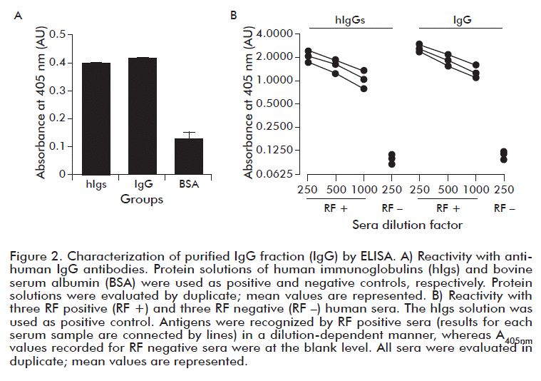
The IgG fraction      was corroborated in the purified protein preparation by ELISA signals at least      three-fold higher than those registered with an unrelated control protein      (<a href="/img/revistas/bta/v35n3/f0201318.gif">Figure 2A</a>) and at      the level of those recorded for the hIgs solution. In this experiment, GBS      was used as coating buffer to favor IgG binding at isoelectric point to polystyrene      plates. </font></P >       
<P   ><font size="2" face="Verdana, Arial, Helvetica, sans-serif">In a previous work      [5], there was developed a RF detection reagent consisting on hIgs adsorbed      to polystyrene spheres. In that case, 40 % of unspecific agglutination was      observed, which was avoided after serum-complement inactivation. These false-positive      results were then attributed to serum-complement activation, due to the presence      of IgM in the hIgs solution used as RF target. For this reason, in the present      work, a series of experiments were conducted aimed to detect the possible      presence of IgM in the purified IgG solution. As expected, the IgG purification      method applied rendered negative results for IgM presence in all the experiments.      This confirmed that purified IgG solution was IgM-free or at least that IgM      levels were below the detection limits of the assays. These experiments included      ELISA tests using GBS (pH 8.2), PBS (pH 7.4) or PBS at pH 6.45 in the coating      step. The pH was lowered to favor IgM binding to polystyrene plates at isoelectric      point. The use of a pre-coating step with Poly-L-Lysine was included to favor      binding of IgM, of higher carbohydrate content than IgG. A dot blot immunoassay      testing 50 &mu;g of antigen also rendered negative results for IgM presence,      whilst confirming IgG content (data not shown). Antigenicity of purified IgG      was demonstrated by the dilution-dependent recognition of the antigen by three      RF positive sera. ELISA signals at blank (GBS) level were recorded for three      RF negative sera (<a href="/img/revistas/bta/v35n3/f0201318.gif">Figure      2B</a>). </font></P >       
<P   ><font size="2" face="Verdana, Arial, Helvetica, sans-serif">Saturation of polystyrene      sphere surfaces was found at 26 &mu;g of purified IgG per mg of spheres, in      close agreement with theoretical calculations [9]. The developed latex reagent      was calibrated to a detection limit of 8 IU/mL, to match the detection limit      of the commercial RF detection kit used for comparison in the present study.      After calibration, negative and positive reactions were verified with control      reagents included in the commercial kit. </font></P >       <P   ><font size="2" face="Verdana, Arial, Helvetica, sans-serif">Negative LAT results      were recorded for the 30 RF negative sera using both the commercial kit and      the detection system prepared in the present work using Multilatex&reg; and      purified IgG as RF target. The use of purified IgG circumvented the unspecific      agglutination with RF negative sera previously obtained with the 480 nm-diameter      polystyrene spheres-based reagent [7]. This emphasized the importance of IgG      purity on the performance of polystyrene latex-based reagents to detect RF.
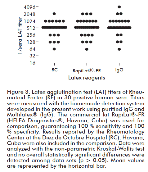
      </font></P >       <P   ><font size="2" face="Verdana, Arial, Helvetica, sans-serif">As shown in <a href="/img/revistas/bta/v35n3/f0301318.gif">Figure      3</a>, all FR positive sera gave positive results with both latex reagents.      Serum titers measured with the commercial reagent were similar (p &gt; 0.05)      to those informed by the RC. Titers measured with the homemade detection system      were similar (p &gt; 0.05) to those measured both at the RC and confirmed      in the present work with the commercial system tested. </font></P >       
<P   ><font size="2" face="Verdana, Arial, Helvetica, sans-serif">In summary, a latex-based      system was implemented to detect RF using Multilatex&reg; as solid support      and purified hIgG, under the defined and tested experimental conditions. It      gave results similar to those obtained with a commercial system for RF detection,      when tested against a panel of 30 positive and 30 negative human sera. Future      work will address the validation of the system and whether could it be possible      to implement this system for RA diagnosis in the clinics. </font></P >       <P   >&nbsp;</P >       <P   > </P >   <FONT size="+1" color="#000000"><B>        ]]></body>
<body><![CDATA[<P   ><font size="3" face="Verdana, Arial, Helvetica, sans-serif">ACKNOWLEDGEMENTS      </font></P >   </B>        <P   ><font size="2" face="Verdana, Arial, Helvetica, sans-serif"><FONT color="#211E1F">The      authors thank MSc. Juan A. Perez, from the Center of Immunoassay, Havana,      Cuba, for kind supplying the commercial latex-based RapiLat&reg;-FR kit for      RF detection and for fruitful discussions. Thanks to Prof. Dr. Carlos Peniche,      from the University of Havana, Havana, Cuba, for the critical reading of the      original manuscript. This work was supported by the Center of Biomaterials      of the University of Havana, Havana, Cuba, by the Finlay Vaccines Institute,      Havana, Cuba and by the Cuban National Program for the Development of Basic      Sciences (PNCB project 2015-2018, Project code P223LH001-061). </font></font></P >       <P   >&nbsp;</P >   <FONT size="+1" color="#211E1F"><FONT size="+1" color="#000000"><B>        <P   ><font size="3" face="Verdana, Arial, Helvetica, sans-serif">CONFLICTS OF INTEREST      STATEMENT </font></P >   </B>        <P   ><font size="2" face="Verdana, Arial, Helvetica, sans-serif"><FONT color="#211E1F">The      authors declare that there are no conflicts of interest. </font></font></P >       <P   >&nbsp;</P >   <FONT size="+1" color="#211E1F">        <P   ><font size="2" face="Verdana, Arial, Helvetica, sans-serif"><B><font size="3">REFERENCES</font>      </b></font></P >   <B>        <P   ></P >   </B> <FONT size="+1" color="#000000">        <!-- ref --><P   ><font size="2" color="#211E1F" face="Verdana, Arial, Helvetica, sans-serif">1.      Nishimura K, Sugiyama D, Kogata Y, Tsuji G, Nakazawa T, Kawano S, et al. Meta-analysis:      diagnostic accuracy of anti-cyclic citrullinated peptide antibody and rheumatoid      factor for rheumatoid arthritis. Ann Intern Med. 2007;146(11):797-808.     </font></P >   <FONT size="+1" color="#211E1F">        ]]></body>
<body><![CDATA[<!-- ref --><P   ><font size="2" face="Verdana, Arial, Helvetica, sans-serif">2. Artandi SE, Calame      KL, Morrison SL, Bonagura VR. Monoclonal IgM rheumatoid factors bind IgG at      a discontinuous epitope comprised of amino acid loops from heavychain constant-region      domains 2 and 3. Proc Natl Acad Sci U S A. 1992;89(1):94-8.     </font></P >       <P   ><font size="2" face="Verdana, Arial, Helvetica, sans-serif">3. Aletaha D, Neogi      T, Silman AJ, Funovits J, Felson DT, Bingham CO 3rd, et al. 2010 Rheumatoid      arthritis classification criteria: an American College of Rheumatology/ European      League Against Rheumatism collaborative initiative. Arthritis Rheum. 2010;62:2569-81.      </font></P >       <!-- ref --><P   ><font size="2" face="Verdana, Arial, Helvetica, sans-serif">4. Molina-Bol&iacute;var      JA, Galisteo-Gonz&aacute;lez F. Latex immunoagglutination assays. J Macromol      Sci, Part C-Polymer Rev. 2005;45:59-98.     </font></P >       <!-- ref --><P   ><font size="2" face="Verdana, Arial, Helvetica, sans-serif">5. Marrero G, Delgado      LP, Caroll H, Ortiz N, Musacchio A, Menendez T. Development of a polystyrene      latex-based reagent for rheumatoid factor detection. J Pol Eng. 2016;36:239-43.          </font></P >       <P   ><font size="2" face="Verdana, Arial, Helvetica, sans-serif">6. Lovell PA and      S. E-AM. Emulsion Polymerization and Emulsion Polymers. Chichester: John Wiley      and Sons Press; 1997. </font></P >       <!-- ref --><P   ><font size="2" face="Verdana, Arial, Helvetica, sans-serif">7. Marrero G, Ramirez-Bencomo      F, Delgado LP, Musacchio A, Gonzalez-Aznar E, Otero-Alfaro O, et al. Polystyrene      latex synthesis and application in Neisseria meningitidis serogroup W serotyping.      Rev Cub Inv Biomed. 2017;36(1):1-12.     </font></P >       ]]></body>
<body><![CDATA[<!-- ref --><P   ><font size="2" face="Verdana, Arial, Helvetica, sans-serif">8. Laemmli UK. Cleavage      of structural proteins during the assembly of the head of bacteriophage T4.      Nature. 1970;227(5259):680-5.     </font></P >       <!-- ref --><P   ><font size="2" face="Verdana, Arial, Helvetica, sans-serif">9. Bangs laboratories      Inc. Technical note 204. Adsorption to Microspheres. Rev. # 003. Fishers:      Bangs laboratories Inc.; 1999.     </font></P >       <!-- ref --><P   ><font size="2" face="Verdana, Arial, Helvetica, sans-serif">10. Anderson SG,      Bentzon MW, Houba V, Krag P. International reference preparation of rheumatoid      arthritis serum. Bull World Health Organ. 1970;42(2):311-8.     </font></P >       <P   >&nbsp;</P >       <P   >&nbsp;</P >       <P   ><font size="2" face="Verdana, Arial, Helvetica, sans-serif">Received in August,      2018.    <br>     Accepted in September, 2018.</font></P >       ]]></body>
<body><![CDATA[<P   >&nbsp;</P >       <P   >&nbsp;</P >       <P   ><font size="2" face="Verdana, Arial, Helvetica, sans-serif"><i>Tamara Menendez</i>.      <font color="#000000">Center of Biomaterials, University of Havana. Ave. Universidad      entre Ronda y G, Plaza, Havana 10400, Cuba.</font></font><font size="2" color="#211E1F" face="Verdana, Arial, Helvetica, sans-serif">      </font><font size="2" color="#000000" face="Verdana, Arial, Helvetica, sans-serif">E-mail:      <a href="mailto:tamara@biomat.uh.cu"> <font color="#0000FF">tamara@biomat.uh.cu</font></a></font>.</P >   </font></font></font></font></font></font></font></font></font></font></font></font></font></font></font></font></font></font></font></font></font></font></font></font></font></font></DIV >      ]]></body><back>
<ref-list>
<ref id="B1">
<label>1</label><nlm-citation citation-type="journal">
<person-group person-group-type="author">
<name>
<surname><![CDATA[Nishimura]]></surname>
<given-names><![CDATA[K]]></given-names>
</name>
<name>
<surname><![CDATA[Sugiyama]]></surname>
<given-names><![CDATA[D]]></given-names>
</name>
<name>
<surname><![CDATA[Kogata]]></surname>
<given-names><![CDATA[Y]]></given-names>
</name>
<name>
<surname><![CDATA[Tsuji]]></surname>
<given-names><![CDATA[G]]></given-names>
</name>
<name>
<surname><![CDATA[Nakazawa]]></surname>
<given-names><![CDATA[T]]></given-names>
</name>
<name>
<surname><![CDATA[Kawano]]></surname>
<given-names><![CDATA[S]]></given-names>
</name>
</person-group>
<article-title xml:lang="en"><![CDATA[Meta-analysis: diagnostic accuracy of anti-cyclic citrullinated peptide antibody and rheumatoid factor for rheumatoid arthritis]]></article-title>
<source><![CDATA[Ann Intern Med]]></source>
<year>2007</year>
<volume>146</volume>
<numero>11</numero>
<issue>11</issue>
<page-range>797-808</page-range></nlm-citation>
</ref>
<ref id="B2">
<label>2</label><nlm-citation citation-type="journal">
<person-group person-group-type="author">
<name>
<surname><![CDATA[Artandi]]></surname>
<given-names><![CDATA[SE]]></given-names>
</name>
<name>
<surname><![CDATA[Calame]]></surname>
<given-names><![CDATA[KL]]></given-names>
</name>
<name>
<surname><![CDATA[Morrison]]></surname>
<given-names><![CDATA[SL]]></given-names>
</name>
<name>
<surname><![CDATA[Bonagura]]></surname>
<given-names><![CDATA[VR]]></given-names>
</name>
</person-group>
<article-title xml:lang="en"><![CDATA[Monoclonal IgM rheumatoid factors bind IgG at a discontinuous epitope comprised of amino acid loops from heavychain constant-region domains 2 and 3]]></article-title>
<source><![CDATA[Proc Natl Acad Sci U S A]]></source>
<year>1992</year>
<volume>89</volume>
<numero>1</numero>
<issue>1</issue>
<page-range>94-8</page-range></nlm-citation>
</ref>
<ref id="B3">
<label>3</label><nlm-citation citation-type="journal">
<person-group person-group-type="author">
<name>
<surname><![CDATA[Aletaha]]></surname>
<given-names><![CDATA[D]]></given-names>
</name>
<name>
<surname><![CDATA[Neogi]]></surname>
<given-names><![CDATA[T]]></given-names>
</name>
<name>
<surname><![CDATA[Silman]]></surname>
<given-names><![CDATA[AJ]]></given-names>
</name>
<name>
<surname><![CDATA[Funovits]]></surname>
<given-names><![CDATA[J]]></given-names>
</name>
<name>
<surname><![CDATA[Felson]]></surname>
<given-names><![CDATA[DT]]></given-names>
</name>
<name>
<surname><![CDATA[Bingham]]></surname>
<given-names><![CDATA[CO]]></given-names>
</name>
</person-group>
<article-title xml:lang="en"><![CDATA[2010 Rheumatoid arthritis classification criteria: an American College of Rheumatology/European League Against Rheumatism collaborative initiative]]></article-title>
<source><![CDATA[Arthritis Rheum]]></source>
<year>2010</year>
<volume>62</volume>
<page-range>2569-81</page-range></nlm-citation>
</ref>
<ref id="B4">
<label>4</label><nlm-citation citation-type="journal">
<person-group person-group-type="author">
<name>
<surname><![CDATA[Molina-Bolívar]]></surname>
<given-names><![CDATA[JA]]></given-names>
</name>
<name>
<surname><![CDATA[Galisteo-González]]></surname>
<given-names><![CDATA[F]]></given-names>
</name>
</person-group>
<article-title xml:lang="en"><![CDATA[Latex immunoagglutination assays]]></article-title>
<source><![CDATA[J Macromol Sci, Part C-Polymer Rev]]></source>
<year>2005</year>
<volume>45</volume>
<page-range>59-98</page-range></nlm-citation>
</ref>
<ref id="B5">
<label>5</label><nlm-citation citation-type="journal">
<person-group person-group-type="author">
<name>
<surname><![CDATA[Marrero]]></surname>
<given-names><![CDATA[G]]></given-names>
</name>
<name>
<surname><![CDATA[Delgado]]></surname>
<given-names><![CDATA[LP]]></given-names>
</name>
<name>
<surname><![CDATA[Caroll]]></surname>
<given-names><![CDATA[H]]></given-names>
</name>
<name>
<surname><![CDATA[Ortiz]]></surname>
<given-names><![CDATA[N]]></given-names>
</name>
<name>
<surname><![CDATA[Musacchio]]></surname>
<given-names><![CDATA[A]]></given-names>
</name>
<name>
<surname><![CDATA[Menendez]]></surname>
<given-names><![CDATA[T]]></given-names>
</name>
</person-group>
<article-title xml:lang="en"><![CDATA[Development of a polystyrene latex-based reagent for rheumatoid factor detection]]></article-title>
<source><![CDATA[J Pol Eng]]></source>
<year>2016</year>
<volume>36</volume>
<page-range>239-43</page-range></nlm-citation>
</ref>
<ref id="B6">
<label>6</label><nlm-citation citation-type="book">
<person-group person-group-type="author">
<name>
<surname><![CDATA[Lovell]]></surname>
<given-names><![CDATA[PAS]]></given-names>
</name>
<name>
<surname><![CDATA[E-AM]]></surname>
<given-names><![CDATA[S]]></given-names>
</name>
</person-group>
<source><![CDATA[Emulsion Polymerization and Emulsion Polymers]]></source>
<year>1997</year>
<publisher-loc><![CDATA[Chichester ]]></publisher-loc>
<publisher-name><![CDATA[John Wiley and Sons Press]]></publisher-name>
</nlm-citation>
</ref>
<ref id="B7">
<label>7</label><nlm-citation citation-type="journal">
<person-group person-group-type="author">
<name>
<surname><![CDATA[Marrero]]></surname>
<given-names><![CDATA[G]]></given-names>
</name>
<name>
<surname><![CDATA[Ramirez-Bencomo]]></surname>
<given-names><![CDATA[F]]></given-names>
</name>
<name>
<surname><![CDATA[Delgado]]></surname>
<given-names><![CDATA[LP]]></given-names>
</name>
<name>
<surname><![CDATA[Musacchio]]></surname>
<given-names><![CDATA[A]]></given-names>
</name>
<name>
<surname><![CDATA[Gonzalez-Aznar]]></surname>
<given-names><![CDATA[E]]></given-names>
</name>
<name>
<surname><![CDATA[Otero-Alfaro]]></surname>
<given-names><![CDATA[O]]></given-names>
</name>
</person-group>
<article-title xml:lang="en"><![CDATA[Polystyrene latex synthesis and application in Neisseria meningitidis serogroup W serotyping]]></article-title>
<source><![CDATA[Rev Cub Inv Biomed]]></source>
<year>2017</year>
<volume>36</volume>
<numero>1</numero>
<issue>1</issue>
<page-range>1-12</page-range></nlm-citation>
</ref>
<ref id="B8">
<label>8</label><nlm-citation citation-type="journal">
<person-group person-group-type="author">
<name>
<surname><![CDATA[Laemmli]]></surname>
<given-names><![CDATA[UK]]></given-names>
</name>
</person-group>
<article-title xml:lang="en"><![CDATA[Cleavage of structural proteins during the assembly of the head of bacteriophage T4]]></article-title>
<source><![CDATA[Nature]]></source>
<year>1970</year>
<volume>227</volume>
<numero>5259</numero>
<issue>5259</issue>
<page-range>680-5</page-range></nlm-citation>
</ref>
<ref id="B9">
<label>9</label><nlm-citation citation-type="book">
<collab>Bangs laboratories Inc.</collab>
<source><![CDATA[Technical note 204. Adsorption to Microspheres. Rev. # 003]]></source>
<year>1999</year>
<publisher-loc><![CDATA[Fishers ]]></publisher-loc>
<publisher-name><![CDATA[Bangs laboratories Inc]]></publisher-name>
</nlm-citation>
</ref>
<ref id="B10">
<label>10</label><nlm-citation citation-type="journal">
<person-group person-group-type="author">
<name>
<surname><![CDATA[Anderson]]></surname>
<given-names><![CDATA[SG]]></given-names>
</name>
<name>
<surname><![CDATA[Bentzon]]></surname>
<given-names><![CDATA[MW]]></given-names>
</name>
<name>
<surname><![CDATA[Houba]]></surname>
<given-names><![CDATA[V]]></given-names>
</name>
<name>
<surname><![CDATA[Krag]]></surname>
<given-names><![CDATA[P]]></given-names>
</name>
</person-group>
<article-title xml:lang="en"><![CDATA[International reference preparation of rheumatoid arthritis serum]]></article-title>
<source><![CDATA[Bull World Health Organ]]></source>
<year>1970</year>
<volume>42</volume>
<numero>2</numero>
<issue>2</issue>
<page-range>311-8</page-range></nlm-citation>
</ref>
</ref-list>
</back>
</article>
