1 Doctor en Ciencias Médicas. Especialista de II Grado en Neurocirugía.
2 Especialista de I Grado en Neurocirugía.
3 Especialista de I Grado en Neurología.
Descriptores DeCS: IMPLANTE ENDOOSEO/métodos; HIDROXIAPATITAS/uso terapéutico; VERTEBRAS CERVICALES/cirugía; DISCO INTERVERTEBRAL/trasplante.
En las intervenciones sobre la columna cervical, tanto por lesiones traumáticas como no traumáticas, es frecuente la necesidad de emplear injertos, para sustituir cuerpos vertebrales o discos incompetentes. Los de mayor uso son los que se obtienen de fragmentos de hueso autógeno, de cresta ilíaca, bóveda craneal o costillas.
Hay varios problemas con estos injertos óseos: requieren una operación adicional sobre el paciente, con mayor pérdida de sangre; hay más posibilidades de sepsis y dolor. En el posoperatorio pueden aparecer inestabilidad pélvica, fractura por fatiga, hernia del músculo ilíaco, fístulas y lesión ureteral, complicaciones que pueden provocar un retraso en la rehabilitación del paciente.1-5
Es por todo ello que se ha experimentado con el uso de materiales biocompatibles y biointegrales que sean sustituidos por hueso endógeno y entre ellos, ocupan un lugar muy importante las cerámicas de fosfatos de calcio.
La hidroxiapatita (HA) ha sido empleada con éxito como implante en operaciones de la columna, en los últimos 10 años,6-9 pero, en Cuba, no hay experiencia documentada sobre su empleo en este tipo de operaciones.
Motivados por los buenos resultados de su empleo en cirugía maxilofacial en nuestros hospitales, decidimos en 1990 obtener bloques de HA para emplearlos como implantes, en sustitución de cuerpos vertebrales o discos incompetentes; verificar su resistencia y capacidad para orientar y facilitar que el hueso nuevo llegue hacia la región afectada, es decir, sus posibilidades osteoconductoras. ]]>
Su aplicación práctica sólo se pudo comenzar en 1993, al obtener láminas de HA fabricadas por el Centro de Biomateriales de la Universidad de La Habana.En este trabajo se informan las primeras experiencias de su empleo en un ensayo clínico, para verificar si las láminas de HA de 3 y 6 mm de espesor y porosidad del 50 % tienen la resistencia necesaria para ser empleadas como implantes en la columna cervical baja (C3 a C7), para sustituir discos intervertebrales incompetentes.
Los implantes utilizados en nuestra serie, fueron fabricados a partir de bloques de HA sintética denominada POROSIT, que se produce en el Centro de Biomateriales de la Universidad de La Habana. Este material ha pasado satisfactoriamente una serie de ensayos "in vitro" e "in vivo" que demostraron su carencia de citotoxicidad10 y de actividad hemolítica, así como su biocompatibilidad.11
Para la obtención de éstos se emplearon como materias primas el hidróxido de calcio, y el ácido ortofosfórico, los que se hicieron reaccionar en condiciones controladas de acuerdo con la siguiente ecuación química:
10Ca (OH)2(n) + 6H3PO4 (an) -Ca 10 (PO4)6 (OH) 2(n) + 10 H20
y se obtuvo un polvo que posteriormente fue modelado en láminas, con una porosidad del 50 % y las siguientes medidas: largo 22 mm; ancho 9 mm y espesor 3 mm, y bloques de 22 x 9 x x 6 mm.
DESCRIPCIÓN DE LA TÉCNICA QUIRÚRGICA
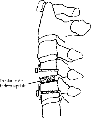
Se realizó abordaje anterolateral a la región cervical. Después de exponer la cara anterior de los cuerpos vertebrales y de haber identificado el interespacio lesionado, se realizó discectomía y descompresión, en dependencia del caso y se colocaron las láminas de HA (figura), después de modelarlas y se
ajustaron a la profundidad del espacio, en sustitución del disco intervertebral. Sólo se empleó la técnica de instrumentación con osteosíntesis, en pacientes con inestabilidad de columna cervical. En estos casos se utilizaron láminas metálicas tipo A-O "Senegas" y tornillos que incluyeron las corticales anteriores y posteriores de los cuerpos vertebrales adyacentes.
clínico, para detectar signos de resorción u osteomielitis, desplazamientos y/o aplastamientos del implante, luxación vertebral, cifosis u otras alteraciones atribuibles al implante, así como "osteo-conducción aparente", y se aceptó como tal, la desaparición imagenológica de la interfase hueso-implante y desaparición o disminución de la porosidad de la HA.
El injerto fue colocado en el espacio C3/4 en 2 pacientes, C4/5 en 3 casos, C5/6 en 3 y C6/7 en 4 pacientes (tabla 1).
| No | | ||||
| Trauma Paciente | ]]> HC | | | | |
| 1. AAG | | | | | |
| 2. JRP | ]]> 767207 | | | | |
| 3. IGM | | | | | |
| 4. RMG | ]]> 768691 | | | | |
| 5. AHD | | | | | |
| 6. GLT | ]]> 777973 | | | | |
| 7. MMD | | | | | |
| 8. APA | ]]> 778146 | | | | |
| 9. REG | | | | | |
| 10. JGR | ]]> 784595 | | | | |
La tabla número 2, muestra las complicaciones detectadas en los controles clínicos y radiológicos.
En la tabla número 3, se presenta la evaluación de la estabilidad y osteoconducción aparente, realizada en los controles clínicos y radiológicos.
En la tabla número 4, se relacionan los resultados obtenidos en el conjunto de la serie: excelentes en 9 pacientes y buenos en 1. ]]>
| Evaluación | | |
| Excelentes | | |
| Buenos | | |
| Regulares | | ]]> ? |
| Malos | | |
| Total | | |
Koyama y Handa,7,8 sin embargo, utilizaron la HA porosa para laminoplastia en 60 pacientes con estenosis del canal, sin complicaciones con los implantes.
Recientemente Senter et al.9 investigaron el uso de la HA densa en forma de bloques en disectomía cervical anterior y fusión; compararon los resultados en 75 pacientes con injertos de hueso ilíaco, con los obtenidos en 84 casos con injertos de HA densa; encontraron que en el primer grupo había 2 pacientes con desplazamientos o compresión de los fragmentos óseos, mientras que en el segundo grupo existían 5 casos. No obstante, observaron que en los pacientes en los que se empleó hueso como injerto, ocurrieron numerosas complicaciones asociadas con la zona de donde se extrajo el injerto como: infecciones, parestesia dolorosa y dolores crónicos. Los autores concluyeron en que el empleo de HA densa en forma de bloques, puede ser superior al uso de hueso autógeno en las fusiones cervicales anteriores.
Entre 1981 y 1986 Heise et al.13 usaron HA porosa (con poros entre 200 y 600?m) para la fusión espinal en 18 pacientes, y concluyeron en que su uso era de utilidad.
La HA por sí misma no es osteogénica, pues no induce la formación de hueso en sitios ectópicos, ni estimula un crecimiento óseo más rápido en los sitios de implante.14 Pero constituye una matriz física, en la que se deposita hueso nuevo y tiene propiedades de orientación que hacen posible que el hueso llegue a sitios que, de otro modo, no hubiese alcanzado. Por eso se usa el término de osteoconducción.
La acumulación de iones de fósforo y calcio en la superficie de los implantes ocurre por la libre difusión con el hueso adyacente, por ser idénticos al del hueso natural. ]]>
Este proceso toma parte en la formación de hueso nuevo entre la superficie del implante y el tejido óseo adyacente.Un factor importante que se tendrá en cuenta en la formación ósea es el grado de porosidad del implante. A medida que el implante es más poroso presenta más posibilidades de formación de tejido fibrovascular que posteriormente se osifica, pero también es menos resistente a la compresión y viceversa.15 En el caso de la utilización de implantes artificiales en la columna vertebral, donde existen grandes fuerzas de compresión, es necesario lograr un implante que sea lo suficientemente resistente para soportar la compresión y lo suficientemente poroso para permitir una buena restauración ósea. Es por eso que en este estudio utilizamos láminas de HA con una porosidad del 50 %.
Lo limitado de nuestra serie, de sólo 10 pacientes, impide realizar un análisis porcentual significativo. Sin embargo, la obtención de excelentes resultados en 9 de ellos y bueno en 1, es un indicador muy interesante.
Cinco de nuestros pacientes tenían una evolución posoperatoria entre 12 y 15 meses al realizar este informe. De los 5 restantes uno tiene 8 meses de evolución, 3 tienen 6 meses y uno, sólo 3.
Tres de nuestros pacientes presentaron complicaciones. En los últimos controles radiológicos del pacientes número 4, se observó el aflojamiento de uno de los 5 tornillos, pero esto no tiene relación con el implante, ni produjo cambios en su estabilidad y osteoconducción, que fueron excelentes.
El paciente número 8, en el que se emplearon implantes en 2 interespacios (C4/5/6), mostró fragmentación y migración parcial lateral de la lámina de
HA del interespacio superior; complicación informada también por otros autores.6,12 En este caso no hubo repercusión clínica y se mantuvo la estabilidad, pero la osteoconducción fue considerada parcial, en el momento del último control, porque se produjo sólo a partir de los fragmentos no migrados (tablas 2 y 3).
| Paciente | ]]> HC | | | | Complicación |
| 1. AAG | | | | | |
| 2. JRP | | | ]]> M | | |
| 3. IGM | | | | | |
| 4. RMG | | | | | Aflojamiento de un tornillo |
| 5. AHD | ]]> 770541 | | | | |
| 6. GLT | | | | | |
| 7. MMD | | | ]]> F | | Parálisis N. Recurrente |
| 8. APA | | | | | Fragmentación y migración parcial de lámina C4/5 |
| 9. REG | | | | | |
| 10. JGR | ]]> 784595 | | | |
| Paciente | | | | | ]]> E O | |
| 1. AAG | | | | | | |
| 2. JRP | | | ]]> M | | | |
| 3. IGM | | | | | | |
| 4. RMG | ]]> 768691 | | | | | |
| 5. AHD | | | | | ]]> X | |
| 6. GLT | | | | | | |
| 7. MMD | | | ]]> F | | | |
| 8. APA | | | | | | |
| 9. REG | ]]> 781632 | | | | | |
| 10. JGR | | | | | ]]> X | |
En un futuro próximo, esperamos obtener implantes con la configuración y dimensiones específicas, que permitan una correlación más exacta entre las interfases injerto-hueso y una distribución vectorial más uniforme de las fuerzas dinámicas en el interespacio.
En conclusión podemos decir, que logramos utilizar HA sintética en forma de láminas y bloques de configuración y dimensiones no específicas que fueron adaptados, transoperatoriamente, para sustituir discos intervertebrales incompetentes en la columna cervical baja; verificamos que las láminas de HA de 3 y 6 mm de espesor, 9 mm de ancho y 50 % de porosidad, tienen la resistencia necesaria para ser empleadas como implantes en la columna cervical baja (C3-7) y es recomendable la fabricación de láminas y bloques de HA de configuración y dimensiones específicas para la sustitución de discos intervertebrales incompetentes.
Subject headings: DENTAL IMPLANTATION, ENDOSSEOUS/methods; HYDROXYAPATITES/therapeutic use; CERVICAL VERTEBRAE/surgery; INTERVERTEBRAL DISK/ transplantation.
Dr. Ramiro Pereira Riverón. Hospital Universitario "General Calixto García". Calle 29 esquina a J, municipio Plaza de la Revolución. Ciudad de La Habana, Cuba. ]]>