<?xml version="1.0" encoding="ISO-8859-1"?><article xmlns:mml="http://www.w3.org/1998/Math/MathML" xmlns:xlink="http://www.w3.org/1999/xlink" xmlns:xsi="http://www.w3.org/2001/XMLSchema-instance">
<front>
<journal-meta>
<journal-id>0034-7507</journal-id>
<journal-title><![CDATA[Revista Cubana de Estomatología]]></journal-title>
<abbrev-journal-title><![CDATA[Rev Cubana Estomatol]]></abbrev-journal-title>
<issn>0034-7507</issn>
<publisher>
<publisher-name><![CDATA[Editorial Ciencias Médicas]]></publisher-name>
</publisher>
</journal-meta>
<article-meta>
<article-id>S0034-75072010000400006</article-id>
<title-group>
<article-title xml:lang="en"><![CDATA[Primary and secondary impaction of four primary molar teeth in a single patient]]></article-title>
<article-title xml:lang="es"><![CDATA[Retención primaria y secundaria de los cuatro primeros molares en un paciente]]></article-title>
</title-group>
<contrib-group>
<contrib contrib-type="author">
<name>
<surname><![CDATA[Ramos Chrcanovic]]></surname>
<given-names><![CDATA[Bruno]]></given-names>
</name>
<xref ref-type="aff" rid="A01"/>
</contrib>
<contrib contrib-type="author">
<name>
<surname><![CDATA[Rodrigues Antunes Souza]]></surname>
<given-names><![CDATA[Ana Cristina]]></given-names>
</name>
<xref ref-type="aff" rid="A01"/>
</contrib>
<contrib contrib-type="author">
<name>
<surname><![CDATA[Mascarenhas Paixão]]></surname>
<given-names><![CDATA[Ricardo]]></given-names>
</name>
<xref ref-type="aff" rid="A01"/>
</contrib>
<contrib contrib-type="author">
<name>
<surname><![CDATA[Napier Souza]]></surname>
<given-names><![CDATA[Leandro]]></given-names>
</name>
<xref ref-type="aff" rid="A01"/>
</contrib>
</contrib-group>
<aff id="A01">
<institution><![CDATA[,Centro Universitário Newton Paiva  ]]></institution>
<addr-line><![CDATA[Belo Horizonte Minas Gerais]]></addr-line>
<country>Brazil</country>
</aff>
<pub-date pub-type="pub">
<day>00</day>
<month>12</month>
<year>2010</year>
</pub-date>
<pub-date pub-type="epub">
<day>00</day>
<month>12</month>
<year>2010</year>
</pub-date>
<volume>47</volume>
<numero>4</numero>
<fpage>439</fpage>
<lpage>446</lpage>
<copyright-statement/>
<copyright-year/>
<self-uri xlink:href="http://scielo.sld.cu/scielo.php?script=sci_arttext&amp;pid=S0034-75072010000400006&amp;lng=en&amp;nrm=iso"></self-uri><self-uri xlink:href="http://scielo.sld.cu/scielo.php?script=sci_abstract&amp;pid=S0034-75072010000400006&amp;lng=en&amp;nrm=iso"></self-uri><self-uri xlink:href="http://scielo.sld.cu/scielo.php?script=sci_pdf&amp;pid=S0034-75072010000400006&amp;lng=en&amp;nrm=iso"></self-uri><abstract abstract-type="short" xml:lang="en"><p><![CDATA[The lack of eruption of a primary tooth can be considered rare. In primary impaction, the primary tooth not only has never appeared in the oral cavity, but also is always covered by a more or less thick layer of bone. Secondary impaction, which is relatively more common, denotes teeth that at one time erupted into the mouth, but subsequently clinically appear to have receded from this position. The purpose of this paper is to present a case of primary and secondary impaction of four primary molar teeth in a single patient.]]></p></abstract>
<abstract abstract-type="short" xml:lang="es"><p><![CDATA[La falta de erupción de un diente temporal puede ser considerada una situación rara. En la retención primaria, no sólo el diente temporal nunca ha aparecido en la cavidad bucal, sino que también está cubierto por una capa más o menos gruesa del hueso. La retención secundaria, que es relativamente más común, se refiere a los dientes que inició su erupción en la boca, pero clínicamente quedan atrapados en esta posición. El propósito de este trabajo es presentar un caso de retención primaria y secundaria de cuatro molares primarios en un paciente.]]></p></abstract>
<kwd-group>
<kwd lng="en"><![CDATA[Tooth ankylosis]]></kwd>
<kwd lng="en"><![CDATA[impacted tooth]]></kwd>
<kwd lng="en"><![CDATA[unerupted tooth]]></kwd>
<kwd lng="en"><![CDATA[primary dentition]]></kwd>
<kwd lng="es"><![CDATA[:anquilosis del diente]]></kwd>
<kwd lng="es"><![CDATA[impacción]]></kwd>
<kwd lng="es"><![CDATA[diente no erupcionado]]></kwd>
<kwd lng="es"><![CDATA[la dentición primaria]]></kwd>
</kwd-group>
</article-meta>
</front><body><![CDATA[ <p align="right"><font face="Verdana, Arial, Helvetica, sans-serif" size="2">    <b>PRESENTACI&Oacute;N DE CASOS</b></font></p>     <p>&nbsp;</p>     <p><font face="Verdana, Arial, Helvetica, sans-serif" size="4"><b>Primary and    secondary impaction of four primary molar teeth in a single patient</b></font><font face="Verdana, Arial, Helvetica, sans-serif" size="2">    </font></p>     <p>&nbsp;</p> <B>    <P>  </B>     <P><b><font face="Verdana, Arial, Helvetica, sans-serif" size="3">Retenci&oacute;n    primaria y secundaria de los cuatro primeros molares en un paciente</font> </b> <B>    <P>     <P>     <P>      <P><font face="Verdana, Arial, Helvetica, sans-serif" size="2">Bruno Ramos Chrcanovic<SUP>I</SUP>;    Ana Cristina Rodrigues Antunes Souza<SUP>II</SUP>; Ricardo Mascarenhas Paix&atilde;o<SUP>III</SUP>;    Leandro Napier Souza<SUP>IV</SUP></font> </B>      ]]></body>
<body><![CDATA[<P>      <P><font face="Verdana, Arial, Helvetica, sans-serif" size="2"><SUP>I</SUP>Doctor    in Dental Surgery. Private practice in General Dentistry, Belo Horizonte, Minas    Gerais, Brazil.    <br>   </font><font face="Verdana, Arial, Helvetica, sans-serif" size="2"><SUP>II</SUP>Master    in Sciences. Doctor in Dental Surgery. Adjunct professor of Head and Neck Anatomy    and Orthodontics. Centro Universit&aacute;rio Newton Paiva, Belo Horizonte,    Minas Gerais, Brazil.    <br>   </font><font face="Verdana, Arial, Helvetica, sans-serif" size="2"><SUP>III</SUP>Doctor    in Dental Surgery. Private practice in Orthodontics, Belo Horizonte, Minas Gerais,    Brazil.    <br>   </font><font face="Verdana, Arial, Helvetica, sans-serif" size="2"><SUP>IV</SUP>Master    in Sciences. Doctor in Dental Surgery. Adjunct professor of Oral and Maxillofacial    Surgery. Centro Universit&aacute;rio Newton Paiva, Belo Horizonte, Minas Gerais,    Brazil.</font>      <P>     <P>     <P><hr size="1" noshade>     <p><font face="Verdana, Arial, Helvetica, sans-serif" size="2"><B>ABSTRACT</B>    </font> </p>     <P><font face="Verdana, Arial, Helvetica, sans-serif" size="2">The lack of eruption    of a primary tooth can be considered rare. In primary impaction, the primary    tooth not only has never appeared in the oral cavity, but also is always covered    by a more or less thick layer of bone. Secondary impaction, which is relatively    more common, denotes teeth that at one time erupted into the mouth, but subsequently    clinically appear to have receded from this position. The purpose of this paper    is to present a case of primary and secondary impaction of four primary molar    teeth in a single patient. </font>     ]]></body>
<body><![CDATA[<P>      <P><font face="Verdana, Arial, Helvetica, sans-serif" size="2"><I>Key words</I>:    Tooth ankylosis, impacted tooth, unerupted tooth, primary dentition. <hr size="1" noshade></font>     <P>      <P><font face="Verdana, Arial, Helvetica, sans-serif" size="2"><B>RESUMEN</B>    </font>     <P><font face="Verdana, Arial, Helvetica, sans-serif" size="2">La falta de erupci&oacute;n    de un diente temporal puede ser considerada una situaci&oacute;n rara. En la    retenci&oacute;n primaria, no s&oacute;lo el diente temporal nunca ha aparecido    en la cavidad bucal, sino que tambi&eacute;n est&aacute; cubierto por una capa    m&aacute;s o menos gruesa del hueso. La retenci&oacute;n secundaria, que es    relativamente m&aacute;s com&uacute;n, se refiere a los dientes que inici&oacute;    su erupci&oacute;n en la boca, pero cl&iacute;nicamente quedan atrapados en    esta posici&oacute;n. El prop&oacute;sito de este trabajo es presentar un caso    de retenci&oacute;n primaria y secundaria de cuatro molares primarios en un    paciente. </font>      <P>      <P><font face="Verdana, Arial, Helvetica, sans-serif" size="2"><I>Palabras clave:    </I>anquilosis del diente, impacci&oacute;n, diente no erupcionado, la dentici&oacute;n    primaria.<hr size="1" noshade></font>      <p>&nbsp;</p>     <p>&nbsp;</p>     <P>      ]]></body>
<body><![CDATA[<P>      <P><font face="Verdana, Arial, Helvetica, sans-serif" size="2"><B><font size="3">INTRODUCTION</font></B>    </font>      <P><font face="Verdana, Arial, Helvetica, sans-serif" size="2">Eruption failure    may affect one or a number of teeth, in either the primary or the permanent    dentition, and can be partial or complete, depending upon the underlying etiology.    The majority of unerupted teeth are permanent teeth, and they may fail to erupt    either as a result of mechanical obstruction, being it from idiopathic or pathological    origin, or because of disruption to the eruptive mechanism itself.<SUP>1</SUP>    </font>     <P><font face="Verdana, Arial, Helvetica, sans-serif" size="2">The lack of eruption    of a primary tooth can be considered rare. Despite of, primary teeth are ankylosed    much more frequently than permanent ones. The tooth in question is almost always    a molar and frequently in the mandible.<SUP>2-5</SUP> Extraction has been widely    recommended as treatment, in order to prevent complications. </font>     <P><font face="Verdana, Arial, Helvetica, sans-serif" size="2"><I>Primary impaction</I>    is rare: in primary impaction, the primary tooth not only has never appeared    in the oral cavity, but is also always covered by a more or less thick layer    of bone.<SUP>6</SUP> Primary impaction has been defined as the prevention (often    by a physical barrier) of the eruption of a tooth at the expected times. This    obstacle could be a bud of a tooth, an odontoma, a cyst, a tumor, or a dento-maxillo    disharmony or malposition and/or malformation of the bud of the tooth itself.<SUP>4</SUP>    </font>     <P><font face="Verdana, Arial, Helvetica, sans-serif" size="2">The following are    criteria for a correct diagnosis of primary impaction of primary teeth:<SUP>6</SUP>    (a) Deep retention into the bone; (b) Absence of caries or restorations of the    crown; (c) No resorption of the roots; (d) Frequent passing of the corresponding    permanent tooth; and (e) Possible retention and malposition of the corresponding    adjacent permanent tooth. </font>     <P><font face="Verdana, Arial, Helvetica, sans-serif" size="2"><I>Secondary impaction:</I>    which is relatively more common, denotes teeth which at one time erupted into    the mouth, but subsequently clinically appear to have receded from this position    (Albers, 1986), i.e., the cessation of further eruption after a tooth has penetrated    the oral mucosa.<SUP>1</SUP> </font>     <P><font face="Verdana, Arial, Helvetica, sans-serif" size="2">Although the tooth    submerges and appears to move apically, there is no evidence whatsoever to demonstrate    any actual apical displacement of a submerged tooth. So, this can be discarded    as a cause. The submersion must be due to occlusal movement of the neighbouring    teeth while the affected tooth moves less or not at all. This may be caused    by: (a) failure or reduction of the eruptive force; (b) obstruction of the erupting    tooth or (c) fixation of the tooth to the surrounding tissue so that it cannot    move.<SUP>7</SUP> In case of severe submersion, clinical disturbance may include    incomplete alveolar process development, lack of normal mesial drift, non-response    to orthodontic forces, retained primary teeth with or without a successor and    impaction of the successor, a depressed tooth with tipping adjacent teeth, supra-eruption    of opposing teeth, lateral open bite and higher frequency of crossbites.<SUP>8-9</SUP>    </font>     <P><font face="Verdana, Arial, Helvetica, sans-serif" size="2">The exact factor    of primary teeth submersion has not yet determined. Suggested factors are ankylosis,    congenitally missing permanent teeth, defects in the periodontal membrane, trauma,    injury of the periodontal ligament, precocious eruption of the first permanent    molar, defective eruptive force, or a combination of these factors. From all    of the cited etiological factors, ankylosis seems to be involved in the majority    of cases or at least as a coexisting factor.<SUP>10</SUP> </font>      <P><font face="Verdana, Arial, Helvetica, sans-serif" size="2">The purpose of    this paper is to present a case with primary and secondary impaction of four    primary second molars in the same patient.</font>     ]]></body>
<body><![CDATA[<P>      <P>      <P><font face="Verdana, Arial, Helvetica, sans-serif" size="2"><B><font size="3">CASE    REPORT</font></B> </font>      <P><font face="Verdana, Arial, Helvetica, sans-serif" size="2">
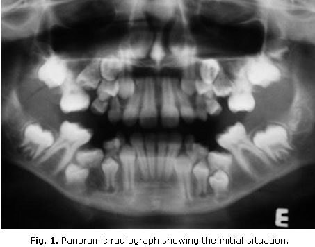
A 10-year-old girl    was referred to an Orthodontic Clinic for evaluation. Clinical examination revealed    that the two superior second primary molars were missing, and the two inferior    second primary molars were submerged and ankylosed. A panoramic radiograph,    taken on that occasion, disclosed the presence of the &quot;missing&quot; two    superior second primary molars near the maxillary sinus. It also showed impaction    of the two superior second pre-molars by the first superior permanent molars.    There was no dental agenesis (<a href="#fig1_06">figure 1</a>). The parents    discarded a familiar history. So, the patient was referred to an oral and maxillofacial    surgeon to perform a surgical treatment as first phase of the treatment. At    the first surgical treatment, the inferior second primary molars were removed.
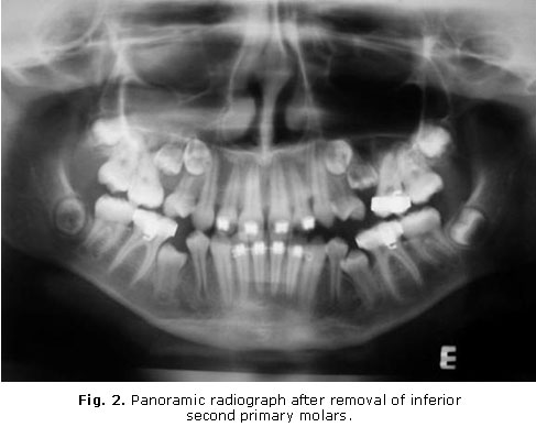
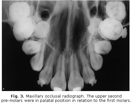
</font>      <P align="center"><a name="fig1_06"></a><img src="/img/revistas/est/v47n4/f0106410.jpg" width="453" height="356">      
<P><font face="Verdana, Arial, Helvetica, sans-serif" size="2"><B>    <br>   </B><a href="#fig2_06">Figure 2</a> shows the patient at the age of 12. The    inferior canines and first pre-molars were already at the occlusal plane and    the second pre-molars in their normal path. At this age an occlusal radiograph    of maxilla was made (<a href="#fig3_06">figure 3</a>). The upper second pre-molars    were in palatal position in relation to the first molars. So, it was accomplished    the extraction of the upper second primary molars and upper second pre-molars    as recommended by her orthodontist. After removal of the four ankylosed primary    second molars and four second premolars the patient is under current orthodontic    treatment.</font>      <P align="center"><a name="fig2_06"></a><img src="/img/revistas/est/v47n4/f0206410.jpg" width="487" height="388">      
<P align="center"><a name="fig3_06"></a><img src="/img/revistas/est/v47n4/f0306410.jpg" width="426" height="337">      
<P align="center">     ]]></body>
<body><![CDATA[<P>      <P><font face="Verdana, Arial, Helvetica, sans-serif" size="2"><B><font size="3">DISCUSSION</font></B>    </font>      <P><font face="Verdana, Arial, Helvetica, sans-serif" size="2">The malposition    of the permanent tooth, sometimes noticed, can by no means be the cause of the    primary impaction, if anything is the result of it. The primary tooth in fact    is never originally located in a deeper position than the permanent tooth, but    beside it, on the palatal or lingual side, slightly closer to the occlusal plane.<SUP>11</SUP>    This fact occurred in the present case report. As shown in <a href="#fig3_06">figure    3</a>, the permanent superior second pre-molars are on the palatal side of the    impacted primary second molars. In primary impaction, the permanent tooth passes    the corresponding primary tooth, probably prematurely ankylosed, and may lose    its correct expected position,<SUP>11</SUP> though this really not happened    in the present case (<a href="#fig1_06">figure 1</a>). </font>      <P><font face="Verdana, Arial, Helvetica, sans-serif" size="2">It is not reasonable    to suggest that the force of eruption should be affected in such a localized    manner when the force is adequate elsewhere in the jaws, nor does a locally    less effective force explain why two, three or four quadrants of the same dentition    should be affected. It is likely that the obstruction/impaction occurs only    after submersion has taken place and results from the tilting of the neighbouring    teeth into the gap so created. Ankylosis provides an excellent reason for the    teeth being held back and prevented from erupting. No other explanation can    do this.<SUP>6-7</SUP> Submersion of deciduous teeth is caused by ankylosis    which prevents further eruption, so that the neighboring teeth continue to erupt    past them, carrying with them the soft tissue and bone, to submerge the ankylosed    tooth.<SUP>4-7</SUP> Spontaneous reactivation of the eruption of ankylosed molars    occurs occasionally, as a consequence of resorption of the area of ankylosis.<SUP>7</SUP>    It is likely that ankylosis also plays a leading role in primary impaction.    The only difference is that occurs very early, always before the emergence of    the tooth.<SUP>5,11</SUP> </font>      <P><font face="Verdana, Arial, Helvetica, sans-serif" size="2">Tooth ankylosis    is defined as a fusion of the cementum and/or dentin with alveolar bone, with    obliteration of the periodontal membrane, with occurs either before or after    tooth eruption.<SUP>3,12</SUP> Several conditions have been proposed as causes    of ankylosis: (a) Genetic tendency;<SUP>13</SUP> (b) Traumatic injury to the    bone and/or periodontal ligament;<SUP>12</SUP> (c) Development disturbances    in the periodontium;<SUP>14</SUP> (d) Deficient local vertical bone growth;<SUP>15</SUP>    and (e) Disturbed local metabolism with disorder in the process of normal resorption    and hard tissue repair.<SUP>2</SUP> Congenital absence of permanent teeth has    also been proposed as the cause of primary tooth ankylosis, but it cannot be    accepted as sufficient, because such absence is not always associated with ankylosis    of the corresponding primary molars.<SUP>3</SUP> </font>     <P><font face="Verdana, Arial, Helvetica, sans-serif" size="2">The dental, medical    and familial history of our patient did not contribute to explain the reasons    of submergence. Therefore, only the development disturbances in the periodontium<SUP>14</SUP>    and/or the disturbed local metabolism with disorder in the process of normal    resorption and hard tissue repair<SUP>2 </SUP>might explain the impactions in    the present case. </font>     <P><font face="Verdana, Arial, Helvetica, sans-serif" size="2">Radiographically,    obliteration of the periodontal ligament space is noted in ankylosis, as also    noted here, especially with the inferior primary molars (<a href="#fig1_06">figure    1</a>). The roots are less radio-opaque and as the ankylosis progresses they    are less distinguished from surrounding bone. </font>      <P><font face="Verdana, Arial, Helvetica, sans-serif" size="2">Submerged primary    molars may cause several problems in dental arch such as space loss, tipping    of adjacent teeth, supra-eruption of the antagonists and dislocations of permanent    teeth lying under the primary tooth.<SUP>9</SUP> According to Kurol and Thilander,<SUP>16</SUP>    these disturbances have no long-term effects on occlusion. On other hand, Becker    and Shochat<SUP>17</SUP> have detected a significant deviation of the dental    inter-incisor midline toward the affected side. In the present case, there was    no deviation of the dental inter-incisor midline, probably due to the bilateral    and symmetrical occurrence, but a mild malocclusion occurred. </font>     <P><font face="Verdana, Arial, Helvetica, sans-serif" size="2">Diagnostic criteria    of impacted primary teeth include the age of the patient, condition and occlusal    status of the infraoccluded tooth, root resorption and adjacent alveolar bone    levels.<SUP>18</SUP> The treatment plan of a submerged primary tooth depends    on degree of abnormality, the present of its successor permanent teeth and time    of onset.<SUP>8-9</SUP> Distinguishing between lack of eruption due to some    external interference and primary failure of eruption is important clinically,    because the latter condition does not respond to the application of orthodontic    force.<SUP>19</SUP> </font>     <P><font face="Verdana, Arial, Helvetica, sans-serif" size="2">Early tooth extraction    for the treatment of submerged teeth is recommended. In previous research it    was found that no primary molars in infraocclusion with agenesis of the successor    exfoliated spontaneously, in contrast to primary molars for which the successor    was present, in which its exfoliation occurs by the erupting successor resorbing    the area of fusion.<SUP>8</SUP> Considering this affirmation, the teeth was    removed in the present case because extensive bony ankylosis might have prevented    normal exfoliation, causing future alignment problems,<SUP>8,16</SUP> and also    because early extraction provides more time for spontaneous mesial drift, and    also a more favorable age for the orthodontic treatment that is usually necessary.<SUP>20</SUP>    Active orthodontic force will most likely result in localized ankylosis and    failure to extrude an affected tooth into occlusion, a finding that is essentially    diagnostic.<SUP>21</SUP> </font>      ]]></body>
<body><![CDATA[<P><font face="Verdana, Arial, Helvetica, sans-serif" size="2">In the presented    case the impacted upper second premolars and primary molars teeth were removed    because of the deeper location inside bone and great lack of arch perimeter.    The preferable treatment option for the inferior impacted teeth was extraction    only of the primary molars because the submerged deciduous mandibular second    molars probably inhibited movement of the teeth germ of the second premolars,    which were in the correct position of eruption. Therefore, the mandibular second    pre-molar erupted correctly with orthodontic traction. If this therapy were    not performed, the patient could have experienced future problems with the definitive    orthodontic treatment. </font>     <P><font face="Verdana, Arial, Helvetica, sans-serif" size="2">Alternatively,    a localized bony osteotomy and orthodontic extrusion of the whole segment would    seem to be the only option if an occlusal position of the tooth or teeth is    to be obtained. If some eruption of the tooth has occurred, a coronal build-up    may be the treatment of choice, in this case accepting the vertical position    of the affected tooth but achieving occlusion via the restoration. Cases where    multiple teeth are involved are more difficult to manage; the only available    method of bringing them into occlusion is a segmental osteotomy.<SUP>21</SUP>    Careful planning in these cases is essential to ensure that no damage is caused    to adjacent teeth. While surgical repositioning may not move teeth into an entirely    acceptable position, it will certainly aid prosthetic management. </font>     <P><font face="Verdana, Arial, Helvetica, sans-serif" size="2">The management    of a tooth eruption failure is difficult, not least because diagnosis of this    condition relies principally upon exclusion, where all possible causative factors    have been considered and eliminated. The possibility of a number of complications    resulting from infra-occlusion/impaction of primary molars (tipping of the neighboring    teeth, loss of space, and over-eruption of the antagonist),<SUP>16</SUP> may    indicate a need for early intervention by extracting the submerged/impacted    tooth and the institution of a comprehensive orthodontic treatment. If this    is not undertaken, periodical observation is mandatory. The treatment options    should always be discussed with the parents, and the possible complications    clearly explained, if the condition is left unattended.<SUP>19</SUP></font>     <P>      <P>      <P><font face="Verdana, Arial, Helvetica, sans-serif" size="2"><B><font size="3">BIBLIOGRAPHIC    REFERENCES</font></B> </font>      <!-- ref --><P><font face="Verdana, Arial, Helvetica, sans-serif" size="2">1. Raghoebar GM,    Boering G, Vissink A, Stegenga B. Eruption disturbances of permanent olars:    a review. J Oral Pathol Med. 1991;20:159-66. </font>    <!-- ref --><P><font face="Verdana, Arial, Helvetica, sans-serif" size="2">2. Biederman W.    The problem of the ankylosed tooth. Dent Clinic North Am. 1968;409-24. </font>    <!-- ref --><P><font face="Verdana, Arial, Helvetica, sans-serif" size="2">3. Brearley LJ,    McKibben DH. Ankylosis of primary molar teeth. I. Prevalence and Characteristics.    ASDC J Dent Child 1973;40:54-63. </font>    <!-- ref --><P><font face="Verdana, Arial, Helvetica, sans-serif" size="2">4. Bordais P, Gineste    P, Grant J, Marchand J. Les dents incluses. Encycl M&eacute;d Chir. Paris: Ed    Stomatologie; 1980. p. 10-2. </font>    <!-- ref --><P><font face="Verdana, Arial, Helvetica, sans-serif" size="2">5. Albers DD. Ankylosis    of teeth in the developing dentition. Quintessence Int. 1986;17:303-8. </font>    <!-- ref --><P><font face="Verdana, Arial, Helvetica, sans-serif" size="2">6. Bianchi SD,    Roccuzzo M. Primary impaction of primary teeth: a review and report of three    cases. J Clin Pediatric Dent. 1991;15:165-8. </font>    <!-- ref --><P><font face="Verdana, Arial, Helvetica, sans-serif" size="2">7. Darling AI,    Levers BG. Submerged human deciduous molars and ankylosis. Arch Oral Biol. 1973;18:1021-40.    </font>    <!-- ref --><P><font face="Verdana, Arial, Helvetica, sans-serif" size="2">8. Ekim SL, Hatibovic-Kofman    S. A treatment decision-making model for infraoccluded primary molars. Int J    Paediatr Dent. 2001;11:340-6. </font>    <!-- ref --><P><font face="Verdana, Arial, Helvetica, sans-serif" size="2">9. Altay N, Cengiz    SB. Space-regaining treatment for a submerged primary molar: a case report.    Int J Paediatr Dent. 2002;12:286-9. </font>    <!-- ref --><P><font face="Verdana, Arial, Helvetica, sans-serif" size="2">10. Antoniades    K, Kavadia S, Milioti K, Antoniades V, Markovitsi E. Submerged teeth. J Clin    Pediatr Dent. 2002;26:239-42. </font>    <!-- ref --><P><font face="Verdana, Arial, Helvetica, sans-serif" size="2">11. Jameson GD,    Burke PH. Inversion of second deciduous molar and second premolar. Br Dent J.    1987;162:265-6. </font>    <!-- ref --><P><font face="Verdana, Arial, Helvetica, sans-serif" size="2">12. Henderson HZ.    Ankylosis of primary molars: a clinical, radiographic, and histologic study.    ASDC J Dent Child. 1979;46:117-22. </font>    <!-- ref --><P><font face="Verdana, Arial, Helvetica, sans-serif" size="2">13. Via WF. Submerged    deciduous molars: familial tendencies. JADA. 1964;69:127-9. </font>    <!-- ref --><P><font face="Verdana, Arial, Helvetica, sans-serif" size="2">14. Kurol J, Magnusson    BC. Infraocclusion of primary molars: an epidemiological study. Scand J Dent    Res. 1984;92:564-76. </font>    <!-- ref --><P><font face="Verdana, Arial, Helvetica, sans-serif" size="2">15. Glucksman DD.    Localized vertical growth disturbance. JADA. 1942;29:184-7. </font>    <!-- ref --><P><font face="Verdana, Arial, Helvetica, sans-serif" size="2">16. Kurol J, Thilander    B. Infra-occlusion of primary molars and the effect on occlusal development.    A longitudinal study. Eur J Orthod. 1984;6:277-93. </font>    <!-- ref --><P><font face="Verdana, Arial, Helvetica, sans-serif" size="2">17. Becker A, Shochat    S. Submergence of a decisuous tooth: its ramifications on the dentition and    treatment of the resulting malocclusion. Am J Orthod. 1982;81:240-4. </font>    <!-- ref --><P><font face="Verdana, Arial, Helvetica, sans-serif" size="2">18. Sidhu HK, Ali    A. Hypodontia, ankylosis and infraocclusion: report of a case restored with    a fibre-reinforced ceromeric bridge. Br Dent J. 2001;191:613-6. </font>    <!-- ref --><P><font face="Verdana, Arial, Helvetica, sans-serif" size="2">19. Ben-Bassat    Y, Brin I, Fuks AB. Occlusal disturbances resulting from neglected submerged    primary molars. ASDC J Dent Child. 1991;58:129-33. </font>    <!-- ref --><P><font face="Verdana, Arial, Helvetica, sans-serif" size="2">20. Steiner-Oliveira    C, Gavi&atilde;o MB, dos Santos MN. Steiner-Oliveira C, Gavi&atilde;o MB, dos    Santos MN. Congenital agenesis of premolars associated with submerged primary    molars and a peg-shaped lateral incisor: a case report. Quintessence Int. 2007;38:435-8.    </font>    <!-- ref --><P><font face="Verdana, Arial, Helvetica, sans-serif" size="2">21. Proffit WR,    Vig KWL. Primary failure of eruption: a possible cause of posterior open-bite.    Am J Orthod. 1981;80:173-80.</font>    <P>     <P>     <P><font face="Verdana, Arial, Helvetica, sans-serif" size="2">Recibido: 15 de    junio de 2010.    ]]></body>
<body><![CDATA[<br>   </font><font face="Verdana, Arial, Helvetica, sans-serif" size="2">Aprobado:    20 de septiembre de 2010.</font>     <P>     <P>     <P><font face="Verdana, Arial, Helvetica, sans-serif" size="2"><i>Dr. Leandro    Napier Souza.</i><b> </b>Centro Universit&aacute;rio Newton Paiva, Belo Horizonte,    Minas Gerais, Brazil. E-mail: <a href="mailto:leandronapierdesouza@gmail.com">leandronapierdesouza@gmail.com</a></font>       ]]></body><back>
<ref-list>
<ref id="B1">
<label>1</label><nlm-citation citation-type="journal">
<person-group person-group-type="author">
<name>
<surname><![CDATA[Raghoebar]]></surname>
<given-names><![CDATA[GM]]></given-names>
</name>
<name>
<surname><![CDATA[Boering]]></surname>
<given-names><![CDATA[G]]></given-names>
</name>
<name>
<surname><![CDATA[Vissink]]></surname>
<given-names><![CDATA[A]]></given-names>
</name>
<name>
<surname><![CDATA[Stegenga]]></surname>
<given-names><![CDATA[B]]></given-names>
</name>
</person-group>
<article-title xml:lang="en"><![CDATA[Eruption disturbances of permanent olars: a review]]></article-title>
<source><![CDATA[J Oral Pathol Med]]></source>
<year>1991</year>
<volume>20</volume>
<page-range>159-66</page-range></nlm-citation>
</ref>
<ref id="B2">
<label>2</label><nlm-citation citation-type="journal">
<person-group person-group-type="author">
<name>
<surname><![CDATA[Biederman]]></surname>
<given-names><![CDATA[W]]></given-names>
</name>
</person-group>
<article-title xml:lang="en"><![CDATA[The problem of the ankylosed tooth]]></article-title>
<source><![CDATA[Dent Clinic North Am]]></source>
<year>1968</year>
<page-range>409-24</page-range></nlm-citation>
</ref>
<ref id="B3">
<label>3</label><nlm-citation citation-type="journal">
<person-group person-group-type="author">
<name>
<surname><![CDATA[Brearley]]></surname>
<given-names><![CDATA[LJ]]></given-names>
</name>
<name>
<surname><![CDATA[McKibben]]></surname>
<given-names><![CDATA[DH]]></given-names>
</name>
</person-group>
<article-title xml:lang="en"><![CDATA[Ankylosis of primary molar teeth. I: Prevalence and Characteristics]]></article-title>
<source><![CDATA[ASDC J Dent Child]]></source>
<year>1973</year>
<volume>40</volume>
<page-range>54-63</page-range></nlm-citation>
</ref>
<ref id="B4">
<label>4</label><nlm-citation citation-type="book">
<person-group person-group-type="author">
<name>
<surname><![CDATA[Bordais]]></surname>
<given-names><![CDATA[P]]></given-names>
</name>
<name>
<surname><![CDATA[Gineste]]></surname>
<given-names><![CDATA[P]]></given-names>
</name>
<name>
<surname><![CDATA[Grant]]></surname>
<given-names><![CDATA[J]]></given-names>
</name>
<name>
<surname><![CDATA[Marchand]]></surname>
<given-names><![CDATA[J]]></given-names>
</name>
</person-group>
<source><![CDATA[Les dents incluses: Encycl Méd Chir]]></source>
<year>1980</year>
<page-range>10-2</page-range><publisher-loc><![CDATA[Paris ]]></publisher-loc>
<publisher-name><![CDATA[Ed Stomatologie]]></publisher-name>
</nlm-citation>
</ref>
<ref id="B5">
<label>5</label><nlm-citation citation-type="journal">
<person-group person-group-type="author">
<name>
<surname><![CDATA[Albers]]></surname>
<given-names><![CDATA[DD]]></given-names>
</name>
</person-group>
<article-title xml:lang="en"><![CDATA[Ankylosis of teeth in the developing dentition]]></article-title>
<source><![CDATA[Quintessence Int]]></source>
<year>1986</year>
<volume>17</volume>
<page-range>303-8</page-range></nlm-citation>
</ref>
<ref id="B6">
<label>6</label><nlm-citation citation-type="journal">
<person-group person-group-type="author">
<name>
<surname><![CDATA[Bianchi]]></surname>
<given-names><![CDATA[SD]]></given-names>
</name>
<name>
<surname><![CDATA[Roccuzzo]]></surname>
<given-names><![CDATA[M]]></given-names>
</name>
</person-group>
<article-title xml:lang="en"><![CDATA[Primary impaction of primary teeth: a review and report of three cases]]></article-title>
<source><![CDATA[J Clin Pediatric Dent]]></source>
<year>1991</year>
<volume>15</volume>
<page-range>165-8</page-range></nlm-citation>
</ref>
<ref id="B7">
<label>7</label><nlm-citation citation-type="journal">
<person-group person-group-type="author">
<name>
<surname><![CDATA[Darling]]></surname>
<given-names><![CDATA[AI]]></given-names>
</name>
<name>
<surname><![CDATA[Levers]]></surname>
<given-names><![CDATA[BG]]></given-names>
</name>
</person-group>
<article-title xml:lang="en"><![CDATA[Submerged human deciduous molars and ankylosis]]></article-title>
<source><![CDATA[Arch Oral Biol]]></source>
<year>1973</year>
<volume>18</volume>
<page-range>1021-40</page-range></nlm-citation>
</ref>
<ref id="B8">
<label>8</label><nlm-citation citation-type="journal">
<person-group person-group-type="author">
<name>
<surname><![CDATA[Ekim]]></surname>
<given-names><![CDATA[SL]]></given-names>
</name>
<name>
<surname><![CDATA[Hatibovic-Kofman]]></surname>
<given-names><![CDATA[S]]></given-names>
</name>
</person-group>
<article-title xml:lang="en"><![CDATA[A treatment decision-making model for infraoccluded primary molars]]></article-title>
<source><![CDATA[Int J Paediatr Dent]]></source>
<year>2001</year>
<volume>11</volume>
<page-range>340-6</page-range></nlm-citation>
</ref>
<ref id="B9">
<label>9</label><nlm-citation citation-type="journal">
<person-group person-group-type="author">
<name>
<surname><![CDATA[Altay]]></surname>
<given-names><![CDATA[N]]></given-names>
</name>
<name>
<surname><![CDATA[Cengiz]]></surname>
<given-names><![CDATA[SB]]></given-names>
</name>
</person-group>
<article-title xml:lang="en"><![CDATA[Space-regaining treatment for a submerged primary molar: a case report]]></article-title>
<source><![CDATA[Int J Paediatr Dent]]></source>
<year>2002</year>
<volume>12</volume>
<page-range>286-9</page-range></nlm-citation>
</ref>
<ref id="B10">
<label>10</label><nlm-citation citation-type="journal">
<person-group person-group-type="author">
<name>
<surname><![CDATA[Antoniades]]></surname>
<given-names><![CDATA[K]]></given-names>
</name>
<name>
<surname><![CDATA[Kavadia]]></surname>
<given-names><![CDATA[S]]></given-names>
</name>
<name>
<surname><![CDATA[Milioti]]></surname>
<given-names><![CDATA[K]]></given-names>
</name>
<name>
<surname><![CDATA[Antoniades]]></surname>
<given-names><![CDATA[V]]></given-names>
</name>
<name>
<surname><![CDATA[Markovitsi]]></surname>
<given-names><![CDATA[E]]></given-names>
</name>
</person-group>
<article-title xml:lang="en"><![CDATA[Submerged teeth]]></article-title>
<source><![CDATA[J Clin Pediatr Dent]]></source>
<year>2002</year>
<volume>26</volume>
<page-range>239-42</page-range></nlm-citation>
</ref>
<ref id="B11">
<label>11</label><nlm-citation citation-type="journal">
<person-group person-group-type="author">
<name>
<surname><![CDATA[Jameson]]></surname>
<given-names><![CDATA[GD]]></given-names>
</name>
<name>
<surname><![CDATA[Burke]]></surname>
<given-names><![CDATA[PH]]></given-names>
</name>
</person-group>
<article-title xml:lang="en"><![CDATA[Inversion of second deciduous molar and second premolar]]></article-title>
<source><![CDATA[Br Dent J]]></source>
<year>1987</year>
<volume>162</volume>
<page-range>265-6</page-range></nlm-citation>
</ref>
<ref id="B12">
<label>12</label><nlm-citation citation-type="journal">
<person-group person-group-type="author">
<name>
<surname><![CDATA[Henderson]]></surname>
<given-names><![CDATA[HZ]]></given-names>
</name>
</person-group>
<article-title xml:lang="en"><![CDATA[Ankylosis of primary molars: a clinical, radiographic, and histologic study]]></article-title>
<source><![CDATA[ASDC J Dent Child]]></source>
<year>1979</year>
<volume>46</volume>
<page-range>117-22</page-range></nlm-citation>
</ref>
<ref id="B13">
<label>13</label><nlm-citation citation-type="journal">
<person-group person-group-type="author">
<name>
<surname><![CDATA[Via]]></surname>
<given-names><![CDATA[WF]]></given-names>
</name>
</person-group>
<article-title xml:lang="en"><![CDATA[Submerged deciduous molars: familial tendencies]]></article-title>
<source><![CDATA[JADA]]></source>
<year>1964</year>
<volume>69</volume>
<page-range>127-9</page-range></nlm-citation>
</ref>
<ref id="B14">
<label>14</label><nlm-citation citation-type="journal">
<person-group person-group-type="author">
<name>
<surname><![CDATA[Kurol]]></surname>
<given-names><![CDATA[J]]></given-names>
</name>
<name>
<surname><![CDATA[Magnusson]]></surname>
<given-names><![CDATA[BC]]></given-names>
</name>
</person-group>
<article-title xml:lang="en"><![CDATA[Infraocclusion of primary molars: an epidemiological study]]></article-title>
<source><![CDATA[Scand J Dent Res]]></source>
<year>1984</year>
<volume>92</volume>
<page-range>564-76</page-range></nlm-citation>
</ref>
<ref id="B15">
<label>15</label><nlm-citation citation-type="journal">
<person-group person-group-type="author">
<name>
<surname><![CDATA[Glucksman]]></surname>
<given-names><![CDATA[DD]]></given-names>
</name>
</person-group>
<article-title xml:lang="en"><![CDATA[Localized vertical growth disturbance]]></article-title>
<source><![CDATA[JADA]]></source>
<year>1942</year>
<volume>29</volume>
<page-range>184-7</page-range></nlm-citation>
</ref>
<ref id="B16">
<label>16</label><nlm-citation citation-type="journal">
<person-group person-group-type="author">
<name>
<surname><![CDATA[Kurol]]></surname>
<given-names><![CDATA[J]]></given-names>
</name>
<name>
<surname><![CDATA[Thilander]]></surname>
<given-names><![CDATA[B]]></given-names>
</name>
</person-group>
<article-title xml:lang="en"><![CDATA[Infra-occlusion of primary molars and the effect on occlusal development: A longitudinal study]]></article-title>
<source><![CDATA[Eur J Orthod]]></source>
<year>1984</year>
<volume>6</volume>
<page-range>277-93</page-range></nlm-citation>
</ref>
<ref id="B17">
<label>17</label><nlm-citation citation-type="journal">
<person-group person-group-type="author">
<name>
<surname><![CDATA[Becker]]></surname>
<given-names><![CDATA[A]]></given-names>
</name>
<name>
<surname><![CDATA[Shochat]]></surname>
<given-names><![CDATA[S]]></given-names>
</name>
</person-group>
<article-title xml:lang="en"><![CDATA[Submergence of a decisuous tooth: its ramifications on the dentition and treatment of the resulting malocclusion]]></article-title>
<source><![CDATA[Am J Orthod]]></source>
<year>1982</year>
<volume>81</volume>
<page-range>240-4</page-range></nlm-citation>
</ref>
<ref id="B18">
<label>18</label><nlm-citation citation-type="journal">
<person-group person-group-type="author">
<name>
<surname><![CDATA[Sidhu]]></surname>
<given-names><![CDATA[HK]]></given-names>
</name>
<name>
<surname><![CDATA[Ali]]></surname>
<given-names><![CDATA[A]]></given-names>
</name>
</person-group>
<article-title xml:lang="en"><![CDATA[Hypodontia, ankylosis and infraocclusion: report of a case restored with a fibre-reinforced ceromeric bridge]]></article-title>
<source><![CDATA[Br Dent J]]></source>
<year>2001</year>
<volume>191</volume>
<page-range>613-6</page-range></nlm-citation>
</ref>
<ref id="B19">
<label>19</label><nlm-citation citation-type="journal">
<person-group person-group-type="author">
<name>
<surname><![CDATA[Ben-Bassat]]></surname>
<given-names><![CDATA[Y]]></given-names>
</name>
<name>
<surname><![CDATA[Brin]]></surname>
<given-names><![CDATA[I]]></given-names>
</name>
<name>
<surname><![CDATA[Fuks]]></surname>
<given-names><![CDATA[AB]]></given-names>
</name>
</person-group>
<article-title xml:lang="en"><![CDATA[Occlusal disturbances resulting from neglected submerged primary molars]]></article-title>
<source><![CDATA[ASDC J Dent Child]]></source>
<year>1991</year>
<volume>58</volume>
<page-range>129-33</page-range></nlm-citation>
</ref>
<ref id="B20">
<label>20</label><nlm-citation citation-type="journal">
<person-group person-group-type="author">
<name>
<surname><![CDATA[Steiner-Oliveira]]></surname>
<given-names><![CDATA[C]]></given-names>
</name>
<name>
<surname><![CDATA[Gavião]]></surname>
<given-names><![CDATA[MB]]></given-names>
</name>
<name>
<surname><![CDATA[dos Santos]]></surname>
<given-names><![CDATA[MN]]></given-names>
</name>
<name>
<surname><![CDATA[Steiner-Oliveira]]></surname>
<given-names><![CDATA[C]]></given-names>
</name>
<name>
<surname><![CDATA[Gavião]]></surname>
<given-names><![CDATA[MB]]></given-names>
</name>
<name>
<surname><![CDATA[dos Santos]]></surname>
<given-names><![CDATA[MN]]></given-names>
</name>
</person-group>
<article-title xml:lang="en"><![CDATA[Congenital agenesis of premolars associated with submerged primary molars and a peg-shaped lateral incisor: a case report]]></article-title>
<source><![CDATA[Quintessence Int]]></source>
<year>2007</year>
<volume>38</volume>
<page-range>435-8</page-range></nlm-citation>
</ref>
<ref id="B21">
<label>21</label><nlm-citation citation-type="journal">
<person-group person-group-type="author">
<name>
<surname><![CDATA[Proffit]]></surname>
<given-names><![CDATA[WR]]></given-names>
</name>
<name>
<surname><![CDATA[Vig]]></surname>
<given-names><![CDATA[KWL]]></given-names>
</name>
</person-group>
<article-title xml:lang="en"><![CDATA[Primary failure of eruption: a possible cause of posterior open-bite]]></article-title>
<source><![CDATA[Am J Orthod]]></source>
<year>1981</year>
<volume>80</volume>
<page-range>173-80</page-range></nlm-citation>
</ref>
</ref-list>
</back>
</article>
