<?xml version="1.0" encoding="ISO-8859-1"?><article xmlns:mml="http://www.w3.org/1998/Math/MathML" xmlns:xlink="http://www.w3.org/1999/xlink" xmlns:xsi="http://www.w3.org/2001/XMLSchema-instance">
<front>
<journal-meta>
<journal-id>0034-7507</journal-id>
<journal-title><![CDATA[Revista Cubana de Estomatología]]></journal-title>
<abbrev-journal-title><![CDATA[Rev Cubana Estomatol]]></abbrev-journal-title>
<issn>0034-7507</issn>
<publisher>
<publisher-name><![CDATA[Editorial Ciencias Médicas]]></publisher-name>
</publisher>
</journal-meta>
<article-meta>
<article-id>S0034-75072011000100011</article-id>
<title-group>
<article-title xml:lang="es"><![CDATA[The importance of early diagnosis of large lipomas in the maxillofacial region]]></article-title>
<article-title xml:lang="en"><![CDATA[Importancia del diagnóstico precoz de los lipomas de células grandes en la región maxilofacial]]></article-title>
</title-group>
<contrib-group>
<contrib contrib-type="author">
<name>
<surname><![CDATA[Furtado de Carvalho]]></surname>
<given-names><![CDATA[Matheus]]></given-names>
</name>
<xref ref-type="aff" rid="A01"/>
</contrib>
<contrib contrib-type="author">
<name>
<surname><![CDATA[Pinheiro Junqueira]]></surname>
<given-names><![CDATA[Thiago]]></given-names>
</name>
<xref ref-type="aff" rid="A01"/>
</contrib>
<contrib contrib-type="author">
<name>
<surname><![CDATA[Reis de Souza]]></surname>
<given-names><![CDATA[Rafael]]></given-names>
</name>
<xref ref-type="aff" rid="A02"/>
</contrib>
<contrib contrib-type="author">
<name>
<surname><![CDATA[Marques Capistrano]]></surname>
<given-names><![CDATA[Hermínia]]></given-names>
</name>
<xref ref-type="aff" rid="A02"/>
</contrib>
<contrib contrib-type="author">
<name>
<surname><![CDATA[das Graças Afonso Miranda Chaves]]></surname>
<given-names><![CDATA[Maria]]></given-names>
</name>
<xref ref-type="aff" rid="A03"/>
</contrib>
</contrib-group>
<aff id="A02">
<institution><![CDATA[,Pontifícia Universidade Católica de Minas de Gerais  ]]></institution>
<addr-line><![CDATA[ ]]></addr-line>
<country>Brazil</country>
</aff>
<aff id="A03">
<institution><![CDATA[,Universidade Estadual Paulista  ]]></institution>
<addr-line><![CDATA[ ]]></addr-line>
<country>Brazil</country>
</aff>
<aff id="A01">
<institution><![CDATA[,Universidade Federal de Juiz de Fora  ]]></institution>
<addr-line><![CDATA[ ]]></addr-line>
</aff>
<pub-date pub-type="pub">
<day>00</day>
<month>03</month>
<year>2011</year>
</pub-date>
<pub-date pub-type="epub">
<day>00</day>
<month>03</month>
<year>2011</year>
</pub-date>
<volume>48</volume>
<numero>1</numero>
<fpage>77</fpage>
<lpage>83</lpage>
<copyright-statement/>
<copyright-year/>
<self-uri xlink:href="http://scielo.sld.cu/scielo.php?script=sci_arttext&amp;pid=S0034-75072011000100011&amp;lng=en&amp;nrm=iso"></self-uri><self-uri xlink:href="http://scielo.sld.cu/scielo.php?script=sci_abstract&amp;pid=S0034-75072011000100011&amp;lng=en&amp;nrm=iso"></self-uri><self-uri xlink:href="http://scielo.sld.cu/scielo.php?script=sci_pdf&amp;pid=S0034-75072011000100011&amp;lng=en&amp;nrm=iso"></self-uri><abstract abstract-type="short" xml:lang="en"><p><![CDATA[Lipoma is a benign tumor composed of proliferation of mature fat cells interspersed by fibrous connective tissue, blood vessels and muscles, delimited by a thin capsule. Although it represents a mesenchymal neoplasm most common human body, are rare occurrences in the oral cavity. Presents clinical and histopathological variables that do not alter their prognosis. The pathogenesis is still uncertain, although some authors consider heredity and endocrine disorders as possible causes. Occurs with greater prevalence in obese people, although their metabolism is completely independent of the normal body lipid metabolism. The clinical diagnosis of oral lipoma is the view of a nodular mass, soft, asymptomatic, flat surface, without ulceration and limited growth. The continuing growth of the lesion may cause difficulty in chewing, speech, dental adaptation and change in facial aesthetics of the patient, requiring surgical excision of the lesion. The final diagnosis is by histopathological examination. Aims to present a literature review and clinical cases of a retrospective study of 61 cases of lipomas diagnosed in pathological service between 1978 and 2009, among the 10 573 reports during that same period. It emphasizes the special cases of large lipomas of the maxillofacial region, and the importance of early diagnosis of these lesions. A dental surgeon should be able to diagnose lipomas in an early stage in the maxillofacial area avoiding a massive growth of these lesions.]]></p></abstract>
<abstract abstract-type="short" xml:lang="es"><p><![CDATA[El lipoma es un tumor benigno compuesto por la proliferación de células adiposas maduras entremezcladas con el tejido conectivo fibroso, los vasos sanguíneos y/o músculos, delimitado por una fina cápsula. Aunque representa un neoplasma del mesénquima muy común del cuerpo humano, tiene raras ocurrencias en la cavidad bucal. Presenta variables clínicas e histopatológicas que no alteran su pronóstico. La patogénesis sigue siendo incierta aunque algunos autores consideran que los trastornos hereditarios y endocrinos son causas posibles. Ocurre con una mayor prevalencia en las personas obesas, aunque su metabolismo es totalmente independiente del metabolismo normal de los lípidos corporales. El diagnóstico clínico del lipoma bucal está relacionado con una masa nodular, blanda, asintomática, de superficie plana, sin ulceración y de crecimiento limitado. El continuo crecimiento de la lesión pudiera crear dificultad al masticar, al hablar, en la adaptación dental y cambio en la estética facial del paciente requiriendo la escisión de la lesión. Presentar una revisión de la literatura y de los casos clínicos de un estudio retrospectivo de 61 casos de lipomas diagnosticados en el servicio de patología entre 1978 y 2009 entre los 10 573 informes hechos durante ese mismo período. Se enfatizan los casos especiales de lipomas grandes de la región maxilofacial y la importancia del diagnóstico temprano de estas lesiones. Un cirujano dental debe ser capaz de diagnosticar los lipomas en una etapa temprana en el área maxilofacial para evitar un crecimiento masivo de estas lesiones.]]></p></abstract>
<kwd-group>
<kwd lng="es"><![CDATA[lipoma]]></kwd>
<kwd lng="es"><![CDATA[oral surgery]]></kwd>
<kwd lng="es"><![CDATA[diagnosis]]></kwd>
<kwd lng="es"><![CDATA[Lipoma]]></kwd>
<kwd lng="es"><![CDATA[cirugía bucal]]></kwd>
<kwd lng="es"><![CDATA[diagnóstico]]></kwd>
</kwd-group>
</article-meta>
</front><body><![CDATA[ <p align="right"><font face="Verdana, Arial, Helvetica, sans-serif" size="2">    <B>PRESENTACI&Oacute;N DE CASOS</B></font></p>     <p>&nbsp;</p>     <p><font face="Verdana, Arial, Helvetica, sans-serif" size="4"><b>The importance    of early diagnosis of large lipomas in the maxillofacial region</b></font></p>     <p>&nbsp;</p>     <p><font face="Verdana, Arial, Helvetica, sans-serif" size="3"><b>Importancia    del diagn&oacute;stico precoz de los lipomas de c&eacute;lulas grandes en la    regi&oacute;n maxilofacial</b></font></p>     <p>&nbsp;</p>     <p>&nbsp;</p> <B>    <P>      <P>      <P><font face="Verdana, Arial, Helvetica, sans-serif" size="2">Matheus Furtado    de Carvalho<SUP>I</SUP>; Thiago Pinheiro Junqueira<SUP>II</SUP>; Rafael Reis    de Souza<SUP>III</SUP>; Herm&iacute;nia Marques Capistrano<SUP>IV</SUP>; Maria    das Gra&ccedil;as Afonso Miranda Chaves<SUP>V</SUP></font> </B>      ]]></body>
<body><![CDATA[<P>      <P><font face="Verdana, Arial, Helvetica, sans-serif" size="2"><SUP>I</SUP>Doctor    in Dental Surgery. Universidade Federal de Juiz de Fora, Brazil.    <br>   </font><font face="Verdana, Arial, Helvetica, sans-serif" size="2"><SUP>II</SUP>Bachelor in Sciences. Universidade Federal de Juiz de For a, Brazil.    <br>   </font><font face="Verdana, Arial, Helvetica, sans-serif" size="2"><SUP>III</SUP>Doctor in Dental Surgery. Pontif&iacute;cia Universidade Cat&oacute;lica    de Minas Gerais, Brazil.    <br>   </font><font face="Verdana, Arial, Helvetica, sans-serif" size="2"><SUP>IV</SUP>Doctor    in Philosiphy (Animal Sciences). Universidade Federal de Minas Gerais. Professor.    Pontif&iacute;cia Universidade Cat&oacute;lica de Minas Gerais, Brazil.    <br>   </font><font face="Verdana, Arial, Helvetica, sans-serif" size="2"><SUP>V</SUP>Doctor    in Philosiphy (Oral Biopatology). Universidade Estadual Paulista. Professor.    Universidade Federal de Juiz de For a, Brazil.</font>     <P>     <P>     <P><hr size="1" noshade>     <p><font face="Verdana, Arial, Helvetica, sans-serif" size="2"><B>ABSTRACT</B></font>  </p>     ]]></body>
<body><![CDATA[<P><font face="Verdana, Arial, Helvetica, sans-serif" size="2">Lipoma is a benign    tumor composed of proliferation of mature fat cells interspersed by fibrous    connective tissue, blood vessels and muscles, delimited by a thin capsule. Although    it represents a mesenchymal neoplasm most common human body, are rare occurrences    in the oral cavity. Presents clinical and histopathological variables that do    not alter their prognosis. The pathogenesis is still uncertain, although some    authors consider heredity and endocrine disorders as possible causes. Occurs    with greater prevalence in obese people, although their metabolism is completely    independent of the normal body lipid metabolism. The clinical diagnosis of oral    lipoma is the view of a nodular mass, soft, asymptomatic, flat surface, without    ulceration and limited growth. The continuing growth of the lesion may cause    difficulty in chewing, speech, dental adaptation and change in facial aesthetics    of the patient, requiring surgical excision of the lesion. The final diagnosis    is by histopathological examination. Aims to present a literature review and    clinical cases of a retrospective study of 61 cases of lipomas diagnosed in    pathological service between 1978 and 2009, among the 10 573 reports during    that same period. It emphasizes the special cases of large lipomas of the maxillofacial    region, and the importance of early diagnosis of these lesions. A dental surgeon    should be able to diagnose lipomas in an early stage in the maxillofacial area    avoiding a massive growth of these lesions. </font>      <P>      <P><font face="Verdana, Arial, Helvetica, sans-serif" size="2"><I>Key words</I>:    lipoma, oral surgery, diagnosis.<hr size="1" noshade></font>      <P><font face="Verdana, Arial, Helvetica, sans-serif" size="2"><b>RESUMEN</b></font>      <P><font face="Verdana, Arial, Helvetica, sans-serif" size="2">El lipoma es un    tumor benigno compuesto por la proliferaci&oacute;n de c&eacute;lulas adiposas    maduras entremezcladas con el tejido conectivo fibroso, los vasos sangu&iacute;neos    y/o m&uacute;sculos, delimitado por una fina c&aacute;psula. Aunque representa    un neoplasma del mes&eacute;nquima muy com&uacute;n del cuerpo humano, tiene    raras ocurrencias en la cavidad bucal. Presenta variables cl&iacute;nicas e    histopatol&oacute;gicas que no alteran su pron&oacute;stico. La patog&eacute;nesis    sigue siendo incierta aunque algunos autores consideran que los trastornos hereditarios    y endocrinos son causas posibles. Ocurre con una mayor prevalencia en las personas    obesas, aunque su metabolismo es totalmente independiente del metabolismo normal    de los l&iacute;pidos corporales. El diagn&oacute;stico cl&iacute;nico del lipoma    bucal est&aacute; relacionado con una masa nodular, blanda, asintom&aacute;tica,    de superficie plana, sin ulceraci&oacute;n y de crecimiento limitado. El continuo    crecimiento de la lesi&oacute;n pudiera crear dificultad al masticar, al hablar,    en la adaptaci&oacute;n dental y cambio en la est&eacute;tica facial del paciente    requiriendo la escisi&oacute;n de la lesi&oacute;n.<b> </b>Presentar una revisi&oacute;n    de la literatura y de los casos cl&iacute;nicos de un estudio retrospectivo    de 61 casos de lipomas diagnosticados en el servicio de patolog&iacute;a entre    1978 y 2009 entre los 10 573 informes hechos durante ese mismo per&iacute;odo.    Se enfatizan los casos especiales de lipomas grandes de la regi&oacute;n maxilofacial    y la importancia del diagn&oacute;stico temprano de estas lesiones.<b> </b>Un    cirujano dental debe ser capaz de diagnosticar los lipomas en una etapa temprana    en el &aacute;rea maxilofacial para evitar un crecimiento masivo de estas lesiones.</font>      <P><font face="Verdana, Arial, Helvetica, sans-serif" size="2"><i>Palabras clave</i>:    Lipoma, cirug&iacute;a bucal, diagn&oacute;stico.<hr size="1" noshade></font>     <p>&nbsp;</p>    <P>     <P><font face="Verdana, Arial, Helvetica, sans-serif" size="2"><B><font size="3">INTRODUCTION</font></B>    </font>      <P><font face="Verdana, Arial, Helvetica, sans-serif" size="2">Lipomas are benign    tumors of mesenchymal origin consisting of mature fat cells which usually are    involved by a thin fibrous capsule.<SUP>1 </SUP>There are several forms and    dimensions in this pathology depending of location, evolution time which may    cause orofacial deformity in some patients.<SUP>2 </SUP>Lipoma's etiology remains    uncertain, even after pointing endocrine changes and heritance factors as possible    causes.<SUP>1</SUP> </font>      ]]></body>
<body><![CDATA[<P><font face="Verdana, Arial, Helvetica, sans-serif" size="2">It is known that    lipomas are mainly present I n the fifth and sixth decade of life,<SUP>1 </SUP>being    rarely found during childhood.<SUP>3,4 </SUP>Some authors reported that there    is major incidence in males,<SUP>3,5</SUP> not having any ethnical relation.<SUP>5    </SUP>Lipomas represent the most common tumor of soft tissues being between    15 and 20 % in oral cavity. Many studies have reported the buccal mucosa as    the site with major occurrence of these lesions, but it has also been seen in    lips, tongue, palate, buccal vestibule, floor of the mouth and parotid region.<SUP>1,3    </SUP>Clinically, lipoma is a superficial lesion having slow growth, generally    asymptomatic, well defined, sessile or pedunculated base, lobule or unique mass,    and yellow coloration.<SUP>1,3 </SUP>Some imaging examinations may be used to    complement the diagnosis, being necessary to do an incisional or excisional    biopsy to confirm the diagnosis and by histopathology. In order to do a clinic    diagnosis, yellow coloration of lipomas should be observed as well as an event    of fluctuation of the lesion when it is soaked in a recipient with formol 10    %, this lesion will be on the liquid surface due to its lower density than a    fixer solution.<SUP>5,6-8</SUP> </font>      <P><font face="Verdana, Arial, Helvetica, sans-serif" size="2">The treatment of    Lipomas in the maxillofacial region, including all the histopathologic varieties,    is a simple surgical removal. The recurrence is rare<SUP>1</SUP> and malignant    transformation neither.<SUP>6 </SUP>Although, lipoma's growth is usually limited,    it may grow in large proportions which can interfere in the speech, mastication,    dental prosthesis adaption and facial aesthetics that reinforce the necessity    to realize a surgical removal.<SUP>7,8 </SUP>The present study aims to show    a retrospective study of 61 cases of lipoma which were diagnosed by Anatomic    Pathological Center of Catholic University of Minas Gerais (PUCMG) from 1978    to 2009 within 10 573 histopathologic reports realized in this period. In addition,    this study will emphasize some particularities in cases of large lipomas in    the maxillofacial region and the importance of an early diagnosis of these lesions.</font>      <P>      <P>      <P><font face="Verdana, Arial, Helvetica, sans-serif" size="2"><B><font size="3">CASE    REPORTS</font></B> </font>      <P><font face="Verdana, Arial, Helvetica, sans-serif" size="2"><I>Case 1</I> </font>     <P><font face="Verdana, Arial, Helvetica, sans-serif" size="2">
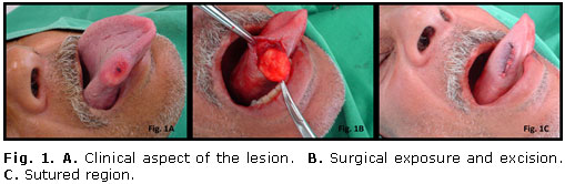
Male, 61 years    old, complaining of enlargement on the lateral border of his tongue with approximately    30 days. After having the incisional biopsy done and histopathology confirmation    of the diagnosis, surgical removal of entire lesion was chosen. The lesion had    the characteristic features of lipoma such as yellow coloration, capsule, soft    consistency and asymptomatic (<a href="#fig1_11">Fig. 1</a>).</font>     <P align="center"><a name="fig1_11"></a><img src="/img/revistas/est/v48n1/f0111111.jpg" width="509" height="166">     
<P>    <br>     ]]></body>
<body><![CDATA[<P><font face="Verdana, Arial, Helvetica, sans-serif" size="2"><I>Case 2</I> </font>     <P><font face="Verdana, Arial, Helvetica, sans-serif" size="2">
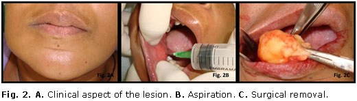
Female, 36 years    old, 10 years of lesion, complaining of facial deformity in the right side of    her face. After an aspirative punch and incisional biopsy, it was chosen to    remove the whole lesion which was not well defined, mixing with fibrous conjunctive    tissue and muscle tissue in buccal mucosa region (<a href="#fig2_11">Fig. 2</a>).</font>     <P align="center"><a name="fig2_11"></a><img src="/img/revistas/est/v48n1/f0211111.jpg" width="520" height="148">     
<P>      <P><font face="Verdana, Arial, Helvetica, sans-serif" size="2"><I>    <br>   Case 3 </I> </font>      <P><font face="Verdana, Arial, Helvetica, sans-serif" size="2">
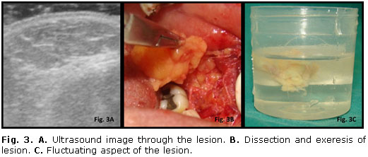
Female, 38 years    old, does not know about the start of lesion, complaining of pain on the mastication.    In this case, it was chosen the excisional biopsy of the lesion which was lobule,    streaking with a fat tissue in the chew, difficulting the dissection and lesion    removal (<a href="#fig3_11">Fig. 3</a>).</font>     <P align="center"><a name="fig3_11"></a><img src="/img/revistas/est/v48n1/f0311111.jpg" width="521" height="222">     
<P>    <br>     ]]></body>
<body><![CDATA[<P><font face="Verdana, Arial, Helvetica, sans-serif" size="2"><I>Case 4</I> </font>     <P><font face="Verdana, Arial, Helvetica, sans-serif" size="2">
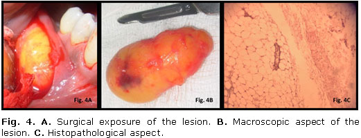
Female, 55 years    old, does not know about the start of lesion, complaining of pain, complaining    of the difficulty to fit her lower partial denture. In this case, an excisional    biopsy was done once the lesion was well defined by a fibrous capsule (<a href="#fig4_11">Fig.    4</a>).</font>     <P align="center"><a name="fig4_11"></a><img src="/img/revistas/est/v48n1/f0411111.jpg" width="521" height="199">     
<P>      <P><font face="Verdana, Arial, Helvetica, sans-serif" size="2">    <br>   All the cases described above have been in follow up period, without signs of    recurrence and functional/aesthetic commitment.</font>     <P>      <P>      <P><font face="Verdana, Arial, Helvetica, sans-serif" size="2"><B><font size="3">DISCUSSION</font></B>    </font>      <P><font face="Verdana, Arial, Helvetica, sans-serif" size="2">The etiology of    lipomas is still discussed. Trauma and infection were also indicated as possible    etiological agents. However, any factor has been established as a cause to lipoma's    onset. Lipoma has a higher incidence in obese people even having metabolism    independent to a lipid's metabolism. It is known that the occurrence of multiple    lipomas may be associated with Cowden syndrome or multiple hamartoma syndrome.<SUP>9</SUP>    </font>     ]]></body>
<body><![CDATA[<P><font face="Verdana, Arial, Helvetica, sans-serif" size="2">Even though lipomas    mainly occur in the subcutaneous tissue, it might locate in a profound and uncommon    area into facial spaces. This fact requires imaging examinations such as computadorized    tomography, magnetic resonance imaging and ultrasound in order to help with    diagnosis and treatment.<SUP>10,11</SUP> </font>      <P><font face="Verdana, Arial, Helvetica, sans-serif" size="2">Being a rare benign    mesenchymal neoplasm in the oral cavity, generally, there are some features    in lipoma that facilitates the diagnosis. In addition to yellow coloration,    the lower density than a fixer solution, making a surgical piece fluctuates    when soaked in recipient. However<I>, Souza et al</I>,<SUP>12 </SUP>reported    a lipoma case which did not observe these features.<SUP>12 </SUP>According to    some authors, these facts are probably found due to the deepness of lesion relating    to surfacing epithelium that difficults the translucence of typical yellow coloration    of lipoma lesions. The event of non fluctuation in formol solution 10 % may    be explained because a great quality of perilesional hemangiomatosis connective    tissue which gives greater weight to a surgical piece. These findings provide    more importance to the histopathologic evaluation to confirm the diagnosis.    </font>      <P><font face="Verdana, Arial, Helvetica, sans-serif" size="2">According to <i>Avelar    et al</i>,<SUP>5</SUP> it is not possible to distinguish microscopically a lipoma    from normal fat tissue, being possible only to see the metabolism process once    the tumors are not used as a source of energy.<SUP> </SUP>Based on histopathologic    findings, lipomas may be classified as simple, fibrolipoma, angiolipoma, intramuscular    (infiltrating) lipomas, pleomorphic lipomas, salivary glands lipoma (sialolipomas),    mixoid lipomas and atypical lipomas.<SUP>13</SUP> According to <i>Avelar et    al</i>,<SUP>5</SUP> simple lipomas are the most common histopathologic type,    being rare in children.<SUP> </SUP>Lipoblastoma and lipoblatomatosis are more    frequent seen in these age group. </font>      <P><font face="Verdana, Arial, Helvetica, sans-serif" size="2">Differential diagnosis    should be done between lipoma and dermoid and epidermoid cysts, lymphoepithelial    cysts, mucocele, ranula, pleomorphic adenoma and mucoepidermoid carcinoma.<SUP>14</SUP>    Mesenchymal neoplasm should be also included in the differential diagnosis.<SUP>5    </SUP>Oral lymphoepithelial cyst differs from oral lipoma due to smaller nodules    and usually occurs between the first and third decade of life. In addition,    a majority of these cysts in oral cavity is found in soft palate, pharynx mucosa    and lymph nodes.<SUP>15 </SUP>Dermoid and epidermoid cyst are also found as    submucous nodules which usually occur in the midline of mouth floor,<SUP>16</SUP>    in different locations on the oral mucosa. Therefore, it is important to conduct    a thorough clinical diagnosis and histopathologic evaluation to confirm diagnosis.    Even though, surgical removal is considered as a protocol for treating these    lesions, other methods have been applied with this aim. <I>Martorelli et al</I><SUP>17    </SUP>indicates the use of laser and electric surgery to treat intra oral lipomas.<SUP>    </SUP>Different microscopic forms have the same prognosis. However, it is important    to warn about the higher rate of recurrence of intramuscular lipomas because    of their infiltrative growth pattern, and it is a rare condition in the oral    and maxillofacial region.<SUP>18</SUP> </font>      <P><font face="Verdana, Arial, Helvetica, sans-serif" size="2">A dental surgeon    should be able to diagnose lipomas in an early stage in the maxillofacial area    avoiding a massive growth of these lesions. It will be essential to prevent    any aesthetic and functional disturbances in patients. An adequate treatment    and postsurgical follow up in lipomas are fundamental to reestablish the region    and monitor any possible chances of recurrence.</font>     <P>      <P>      <P><font face="Verdana, Arial, Helvetica, sans-serif" size="2"><B><font size="3">BIBLIOGRAPHIC    REFERENCES</font></B> </font>      <!-- ref --><P><font face="Verdana, Arial, Helvetica, sans-serif" size="2">1. Fregnani ER,    Pires FR, Falzoni R, Lopes MA, Vargas PA. Lipomas of the oral cavity: clinical    findings, histological classification and proliferative activity of 46 cases.    Int J Oral Maxillofac Surg. 2003;32(1):49-53. </font>    <!-- ref --><P><font face="Verdana, Arial, Helvetica, sans-serif" size="2">2. Chidzonga MM,    Mahomva L, Marimo C. Gigantic tongue lipoma: a case report. Med Oral Patol Oral    Cir Bucal. 2006;11(5):437-9. </font>    <!-- ref --><P><font face="Verdana, Arial, Helvetica, sans-serif" size="2">3. Furlong MA,    Fanburg-Smith JC, Childers ELB. Lipoma of the oral and maxillofacial region:    Site and subclassification of 125 cases. Oral Surg Oral Med Oral Pathol Oral    Radiol Endod. 2004;98(4):441-50. </font>    <!-- ref --><P><font face="Verdana, Arial, Helvetica, sans-serif" size="2">4. Morais HHA,    Vajgel A, Rocha NS, Carvalho RWF, Caubi AF, Vasconcellos RJH. Congenital lipoma    of the lip: a case report. Journal of Oral Science. 2009;51(3):489-91. </font>    <!-- ref --><P><font face="Verdana, Arial, Helvetica, sans-serif" size="2">5. Avelar RL, Carvalho    RWF, Falc&atilde;o PGCB, Antunes AA, Andrade ESS. Lipomas in the oral and maxillofacial    region: retrospective study of 16 years in Brazil. Revista Portuguesa de Estomatologia,    Medicina Dent&aacute;ria e Cirurgia Maxilofacial. 2008;49(4):207-11. </font>    <!-- ref --><P><font face="Verdana, Arial, Helvetica, sans-serif" size="2">6. Prado R, Ribeiro    DPB, Fontoura RA, Sampaio RKPL, Moreira LC. A case of sublingual lipoma. Revista    Brasileira de Odontologia. 1998;55(4):226-8. </font>    <!-- ref --><P><font face="Verdana, Arial, Helvetica, sans-serif" size="2">7. Greer RO, Richardson    JF. The nature of lipomas and their significance in the oral cavity: a review    and report of cases. Oral Surg Oral Med Oral Pathol. 1973;36(4):551-5. </font>    <!-- ref --><P><font face="Verdana, Arial, Helvetica, sans-serif" size="2">8. Band&eacute;ca    MC, P&aacute;dua JM, Nadalin MR, Oz&oacute;rio JEV, Silva-Sousa YTC, Perez DEC.    Oral soft tissue lipomas: a case series. J Canad Dental Assoc. 2007;73(5):431-4.    </font>    <!-- ref --><P><font face="Verdana, Arial, Helvetica, sans-serif" size="2">9. Woodhouse JB,    Delahunt B, English SF, Fraser HH, Ferguson MM. Testicular lipomatosis in Cowden's    syndrome. Modern Pathology. 2005;18(9):1151-6. </font>    <!-- ref --><P><font face="Verdana, Arial, Helvetica, sans-serif" size="2">10. Rimmer J, Singh    A, Irving C, Archer DJ, Rhys-Evans P. Asymtomatic oropharyngeal lipoma complicating    intubation. J Laryngol Otol. 2005;119(6):483-5. </font>    <!-- ref --><P><font face="Verdana, Arial, Helvetica, sans-serif" size="2">11. Pass B, Guttenberg    S, Childers EL, Emery RW. Soft tissue lipoma with the radiographic appearance    of a neoplasm within the mandibular canal. Dentomaxillofac Radiol. 2006;35(4):299-302.    </font>    <!-- ref --><P><font face="Verdana, Arial, Helvetica, sans-serif" size="2">12. Sousa FRN,    Castro AL, Morais NP, Soubhia AMP, Jardim J&uacute;nior EG, Miyahara GI. Lipoma    in the oral mucosa. Rev Cir Traumatol. Buco-Maxilo-fac. 2008;8(3):31-4. </font>    <!-- ref --><P><font face="Verdana, Arial, Helvetica, sans-serif" size="2">13. Said-Al-Naieff    N, Zahurullah FR, Sciubba JJ. Oral spindle cell lipoma. Ann Diagn Pathol. 2001;5(4):207-15.    </font>    <!-- ref --><P><font face="Verdana, Arial, Helvetica, sans-serif" size="2">14. Hattori H.    Atypical lipomatous tumor of the lip with pleomorphic lipoma-like myxoid area,    clinically simulating mucocele. J Oral Pathol Med. 2002;31(9):561-4. </font>    <!-- ref --><P><font face="Verdana, Arial, Helvetica, sans-serif" size="2">15. Flaitz CM.    Oral lymphoepithelial cyst in a young child. Pediatr Dent. 2002;22(5):422-3.    </font>    <!-- ref --><P><font face="Verdana, Arial, Helvetica, sans-serif" size="2">16. Akyol MU, Ozdek    A, Sokmensuer C. Lipoma of the tongue. Otolaryngology-Head Neck Surg. 2000;122(3):461-2.    </font>    <!-- ref --><P><font face="Verdana, Arial, Helvetica, sans-serif" size="2">17. Martorelli    SBF, Gueiros LAC, Albert J&uacute;nior A, Albuquerque RS, Martorelli FO. Intraoral    lipoma of uncommon size. Odontologia. Cl&iacute;n-Cient&iacute;f. 2005;4(1):57-62.    </font>    <!-- ref --><P><font face="Verdana, Arial, Helvetica, sans-serif" size="2">18. Thomas S, Varghese    BT, Sebastian P, Koshy CM, Mathews A, Abraham EK. Intramuscular lipomatosis    of Tongue. Postgrad Med J. 2002;78(919):295-7.</font>    <P>     <P>     <P>     <P>     ]]></body>
<body><![CDATA[<P><font face="Verdana, Arial, Helvetica, sans-serif" size="2">Recibido: 31 de    octubre de 2010.    <br>   </font><font face="Verdana, Arial, Helvetica, sans-serif" size="2">Aprobado:    22 de diciembre de 2010. </font>     <P>     <P>      <P>     <P>      <P>      <P>      <P><font face="Verdana, Arial, Helvetica, sans-serif" size="2"><I>Dr. Matheus    Furtado de Carvalho. </I>Universidade Federal de Juiz de Fora. E-Mail: <U><FONT COLOR="#0000ff"><a href="mailto:matcarodonto@yahoo.com.br">matcarodonto@yahoo.com.br</a></FONT></U></font>      ]]></body><back>
<ref-list>
<ref id="B1">
<label>1</label><nlm-citation citation-type="journal">
<person-group person-group-type="author">
<name>
<surname><![CDATA[Fregnani]]></surname>
<given-names><![CDATA[ER]]></given-names>
</name>
<name>
<surname><![CDATA[Pires]]></surname>
<given-names><![CDATA[FR]]></given-names>
</name>
<name>
<surname><![CDATA[Falzoni]]></surname>
<given-names><![CDATA[R]]></given-names>
</name>
<name>
<surname><![CDATA[Lopes]]></surname>
<given-names><![CDATA[MA]]></given-names>
</name>
<name>
<surname><![CDATA[Vargas]]></surname>
<given-names><![CDATA[PA]]></given-names>
</name>
</person-group>
<article-title xml:lang="en"><![CDATA[Lipomas of the oral cavity: clinical findings, histological classification and proliferative activity of 46 cases]]></article-title>
<source><![CDATA[Int J Oral Maxillofac Surg.]]></source>
<year>2003</year>
<volume>32</volume>
<numero>1</numero>
<issue>1</issue>
<page-range>49-53</page-range></nlm-citation>
</ref>
<ref id="B2">
<label>2</label><nlm-citation citation-type="journal">
<person-group person-group-type="author">
<name>
<surname><![CDATA[Chidzonga]]></surname>
<given-names><![CDATA[MM]]></given-names>
</name>
<name>
<surname><![CDATA[Mahomva]]></surname>
<given-names><![CDATA[L]]></given-names>
</name>
<name>
<surname><![CDATA[Marimo]]></surname>
<given-names><![CDATA[C]]></given-names>
</name>
</person-group>
<article-title xml:lang="en"><![CDATA[Gigantic tongue lipoma: a case report]]></article-title>
<source><![CDATA[Med Oral Patol Oral Cir Bucal]]></source>
<year>2006</year>
<volume>11</volume>
<numero>5</numero>
<issue>5</issue>
<page-range>437-9</page-range></nlm-citation>
</ref>
<ref id="B3">
<label>3</label><nlm-citation citation-type="journal">
<person-group person-group-type="author">
<name>
<surname><![CDATA[Furlong]]></surname>
<given-names><![CDATA[MA]]></given-names>
</name>
<name>
<surname><![CDATA[Fanburg-Smith]]></surname>
<given-names><![CDATA[JC]]></given-names>
</name>
<name>
<surname><![CDATA[Childers]]></surname>
<given-names><![CDATA[ELB]]></given-names>
</name>
</person-group>
<article-title xml:lang="en"><![CDATA[Lipoma of the oral and maxillofacial region: Site and subclassification of 125 cases]]></article-title>
<source><![CDATA[Oral Surg Oral Med Oral Pathol Oral Radiol Endod]]></source>
<year>2004</year>
<volume>98</volume>
<numero>4</numero>
<issue>4</issue>
<page-range>441-50</page-range></nlm-citation>
</ref>
<ref id="B4">
<label>4</label><nlm-citation citation-type="journal">
<person-group person-group-type="author">
<name>
<surname><![CDATA[Morais]]></surname>
<given-names><![CDATA[HHA]]></given-names>
</name>
<name>
<surname><![CDATA[Vajgel]]></surname>
<given-names><![CDATA[A]]></given-names>
</name>
<name>
<surname><![CDATA[Rocha]]></surname>
<given-names><![CDATA[NS]]></given-names>
</name>
<name>
<surname><![CDATA[Carvalho]]></surname>
<given-names><![CDATA[RWF]]></given-names>
</name>
<name>
<surname><![CDATA[Caubi]]></surname>
<given-names><![CDATA[AF]]></given-names>
</name>
<name>
<surname><![CDATA[Vasconcellos]]></surname>
<given-names><![CDATA[RJH]]></given-names>
</name>
</person-group>
<article-title xml:lang="en"><![CDATA[Congenital lipoma of the lip: a case report]]></article-title>
<source><![CDATA[Journal of Oral Science.]]></source>
<year>2009</year>
<volume>51</volume>
<numero>3</numero>
<issue>3</issue>
<page-range>489-91</page-range></nlm-citation>
</ref>
<ref id="B5">
<label>5</label><nlm-citation citation-type="journal">
<person-group person-group-type="author">
<name>
<surname><![CDATA[Avelar]]></surname>
<given-names><![CDATA[RL]]></given-names>
</name>
<name>
<surname><![CDATA[Carvalho]]></surname>
<given-names><![CDATA[RWF]]></given-names>
</name>
<name>
<surname><![CDATA[Falcão]]></surname>
<given-names><![CDATA[PGCB]]></given-names>
</name>
<name>
<surname><![CDATA[Antunes]]></surname>
<given-names><![CDATA[AA]]></given-names>
</name>
<name>
<surname><![CDATA[Andrade]]></surname>
<given-names><![CDATA[ESS]]></given-names>
</name>
</person-group>
<article-title xml:lang="en"><![CDATA[Lipomas in the oral and maxillofacial region: retrospective study of 16 years in Brazil]]></article-title>
<source><![CDATA[Revista Portuguesa de Estomatologia, Medicina Dentária e Cirurgia Maxilofacial]]></source>
<year>2008</year>
<volume>49</volume>
<numero>4</numero>
<issue>4</issue>
<page-range>207-11</page-range></nlm-citation>
</ref>
<ref id="B6">
<label>6</label><nlm-citation citation-type="journal">
<person-group person-group-type="author">
<name>
<surname><![CDATA[Prado]]></surname>
<given-names><![CDATA[R]]></given-names>
</name>
<name>
<surname><![CDATA[Ribeiro]]></surname>
<given-names><![CDATA[DPB]]></given-names>
</name>
<name>
<surname><![CDATA[Fontoura]]></surname>
<given-names><![CDATA[RA]]></given-names>
</name>
<name>
<surname><![CDATA[Sampaio]]></surname>
<given-names><![CDATA[RKPL]]></given-names>
</name>
<name>
<surname><![CDATA[Moreira]]></surname>
<given-names><![CDATA[LC]]></given-names>
</name>
</person-group>
<article-title xml:lang="en"><![CDATA[A case of sublingual lipoma]]></article-title>
<source><![CDATA[Revista Brasileira de Odontologia.]]></source>
<year>1998</year>
<volume>55</volume>
<numero>4</numero>
<issue>4</issue>
<page-range>226-8</page-range></nlm-citation>
</ref>
<ref id="B7">
<label>7</label><nlm-citation citation-type="journal">
<person-group person-group-type="author">
<name>
<surname><![CDATA[Greer]]></surname>
<given-names><![CDATA[RO]]></given-names>
</name>
<name>
<surname><![CDATA[Richardson]]></surname>
<given-names><![CDATA[JF]]></given-names>
</name>
</person-group>
<article-title xml:lang="en"><![CDATA[The nature of lipomas and their significance in the oral cavity: a review and report of cases]]></article-title>
<source><![CDATA[Oral Surg Oral Med Oral Pathol]]></source>
<year>1973</year>
<volume>36</volume>
<numero>4</numero>
<issue>4</issue>
<page-range>551-5</page-range></nlm-citation>
</ref>
<ref id="B8">
<label>8</label><nlm-citation citation-type="journal">
<person-group person-group-type="author">
<name>
<surname><![CDATA[Bandéca]]></surname>
<given-names><![CDATA[MC]]></given-names>
</name>
<name>
<surname><![CDATA[Pádua]]></surname>
<given-names><![CDATA[JM]]></given-names>
</name>
<name>
<surname><![CDATA[Nadalin]]></surname>
<given-names><![CDATA[MR]]></given-names>
</name>
<name>
<surname><![CDATA[Ozório]]></surname>
<given-names><![CDATA[JEV]]></given-names>
</name>
<name>
<surname><![CDATA[Silva-Sousa]]></surname>
<given-names><![CDATA[YTC]]></given-names>
</name>
<name>
<surname><![CDATA[Perez]]></surname>
<given-names><![CDATA[DEC]]></given-names>
</name>
</person-group>
<article-title xml:lang="en"><![CDATA[Oral soft tissue lipomas: a case series]]></article-title>
<source><![CDATA[J Canad Dental Assoc.]]></source>
<year>2007</year>
<volume>73</volume>
<numero>5</numero>
<issue>5</issue>
<page-range>431-4</page-range></nlm-citation>
</ref>
<ref id="B9">
<label>9</label><nlm-citation citation-type="journal">
<person-group person-group-type="author">
<name>
<surname><![CDATA[Woodhouse]]></surname>
<given-names><![CDATA[JB]]></given-names>
</name>
<name>
<surname><![CDATA[Delahunt]]></surname>
<given-names><![CDATA[B]]></given-names>
</name>
<name>
<surname><![CDATA[English]]></surname>
<given-names><![CDATA[SF]]></given-names>
</name>
<name>
<surname><![CDATA[Fraser]]></surname>
<given-names><![CDATA[HH]]></given-names>
</name>
<name>
<surname><![CDATA[Ferguson]]></surname>
<given-names><![CDATA[MM]]></given-names>
</name>
</person-group>
<article-title xml:lang="en"><![CDATA[Testicular lipomatosis in Cowden's syndrome]]></article-title>
<source><![CDATA[Modern Pathology.]]></source>
<year>2005</year>
<volume>18</volume>
<numero>9</numero>
<issue>9</issue>
<page-range>1151-6</page-range></nlm-citation>
</ref>
<ref id="B10">
<label>10</label><nlm-citation citation-type="journal">
<person-group person-group-type="author">
<name>
<surname><![CDATA[Rimmer]]></surname>
<given-names><![CDATA[J]]></given-names>
</name>
<name>
<surname><![CDATA[Singh]]></surname>
<given-names><![CDATA[A]]></given-names>
</name>
<name>
<surname><![CDATA[Irving]]></surname>
<given-names><![CDATA[C]]></given-names>
</name>
<name>
<surname><![CDATA[Archer]]></surname>
<given-names><![CDATA[DJ]]></given-names>
</name>
<name>
<surname><![CDATA[Rhys-Evans]]></surname>
<given-names><![CDATA[P]]></given-names>
</name>
</person-group>
<article-title xml:lang="en"><![CDATA[Asymtomatic oropharyngeal lipoma complicating intubation]]></article-title>
<source><![CDATA[J Laryngol Otol.]]></source>
<year>2005</year>
<volume>119</volume>
<numero>6</numero>
<issue>6</issue>
<page-range>483-5</page-range></nlm-citation>
</ref>
<ref id="B11">
<label>11</label><nlm-citation citation-type="journal">
<person-group person-group-type="author">
<name>
<surname><![CDATA[Pass]]></surname>
<given-names><![CDATA[B]]></given-names>
</name>
<name>
<surname><![CDATA[Guttenberg]]></surname>
<given-names><![CDATA[S]]></given-names>
</name>
<name>
<surname><![CDATA[Childers]]></surname>
<given-names><![CDATA[EL]]></given-names>
</name>
<name>
<surname><![CDATA[Emery]]></surname>
<given-names><![CDATA[RW]]></given-names>
</name>
</person-group>
<article-title xml:lang="en"><![CDATA[Soft tissue lipoma with the radiographic appearance of a neoplasm within the mandibular canal]]></article-title>
<source><![CDATA[Dentomaxillofac Radiol.]]></source>
<year>2006</year>
<volume>35</volume>
<numero>4</numero>
<issue>4</issue>
<page-range>299-302</page-range></nlm-citation>
</ref>
<ref id="B12">
<label>12</label><nlm-citation citation-type="journal">
<person-group person-group-type="author">
<name>
<surname><![CDATA[Sousa]]></surname>
<given-names><![CDATA[FRN]]></given-names>
</name>
<name>
<surname><![CDATA[Castro]]></surname>
<given-names><![CDATA[AL]]></given-names>
</name>
<name>
<surname><![CDATA[Morais]]></surname>
<given-names><![CDATA[NP]]></given-names>
</name>
<name>
<surname><![CDATA[Soubhia]]></surname>
<given-names><![CDATA[AMP]]></given-names>
</name>
<name>
<surname><![CDATA[Jardim Júnior]]></surname>
<given-names><![CDATA[EG]]></given-names>
</name>
<name>
<surname><![CDATA[Miyahara]]></surname>
<given-names><![CDATA[GI]]></given-names>
</name>
</person-group>
<article-title xml:lang="en"><![CDATA[Lipoma in the oral mucosa: Rev Cir Traumatol]]></article-title>
<source><![CDATA[Buco-Maxilo-fac.]]></source>
<year>2008</year>
<volume>8</volume>
<numero>3</numero>
<issue>3</issue>
<page-range>31-4</page-range></nlm-citation>
</ref>
<ref id="B13">
<label>13</label><nlm-citation citation-type="journal">
<person-group person-group-type="author">
<name>
<surname><![CDATA[Said-Al-Naieff]]></surname>
<given-names><![CDATA[N]]></given-names>
</name>
<name>
<surname><![CDATA[Zahurullah]]></surname>
<given-names><![CDATA[FR]]></given-names>
</name>
<name>
<surname><![CDATA[Sciubba]]></surname>
<given-names><![CDATA[JJ]]></given-names>
</name>
</person-group>
<article-title xml:lang="en"><![CDATA[Oral spindle cell lipoma]]></article-title>
<source><![CDATA[Ann Diagn Pathol.]]></source>
<year>2001</year>
<volume>5</volume>
<numero>4</numero>
<issue>4</issue>
<page-range>207-15</page-range></nlm-citation>
</ref>
<ref id="B14">
<label>14</label><nlm-citation citation-type="journal">
<person-group person-group-type="author">
<name>
<surname><![CDATA[Hattori]]></surname>
<given-names><![CDATA[H]]></given-names>
</name>
</person-group>
<article-title xml:lang="en"><![CDATA[Atypical lipomatous tumor of the lip with pleomorphic lipoma-like myxoid area, clinically simulating mucocele]]></article-title>
<source><![CDATA[J Oral Pathol Med.]]></source>
<year>2002</year>
<volume>31</volume>
<numero>9</numero>
<issue>9</issue>
<page-range>561-4</page-range></nlm-citation>
</ref>
<ref id="B15">
<label>15</label><nlm-citation citation-type="journal">
<person-group person-group-type="author">
<name>
<surname><![CDATA[Flaitz]]></surname>
<given-names><![CDATA[CM]]></given-names>
</name>
</person-group>
<article-title xml:lang="en"><![CDATA[Oral lymphoepithelial cyst in a young child]]></article-title>
<source><![CDATA[Pediatr Dent.]]></source>
<year>2002</year>
<volume>22</volume>
<numero>5</numero>
<issue>5</issue>
<page-range>422-3</page-range></nlm-citation>
</ref>
<ref id="B16">
<label>16</label><nlm-citation citation-type="journal">
<person-group person-group-type="author">
<name>
<surname><![CDATA[Akyol]]></surname>
<given-names><![CDATA[MU]]></given-names>
</name>
<name>
<surname><![CDATA[Ozdek]]></surname>
<given-names><![CDATA[A]]></given-names>
</name>
<name>
<surname><![CDATA[Sokmensuer]]></surname>
<given-names><![CDATA[C]]></given-names>
</name>
</person-group>
<article-title xml:lang="en"><![CDATA[Lipoma of the tongue]]></article-title>
<source><![CDATA[Otolaryngology-Head Neck Surg.]]></source>
<year>2000</year>
<volume>122</volume>
<numero>3</numero>
<issue>3</issue>
<page-range>461-2</page-range></nlm-citation>
</ref>
<ref id="B17">
<label>17</label><nlm-citation citation-type="journal">
<person-group person-group-type="author">
<name>
<surname><![CDATA[Martorelli]]></surname>
<given-names><![CDATA[SBF]]></given-names>
</name>
<name>
<surname><![CDATA[Gueiros]]></surname>
<given-names><![CDATA[LAC]]></given-names>
</name>
<name>
<surname><![CDATA[Albert Júnior]]></surname>
<given-names><![CDATA[A]]></given-names>
</name>
<name>
<surname><![CDATA[Albuquerque]]></surname>
<given-names><![CDATA[RS]]></given-names>
</name>
<name>
<surname><![CDATA[Martorelli]]></surname>
<given-names><![CDATA[FO]]></given-names>
</name>
</person-group>
<article-title xml:lang="en"><![CDATA[Intraoral lipoma of uncommon size: Odontologia]]></article-title>
<source><![CDATA[Clín-Científ.]]></source>
<year>2005</year>
<volume>4</volume>
<numero>1</numero>
<issue>1</issue>
<page-range>57-62</page-range></nlm-citation>
</ref>
<ref id="B18">
<label>18</label><nlm-citation citation-type="journal">
<person-group person-group-type="author">
<name>
<surname><![CDATA[Thomas]]></surname>
<given-names><![CDATA[S]]></given-names>
</name>
<name>
<surname><![CDATA[Varghese]]></surname>
<given-names><![CDATA[BT]]></given-names>
</name>
<name>
<surname><![CDATA[Sebastian]]></surname>
<given-names><![CDATA[P]]></given-names>
</name>
<name>
<surname><![CDATA[Koshy]]></surname>
<given-names><![CDATA[CM]]></given-names>
</name>
<name>
<surname><![CDATA[Mathews]]></surname>
<given-names><![CDATA[A]]></given-names>
</name>
<name>
<surname><![CDATA[Abraham]]></surname>
<given-names><![CDATA[EK]]></given-names>
</name>
</person-group>
<article-title xml:lang="en"><![CDATA[Intramuscular lipomatosis of Tongue]]></article-title>
<source><![CDATA[Postgrad Med J.]]></source>
<year>2002</year>
<volume>78</volume>
<numero>919</numero>
<issue>919</issue>
<page-range>295-7</page-range></nlm-citation>
</ref>
</ref-list>
</back>
</article>
