<?xml version="1.0" encoding="ISO-8859-1"?><article xmlns:mml="http://www.w3.org/1998/Math/MathML" xmlns:xlink="http://www.w3.org/1999/xlink" xmlns:xsi="http://www.w3.org/2001/XMLSchema-instance">
<front>
<journal-meta>
<journal-id>0034-7507</journal-id>
<journal-title><![CDATA[Revista Cubana de Estomatología]]></journal-title>
<abbrev-journal-title><![CDATA[Rev Cubana Estomatol]]></abbrev-journal-title>
<issn>0034-7507</issn>
<publisher>
<publisher-name><![CDATA[Editorial Ciencias Médicas]]></publisher-name>
</publisher>
</journal-meta>
<article-meta>
<article-id>S0034-75072012000400010</article-id>
<title-group>
<article-title xml:lang="en"><![CDATA[Lymphoepithelial cyst in the palatoglossus arch]]></article-title>
<article-title xml:lang="es"><![CDATA[Quiste linfoepitelial en el arco palatogloso]]></article-title>
</title-group>
<contrib-group>
<contrib contrib-type="author">
<name>
<surname><![CDATA[Marçal Vieira]]></surname>
<given-names><![CDATA[Evanice Maria]]></given-names>
</name>
<xref ref-type="aff" rid="A01"/>
</contrib>
<contrib contrib-type="author">
<name>
<surname><![CDATA[Canavarros Palma]]></surname>
<given-names><![CDATA[Vinícius]]></given-names>
</name>
<xref ref-type="aff" rid="A01"/>
</contrib>
<contrib contrib-type="author">
<name>
<surname><![CDATA[Delle Vedove Semenoff]]></surname>
<given-names><![CDATA[Tereza Aparecida]]></given-names>
</name>
<xref ref-type="aff" rid="A01"/>
</contrib>
<contrib contrib-type="author">
<name>
<surname><![CDATA[Fábio Aranha]]></surname>
<given-names><![CDATA[Andreza Maria]]></given-names>
</name>
<xref ref-type="aff" rid="A01"/>
</contrib>
<contrib contrib-type="author">
<name>
<surname><![CDATA[Baratto-Filho]]></surname>
<given-names><![CDATA[Flares]]></given-names>
</name>
<xref ref-type="aff" rid="A02"/>
</contrib>
<contrib contrib-type="author">
<name>
<surname><![CDATA[Henrique Borges]]></surname>
<given-names><![CDATA[Álvaro]]></given-names>
</name>
<xref ref-type="aff" rid="A01"/>
</contrib>
</contrib-group>
<aff id="A02">
<institution><![CDATA[,University of Curitiba  ]]></institution>
<addr-line><![CDATA[Paraná ]]></addr-line>
<country>Brazil</country>
</aff>
<aff id="A01">
<institution><![CDATA[,University of Cuiabá  ]]></institution>
<addr-line><![CDATA[Cuiabá Mato Grosso]]></addr-line>
<country>Brazil</country>
</aff>
<pub-date pub-type="pub">
<day>00</day>
<month>12</month>
<year>2012</year>
</pub-date>
<pub-date pub-type="epub">
<day>00</day>
<month>12</month>
<year>2012</year>
</pub-date>
<volume>49</volume>
<numero>4</numero>
<fpage>335</fpage>
<lpage>340</lpage>
<copyright-statement/>
<copyright-year/>
<self-uri xlink:href="http://scielo.sld.cu/scielo.php?script=sci_arttext&amp;pid=S0034-75072012000400010&amp;lng=en&amp;nrm=iso"></self-uri><self-uri xlink:href="http://scielo.sld.cu/scielo.php?script=sci_abstract&amp;pid=S0034-75072012000400010&amp;lng=en&amp;nrm=iso"></self-uri><self-uri xlink:href="http://scielo.sld.cu/scielo.php?script=sci_pdf&amp;pid=S0034-75072012000400010&amp;lng=en&amp;nrm=iso"></self-uri><abstract abstract-type="short" xml:lang="en"><p><![CDATA[The objective of this study was to describe a case of a lymphoepithelial cyst in the palatoglossus arch. A 16-years-old black man said that he was observed a lesion in his mouth. On the physical exam, a pedicled, consistent, smooth surface 1.5 x 1 cm lesion, similar in color to the adjacent mucosa, was found. The lesion was surgically removed and the microscopic exam showed mucosal fragments with pedicled lesion; the cystic cavity sometimes lined with pseudostratified cylindrical epithelium and others with stratified squamous flat interface of the epithelium. Around the cyst, a well-delimited mass of lymphoid tissue, presenting lymphoid follicles, was also seen. Lymphoepithelial cyst has clinical characteristics similar to those of others lesions that occur in the oral cavity. The diagnosis should be based on conservative biopsy, with total removal of lesion.]]></p></abstract>
<abstract abstract-type="short" xml:lang="es"><p><![CDATA[El objetivo de este estudio fue describir un caso clínico de quiste linfoepitelial en el arco palatoglosso. Paciente de 16 años de edad, sexo masculino, raza negra, que durante la anamnesis relató que observó una lesión en la boca. Al examen físico se observó una lesión pediculada, de consistencia firme, superficie lisa, de 1,5 x 1,0 cm de tamaño, con color similar al de la mucosa adyacente. La lesión fue removida quirúrgicamente y el examen microscópico mostró fragmentos de mucosa con lesión pediculada, la cavidad del quiste a veces revestida por epitelio cilíndrico pseudoestratificado y otras veces por la interfase plana del epitelio escamosa estratificado. Alrededor del quiste, una masa bien delimitada de tejido linfoide con presencia de folículos linfoides. El quiste linfoepitelial tiene las mismas características clínicas de otras lesiones que ocurren en la cavidad oral, el diagnóstico debe ser realizado por biopsia, de manera conservadora, con eliminación total de la lesión.]]></p></abstract>
<kwd-group>
<kwd lng="en"><![CDATA[pathology]]></kwd>
<kwd lng="en"><![CDATA[oral pathology]]></kwd>
<kwd lng="en"><![CDATA[cyst]]></kwd>
<kwd lng="en"><![CDATA[lymphoepithelial cyst]]></kwd>
<kwd lng="es"><![CDATA[patología]]></kwd>
<kwd lng="es"><![CDATA[patología oral]]></kwd>
<kwd lng="es"><![CDATA[quiste]]></kwd>
<kwd lng="es"><![CDATA[quiste linfoepitelial]]></kwd>
</kwd-group>
</article-meta>
</front><body><![CDATA[ <div align="right">     <p><font face="Verdana, Arial, Helvetica, sans-serif" size="2">  <b>PRESENTACI&Oacute;N DE CASO</b></font></p>    <p>&nbsp; </p></div>    <P>     <P><b><font face="Verdana, Arial, Helvetica, sans-serif" size="4">Lymphoepithelial  cyst in the palatoglossus arch </font></b>     <P>&nbsp;     <P>     <P>     <p><font face="Verdana, Arial, Helvetica, sans-serif" size="2"><B><font size="3">Quiste  linfoepitelial en el arco palatogloso </font></B></font></p>    <p>&nbsp;</p>    ]]></body>
<body><![CDATA[<p><B> </B></p><B>      <P>     <P>     <P><font face="Verdana, Arial, Helvetica, sans-serif" size="2">Evanice  Maria Mar&ccedil;al Vieira,<SUP>I </SUP>Vin&iacute;cius Canavarros Palma,<SUP>I  </SUP>Tereza Aparecida Delle Vedove Semenoff,<SUP>I </SUP>Andreza Maria F&aacute;bio  Aranha,<SUP>I </SUP>Flares Baratto-Filho,<SUP>II</SUP> &Aacute;lvaro Henrique  Borges<SUP>I </SUP></font> </B>    <P><B>&nbsp; </b><SUP>     <P> </SUP>     <P><font face="Verdana, Arial, Helvetica, sans-serif" size="2"><SUP>I</sup>  University of Cuiab&aacute;, Cuiab&aacute;, Mato Grosso, Brazil.    <br> </font><font face="Verdana, Arial, Helvetica, sans-serif" size="2"><SUP>II</SUP>  University of Curitiba, Paran&aacute;, Brazil. </font>     <P>&nbsp;     <P>&nbsp;     ]]></body>
<body><![CDATA[<P>&nbsp; <hr size="1" noshade>      <P><font face="Verdana, Arial, Helvetica, sans-serif" size="2"><b>ABSTRACT</b>  </font>     <p><font face="Verdana, Arial, Helvetica, sans-serif" size="2">The objective  of this study was to describe a case of a lymphoepithelial cyst<b> </b>in the  palatoglossus arch<b>. </b>A 16-years-old black man said that he was observed  a lesion in his mouth. On the physical exam, a pedicled, consistent, smooth surface  1.5 x 1 cm lesion, similar in color to the adjacent mucosa, was found. The lesion  was surgically removed and the microscopic exam showed mucosal fragments with  pedicled lesion; the cystic cavity sometimes lined with pseudostratified cylindrical  epithelium and others with stratified squamous flat interface of the epithelium.  Around the cyst, a     <br> well-delimited mass of lymphoid tissue, presenting lymphoid  follicles, was also seen. Lymphoepithelial cyst has clinical characteristics similar  to those of others lesions that occur in the oral cavity. The diagnosis should  be based on conservative biopsy, with total removal of lesion. </font>     <p><font face="Verdana, Arial, Helvetica, sans-serif" size="2"><b>Key  words:</b> pathology, oral pathology, cyst, lymphoepithelial cyst. </font> <FONT  COLOR="#333333"></FONT> <hr size="1" noshade>     <P>     <P><b><font face="Verdana, Arial, Helvetica, sans-serif" size="2">RESUMEN  </font></b><font face="Verdana, Arial, Helvetica, sans-serif" size="2">    <br>     <br>  El objetivo de este estudio fue describir un caso cl&iacute;nico de quiste linfoepitelial  en el arco palatoglosso. Paciente de 16 a&ntilde;os de edad, sexo masculino, raza  negra, que durante la anamnesis relat&oacute; que observ&oacute; una lesi&oacute;n  en la boca. Al examen f&iacute;sico se observ&oacute; una lesi&oacute;n pediculada,  de consistencia firme, superficie lisa, de 1,5 x 1,0 cm de tama&ntilde;o, con  color similar al de la mucosa adyacente. La lesi&oacute;n fue removida quir&uacute;rgicamente  y el examen microsc&oacute;pico mostr&oacute; fragmentos de mucosa con lesi&oacute;n  pediculada, la cavidad del quiste a veces revestida por epitelio cil&iacute;ndrico  pseudoestratificado y otras veces por la interfase plana del epitelio escamosa  estratificado. Alrededor del quiste, una masa bien delimitada de tejido linfoide  con presencia de fol&iacute;culos linfoides. El quiste linfoepitelial tiene las  mismas caracter&iacute;sticas cl&iacute;nicas de otras lesiones que ocurren en  la cavidad oral, el diagn&oacute;stico debe ser realizado por biopsia, de manera  conservadora, con eliminaci&oacute;n total de la lesi&oacute;n. </font>     <p><font face="Verdana, Arial, Helvetica, sans-serif" size="2"><b>Palabras  clave:</b> patolog&iacute;a, patolog&iacute;a oral, quiste, quiste linfoepitelial.    ]]></body>
<body><![CDATA[<br>  </font><font face="Verdana, Arial, Helvetica, sans-serif"> </font> </p>    <P>&nbsp; <hr size="1" noshade>      <P>&nbsp;     <P>     <P>     <P>     <P>     <P>     <P>     <P>&nbsp;     ]]></body>
<body><![CDATA[<P>&nbsp;     <P><font face="Verdana, Arial, Helvetica, sans-serif" size="3"><B>INTRODUCTION</B>  </font><font face="Verdana, Arial, Helvetica, sans-serif" size="2"> </font>     <P><font face="Verdana, Arial, Helvetica, sans-serif" size="2">Lymphoepithelial  cyst, also called branquial cyst, is a lesion in the oral cavity with rare incidence.<SUP>1-6</SUP>  Histologically, it consists in a cystic cavity usually coated by stratified squamous  epithelium, lined by a well-defined mass of lymphoid tissue, organized by the  presence of many follicles and typical germinative centers.<SUP>1,7</SUP> The  stratified squamous epithelium is thin, rectified, without epithelial crests,  and it is usually parakeratinized.<SUP>2,3 </SUP>The cystic lumem often contains  desquamated epithelial cells, lymphocytes and an amorphous eosinophilic material.<SUP>1-3</SUP>  </font>     <P><font face="Verdana, Arial, Helvetica, sans-serif" size="2">Although  the lesion pathogenesis is not clear, some authors have suggested that the developmental  lesion derived from the remaining of the second branquial arch, while others have  postulated the relation to ectopic focus of mucosal epithelium or salivary glands  which was trapped in lymphoid tissue and after proliferated, forming the cysts.<SUP>7,8</SUP>  Others authors have reported that the epithelium of tonsillar crypts can be obstructed  and form cysts involved by lymphoid tissue, surrounding it.<SUP>1,2,7 </SUP>Clinically,  the lymphoepithelial cist is described as asymptomatic submucosal mass, mobile,  yellow, present by months or years, and a variable size from milimeters to 1.5  centimeters.<SUP>2,6,7</SUP> In mouth, its occurrence is common in mouth floors  and tongue, in ventral or postero-lateral portions.<SUP>4,6,9 </SUP>The greater  prevalence of this lesion occurs in female gender, in rate over to 2:1<SUP>4</SUP>,  and age between 15 to 65 years old.<SUP>4-7</SUP> </font>     <P><font face="Verdana, Arial, Helvetica, sans-serif" size="2">The  lymphoepithelial cysts are benign, successfully treated by surgical excision.  Relapses are rare and they can be resulted by the residual remaining left when  the lesion was simply aspirated or drained.<SUP>2,7,9,10</SUP> The aim of this  study was to report a clinical case of lymphoepithelial cyst with rare occurence  in the palatoglossus arch. </font>     <P>     <P><b><font face="Verdana, Arial, Helvetica, sans-serif" size="2">CASE  REPORT </font></b>     <P><font face="Verdana, Arial, Helvetica, sans-serif" size="2">
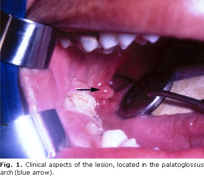
A  16-years-old black man, came to the Semiology Clinic at Dental School of the University  of Cuiab&aacute;, Cuiab&aacute;-MT, Brazil, with chief complaint of &#147;small  ball inside the mouth&#148;. During the anamnesis, related that he ever observed  this lesion, but only the last months he felt worried about the possibility that  could be something with malignat characteristics. The lesion showed a characteristic  pedicle, firm consistency, smooth surface, similar in color to the adjacent mucosa  and measuring 1,5 x 1,0 cm (<a href="#fig1">Fig. 1</a>). Because of the clinical  characteristics and localization, the diagnostic hypotheses included fibroma,  hyperplastic lymphoid aggregate and lymphoepithelial cyst. </font>     <P align="center">  <font face="Verdana, Arial, Helvetica, sans-serif" size="2"><B><a name="fig1"></a>  <img src="/img/revistas/est/v49n4/f0110412.jpg" width="420" height="370">  </B></font>     
<P><font face="Verdana, Arial, Helvetica, sans-serif" size="2">
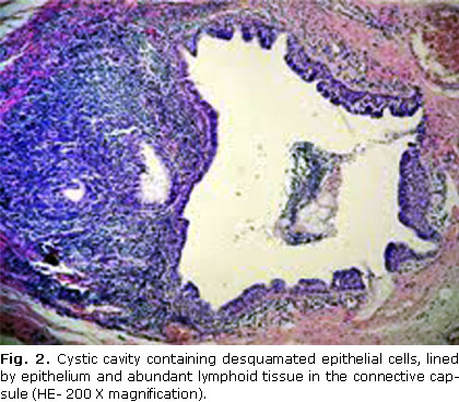
The  treatment administered in the present case was surgical. The collected material  was sent to be analysed in the Surgical Pathology Laboratory of the University  of Cuiab&aacute;. The microscopic exam showed fragments of the mucosa, presenting  in the lamina propria and subepithelial region. The cystic cavity was sometimes  lined by pseudostratified cylindrical epithelium and others by stratified squamous  flat interface (<a href="#fig2">Fig.2)</a>. Around the cyst, a well-delimited  mass of lymphoid tissue, presenting discrete lymphoid follicles organized with  many germinative centers, distributed in an abundant manner, in one of the faces  of the lesion (<a href="#fig3">Fig. 3</a>). These characteristics are compatible  to oral lymphoepithelial cyst. The two-year follow up evolved favorably, and a  relapse was not observed. </font>     ]]></body>
<body><![CDATA[<br>     <P>&nbsp;     <P align="center"><a name="fig2"></a><img src="/img/revistas/est/v49n4/f0210412.jpg" width="420" height="368">      
<P align="center">    <br> <a name="fig3"></a><img src="/img/revistas/est/v49n4/f0310412.jpg" width="420" height="372"><font face="Verdana, Arial, Helvetica, sans-serif" size="2"><B>  </B></font> <B>     
<P>    <br>     <br>     <br>     <P><font face="Verdana, Arial, Helvetica, sans-serif" size="3" color="#000000">DISCUSSION</font>  </B>     ]]></body>
<body><![CDATA[<P>     <P><font face="Verdana, Arial, Helvetica, sans-serif" size="2">Considering  the clinical aspects (pedicled form of the lesion, similar color to the adjacent  mucosa and firm consistency), at first moment, diagnostic hypothesis of fibroma  was considered. In the literature, there are reports of differential diagnostic  with lipoma, when the staining is clearer and soft consistency.<SUP>2,10</SUP>  Depending the localization, it should be differentiated by the mucocele.<SUP>1,3,5,9  </SUP>About 50 to 80% of the cases of lymphoepithelial cysts ocurr in the bucal  cavity floor,<SUP>1,4,6 </SUP>followed by ventral and posterior surfaces of the  tongue.<SUP>2,4,7</SUP> In this case report, the lesion occured in the palatoglossus  arch, with rare frequence (4 to 5 %), such as in soft palate<B>, </B>vestibular  region of the mandibular mucosa and retromolar region.<SUP>4,7</SUP> </font>     <P><font face="Verdana, Arial, Helvetica, sans-serif" size="2">The  histological aspects showed cyst lined by pseudostratified or stratified squamous  epithelium. Small focus of lymphocytic exocytosis and epithelial cells destruction  were observed in the area of the stratified paviment epithelium. The cyst was  not regularly surrounded by well-delimited and organized lymphoid tissue, presenting  several follicles, some with evident germinative centers. The lymphoid tissue  was abundant in the face of the cyst in contact with the squamous epithelium,  and scarce in the pseudostratified cylindrical epithelium. The lymphoepithelial  cyst is usually lined by stratified squamous epithelium, with desquamated epithelial  cells to the cyst lumen.<SUP>1,2,6,7</SUP> However, several authors reported coating<B>  </B>by cylindrical epithelium, ciliate or not, sometimes with mucin cells, in  monolayers or pseudostratified, and even the occurrence of small areas without  epithelial lining.<SUP>1,6</SUP> The main characteristic is related to the presence  of the lymphoid tissue in the cyst wall<SUP>1,2,6,7</SUP> with lymphoid follicles  formation and germinative centers.<SUP>2</SUP> The situation of lymphoid tissue  abundance in one portion of the cyst wall was reported by some authors.<SUP>1,2,10</SUP>  </font>     <P><font face="Verdana, Arial, Helvetica, sans-serif" size="2">Because  this lesion is harmless, the necessity of biopsy or surgical remotion can be discussed,  mainly when it is well-characterized and the diagnosis can be clinically defined.<SUP>2,4-6</SUP>  However, the lymphoepithelial cyst is commonly treated by the surgical remotion<SUP>1,2,4,5,7,9</SUP>,  as it was in this case report. All injuries must be cautiously diagnosed and when  there are doubts about the differential diagnosis, it is necessary to perform  a biopsy and microscopic examination to establish a conclusive position.<SUP>3,4,6  </SUP></font>     <P>&nbsp;     <P>     <P><font face="Verdana, Arial, Helvetica, sans-serif" size="3"><font size="2">CONCLUSION</font>  </font>     <P>     <P><font face="Verdana, Arial, Helvetica, sans-serif" size="2">By  the fact that lymphoepithelial cyst has similar clinical characteristics to other  lesions that occur in the oral cavity and considering its rare occurrence, it  is important to emphasize that the diagnosis should be done by biopsy, in a conservative  way, removing it totally. </font>     <P>&nbsp;     ]]></body>
<body><![CDATA[<P>     <P>     <P><font face="Verdana, Arial, Helvetica, sans-serif" size="3"><B>REFERENCES</B>  </font><font face="Verdana, Arial, Helvetica, sans-serif" size="2"> </font>     <!-- ref --><P><font face="Verdana, Arial, Helvetica, sans-serif" size="2">1.Khelemsky  R, Mandel L. Lymphoepithelial cyst of mouth floor. J Oral Maxillofac Surg. 2010;68(12):3055-7.      </font>     <!-- ref --><P><font face="Verdana, Arial, Helvetica, sans-serif" size="2">2.Neville  BW, Damm DD, Allen CM, Bouquot JE. Oral and Maxillofacial Pathology. Saunders/Elsevier;  2009.     </font>     <!-- ref --><P><font face="Verdana, Arial, Helvetica, sans-serif" size="2">3.Gallagher  G, Kabani S, Noonan V. Oral lymphoepithelial cyst. J Mass Dent Soc. 2006;54(4):52.      </font>     <!-- ref --><P><font face="Verdana, Arial, Helvetica, sans-serif" size="2">4.Yang  X, Ow A, Zhang CP, Wang LZ, Yang WJ, Hu YJ, Zhong LP. Clinical analysis of 120  cases of intraoral lymphoepithelial cyst. Oral Surg Oral Med Oral Pathol Oral  Radiol. 2012;113(4):448-52.     </font>     <!-- ref --><P><font face="Verdana, Arial, Helvetica, sans-serif" size="2">5.Silva  MM, Castro AL, Soubhia AMP, Crivelini MM. Lymphoepithelial cyst in jugal mucosa.  Int. J. Odontostomat. 2011;5(1):55-8.     </font>     <!-- ref --><P><font face="Verdana, Arial, Helvetica, sans-serif" size="2">6.Khelemsky  R, Mandel L. Lymphoepithelial cyst of mouth floor. J Oral Maxillofac Surg. 2010;68:3055-7.      </font>     <!-- ref --><P><font face="Verdana, Arial, Helvetica, sans-serif" size="2">7.Stramandinoli-Zanicotti  RT, de Castro &Aacute;vila LF, de Azevedo Izidoro AC, Izidoro FA, Schussel JL.  Lymphoepithelial cysts of oral mucosa: two cases in different regions. Bull Tokyo  Dent Coll. 2012;53(1):17-22.     </font>     <!-- ref --><P><font face="Verdana, Arial, Helvetica, sans-serif" size="2">8.L&oacute;pez-Jornet  P. Oral lymphoepithelial cyst. Ann Dermatol Venereol. 2007;134(6-7):588.     </font>      <!-- ref --><P><font face="Verdana, Arial, Helvetica, sans-serif" size="2">9.Nonaka CF, Henriques  AC, de Matos FR, de Souza LB, Pinto LP. Nonodontogenic cysts of the oral and maxillofacial  region: demographic profile in a Brazilian population over a 40-year period. Eur  Arch Otorhinolaryngol. 2011;268(6):917-22.     </font>     <!-- ref --><P><font face="Verdana, Arial, Helvetica, sans-serif" size="2">10.Varnholt  H, Thompson LDR, Pantanowitz L.Salivary gland lymphoepithelial cysts. Ear, Nose  &amp; Throat Journal. 2007;86(5):265.     </font>     <P>&nbsp;     <P>&nbsp;     <P>&nbsp;     <P>     <P>     <P><font face="Verdana, Arial, Helvetica, sans-serif" size="2">Recibido:  19 de agosto de 2012.     <br> </font><font face="Verdana, Arial, Helvetica, sans-serif" size="2">Aprobado:  5 de septiembre de 2012. </font>     ]]></body>
<body><![CDATA[<P>&nbsp;     <P>&nbsp;     <P>     <P>     <P>     <P><font face="Verdana, Arial, Helvetica, sans-serif" size="2"><I>Dr.  Evanice Maria Mar&ccedil;al Vieira.</I> University of Cuiab&aacute;, Cuiab&aacute;,  Mato Grosso, Brazil. Correo electr&oacute;nico:<a href="malto:evanice_mmv@yahoo.com.br">  evanice_mmv@yahoo.com.br </a></font>       ]]></body><back>
<ref-list>
<ref id="B1">
<label>1</label><nlm-citation citation-type="journal">
<person-group person-group-type="author">
<name>
<surname><![CDATA[Khelemsky]]></surname>
<given-names><![CDATA[R]]></given-names>
</name>
<name>
<surname><![CDATA[Mandel]]></surname>
<given-names><![CDATA[L]]></given-names>
</name>
</person-group>
<article-title xml:lang="en"><![CDATA[Lymphoepithelial cyst of mouth floor]]></article-title>
<source><![CDATA[J Oral Maxillofac Surg.]]></source>
<year>2010</year>
<volume>68</volume>
<numero>12</numero>
<issue>12</issue>
<page-range>3055-7</page-range></nlm-citation>
</ref>
<ref id="B2">
<label>2</label><nlm-citation citation-type="book">
<person-group person-group-type="author">
<name>
<surname><![CDATA[Neville]]></surname>
<given-names><![CDATA[BW]]></given-names>
</name>
<name>
<surname><![CDATA[Damm]]></surname>
<given-names><![CDATA[DD]]></given-names>
</name>
<name>
<surname><![CDATA[Allen]]></surname>
<given-names><![CDATA[CM]]></given-names>
</name>
<name>
<surname><![CDATA[Bouquot]]></surname>
<given-names><![CDATA[JE]]></given-names>
</name>
</person-group>
<source><![CDATA[Oral and Maxillofacial Pathology]]></source>
<year>2009</year>
<publisher-name><![CDATA[Saunders/Elsevier]]></publisher-name>
</nlm-citation>
</ref>
<ref id="B3">
<label>3</label><nlm-citation citation-type="journal">
<person-group person-group-type="author">
<name>
<surname><![CDATA[Gallagher]]></surname>
<given-names><![CDATA[G]]></given-names>
</name>
<name>
<surname><![CDATA[Kabani]]></surname>
<given-names><![CDATA[S]]></given-names>
</name>
<name>
<surname><![CDATA[Noonan]]></surname>
<given-names><![CDATA[V]]></given-names>
</name>
</person-group>
<article-title xml:lang="en"><![CDATA[Oral lymphoepithelial cyst]]></article-title>
<source><![CDATA[J Mass Dent Soc.]]></source>
<year>2006</year>
<volume>54</volume>
<numero>4</numero>
<issue>4</issue>
<page-range>52</page-range></nlm-citation>
</ref>
<ref id="B4">
<label>4</label><nlm-citation citation-type="journal">
<person-group person-group-type="author">
<name>
<surname><![CDATA[Yang]]></surname>
<given-names><![CDATA[X]]></given-names>
</name>
<name>
<surname><![CDATA[Ow]]></surname>
<given-names><![CDATA[A]]></given-names>
</name>
<name>
<surname><![CDATA[Zhang]]></surname>
<given-names><![CDATA[CP]]></given-names>
</name>
<name>
<surname><![CDATA[Wang]]></surname>
<given-names><![CDATA[LZ]]></given-names>
</name>
<name>
<surname><![CDATA[Yang]]></surname>
<given-names><![CDATA[WJ]]></given-names>
</name>
<name>
<surname><![CDATA[Hu]]></surname>
<given-names><![CDATA[YJ]]></given-names>
</name>
<name>
<surname><![CDATA[Zhong]]></surname>
<given-names><![CDATA[LP]]></given-names>
</name>
</person-group>
<article-title xml:lang="en"><![CDATA[Clinical analysis of 120 cases of intraoral lymphoepithelial cyst]]></article-title>
<source><![CDATA[Oral Surg Oral Med Oral Pathol Oral Radiol]]></source>
<year>2012</year>
<volume>113</volume>
<numero>4</numero>
<issue>4</issue>
<page-range>448-52</page-range></nlm-citation>
</ref>
<ref id="B5">
<label>5</label><nlm-citation citation-type="journal">
<person-group person-group-type="author">
<name>
<surname><![CDATA[Silva]]></surname>
<given-names><![CDATA[MM]]></given-names>
</name>
<name>
<surname><![CDATA[Castro]]></surname>
<given-names><![CDATA[AL]]></given-names>
</name>
<name>
<surname><![CDATA[Soubhia]]></surname>
<given-names><![CDATA[AMP]]></given-names>
</name>
<name>
<surname><![CDATA[Crivelini]]></surname>
<given-names><![CDATA[MM]]></given-names>
</name>
</person-group>
<article-title xml:lang="en"><![CDATA[Lymphoepithelial cyst in jugal mucosa: Int]]></article-title>
<source><![CDATA[J. Odontostomat.]]></source>
<year>2011</year>
<volume>5</volume>
<numero>1</numero>
<issue>1</issue>
<page-range>55-8</page-range></nlm-citation>
</ref>
<ref id="B6">
<label>6</label><nlm-citation citation-type="journal">
<person-group person-group-type="author">
<name>
<surname><![CDATA[Khelemsky]]></surname>
<given-names><![CDATA[R]]></given-names>
</name>
<name>
<surname><![CDATA[Mandel]]></surname>
<given-names><![CDATA[L]]></given-names>
</name>
</person-group>
<article-title xml:lang="en"><![CDATA[Lymphoepithelial cyst of mouth floor]]></article-title>
<source><![CDATA[J Oral Maxillofac Surg.]]></source>
<year>2010</year>
<volume>68</volume>
<page-range>3055-7</page-range></nlm-citation>
</ref>
<ref id="B7">
<label>7</label><nlm-citation citation-type="journal">
<person-group person-group-type="author">
<name>
<surname><![CDATA[Stramandinoli-Zanicotti]]></surname>
<given-names><![CDATA[RT]]></given-names>
</name>
<name>
<surname><![CDATA[de Castro Ávila]]></surname>
<given-names><![CDATA[LF]]></given-names>
</name>
<name>
<surname><![CDATA[de Azevedo Izidoro]]></surname>
<given-names><![CDATA[AC]]></given-names>
</name>
<name>
<surname><![CDATA[Izidoro]]></surname>
<given-names><![CDATA[FA]]></given-names>
</name>
<name>
<surname><![CDATA[Schussel]]></surname>
<given-names><![CDATA[JL]]></given-names>
</name>
</person-group>
<article-title xml:lang="en"><![CDATA[Lymphoepithelial cysts of oral mucosa: two cases in different regions]]></article-title>
<source><![CDATA[Bull Tokyo Dent Coll.]]></source>
<year>2012</year>
<volume>53</volume>
<numero>1</numero>
<issue>1</issue>
<page-range>17-22</page-range></nlm-citation>
</ref>
<ref id="B8">
<label>8</label><nlm-citation citation-type="journal">
<person-group person-group-type="author">
<name>
<surname><![CDATA[López-Jornet]]></surname>
<given-names><![CDATA[P]]></given-names>
</name>
</person-group>
<article-title xml:lang="en"><![CDATA[Oral lymphoepithelial cyst]]></article-title>
<source><![CDATA[Ann Dermatol Venereol.]]></source>
<year>2007</year>
<volume>134</volume>
<numero>6-7</numero>
<issue>6-7</issue>
<page-range>588</page-range></nlm-citation>
</ref>
<ref id="B9">
<label>9</label><nlm-citation citation-type="journal">
<person-group person-group-type="author">
<name>
<surname><![CDATA[Nonaka]]></surname>
<given-names><![CDATA[CF]]></given-names>
</name>
<name>
<surname><![CDATA[Henriques]]></surname>
<given-names><![CDATA[AC]]></given-names>
</name>
<name>
<surname><![CDATA[de Matos]]></surname>
<given-names><![CDATA[FR]]></given-names>
</name>
<name>
<surname><![CDATA[de Souza]]></surname>
<given-names><![CDATA[LB]]></given-names>
</name>
<name>
<surname><![CDATA[Pinto]]></surname>
<given-names><![CDATA[LP]]></given-names>
</name>
</person-group>
<article-title xml:lang="en"><![CDATA[Nonodontogenic cysts of the oral and maxillofacial region: demographic profile in a Brazilian population over a 40-year period]]></article-title>
<source><![CDATA[Eur Arch Otorhinolaryngol.]]></source>
<year>2011</year>
<volume>268</volume>
<numero>6</numero>
<issue>6</issue>
<page-range>917-22</page-range></nlm-citation>
</ref>
<ref id="B10">
<label>10</label><nlm-citation citation-type="journal">
<person-group person-group-type="author">
<name>
<surname><![CDATA[Varnholt]]></surname>
<given-names><![CDATA[H]]></given-names>
</name>
<name>
<surname><![CDATA[Thompson]]></surname>
<given-names><![CDATA[LDR]]></given-names>
</name>
<name>
<surname><![CDATA[Pantanowitz]]></surname>
<given-names><![CDATA[L]]></given-names>
</name>
</person-group>
<article-title xml:lang="en"><![CDATA[Salivary gland lymphoepithelial cysts]]></article-title>
<source><![CDATA[Ear, Nose & Throat Journal.]]></source>
<year>2007</year>
<volume>86</volume>
<numero>5</numero>
<issue>5</issue>
<page-range>265</page-range></nlm-citation>
</ref>
</ref-list>
</back>
</article>
