<?xml version="1.0" encoding="ISO-8859-1"?><article xmlns:mml="http://www.w3.org/1998/Math/MathML" xmlns:xlink="http://www.w3.org/1999/xlink" xmlns:xsi="http://www.w3.org/2001/XMLSchema-instance">
<front>
<journal-meta>
<journal-id>0034-7507</journal-id>
<journal-title><![CDATA[Revista Cubana de Estomatología]]></journal-title>
<abbrev-journal-title><![CDATA[Rev Cubana Estomatol]]></abbrev-journal-title>
<issn>0034-7507</issn>
<publisher>
<publisher-name><![CDATA[Editorial Ciencias Médicas]]></publisher-name>
</publisher>
</journal-meta>
<article-meta>
<article-id>S0034-75072013000400011</article-id>
<title-group>
<article-title xml:lang="en"><![CDATA[Double mental foramina]]></article-title>
<article-title xml:lang="es"><![CDATA[Agujero mentoniano doble]]></article-title>
</title-group>
<contrib-group>
<contrib contrib-type="author">
<name>
<surname><![CDATA[Ventorini Vasconcelos]]></surname>
<given-names><![CDATA[Taruska]]></given-names>
</name>
<xref ref-type="aff" rid="A01"/>
</contrib>
<contrib contrib-type="author">
<name>
<surname><![CDATA[Sampaio Neves]]></surname>
<given-names><![CDATA[Frederico]]></given-names>
</name>
<xref ref-type="aff" rid="A01"/>
</contrib>
<contrib contrib-type="author">
<name>
<surname><![CDATA[Haiter-Neto]]></surname>
<given-names><![CDATA[Francisco]]></given-names>
</name>
<xref ref-type="aff" rid="A01"/>
</contrib>
<contrib contrib-type="author">
<name>
<surname><![CDATA[Queiroz Freitas]]></surname>
<given-names><![CDATA[Deborah]]></given-names>
</name>
<xref ref-type="aff" rid="A01"/>
</contrib>
</contrib-group>
<aff id="A01">
<institution><![CDATA[,Piracicaba Dental School University of Campinas ]]></institution>
<addr-line><![CDATA[São Paulo Piracicaba]]></addr-line>
<country>Brazil</country>
</aff>
<pub-date pub-type="pub">
<day>00</day>
<month>12</month>
<year>2013</year>
</pub-date>
<pub-date pub-type="epub">
<day>00</day>
<month>12</month>
<year>2013</year>
</pub-date>
<volume>50</volume>
<numero>4</numero>
<fpage>443</fpage>
<lpage>448</lpage>
<copyright-statement/>
<copyright-year/>
<self-uri xlink:href="http://scielo.sld.cu/scielo.php?script=sci_arttext&amp;pid=S0034-75072013000400011&amp;lng=en&amp;nrm=iso"></self-uri><self-uri xlink:href="http://scielo.sld.cu/scielo.php?script=sci_abstract&amp;pid=S0034-75072013000400011&amp;lng=en&amp;nrm=iso"></self-uri><self-uri xlink:href="http://scielo.sld.cu/scielo.php?script=sci_pdf&amp;pid=S0034-75072013000400011&amp;lng=en&amp;nrm=iso"></self-uri><abstract abstract-type="short" xml:lang="en"><p><![CDATA[The knowledge of the location, trajectory, and characteristics of the neurovascular bundles in the jaws is fundamental to reduce risk of injuries to this structure during surgical procedures, especially when anatomical variations are present. The presence of anatomical variations associated with the mental foramen has been reported in some cases and is frequently undervalued in clinical procedures. Sensorial disturbances, such as paresthesia in the lower lip or cheeks, may occur as result of pressure on the mental foramen. These anatomical variations can be detected in clinical practice by imaging exams. Computed tomography has been established as a valuable imaging modality capable of providing in-depth information about maxillofacial structures, allowing detailed evaluation of their topography and anatomical variations, such as additional mental foramina. The objective of this article was to describe a case with double mental foramina that only could be observed in computed tomography images. The use of cone beam computed tomography has increased in dentistry, thus anatomical variations that may have an influence on the diagnosis and treatment planning must be recognized. Have a good knowledge of additional mental foramina may contribute to adequate anesthetic techniques and to avoid misdiagnosis of bone lesions and eventual damages to the nerves and vessel during surgical procedures in that region.]]></p></abstract>
<abstract abstract-type="short" xml:lang="es"><p><![CDATA[El conocimiento de la ubicación, trayectoria y características de los haces neurovasculares en la mandíbula es de fundamental importancia para reducir el riesgo de lesión en estas estructuras durante procedimientos quirúrgicos, especialmente cuando hay presencia de variaciones anatómicas. La presencia de estas variaciones anatómicas relacionadas con el agujero mentoniano ha sido reportada en algunos casos y no es frecuentemente valorada en los procedimientos clínicos. Alteraciones sensoriales, tales como parestesias en el labio inferior o en las mejillas, pueden ocurrir como resultado de la presión en el agujero mentoniano. Estas variaciones anatómicas pueden ser detectadas en la práctica clínica a través de los exámenes de diagnóstico por imágenes. La tomografía computarizada se ha establecido como una técnica de imagen útil capaz de proporcionar información detallada de las estructuras maxilofaciales, lo que permite una evaluación minuciosa de su topografía y de las variaciones anatómicas, tales como el agujero mentoniano accesorio. El objetivo de este artículo es describir un caso con agujeros mentonianos dobles que solo pudieron ser observados en las imágenes de tomografía computarizada. El uso de la tomografía computarizada de haz cónico se ha incrementado en la odontología, así las variaciones anatómicas que pueden tener influencia sobre el diagnóstico y planificación del tratamiento pueden ser conocidas. El conocimiento de los forámenes mentonianos adicionales puede contribuir a una adecuada técnica de anestesia y evitar errores diagnósticos de lesiones óseas y daño eventual de los nervios y vasos durante procederes quirúrgicos en la región.]]></p></abstract>
<kwd-group>
<kwd lng="en"><![CDATA[mental foramen]]></kwd>
<kwd lng="en"><![CDATA[mental foramina]]></kwd>
<kwd lng="en"><![CDATA[computed tomography]]></kwd>
<kwd lng="es"><![CDATA[agujero mentoniano]]></kwd>
<kwd lng="es"><![CDATA[agujero mentoniano doble]]></kwd>
<kwd lng="es"><![CDATA[tomografía computarizada]]></kwd>
</kwd-group>
</article-meta>
</front><body><![CDATA[ <P>     <div align="right">       <p><font face="Verdana" size="2"><B>PRESENTACI&Oacute;N DE CASO</B></font></p>       <p>&nbsp;</p> </div> <B>     <P>      <P><font face="Verdana" size="4">Double mental foramina </font>     <P>&nbsp;     <P><font face="Verdana" size="3">Agujero mentoniano doble</font>  </B>      <p>&nbsp;</p>     <p>&nbsp;</p>     ]]></body>
<body><![CDATA[<P>      <P><b><font face="Verdana" size="2">Taruska Ventorini Vasconcelos, Frederico Sampaio    Neves, Francisco Haiter-Neto, Deborah Queiroz Freitas </font></b>     <P><font face="Verdana" size="2">Piracicaba Dental School, University of Campinas.    Piracicaba, S&atilde;o Paulo, Brazil. </font>     <P>&nbsp;     <P>&nbsp; <hr size="1" noshade>     <P>      <P>      <P><font face="Verdana" size="2"><B>ABSTRACT</B> </font>     <P><font face="Verdana" size="2">The knowledge of the location, trajectory, and    characteristics of the neurovascular bundles in the jaws is fundamental to reduce    risk of injuries to this structure during surgical procedures, especially when    anatomical variations are present. The presence of anatomical variations associated    with the mental foramen has been reported in some cases and is frequently undervalued    in clinical procedures. Sensorial disturbances, such as paresthesia in the lower    lip or cheeks, may occur as result of pressure on the mental foramen. These    anatomical variations can be detected in clinical practice by imaging exams.    Computed tomography has been established as a valuable imaging modality capable    of providing in-depth information about maxillofacial structures, allowing detailed    evaluation of their topography and anatomical variations, such as additional    mental foramina. The objective of this article was to describe a case with double    mental foramina that only could be observed in computed tomography images. The    use of cone beam computed tomography has increased in dentistry, thus anatomical    variations that may have an influence on the diagnosis and treatment planning    must be recognized. Have a good knowledge<FONT  COLOR="#424242"> </FONT>of additional mental foramina may contribute to adequate    anesthetic techniques and to avoid misdiagnosis of bone lesions and eventual    damages to the nerves and vessel during surgical procedures in that region.    </font>     <P><font face="Verdana" size="2"><B>Keywords:</B> mental foramen, mental foramina,    computed tomography. </font>  <hr size="1" noshade>     ]]></body>
<body><![CDATA[<P>      <P><font face="Verdana" size="2"><B>RESUMEN</B> </font>     <P><font face="Verdana" size="2">El conocimiento de la ubicaci&oacute;n, trayectoria    y caracter&iacute;sticas de los haces neurovasculares en la mand&iacute;bula    es de fundamental importancia para reducir el riesgo<U> </U>de lesi&oacute;n    en estas estructuras durante procedimientos quir&uacute;rgicos, especialmente    cuando hay presencia de variaciones anat&oacute;micas. La presencia de estas    variaciones anat&oacute;micas relacionadas con el agujero mentoniano ha sido    reportada en algunos casos y no es frecuentemente valorada en los procedimientos    cl&iacute;nicos. Alteraciones sensoriales, tales como parestesias en el labio    inferior o en las mejillas, pueden ocurrir como resultado de la presi&oacute;n    en el agujero mentoniano. Estas variaciones anat&oacute;micas pueden ser detectadas    en la pr&aacute;ctica cl&iacute;nica a trav&eacute;s de los ex&aacute;menes    de diagn&oacute;stico por im&aacute;genes. La tomograf&iacute;a computarizada    se ha establecido como una t&eacute;cnica de imagen &uacute;til capaz de proporcionar    informaci&oacute;n detallada de las estructuras maxilofaciales, lo que permite    una evaluaci&oacute;n minuciosa de su topograf&iacute;a y de las variaciones    anat&oacute;micas, tales como el agujero mentoniano accesorio. El objetivo de    este art&iacute;culo es describir un caso con agujeros mentonianos dobles que    solo pudieron ser observados en las im&aacute;genes de tomograf&iacute;a computarizada.    El uso de la tomograf&iacute;a computarizada de haz c&oacute;nico se ha incrementado    en la odontolog&iacute;a, as&iacute; las variaciones anat&oacute;micas que pueden    tener influencia sobre el diagn&oacute;stico y planificaci&oacute;n del tratamiento    pueden ser conocidas. El conocimiento de los for&aacute;menes mentonianos adicionales    puede contribuir a una adecuada t&eacute;cnica de anestesia y evitar errores    diagn&oacute;sticos de lesiones &oacute;seas y da&ntilde;o eventual de los nervios    y vasos durante procederes quir&uacute;rgicos en la regi&oacute;n. </font>     <P><font face="Verdana" size="2"><B>Palabras clave:</B> agujero mentoniano, agujero    mentoniano doble, tomograf&iacute;a computarizada.</font> <hr size="1" noshade>     <p>&nbsp;</p>    <P>&nbsp;     <P>      <P><font face="Verdana" size="3"><B>INTRODUCTION</B> </font>     <P><font face="Verdana" size="2">Mandibular canal is an anatomical structure that    extends bilaterally from the mandibular foramen to the mental foramen carrying    the inferior alveolar nerves, arteries, and veins. The mental foramen is located    bilaterally on the lateral aspect of the mandible, usually inferiorly to the    interproximal region of the first and second premolars.<SUP>1</SUP> In the premolar    region, the inferior alveolar nerve usually splits in two branches, the mental    nerve and the incisive nerve. The incisive nerve runs intraosseously along with    veins and innervates the anterior mandibular teeth, while the mental nerve emerges    at the mental foramen and divides into four branches: angular (innervation of    the angle of the mouth region), medial and lateral inferior labial (skin of    the lower lip, oral mucosa and gingiva as far posterior as the second premolar),    and mental branch (skin of the mental region).<SUP>2</SUP> </font>     <P><font face="Verdana" size="2">The locations and configuration of the mental    foramen and mandibular canal are important considerations in surgical procedures    and must be identified preoperatively to prevent confusion with bony defects.    To avoid damage, the neurovascular bundle must be identified precisely before    any surgical procedure involving mandible, such as in extraction of third molars,    dental implant treatment, and sagittal split ramus osteotomy.<SUP>3,4</SUP>    However, the presence of anatomical variations like additional mental foramina    is often ignored. Surgical complications might be attributed to the existence    of a true neurovascular supply, and it is indicated close attention to this    variations to reduce the rate of paralysis and hemorrhage in surgical procedures.<SUP>4</SUP>    </font>     ]]></body>
<body><![CDATA[<P><font face="Verdana" size="2">The presence of additional foramens and canals    in the mandible is frequently undervalued in clinical procedures. It is important    to highlight that these anatomical variations may only be pre-surgically detected    on imaging exams, and such detection may have a direct influence on therapeutic    success. Furthermore, the objective of this article was to describe a case with    double mental foramina that only could be observed in computed tomography images.    </font>     <P>&nbsp;     <P>      <P><font face="Verdana" size="3"><B>CASE REPORT</B> </font>     <P><font face="Verdana" size="2"> A 25-year-old asymptomatic male underwent cone    beam computed tomography<U> </U>exam and panoramic radiography for preoperative    third molar assessment. Extraoral and intraoral examinations showed no significant    issues. On panoramic radiography, it was observed the mental foramen with the    normal position, shape and number, without any anatomical variations associated    (<a href="#fig1">Fig. 1</a>). </font>     <P align="center"><img src="/img/revistas/est/v50n4/f0111413.jpg" width="580" height="340"><a name="fig1"></a>     <P><font face="Verdana" size="2">
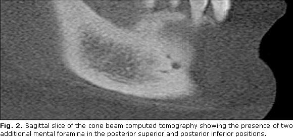
Three-dimensional images were obtained by i-CAT    cone beam computed tomography<U> </U>unit (Imaging Sciences International, Inc,    Hatfield, PA, USA). The sagittal slice (<a href="#fig2">Fig. 2</a>) shows two    bifurcations on the right mandibular canal below the right inferior premolars    originating three exits. Distinct intra-osseous courses could be observed, which    originated three mental foramens separated by bony septa. The additional mental    foramina were located in posterosuperior and posteroinferior positions. On the    left side, all images showed only one foramen without any anatomical variation.    </font>     <P align="center"><img src="/img/revistas/est/v50n4/f0211413.jpg" width="580" height="278"><a name="fig2"></a>     <P>&nbsp;     <P>      ]]></body>
<body><![CDATA[<P><font face="Verdana" size="3"><B>DISCUSSION</B> </font>     <P><font face="Verdana" size="2">The presence of one or more additional foramina    is among the variations described in the literature, which are usually called    mental foramina. It has been assumed that such variation results from the ramification    of the mental nerve before it passes the mental foramen.<SUP>3-7</SUP> It is    important to differentiate the additional mental foramina<U> </U>from a nutritious    foramen. The additional mental foramina is defined as a bony foramen originated    from the mandibular canal. Nutritious foramens, on the other hand, are not originated    from the mandibular canal, and their dimensions are significantly smaller.<SUP>6</SUP>    It was possible to observe that in the present case the anatomical variations    are originated directly from the mandibular canal, being classified as additional    mental foramina. </font>     <P><font face="Verdana" size="2">It is possible to observe in the present case    that, based on the findings of Hu et al.,<SUP>2</SUP> the additional mental    foramina probably carry the ramification that innervates the mental region and    the medial half of inferior lip (mental and medial inferior labial branches,    respectively). The angular and/or lateral inferior labial branches probably    emerge in the posterior superior additional mental foramina, and the mental    branch emerges in the posterior inferior additional mental foramina. This can    be justified due to the both positions of the additional mental foramina presented    in the case report. </font>     <P><font face="Verdana" size="2">This anatomical variation can be detected in    clinical practice using conventional radiographic exams, e.g., periapical and    panoramic radiographs. However, conventional radiographs have several drawbacks,    such as the superimposition of anatomical structures and the distortion and    magnification in panoramic radiography that can lead to errors of identification.    Cone beam computed tomography has been established as a valuable imaging modality    capable of providing in-depth information about maxillofacial structures, which    allows detailed evaluation of their topography and anatomical variations. It    provides reliable high-resolution images and superior technology compared with    its predecessors, and it can generate images with a small slice thickness and    good visualization of bony structures.<SUP>8</SUP> It was possible to observe    this in our case because the presence of the additional mental foramina was    misdiagnosed in the panoramic radiography, being your characteristics (location,    number and position) described only in the cone beam computed tomography. Besides,    we believe it is possible to recognize this anatomical variation when a smaller    voxel size of the cone beam computed tomography is used. </font>     <P><font face="Verdana" size="2">Studies with cone beam computed tomography images    show a similar incidence of additional mental foramina. <I>Oliveira-Santos et    al</I>.<SUP>7</SUP> found 27 out of 285 cases (9.4 %), being two of them bilateral    additional mental foramina<I>. Katakami et al</I>.<SUP>6</SUP> observed 16 out    of 150 cases (10.7 %), with one bilateral case<I>. Naitoh et al</I>.<SUP>4</SUP>    found 11 out of 157 cases (7 %), and two had bilateral occurrence. Using multislice    computed tomography, <I>Haktanir et al</I>.<SUP>5</SUP> observed the presence    of additional mental foramina in 4 out of 100 cases (4 %), with one bilateral    case. Another study conducted by <I>Kalender et al</I>.<SUP>3</SUP> also evaluating    the presence of additional mental foramina on cone beam computed tomography    images showed 27 out of 386 cases (6.5 %). In the present case, the double additional    mental foramina was located only in the right side of the mandible. </font>     <P><font face="Verdana" size="2">The location of the additional mental foramina    were also evaluated and were most commonly located inferior to the mental foramen    (posteroinferior<SUP>4,6,7</SUP> and anterior inferior positions<SUP>3</SUP>).    In the present case, the double additional mental foramina were both located    posterior to the mental foramen (posterior superior and posterior inferior positions).<U>    </U> </font>     <P><font face="Verdana" size="2">Although the most reliable diagnosis of additional    foramina is through direct visualization during surgery, it is not applicable    to clinical practice. Thus, the imaging exams have become extremely important    in detecting such anatomical variations, being helpful for a proper treatment    planning. Some cases are reported in the literature in which the presence of    additional mental foramina is detected on conventional radiographs.<SUP>9,10</SUP>    <I>Ramadhan </I>y otros<SUP>10</SUP> presented a case of two additional mental    foramina in the computed tomography; however, in the panoramic radiography,    it was only observed one foramina. In our case, the additional mental foramina    were only diagnosed by the three-dimensional images. Nevertheless, it has been    demonstrated that two-dimension radiographs may underestimate the presence of    additional mental foramina,<SUP>11</SUP> particularly when their dimensions    are inferior to 1mm. The small sizes of the additional mental foramina presented    in this clinic case, we believe that was the reason for the misdiagnosis in    the panoramic radiography. </font>      <P><font face="Verdana" size="2">Sensorial disturbances, such as paresthesia in    the lower lip or cheeks, may occur as result of pressure on the mental foramen.    Such alterations may be transient or permanent, depending on how much the nerve    is damaged. Cone beam computed tomography examination is very helpful to obtain    information about the maxillofacial structures, trabecular patterns, alveolar    processes, skeletal measurements, and surgical planning of jaw deformities or    implant insertion. It is the most accurate imaging modality for the identification    and localization of the mandibular foramen, mandibular canal, and mental foramen.    The localization of such structures, as well as their eventual anatomical variations,    is of fundamental importance prior to any surgical and anesthetic procedures.    Because the mental nerve supplies the skins of the chin and mucous membrane    of the lower lip and gingiva, in the surgeries of the anterior region of the    mandible, the surgeon should protect this major anatomical structure when the    additional mental foramina are present. </font>     <P><font face="Verdana" size="2">The use of cone beam computed tomography has    increased in dentistry, thus anatomical variations that may have an influence    on the diagnosis and treatment planning must be recognized. Have a good knowledge<FONT COLOR="#424242">    </FONT>of additional mental foramina may contribute to adequate anesthetic techniques    and to avoid misdiagnosis of bone lesions and eventual damages to the nerves    and vessel during surgical procedures in that region. </font>     <P>&nbsp;      ]]></body>
<body><![CDATA[<P>      <P><font face="Verdana" size="3"><B>BIBLIOGRAPHIC REFERENCES</B> </font>      <!-- ref --><P><font face="Verdana" size="2">1. Sawyer DR, Kiely ML, Pyle MA. The frequency    of accessory mental foramina in four ethnic groups. Arch Oral Biol. 1998;43:417-20.        </font>     <!-- ref --><P><font face="Verdana" size="2">2. Hu KS, Yun HS,&#160;Hur MS,&#160;Kwon HJ,&#160;Abe    S,&#160;Kim HJ. Branching patterns and intraosseous course of the mental nerve.    J Oral Maxillofac Surg. 2007;65:2288-94.     </font>     <!-- ref --><P><font face="Verdana" size="2">3. Kalender K, Orhan K, Aksoy U. Evaluation of    the mental foramen and accessory mental foramen in turkish patients using cone-    beam images reconstructed from a volumetric rendering program. Clin Anat. 2011.        Doi: 10.1002/ca.21277. </font>      <!-- ref --><P><font face="Verdana" size="2">4. Naitoh M, Hiraiwa Y,&#160;Aimiya H,&#160;Gotoh    K,&#160;Ariji E. Accessory mental foramen assessment using cone-beam computed    tomography. Oral Surg Oral Med Oral Pathol Oral Radiol Endod. 2009;107:289-94.        </font>     ]]></body>
<body><![CDATA[<!-- ref --><P><font face="Verdana" size="2">5. Haktanir A, Ilgaz K, Turhan-Haktanir N. Evaluation    of mental foramina in adult living crania using MDCT. Surg Radiol Anat. 2010;32:351-6.        </font>     <!-- ref --><P><font face="Verdana" size="2">6. Katakami K, Mishima A, Shiozaki K, Shimoda    S, Hamada Y, Kobayashi K. Characteristics of accessory mental foramina observed    on limited cone-beam computed tomography images. J Endod. 2008;34:1441-5.     </font>     <!-- ref --><P><font face="Verdana" size="2">7. Oliveira-Santos C, Souza PH,&#160;De Azambuja    Berti-Couto S,&#160;Stinkens L,&#160;Moyaert K,&#160;Van Assche N,&#160;Jacobs    R. Characterization of additional mental foramina through cone beam computed    tomography. J Oral Rehabil. 2011;38:595-600.     </font>      <!-- ref --><P><font face="Verdana" size="2">8. Naitoh M, Nakahara K, Suenaga Y,&#160;Gotoh    K,&#160;Kondo S,&#160;Ariji E. Comparison between cone-beam and multislice computed    tomography depicting mandibular neurovascular canal structures. Oral Surg Oral    Med Oral Pathol Oral Radiol Endod. 2010;109:25-31.     </font>     <!-- ref --><P><font face="Verdana" size="2">9. &Ccedil;agiranka LB, Kansu H. Accessory mental    foramen: a case report. J Contemp Dent Pract. 2008;9:98-104.     </font>     ]]></body>
<body><![CDATA[<!-- ref --><P><font face="Verdana" size="2">10. Ramadhan A, Messo E, Hirsch JM. Anatomical    variation of mental foramen. A case report. Stomatologija. 2010;12:93-6.     </font>     <!-- ref --><P><font face="Verdana" size="2">11. Serman NJ. Differentiation of double mental    foramina from extra bony coursing of the incisive branch of the mandibular nerve&#151;an    anatomic study. Refuat Hashinayim. 1987; 5:20-2.     </font>     <P>&nbsp;     <P>&nbsp;      <P>      <P>      <P><font face="Verdana" size="2">Recibido: 11 de julio de 2013.    <br>   Aprobado:</font> <font face="Verdana" size="2">17 de septiembre de 2013.</font>     ]]></body>
<body><![CDATA[<P>&nbsp;     <P>&nbsp;     <P>      <P>      <P>      <P>      <P><font face="Verdana" size="2"><I>Taruska Ventorini Vasconcelos. </I>Av. Limeira,    901, Piracicaba, S&atilde;o Paulo, Brasil. correo electr&oacute;nico: <U><a href="mailto:tataventorini@hotmail.com">tataventorini@hotmail.com</a></U></font>      ]]></body><back>
<ref-list>
<ref id="B1">
<label>1</label><nlm-citation citation-type="journal">
<person-group person-group-type="author">
<name>
<surname><![CDATA[Sawyer]]></surname>
<given-names><![CDATA[DR]]></given-names>
</name>
<name>
<surname><![CDATA[Kiely]]></surname>
<given-names><![CDATA[ML]]></given-names>
</name>
<name>
<surname><![CDATA[Pyle]]></surname>
<given-names><![CDATA[MA]]></given-names>
</name>
</person-group>
<article-title xml:lang="en"><![CDATA[The frequency of accessory mental foramina in four ethnic groups]]></article-title>
<source><![CDATA[Arch Oral Biol]]></source>
<year>1998</year>
<volume>43</volume>
<page-range>417-20</page-range></nlm-citation>
</ref>
<ref id="B2">
<label>2</label><nlm-citation citation-type="journal">
<person-group person-group-type="author">
<name>
<surname><![CDATA[Hu]]></surname>
<given-names><![CDATA[KS]]></given-names>
</name>
<name>
<surname><![CDATA[Yun]]></surname>
<given-names><![CDATA[HS]]></given-names>
</name>
<name>
<surname><![CDATA[Hur]]></surname>
<given-names><![CDATA[MS]]></given-names>
</name>
<name>
<surname><![CDATA[Kwon]]></surname>
<given-names><![CDATA[HJ]]></given-names>
</name>
<name>
<surname><![CDATA[Abe]]></surname>
<given-names><![CDATA[S]]></given-names>
</name>
<name>
<surname><![CDATA[Kim]]></surname>
<given-names><![CDATA[HJ]]></given-names>
</name>
</person-group>
<article-title xml:lang="en"><![CDATA[Branching patterns and intraosseous course of the mental nerve]]></article-title>
<source><![CDATA[J Oral Maxillofac Surg]]></source>
<year>2007</year>
<volume>65</volume>
<page-range>2288-94</page-range></nlm-citation>
</ref>
<ref id="B3">
<label>3</label><nlm-citation citation-type="journal">
<person-group person-group-type="author">
<name>
<surname><![CDATA[Kalender]]></surname>
<given-names><![CDATA[K]]></given-names>
</name>
<name>
<surname><![CDATA[Orhan]]></surname>
<given-names><![CDATA[K]]></given-names>
</name>
<name>
<surname><![CDATA[Aksoy]]></surname>
<given-names><![CDATA[U]]></given-names>
</name>
</person-group>
<article-title xml:lang="en"><![CDATA[Evaluation of the mental foramen and accessory mental foramen in turkish patients using cone- beam images reconstructed from a volumetric rendering program]]></article-title>
<source><![CDATA[Clin Anat]]></source>
<year>2011</year>
</nlm-citation>
</ref>
<ref id="B4">
<label>4</label><nlm-citation citation-type="journal">
<person-group person-group-type="author">
<name>
<surname><![CDATA[Naitoh]]></surname>
<given-names><![CDATA[M]]></given-names>
</name>
<name>
<surname><![CDATA[Hiraiwa]]></surname>
<given-names><![CDATA[Y]]></given-names>
</name>
<name>
<surname><![CDATA[Aimiya]]></surname>
<given-names><![CDATA[H]]></given-names>
</name>
<name>
<surname><![CDATA[Gotoh]]></surname>
<given-names><![CDATA[K]]></given-names>
</name>
<name>
<surname><![CDATA[Ariji]]></surname>
<given-names><![CDATA[E]]></given-names>
</name>
</person-group>
<article-title xml:lang="en"><![CDATA[Accessory mental foramen assessment using cone-beam computed tomography]]></article-title>
<source><![CDATA[Oral Surg Oral Med Oral Pathol Oral Radiol Endod]]></source>
<year>2009</year>
<volume>107</volume>
<page-range>289-94</page-range></nlm-citation>
</ref>
<ref id="B5">
<label>5</label><nlm-citation citation-type="journal">
<person-group person-group-type="author">
<name>
<surname><![CDATA[Haktanir]]></surname>
<given-names><![CDATA[A]]></given-names>
</name>
<name>
<surname><![CDATA[Ilgaz]]></surname>
<given-names><![CDATA[K]]></given-names>
</name>
<name>
<surname><![CDATA[Turhan-Haktanir]]></surname>
<given-names><![CDATA[N]]></given-names>
</name>
</person-group>
<article-title xml:lang="en"><![CDATA[Evaluation of mental foramina in adult living crania using MDCT]]></article-title>
<source><![CDATA[Surg Radiol Anat]]></source>
<year>2010</year>
<volume>32</volume>
<page-range>351-6</page-range></nlm-citation>
</ref>
<ref id="B6">
<label>6</label><nlm-citation citation-type="journal">
<person-group person-group-type="author">
<name>
<surname><![CDATA[Katakami]]></surname>
<given-names><![CDATA[K]]></given-names>
</name>
<name>
<surname><![CDATA[Mishima]]></surname>
<given-names><![CDATA[A]]></given-names>
</name>
<name>
<surname><![CDATA[Shiozaki]]></surname>
<given-names><![CDATA[K]]></given-names>
</name>
<name>
<surname><![CDATA[Shimoda]]></surname>
<given-names><![CDATA[S]]></given-names>
</name>
<name>
<surname><![CDATA[Hamada]]></surname>
<given-names><![CDATA[Y]]></given-names>
</name>
<name>
<surname><![CDATA[Kobayashi]]></surname>
<given-names><![CDATA[K]]></given-names>
</name>
</person-group>
<article-title xml:lang="en"><![CDATA[Characteristics of accessory mental foramina observed on limited cone-beam computed tomography images]]></article-title>
<source><![CDATA[J Endod]]></source>
<year>2008</year>
<volume>34</volume>
<page-range>1441-5</page-range></nlm-citation>
</ref>
<ref id="B7">
<label>7</label><nlm-citation citation-type="journal">
<person-group person-group-type="author">
<name>
<surname><![CDATA[Oliveira-Santos]]></surname>
<given-names><![CDATA[C]]></given-names>
</name>
<name>
<surname><![CDATA[Souza]]></surname>
<given-names><![CDATA[PH]]></given-names>
</name>
<name>
<surname><![CDATA[De Azambuja Berti-Couto]]></surname>
<given-names><![CDATA[S]]></given-names>
</name>
<name>
<surname><![CDATA[Stinkens]]></surname>
<given-names><![CDATA[L]]></given-names>
</name>
<name>
<surname><![CDATA[Moyaert]]></surname>
<given-names><![CDATA[K]]></given-names>
</name>
<name>
<surname><![CDATA[Van Assche]]></surname>
<given-names><![CDATA[N]]></given-names>
</name>
<name>
<surname><![CDATA[Jacobs]]></surname>
<given-names><![CDATA[R]]></given-names>
</name>
</person-group>
<article-title xml:lang="en"><![CDATA[Characterization of additional mental foramina through cone beam computed tomography]]></article-title>
<source><![CDATA[J Oral Rehabil]]></source>
<year>2011</year>
<volume>38</volume>
<page-range>595-600</page-range></nlm-citation>
</ref>
<ref id="B8">
<label>8</label><nlm-citation citation-type="journal">
<person-group person-group-type="author">
<name>
<surname><![CDATA[Naitoh]]></surname>
<given-names><![CDATA[M]]></given-names>
</name>
<name>
<surname><![CDATA[Nakahara]]></surname>
<given-names><![CDATA[K]]></given-names>
</name>
<name>
<surname><![CDATA[Suenaga]]></surname>
<given-names><![CDATA[Y]]></given-names>
</name>
<name>
<surname><![CDATA[Gotoh]]></surname>
<given-names><![CDATA[K]]></given-names>
</name>
<name>
<surname><![CDATA[Kondo]]></surname>
<given-names><![CDATA[S]]></given-names>
</name>
<name>
<surname><![CDATA[Ariji]]></surname>
<given-names><![CDATA[E]]></given-names>
</name>
</person-group>
<article-title xml:lang="en"><![CDATA[Comparison between cone-beam and multislice computed tomography depicting mandibular neurovascular canal structures]]></article-title>
<source><![CDATA[Oral Surg Oral Med Oral Pathol Oral Radiol Endod]]></source>
<year>2010</year>
<volume>109</volume>
<page-range>25-31</page-range></nlm-citation>
</ref>
<ref id="B9">
<label>9</label><nlm-citation citation-type="journal">
<person-group person-group-type="author">
<name>
<surname><![CDATA[Çagiranka]]></surname>
<given-names><![CDATA[LB]]></given-names>
</name>
<name>
<surname><![CDATA[Kansu]]></surname>
<given-names><![CDATA[H]]></given-names>
</name>
</person-group>
<article-title xml:lang="en"><![CDATA[Accessory mental foramen: a case report]]></article-title>
<source><![CDATA[J Contemp Dent Pract]]></source>
<year>2008</year>
<volume>9</volume>
<page-range>98-104</page-range></nlm-citation>
</ref>
<ref id="B10">
<label>10</label><nlm-citation citation-type="journal">
<person-group person-group-type="author">
<name>
<surname><![CDATA[Ramadhan]]></surname>
<given-names><![CDATA[A]]></given-names>
</name>
<name>
<surname><![CDATA[Messo]]></surname>
<given-names><![CDATA[E]]></given-names>
</name>
<name>
<surname><![CDATA[Hirsch]]></surname>
<given-names><![CDATA[JM]]></given-names>
</name>
</person-group>
<article-title xml:lang="en"><![CDATA[Anatomical variation of mental foramen A case report]]></article-title>
<source><![CDATA[Stomatologija]]></source>
<year>2010</year>
<volume>12</volume>
<page-range>93-6</page-range></nlm-citation>
</ref>
<ref id="B11">
<label>11</label><nlm-citation citation-type="journal">
<person-group person-group-type="author">
<name>
<surname><![CDATA[Serman]]></surname>
<given-names><![CDATA[NJ]]></given-names>
</name>
</person-group>
<article-title xml:lang="en"><![CDATA[Differentiation of double mental foramina from extra bony coursing of the incisive branch of the mandibular nerve-an anatomic study]]></article-title>
<source><![CDATA[Refuat Hashinayim]]></source>
<year>1987</year>
<volume>5</volume>
<page-range>20-2</page-range></nlm-citation>
</ref>
</ref-list>
</back>
</article>
