<?xml version="1.0" encoding="ISO-8859-1"?><article xmlns:mml="http://www.w3.org/1998/Math/MathML" xmlns:xlink="http://www.w3.org/1999/xlink" xmlns:xsi="http://www.w3.org/2001/XMLSchema-instance">
<front>
<journal-meta>
<journal-id>0253-570X</journal-id>
<journal-title><![CDATA[Revista de Salud Animal]]></journal-title>
<abbrev-journal-title><![CDATA[Rev Salud Anim.]]></abbrev-journal-title>
<issn>0253-570X</issn>
<publisher>
<publisher-name><![CDATA[Centro Nacional de Sanidad Agropecuaria]]></publisher-name>
</publisher>
</journal-meta>
<article-meta>
<article-id>S0253-570X2008000200003</article-id>
<title-group>
<article-title xml:lang="en"><![CDATA[MOLECULAR CLONING AND EXPRESSION OF A FRAGMENT OF THE GENE CODIFYING FOR THE PROTEIN ERNS OF CLASSICAL SWINE FEVER VIRUS]]></article-title>
<article-title xml:lang="es"><![CDATA[CLONAJE MOLECULAR Y EXPRESIÓN DE UN FRAGMENTO DEL GEN QUE CODIFICA PARA LA PROTEÍNA DEL VIRUS DE LA PESTE PORCINA CLÁSICA, ERNS]]></article-title>
</title-group>
<contrib-group>
<contrib contrib-type="author">
<name>
<surname><![CDATA[Agüero]]></surname>
<given-names><![CDATA[J.A]]></given-names>
</name>
<xref ref-type="aff" rid="A01"/>
</contrib>
<contrib contrib-type="author">
<name>
<surname><![CDATA[Sánchez]]></surname>
<given-names><![CDATA[O]]></given-names>
</name>
<xref ref-type="aff" rid="A02"/>
</contrib>
<contrib contrib-type="author">
<name>
<surname><![CDATA[Barrera]]></surname>
<given-names><![CDATA[Maritza]]></given-names>
</name>
<xref ref-type="aff" rid="A01"/>
</contrib>
<contrib contrib-type="author">
<name>
<surname><![CDATA[Toledo]]></surname>
<given-names><![CDATA[J.R]]></given-names>
</name>
<xref ref-type="aff" rid="A02"/>
</contrib>
</contrib-group>
<aff id="A01">
<institution><![CDATA[,National Center for Animal and Planth Health(CENSA).  ]]></institution>
<addr-line><![CDATA[La Habana ]]></addr-line>
</aff>
<aff id="A02">
<institution><![CDATA[,Centre for Genetic Engineering and Biotechnology (CIGB)  ]]></institution>
<addr-line><![CDATA[Habana ]]></addr-line>
</aff>
<pub-date pub-type="pub">
<day>00</day>
<month>08</month>
<year>2008</year>
</pub-date>
<pub-date pub-type="epub">
<day>00</day>
<month>08</month>
<year>2008</year>
</pub-date>
<volume>30</volume>
<numero>2</numero>
<fpage>85</fpage>
<lpage>92</lpage>
<copyright-statement/>
<copyright-year/>
<self-uri xlink:href="http://scielo.sld.cu/scielo.php?script=sci_arttext&amp;pid=S0253-570X2008000200003&amp;lng=en&amp;nrm=iso"></self-uri><self-uri xlink:href="http://scielo.sld.cu/scielo.php?script=sci_abstract&amp;pid=S0253-570X2008000200003&amp;lng=en&amp;nrm=iso"></self-uri><self-uri xlink:href="http://scielo.sld.cu/scielo.php?script=sci_pdf&amp;pid=S0253-570X2008000200003&amp;lng=en&amp;nrm=iso"></self-uri><abstract abstract-type="short" xml:lang="en"><p><![CDATA[Classical swine fever virus (CSFV), belonging to the genus Pestivirus of the Flaviviridae family, is an enveloped positive stranded RNA virus highly contagious that can cause a fatal disease, characterized by fever, leukopenia and hemorrhage, with substantial economic losses. There is a great demand for a marker vaccine against CSFV. C, Erns, E1, and E2 are the structural proteins of the virus. E2 is the best candidate to be incorporated in vaccine, while Erns becomes an ideal candidate as an antigen in a differential diagnostic test. A synthetic fragment of the Erns gene (codifying for aa 109-160) was subcloned into pET28a vector. The cloning was screened by restriction analysis. The gene was expressed as a his-tag fusion protein in BL21 (DE3) E. coli strain. The recombinant polypeptide formed aggregates of about 7.9; 15.8; 23.7; 31.6; 39.5; 47.5 and 55.3kDa. The protein was not recognized by Western blot using an antibody against the virus. Using an ion metal affinity chromatography procedure, a 90% pure recombinant product was obtained. The potential use of this antigen for detection of CSFV antibodies should be further evaluated.]]></p></abstract>
<abstract abstract-type="short" xml:lang="es"><p><![CDATA[El virus de la fiebre porcina clásica (CSFV), perteneciente al género Pestivirus de la familia Flaviviridae, es un virus ARN positivo, altamente contagioso capaz de causar una enfermedad fatal caracterizada por fiebre, leucopenia y hemorragia, con considerables pérdidas económicas. Existe una gran demanda de una vacuna marcadora contra CSFV. C, Erns, E1 and E2 son las proteínas estructurales del virus. E2 es el mejor candidato para ser incorporado a una vacuna, mientras Erns resulta entonces un candidato ideal como antígeno en un ensayo de diagnóstico diferencial. En el vector pET28a se subclonó un fragmento sintético del gene que codifica para Erns (codificante para los aa 109-160). El clonaje se monitoreó por análisis de restricción. El gen se expresó como una proteína de fusión a histidina en la cepa BL21(DE3) de E. coli. El polipéptido recombinante formó agregados de aproximadamente 7.9; 15.8; 23.7; 31.6; 39.5; 47.5 y 55.3kDa. La proteína no fue reconocida por anticuerpos contra el virus por Western blot. Con el empleo de la cromatografía de afinidad a iones metálicos se obtuvo un producto recombinante con un 90% de pureza. Más adelante debe ser evaluado el empleo potencial de este antígeno para la detección de anticuerpos contra CSFV.]]></p></abstract>
<kwd-group>
<kwd lng="en"><![CDATA[Erns]]></kwd>
<kwd lng="en"><![CDATA[CSFV]]></kwd>
<kwd lng="en"><![CDATA[recombinant protein]]></kwd>
<kwd lng="en"><![CDATA[IMAC]]></kwd>
<kwd lng="es"><![CDATA[Erns]]></kwd>
<kwd lng="es"><![CDATA[CSFV]]></kwd>
<kwd lng="es"><![CDATA[proteína recombinante]]></kwd>
<kwd lng="es"><![CDATA[IMAC]]></kwd>
</kwd-group>
</article-meta>
</front><body><![CDATA[ <p align="right"><font face="Verdana, Arial, Helvetica, sans-serif" size="2"><b>Trabajo    original</b>     <BR>   </font></p>     <p align="left">&nbsp;</p>     <p align="left"><font face="Verdana, Arial, Helvetica, sans-serif" size="4"><b>MOLECULAR    CLONING AND EXPRESSION OF A FRAGMENT OF THE GENE CODIFYING FOR THE PROTEIN ERNS    OF CLASSICAL SWINE FEVER VIRUS</b></font></p>     <p align="left">&nbsp;</p>     <p><b><font face="Verdana, Arial, Helvetica, sans-serif" size="3">CLONAJE MOLECULAR    Y EXPRESI&Oacute;N DE UN FRAGMENTO DEL GEN QUE CODIFICA PARA LA PROTE&Iacute;NA    DEL VIRUS DE LA PESTE PORCINA CL&Aacute;SICA, ERNS</font></b></p>     <p>&nbsp;</p>     <p>&nbsp;</p> <B>     <P><font face="Verdana, Arial, Helvetica, sans-serif" size="2">J.A. Ag&uuml;ero<SUP>*&#134;</SUP>,    O. S&aacute;nchez**, Maritza Barrera* y J.R. Toledo**</font>  </B>      <P> <font face="Verdana, Arial, Helvetica, sans-serif" size="2"><I>*National Centre    for Animal and Planth Health (CENSA). Carretera de Jamaica y Autopista Nacional.    Apto.10. San Jos&eacute; de las Lajas. La Habana. CP. 32700. Cuba.<font face="Verdana, Arial, Helvetica, sans-serif" size="2">Corresponding    author:</font> <font face="Verdana, Arial, Helvetica, sans-serif" size="2">jaaguero@censa.edu.cu</font></I>    <I>**Centre for Genetic Engineering and Biotechnology (CIGB). Ave 31 e/ 158    y 190 Playa. P.O. Box 6162. Habana 10600. </I></font>      ]]></body>
<body><![CDATA[<P>&nbsp;     <P>&nbsp; <hr noshade size="1">     <P><font face="Verdana, Arial, Helvetica, sans-serif" size="2"><B>ABSTRACT</B></font>     <P><font face="Verdana, Arial, Helvetica, sans-serif" size="2">Classical swine    fever virus (CSFV), belonging to the genus Pestivirus of the Flaviviridae family,    is an enveloped positive stranded RNA virus highly contagious that can cause    a fatal disease, characterized by fever, leukopenia and hemorrhage, with substantial    economic losses. There is a great demand for a marker vaccine against CSFV.    C, Erns, E1, and E2 are the structural proteins of the virus. E2 is the best    candidate to be incorporated in vaccine, while Erns becomes an ideal candidate    as an antigen in a differential diagnostic test. A synthetic fragment of the    Erns gene (codifying for aa 109-160) was subcloned into pET28a vector. The cloning    was screened by restriction analysis. The gene was expressed as a his-tag fusion    protein in BL21 (DE3) <I>E. coli</I> strain. The recombinant polypeptide formed    aggregates of about 7.9; 15.8; 23.7; 31.6; 39.5; 47.5 and 55.3kDa. The protein    was not recognized by Western blot using an antibody against the virus. Using    an ion metal affinity chromatography procedure, a 90% pure recombinant product    was obtained. The potential use of this antigen for detection of CSFV antibodies    should be further evaluated. </font>       <P><font face="Verdana, Arial, Helvetica, sans-serif" size="2"><b>Key words:</b>    Erns; CSFV; recombinant protein; IMAC</font> <hr noshade size="1">     <P><font face="Verdana, Arial, Helvetica, sans-serif" size="2"><b>RESUMEN</b></font>     <P><font face="Verdana, Arial, Helvetica, sans-serif" size="2">El virus de la    fiebre porcina cl&aacute;sica (CSFV), perteneciente al g&eacute;nero <I>Pestivirus    </I>de la familia <I>Flaviviridae</I>, es un virus ARN positivo, altamente contagioso    capaz de causar una enfermedad fatal caracterizada por fiebre, leucopenia y    hemorragia, con considerables p&eacute;rdidas econ&oacute;micas. Existe una    gran demanda de una vacuna marcadora contra CSFV. C, Erns, E1 and E2 son las    prote&iacute;nas estructurales del virus. E2 es el mejor candidato para ser    incorporado a una vacuna, mientras Erns resulta entonces un candidato ideal    como ant&iacute;geno en un ensayo de diagn&oacute;stico diferencial. En el vector    pET28a se subclon&oacute; un fragmento sint&eacute;tico del gene que codifica    para Erns (codificante para los aa 109-160). El clonaje se monitore&oacute;    por an&aacute;lisis de restricci&oacute;n. El gen se expres&oacute; como una    prote&iacute;na de fusi&oacute;n a histidina en la cepa BL21(DE3) de <I>E. coli</I>.    El polip&eacute;ptido recombinante form&oacute; agregados de aproximadamente    7.9; 15.8; 23.7; 31.6; 39.5; 47.5 y 55.3kDa. La prote&iacute;na no fue reconocida    por anticuerpos contra el virus por Western blot. Con el empleo de la cromatograf&iacute;a    de afinidad a iones met&aacute;licos se obtuvo un producto recombinante con    un 90% de pureza. M&aacute;s adelante debe ser evaluado el empleo potencial    de este ant&iacute;geno para la detecci&oacute;n de anticuerpos contra CSFV.</font>  <B></B>      <P><font face="Verdana, Arial, Helvetica, sans-serif" size="2"><b>Palabras clave:</b>    Erns; CSFV; prote&iacute;na recombinante; IMAC</font> <hr noshade size="1">     <P>&nbsp;     <P>&nbsp;     ]]></body>
<body><![CDATA[<P><font face="Verdana, Arial, Helvetica, sans-serif" size="2"><b><font size="3">INTRODUCTION</font></b>    </font>     <p><font face="Verdana, Arial, Helvetica, sans-serif" size="2">Classical swine    fever virus (CSFV), Bovine viral diarrhoea virus (BVDV) and Border disease virus<i>    </i>(BDV) belong to the genus<i> Pestivirus </i>of the <i>Flaviviridae </i>family    (4). CSFV is highly contagious and can cause a fatal disease in pigs. Although    pigs can be infected by all these pestiviruses (16; 22), only CSFV induces severe    disease and it is often fatal. The disease is characterized by fever, leukopenia    and hemorrhage, and can run an acute, chronic, or subclinical course substantial    economic losses (18). In several countries, classical swine fever (CSF) outbreaks    are controlled by the eradication of all pigs from infected and neighbouring    farms. Although effective live-attenuated vaccines are available, sometimes    they are not used due to the fact that vaccinated and infected pigs are serologically    indistinguishable. That is why there is a great demand for a marker vaccine    against CSFV. </font>     <p><font face="Verdana, Arial, Helvetica, sans-serif" size="2">Like other members    of the family, CSFV is an enveloped positive stranded RNA virus (13, 3). Similar    to other members of the genus, the 12.5-kb CSFV genome contains a single large    open reading frame encoding a polyprotein precursor of approximately 4,000 amino    acids (aa). The precursor is cleaved co- and post-translationally by cellular    and viral proteases into structural proteins C, Erns, E1, and E2 and nonstructural    proteins NS2, NS3, NS4A, NS4B, NS5A, and NS5B (19). </font>     <p><font face="Verdana, Arial, Helvetica, sans-serif" size="2">Antibodies against    Erns, E2, and NS3 have been demonstrated in infected animals (7; 10; 14; 15;    16), with Erns and E2 capable of inducing neutralizing antibodies (5; 6; 24).    Glycoprotein E2 is the best candidate to be incorporated in vaccine because    it is the most immunogenic protein of pestiviruses and elicits high titers of    neutralizing antibodies after infection (20; 25). Additionally, vaccination    of pigs with E2 has been shown to give protection against a lethal homologous    challenge (1; 5). In this scenario, Erns becomes an ideal candidate as an antigen    in a differential diagnostic test. It has been shown that antibodies to full-length    Erns or even a 37-mer peptide derived from its C-terminal end (aa 191 to 227)    could be used as an indicator of CSFV infection in pigs (7; 14). Recent studies    have indicated that an Erns-based enzymelinked immunosorbent assay (ELISA) can    be used as a companion diagnostic test to identify CSFV-infected pigs in herds    vaccinated with the E2 subunit marker vaccine (14). Recently, an immunodominant    region encompassing three overlapping antigenic regions that induce antibody    responses during CSFV infection: aa 65 to 145, aa 84 to 160 , and aa 109 to    220 was mapped (11) and antibody responses of pigs to defined Erns fragments    (selected from the above mentioned regions), after classical swine fever virus    (CSFV) infection, were studied by using an enzyme-linked immunosorbent assay    (ELISA). In this study, Erns aa 109-145 and Erns aa 109-160 offered the best    specificity and sensitivity (Lin et al., 2005). </font>     <p><font face="Verdana, Arial, Helvetica, sans-serif" size="2">In this work, the    molecular cloning of the DNA segment encoding for the Erns aa 109-160 fragment    is described, as well as its expression and the purification of the product    obtained. </font>     <p>&nbsp;     <p><font face="Verdana, Arial, Helvetica, sans-serif" size="2"><b><font size="3">MATERIALS    AND METHODS </font></b> </font>     <p><font face="Verdana, Arial, Helvetica, sans-serif" size="2"><b>Vectors, bacterial    strains and growth conditions.</b> The XL1 Blue competent cells, as well as    BL21(DE3) competent were obtained from the strain bank of the Centre for Genetic    Engineering and Biotechnology (CIGB) (La Habana, Cuba). The plasmid 050778pPCR-Script    containing the synthetic fragment from the Erns gene fragment, codifying for    the Erns aa 109-160 polypeptide (Brescia strain), was provided by GeneArt. The    fragment was designed in a way that its sequence was optimized for expression    in <i>E.coli</i>, additionally <i>Nde</i> I and <i>BamH</i> I restriction sites    were introduced in its extremes 5&#180;and 3&#180;, for its subsequent, in frame,    cloning in the expression vector pET28a. Competent XL1-Blue cells were separately    transformed with both plasmids and growth O/N at 37&#176;C in Luria-Bertani    (LB) medium supplemented with 100&#181;g of ampicillin/mL (050778pPCR-Script)    and 50&#181;g of kanamycin/mL (pET28a). </font>     <p><font face="Verdana, Arial, Helvetica, sans-serif" size="2"><b>Vector Preparation.</b>    The pET28a plasmid (Novagen) was purified using the Wizard Plus SV Minipreps    DNA Purification System (Promega). 5&#181;g of the vector were digested with    50u each of <i>Nde</i> I and <i>BamH</i> I (Promega) in 100&#181;L of total    volume and in presence of 0.1mg/mL BSA and 10&#181;L of MULTI-CORE<sup>TM</sup>    buffer, the digestion was carried out for 3 hours at 37&#176;C. The product    digested was applied in a low melting point agarose gel at 0.8% from where,    after electrophoresis (constant voltage 100V; in TB buffer), the 5329bp band    corresponding to digested pET28 plasmid, was isolated. This band was purified    using the QIAquick Gel Extraction Kit (QIAGEN). </font>     <p><font face="Verdana, Arial, Helvetica, sans-serif" size="2">When kits where    used, all the procedures were carried out according to the manufacturer's instructions.    </font>     ]]></body>
<body><![CDATA[<p><font face="Verdana, Arial, Helvetica, sans-serif" size="2"><b>Recombinant    plasmid generation.</b> The 050778pPCR-Script plasmid was digested with <i>Nde</i>    I and <i>BamH</i> I restriction enzymes to obtain the Erns gene fragment. For    this, 5&#181;g of the plasmid were digested with 50u each of <i>Nde </i>I and    <i>BamH</i> I (Promega) in 100&#181;L of total volumen and in presence of 0.1mg/mL    BSA and 10&#181;L of MULTI-CORE<sup>TM</sup> buffer; the digestion was carried    out for 3 hours at 37&#176;C. The product digested was applied in a low melting    point agarose gel at 1.5% from where, after electrophoresis, the 173bp band    corresponding to the Erns gene fragment, was isolated. This digestion product    was purified using the QIAquick Gel Extraction Kit (QIAGEN). </font>     <p><font face="Verdana, Arial, Helvetica, sans-serif" size="2">To obtain the construction    pET28a-Erns, the previously digested Erns gene fragment and pET28a vector were    ligated. In the ligation mix the vector and insert concentrations were 31ng/mL    and 5ng/mL, respectively. 4u of T4 ligase (Promega), 2mL of T4 ligasa 10x buffer    (Promega) were employed in a 20mL of total volumen and the reaction was carried    out at 22&#176;C, for 5h. </font>     <p><font face="Verdana, Arial, Helvetica, sans-serif" size="2">For transformation,    Ca<sup>2+</sup> competent cells from XL1-Blue of <i>E. coli</i> were employed,    and transformed as follow: 10&#181;L of the ligation product were added to 150&#181;L    of competent cells. The mix was incubated in ice for 20 min, then 2 min at 42&#176;C    and to finish 5 min again in ice. 1mL of LB medium was added to the mix and    this was incubated 1h at 37&#176;C at 50rpm in an orbital shaker. This culture    was inoculated to LB plates supplemented with 50&#181;g of kanamycin/mL and    the plates were incubated O/N at 37&#176;C. </font>     <p><font face="Verdana, Arial, Helvetica, sans-serif" size="2">The screening of    recombinant clones was done by restriction analysis with <i>Nde </i>I and <i>BamH</i>    I restriction enzymes. </font>     <p><font face="Verdana, Arial, Helvetica, sans-serif" size="2"><b>Expression of    recombinant Erns fragment. </b>For the expression experiments, the method of    autoinduction developed by Studier (2005) (21) was employed with some modifications.    <i>E. coli </i>BL21(DE3) cells harboring the expression construct pET28a-Erns-1    were cultured O/N at 37&#176;C in a Luria-Bertani broth supplemented with 50&#181;g    of kanamycin/mL. Next day, 100mL (in a 1L erlenmeyer) of auto-induction media    (1% triptone; 0.5% yeast extract; 25mM Na<sub>2</sub>HPO<sub>4</sub>; 25mM KH<sub>2</sub>PO<sub>4</sub>;    50mM NH<sub>4</sub>Cl; 5mM Na<sub>2</sub>SO<sub>4</sub>; 0.5% glicerol; 0.05%    glucose; 0.2% alfa-lactose monohydrate; 2mM MgSO<sub>4</sub> heptahydrate; 0.05mM    FeCl<sub>3</sub>) supplemented with 200&#181;g of kanamycin/mL were inoculated    with 10&#181;L of the O/N inoculum and grown for 16 hours at 37&#176;C with    shaking at 310rpm,. The cells were harvested by centrifugation at 5,000 x <i>g    </i>for 10 min and conserved at -20&#176;C. </font>     <p><font face="Verdana, Arial, Helvetica, sans-serif" size="2"><b>Purification    of recombinant proteins by metal ion affinity chromatography (IMAC). </b>Cell    pellets from cultures were resuspended in 10mL of protein extraction buffer    (6M guanidine hydrochloride, 0.1M Na-Phosfate buffer, [pH 8.0]) and lysed with    a French press at 1,500 kgf/cm<sup>2</sup>. The homogenates were spun at 12,000    x <i>g </i>for 30 min at 4&#176;C, and the supernatant was filtered by 0.45&#181;m    and then loaded onto a Protein Purification Resin (Promega), previously equilibrated    with the extraction buffer column (1 by 1 cm) of HisLink&#153;. The column was    washed with buffer A (8M urea, 0.1M Na-Phosfate buffer [pH 8.0]), followed by    buffer B (buffer A plus 0.5 M NaCl [pH 6.3]) and buffer C (buffer B plus 5 mM    imidazole [pH 5.9]). The denatured protein was then refolded on the column by    washes with Tris-buffered saline (Na-Phosfate buffer) (pH 7.4) containing 1    M urea, followed by Na-Phosfate buffer (pH 7.4). The recombinant protein was    eluted and collected with fractions of 10 ml from the column using Na-Phosfate    buffer (pH 7.4) containing 500 mM imidazole. A final washing step was done using    Na-Phosfate buffer (pH 7.4) containing 500 mM imidazole and 8 M urea. The percent    of Erns fragment in each fraction was determined by densitometry of the Coomassie    Blue stained gels with the Molecular Analyst software. Proteins were quantified    by using the Bradford method (2), with bovine serum albumin as a standard. </font>     <p><font face="Verdana, Arial, Helvetica, sans-serif" size="2"><b>Sera.</b> The    sera from pigs experimentally infected with CSFV were provided by the Virology    group of the National Centre for Animal and Plant Health (CENSA) (Cuba). </font>     <p><font face="Verdana, Arial, Helvetica, sans-serif" size="2"><b>SDS-PAGE and    Western blot.</b> Briefly, SDS-PAGE was performed at constant current of 30mA    in a 15% acrylamide gels. The proteins were analyzed either through staining    with Coomassie Blue or by Western blotting. From the 10mL fractions, 10&#181;L    aliquots were taken and analized by SDS-PAGE and Western Blotting. For Western    blots, the proteins were transferred to nitrocellulose membranes using semi-dry    transfer equipment. The membranes were blocked for 1 hour with PBS-BSA 1%. The    blots were probed with swine CSFV antisera from experimentally infected pigs    for 2 hours at 37&#176;C. Bound antibodies were detected by using horseradish    peroxidase (HRP)_conjugated anti_swine IgG antibodies (Sigma) and a 4-chloro-1-    naphthol-H<sub>2</sub>O<sub>2</sub> substrate kit (Bio-Rad), according to the    manufacturer's instructions. </font>     <p>&nbsp;     <p><font face="Verdana, Arial, Helvetica, sans-serif" size="2"><b><font size="3">RESULTS    AND DISCUSSION</font></b> </font>     ]]></body>
<body><![CDATA[<p><font face="Verdana, Arial, Helvetica, sans-serif" size="2">The focus of this    study is the obtainment of a recombinant fragment of the CSFV Erns structural    protein. This polypeptide (Erns aa 109-160) had been previously described as    good antigen candidate for detection of antibodies against the virus, offering,    together with Erns aa 109-145 fragment, the best specificity and sensitivitiy    measured by ELISA, among other fragments from an immunodominant Erns region    (12). In particular, antibody responses to Erns aa 109-160 can be detected as    early as 7 dpi, then this small Erns fragment would offer a diagnostic antigen    superior to the full-length protein, which can only be used to detect CSFV-specific    antibodies in vaccinated or unvaccinated pigs as early as 14 dpi (14). </font>     <p><font face="Verdana, Arial, Helvetica, sans-serif" size="2"> The 173bp band    corresponding to the Erns aa 109-160 gene fragment was sussesfully isolated    from plasmid 050778pPCR-Script (results are not shown) and subcloned in the    pET28a vector using the <i>Nde</i> I and <i>BamH</i> I restriction sites introduced    in the synthetic gene. Four positive recombinant clones were detected by restriction    analysis. </font>     <p><font face="Verdana, Arial, Helvetica, sans-serif" size="2">The initial transformation    was done in XL1-Blue cells of <i>E. coli</i> because this strain facilitates    the propagation and subsequent purification of plasmids, but for protein production,    in order to quarantee greater control and a higher level of the gene expression    it was decided to use BL21(DE3) cells of <i>E. coli</i>, which were transformed    with the four recombinant plasmids. In pET plasmids, the gene expression is    under the control of the T7 RNA polymerase. This enzyme is so specific, active,    and processive that the amount of target RNA produced can be comparable to the    amount of ribosomal RNA in a cell. A problem in using inducible T7 expression    systems is that T7 RNA polymerase is so active that a small basal level can    lead to substantial expression of target protein even in the absence of added    inducer. If the target protein is sufficiently toxic to the host cell, establishment    of the target plasmid in the expression host may be difficult or impossible,    or the expression strain may be unstable or accumulate mutations. In BL21(DE3)-pET    system, this problem is overcome, the lac operator sequence (the binding site    for lac repressor) is placed in both, the start site of a T7 promoter (of the    pET plasmid) and in the lac promoter, under the control of which is the T7 RNA    polimerase in cell genomes. In this way, the basal level of target protein in    uninduced cells is substantially reduced, but induction leads to the typical    high levels of expression (21). The BL21(DE3) clones, obtained from the four    original recombinant plasmids, were thus designated pET28a-Erns1, pET28a-Erns2,    pET28a-Erns3 and pET28a-Erns4. This clones were then tested for protein expression,    using the classical IPTG induction method. In all four cases, a band of approximate    8kDa, corresponding to the expected size, was detected in the induced cell lysates    by SDS-PAGE (<a href="/img/revistas/rsa/v30n2/f0103208.jpg">Fig. 1</a>).    Taking into consideration that the level of expression was similar in all clones,    the work with pET28a-Erns1 clone was continued. </font>      
<p>      <p>      <p>      <p>      <p>      <p>      <p>      ]]></body>
<body><![CDATA[<p>      <p>      <p>      <p><font face="Verdana, Arial, Helvetica, sans-serif" size="2">For the rest of    the experiments, it was decided to test the auto-induction method described    by Studier (2005) (21). Auto-induction allows efficient screening of many clones    in parallel for expression and solubility, as cultures have only to be inoculated    and grown to saturation. Yields of target protein are typically several-fold    higher than those obtained by conventional IPTG induction. In addition, IPTG,    a very expensive reagent, is substituted by the most shipper ?-lactose. In this    system, lactose is used as inductor, and the unintended level of expression    of the target protein in the initial moments of the culture, is abolished by    the addition of glucose to the media. Glucose prevents induction by lactose    by well-studied mechanisms. In the original protocol, expression strains grow    O/N to saturation in non-inducing medium, which is then inoculated into the    autoinduction medium (21). This non-inducing medium was substituted by the normal    LB, because the yields were similar and the LB medium was easier to prepare.    <br>       <br>   The obtained results were categorical. Although the synthesis level of the recombinant    protein was similar when both methods of induction were used (about 10%). When    auto-induction was employed, the culture growth was always higher in at least    3-4 times in comparison with the growth of the IPTG culture, given yields of    target protein several-fold higher than the obtained by conventional IPTG induction    (<a href="/img/revistas/rsa/v30n2/f0203208.jpg">Fig. 2</a>). </font>      
<p>      <p>      <p>      <p>      ]]></body>
<body><![CDATA[<p>      <p>      <p>      <p>      <p>      <p>      <p><font face="Verdana, Arial, Helvetica, sans-serif" size="2">When it was tried    to antigenically confirm the expression of our target polypeptide, Western blot    assayed with anti-CSFV antibodies failed to detect the expression of the protein    obtained, or the reaction was very weak (results are not shown), although the    polypeptide was expressed in abundance, as evidenced by SDS-PAGE. These results    are in agreement with the previously obtained. First, some data showed that    Erns aa 109_145, the consensus region defined by AR1, AR2, and AR3 (three overlapping    antigenic region), and Erns aa 109_160, a fragment 15 amino acids larger than    the consensus region, were not recognized by CSFV antisera on Western blot (11).    Other results showed that both Erns aa 109_145 and Erns aa 109_160, after refolding    and eluting from a Ni-agarose column, were recognized by antibodies in sera    from CSFV-infected animals in ELISA (12). These results provide evidence that    the epitopes located within the consensus region are conformational and, as    they are destroyed under the denaturing conditions of the SDS-PAGE, they cannot    be detected by Western blot. This is consistent with the proposed three-dimensional    model of Erns (8), which predicts the cysteine-rich consensus region to be on    a large surface-exposed loop between helix 6 and helix 7 and likely to form    two disulfide bonds in this region. </font>     <p><font face="Verdana, Arial, Helvetica, sans-serif" size="2">When the recombinant    proteins are expressed at high levels, they usually form insoluble inclusion    bodies (9; 17). For any further use, the proteins should be refolded <i>in vitro</i>.    Surprisingly, when the solubility of our product was checked, it was observed    that our protein was located in the supernatant after the centrifugation of    the lysis suspension, without the use of any chaotropic agent, what meant, that    it was soluble (<a href="/img/revistas/rsa/v30n2/f0303208.jpg">Fig. 3</a>).    Maybe this was influenced by the small size of our polypeptide. </font>      
<p>      <p>      ]]></body>
<body><![CDATA[<p>      <p>      <p>      <p>      <p>      <p>      <p>      <p>      <p><font face="Verdana, Arial, Helvetica, sans-serif" size="2">However, when the    purification experiments started, it was impossible to attach the protein to    the column, and in all cases it was eluted in the first wash buffer. Supposing    that it was due to an effect of the protein conformation, that made the protein    hide its Histidine tale in the inner part of its three dimensional structure,    it was decided to add urea to the lysis buffer. After the addition of at least    1M urea our suspicion was confirmed, the protein could be attached to the quelating    matrix. After this result, it was decided to use, for lysis and protein purification,    a very similar protocol to the one developed by Lin <i>et al.</i> (2005) (12)    that included 6M guanidine hydrochloride in the lysis buffer.    <br>       ]]></body>
<body><![CDATA[<br>   The supernantant thus obtained, was then applied into a His-Link matrix, charged    with Ni ions. The Erns aa 109-160 polypeptide was obtained with more than 90%    of purity after elution with 500mM of imidazole without urea, because the protein    was refolded in the column (<a href="/img/revistas/rsa/v30n2/f0403208.jpg">Fig.    4</a>). The purity was so high, that it was not necessary to use a second anion    exchange purification step, as it was employed in the original work (12). </font>      
<p>      <p>      <p>      <p>      <p>      <p>      <p>      <p>      <p>      ]]></body>
<body><![CDATA[<p>      <p>      <p><font face="Verdana, Arial, Helvetica, sans-serif" size="2">
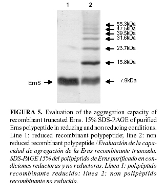
Purification fractions    were first analyzed in reducing conditions by SDS PAGE. In these conditions,    in the Erns aa 109-160 elution fraction, an additional protein of 15.8kDa was    observed. As this protein was not observed in the same fraction of the negative    control (the same purification protocol for an induced culture of the pET28a    plasmid without the insert, was runned in parallel), then, and taking into account    that Erns protein forms a disulfide-bridged homodimer in the virion (8; 23),    it was possible that the 15.8kDa protein was an aggregation product of the Erns    aa 109-160 polypeptide. When samples were analyzed under non-reducing conditions    by SDS-PAGE, a very complex aggregation pattern was observed, seven proteins    with sizes coincided with the possible polymers that could form our truncated    Erns (<a href="/img/revistas/rsa/v30n2/f0503208.jpg">Fig. 5</a>). This    multimerization is not reported by Lin <i>et al. </i>(12). One possible explanation    is the presence of four cysteines in the sequence of the Erns aa 109-160 polypeptide.    Although in the Erns predicted structure, these cysteins do not play any role    in the formation of disulfide bridges between two Erns homodimers, and only    in the formation of intra disulfide bridges (8). The overexpression of this    truncated Erns version in <i>E. coli</i> could induce this rare behaviour. </font>      
<p>      <p>      <p>      <p>      <p>      <p>      <p>      ]]></body>
<body><![CDATA[<p>      <p>      <p>      <p><font face="Verdana, Arial, Helvetica, sans-serif" size="2">Whether this phenomenon    affects or not the antigenicity of the recombinant Erns aa 109-160 fragment    in an ELISA based detection system is an open question. Further experiments    have to be done to elucidate the impact of this aggregation in the antigenicity    of this polypeptide. </font>     <p>&nbsp;     <p><font face="Verdana, Arial, Helvetica, sans-serif" size="2"><b><font size="3">CONCLUSIONS</font></b>    </font>     <p><font face="Verdana, Arial, Helvetica, sans-serif" size="2">As conclusions,    it can be said that the construct obtained, as well as the described methodology,    including the use of the auto induction protein expression system, will permit    the obtainment, in a very simple way, of large quantity of Erns antigen that    could facilitate the development and validation of a test to be used for serological    survey.     <br>       <br>   Of course, the potential use of this polypeptide, as an antigen for detection    of CSFV antibodies will depend on the antigenicity studies that still have to    be carried out. </font>     <p>&nbsp;     ]]></body>
<body><![CDATA[<p><font face="Verdana, Arial, Helvetica, sans-serif" size="2"><b><font size="3">ACKNOWLEDGEMENTS</font></b>    </font>     <p><font face="Verdana, Arial, Helvetica, sans-serif" size="2">The authors are    grateful to Alejandro Martin and the members of the lab of Dengue Vaccine of    the CIGB, for their technical and material assistance. </font>     <p>&nbsp;     <p><font face="Verdana, Arial, Helvetica, sans-serif" size="2"><b><font size="3">REFERENCES</font></b>    </font>     <!-- ref --><p><font face="Verdana, Arial, Helvetica, sans-serif" size="2">1. Bouma A, de    Smit AJ, de Kluijver EP, Terpstra C, Moormann RJ. Efficacy and stability of    a subunit vaccine based on glycoprotein E2 of classical swine fever virus. <i>Vet    Microbiol</i>. 1999;66:101-114. </font>    <!-- ref --><p><font face="Verdana, Arial, Helvetica, sans-serif" size="2">2. Bradford M.    A rapid and sensitive method for the quantitation of microgram quantities of    proteins utilizing the principle of protein-dye binding. <i>Anal Biochem</i>.    1976;72:248-154. </font>    <!-- ref --><p><font face="Verdana, Arial, Helvetica, sans-serif" size="2">3. Colett MS, Larson    R, Gold C, Strick D, Anderson DK, Purchio AF. Molecular cloning and nucleotide    sequence of the pestivirus bovine viral diarrhea virus. <i>Virology</i>. 1988;165:191-199.    </font>    <!-- ref --><p><font face="Verdana, Arial, Helvetica, sans-serif" size="2">4. Francki RIB,    Faquet DL, Knudson DL, Brown F. Fifth report of the International Committee    on the Taxonomy of Viruses. <i>Arch Virol Suppl</i>. 1991;2:223-233. </font>    <!-- ref --><p><font face="Verdana, Arial, Helvetica, sans-serif" size="2">5. Hulst MM, Westra    DF, Wensvoort G, Moormann RJ. Glycoprotein E1 of hog cholera virus expressed    in insect cells protects swine from hog cholera. <i>J Virol</i>. 1993;67:5435-5442.    </font>    <!-- ref --><p><font face="Verdana, Arial, Helvetica, sans-serif" size="2">6. Konig M, Lengsfeld    T, Pauly T, Stark R, Thiel HJ. Classical swine fever virus: independent induction    of protective immunity by two structural glycoproteins. <i>J Virol</i>. 1995;69:6479-6486.    </font>    <!-- ref --><p><font face="Verdana, Arial, Helvetica, sans-serif" size="2">7.Langedijk JP,    Middel WG, Meloen RH, Kramps JA, de Smit JA. Enzyme-linked immunosorbent assay    using a virus type-specific peptide based on a subdomain of envelope protein    Erns for serologic diagnosis of pestivirus infections in swine. <i>J Clin. Microbiol</i>.    2001;39:906-912. </font>    <!-- ref --><p><font face="Verdana, Arial, Helvetica, sans-serif" size="2">8. Langedijk JP,    van Veelen PA, Schaaper WM, de Ru AH, Meloen RH, Hulst MM. A structural model    of pestivirus Erns based on disulfide bond connectivity and homology modeling    reveals an extremely rare vicinal disulfide. <i>J Virol</i>. 2002;76:10383-10392.    </font>    <!-- ref --><p><font face="Verdana, Arial, Helvetica, sans-serif" size="2">9. Li M, Su ZG,    Janson JC. <i>In vitro</i> protein refolding by chromatographic procedures.    <i>Protein Expr Purif</i>. 2004;33(1):1-10. </font>    <!-- ref --><p><font face="Verdana, Arial, Helvetica, sans-serif" size="2">10.Lin M, Lin F,    Mallory M, Clavijo A. Deletions of structural glycoprotein E2 of classical swine    fever virus strain alfort/187 resolve a linear epitope of monoclonal antibody    WH303 and the minimal N-terminal domain essential for binding immunoglobulin    G antibodies of a pig hyperimmune serum. <i>J Virol</i>. 2000;74(24):11619-11625.    </font>    <!-- ref --><p><font face="Verdana, Arial, Helvetica, sans-serif" size="2">11.Lin M, Trottier    E, Pasick J, Sabara Marta. Identification of Antigenic Regions of the Erns Protein    for Pig Antibodies Elicited during Classical Swine Fever Virus Infection. <i>J    Biochem</i>. 2004;136(6):795-804. </font>    <!-- ref --><p><font face="Verdana, Arial, Helvetica, sans-serif" size="2">12.Lin M, Trottier    E, Pasick J. Antibody Responses of Pigs to Defined Erns Fragments after Infection    with Classical Swine Fever Virus. <i>Clinical Diagnostic Lab Immunol</i>. 2005;12(1):180-186.    </font>    <p><font face="Verdana, Arial, Helvetica, sans-serif" size="2">13.Moennig V. Characteristics    of the virus, p. 55-58. In B. Liess (ed.). Classical swine fever and related    viral infections. Martinus Nijhoff Publishing, Boston, Mass. 1988. </font>     <!-- ref --><p><font face="Verdana, Arial, Helvetica, sans-serif" size="2">14.Moormann RJ,    Bouma A, Kramps JA, Terpstra C, de Smit HJ. Development of a classical swine    fever subunit marker vaccine and companion diagnostic test. <i>Vet Microbiol</i>.    2000;73(2-3):209-219. </font>    <!-- ref --><p><font face="Verdana, Arial, Helvetica, sans-serif" size="2">15.Moser C, Ruggli    N, Tratschin JD, Hofmann MA. Detection of antibodies against classical swine    fever virus in swine sera by indirect ELISA using recombinant envelope glycoprotein    E2. <i>Vet Microbiol</i>. 1996;51(1-2):41-53. </font>    <!-- ref --><p><font face="Verdana, Arial, Helvetica, sans-serif" size="2">16.Paton DJ, Ibata    G, Edwards S, Wensvoort G. An ELISA detecting antibody to conserved pestivirus    epitopes. <i>J Virol Methods</i>. 1991; 31:315-324. </font>    <!-- ref --><p><font face="Verdana, Arial, Helvetica, sans-serif" size="2">17.Platis D, Foster    GR. High yield expression, refolding, and characterization of recombinant interferon    alpha2/alpha8 hybrids in <i>Escherichia coli</i>. <i>Protein Expr Purif</i>.    2003;31(2):222-30. </font>    <!-- ref --><p><font face="Verdana, Arial, Helvetica, sans-serif" size="2">18.Pluimers FH,    de Leeuw PW, Smak JA, Elbers AR, Stegeman JA. Classical swine fever in The Netherlands    1997-1998: a description of organisation and measures to eradicate the disease.    <i>Prev Vet Med</i>. 1999;42:139-155. </font>    <p><font face="Verdana, Arial, Helvetica, sans-serif" size="2">19.Rice MC, Lindenbach    BD. Flaviviridae: the viruses and their replication, 2001: p. 991-1041. <i>In    </i>D. M. Knipe and P. M. Howley (ed.), Fields virology. Lippincott Williams    &amp; Wilkins, Philadelphia, Pa. </font>     <!-- ref --><p><font face="Verdana, Arial, Helvetica, sans-serif" size="2">20.Rumenapf T,    Stark R, Meyers G, Thiel HJ. Structural proteins of hog cholera virus expressed    by vaccinia virus: further characterization and induction of protective immunity.    <i>J Virol</i>. 1991;65:589-597. </font>    <!-- ref --><p><font face="Verdana, Arial, Helvetica, sans-serif" size="2">21.Studier FW.    Protein production by auto-induction in high-density shaking cultures. <i>Protein    Expr and Purif.</i> 2005;41:207-234. </font>    <!-- ref --><p><font face="Verdana, Arial, Helvetica, sans-serif" size="2">22.Terpstra C,    Wensvoort G. Natural infections of pigs with bovine viral diarrhoea virus associated    with signs resembling swine fever. <i>Res Vet Sci</i>. 1988;45:137-142. </font>    <!-- ref --><p><font face="Verdana, Arial, Helvetica, sans-serif" size="2">23.Thiel HJ, Stark    R, Weiland E, Rumenapf T, Meyers G. Hog cholera virus: molecular composition    of virions from a pestivirus. <i>J Virol</i>. 1991;65:4705-4712. </font>    <!-- ref --><p><font face="Verdana, Arial, Helvetica, sans-serif" size="2">24.van Rijn PA,    Bossers A, Wensvoort G, Moormann RJ. Classical swine fever virus (CSFV) envelope    glycoprotein E2 containing one structural antigenic unit protects pigs from    lethal CSFV challenge. <i>J Gen Virol</i>. 1996;77:2737-2745. </font>    <!-- ref --><p><font face="Verdana, Arial, Helvetica, sans-serif" size="2">25.van Zijl M,    Wensvoort G, de Kluyver E, Hulst M, van der Gulden H, Gielkens A, et al. Live    attenuated pseudorabies virus expressing envelope glycoprotein E1 of hog cholera    virus protects swine against both pseudorabies and hog cholera. <i>J Virol Methods</i>.    1991;60:103-108. </font>    <p>      ]]></body>
<body><![CDATA[<p>      <p>      <p>      <p>      <p>&nbsp;     <p>&nbsp;     <p><font face="Verdana, Arial, Helvetica, sans-serif" size="2"><b>(Recibido 7-11-2007;    Aceptado 15-2-2008)</b> </font>     <P><font face="Verdana, Arial, Helvetica, sans-serif" size="2">    <BR>       <BR>   </font>      ]]></body>
<body><![CDATA[ ]]></body><back>
<ref-list>
<ref id="B1">
<label>1</label><nlm-citation citation-type="journal">
<person-group person-group-type="author">
<name>
<surname><![CDATA[Bouma]]></surname>
<given-names><![CDATA[A]]></given-names>
</name>
<name>
<surname><![CDATA[de Smit]]></surname>
<given-names><![CDATA[AJ]]></given-names>
</name>
<name>
<surname><![CDATA[de Kluijver]]></surname>
<given-names><![CDATA[EP]]></given-names>
</name>
<name>
<surname><![CDATA[Terpstra]]></surname>
<given-names><![CDATA[C]]></given-names>
</name>
<name>
<surname><![CDATA[Moormann]]></surname>
<given-names><![CDATA[RJ]]></given-names>
</name>
</person-group>
<article-title xml:lang="en"><![CDATA[Efficacy and stability of a subunit vaccine based on glycoprotein E2 of classical swine fever virus]]></article-title>
<source><![CDATA[Vet Microbiol]]></source>
<year>1999</year>
<volume>66</volume>
<page-range>101-114</page-range></nlm-citation>
</ref>
<ref id="B2">
<label>2</label><nlm-citation citation-type="journal">
<person-group person-group-type="author">
<name>
<surname><![CDATA[Bradford]]></surname>
<given-names><![CDATA[M]]></given-names>
</name>
</person-group>
<article-title xml:lang="en"><![CDATA[A rapid and sensitive method for the quantitation of microgram quantities of proteins utilizing the principle of protein-dye binding]]></article-title>
<source><![CDATA[Anal Biochem]]></source>
<year>1976</year>
<volume>72</volume>
<page-range>248-154</page-range></nlm-citation>
</ref>
<ref id="B3">
<label>3</label><nlm-citation citation-type="journal">
<person-group person-group-type="author">
<name>
<surname><![CDATA[Colett]]></surname>
<given-names><![CDATA[MS]]></given-names>
</name>
<name>
<surname><![CDATA[Larson]]></surname>
<given-names><![CDATA[R]]></given-names>
</name>
<name>
<surname><![CDATA[Gold]]></surname>
<given-names><![CDATA[C]]></given-names>
</name>
<name>
<surname><![CDATA[Strick]]></surname>
<given-names><![CDATA[D]]></given-names>
</name>
<name>
<surname><![CDATA[Anderson]]></surname>
<given-names><![CDATA[DK]]></given-names>
</name>
<name>
<surname><![CDATA[Purchio]]></surname>
<given-names><![CDATA[AF]]></given-names>
</name>
</person-group>
<article-title xml:lang="en"><![CDATA[Molecular cloning and nucleotide sequence of the pestivirus bovine viral diarrhea virus]]></article-title>
<source><![CDATA[Virology]]></source>
<year>1988</year>
<volume>165</volume>
<page-range>191-199</page-range></nlm-citation>
</ref>
<ref id="B4">
<label>4</label><nlm-citation citation-type="journal">
<person-group person-group-type="author">
<name>
<surname><![CDATA[Francki]]></surname>
<given-names><![CDATA[RIB]]></given-names>
</name>
<name>
<surname><![CDATA[Faquet]]></surname>
<given-names><![CDATA[DL]]></given-names>
</name>
<name>
<surname><![CDATA[Knudson]]></surname>
<given-names><![CDATA[DL]]></given-names>
</name>
<name>
<surname><![CDATA[Brown]]></surname>
<given-names><![CDATA[F]]></given-names>
</name>
</person-group>
<article-title xml:lang="en"><![CDATA[Fifth report of the International Committee on the Taxonomy of Viruses]]></article-title>
<source><![CDATA[Arch Virol Suppl]]></source>
<year>1991</year>
<volume>2</volume>
<page-range>223-233</page-range></nlm-citation>
</ref>
<ref id="B5">
<label>5</label><nlm-citation citation-type="journal">
<person-group person-group-type="author">
<name>
<surname><![CDATA[Hulst]]></surname>
<given-names><![CDATA[MM]]></given-names>
</name>
<name>
<surname><![CDATA[Westra]]></surname>
<given-names><![CDATA[DF]]></given-names>
</name>
<name>
<surname><![CDATA[Wensvoort]]></surname>
<given-names><![CDATA[G]]></given-names>
</name>
<name>
<surname><![CDATA[Moormann]]></surname>
<given-names><![CDATA[RJ]]></given-names>
</name>
</person-group>
<article-title xml:lang="en"><![CDATA[Glycoprotein E1 of hog cholera virus expressed in insect cells protects swine from hog cholera]]></article-title>
<source><![CDATA[J Virol]]></source>
<year>1993</year>
<volume>67</volume>
<page-range>5435-5442</page-range></nlm-citation>
</ref>
<ref id="B6">
<label>6</label><nlm-citation citation-type="journal">
<person-group person-group-type="author">
<name>
<surname><![CDATA[Konig]]></surname>
<given-names><![CDATA[M]]></given-names>
</name>
<name>
<surname><![CDATA[Lengsfeld]]></surname>
<given-names><![CDATA[T]]></given-names>
</name>
<name>
<surname><![CDATA[Pauly]]></surname>
<given-names><![CDATA[T]]></given-names>
</name>
<name>
<surname><![CDATA[Stark]]></surname>
<given-names><![CDATA[R]]></given-names>
</name>
<name>
<surname><![CDATA[Thiel]]></surname>
<given-names><![CDATA[HJ]]></given-names>
</name>
</person-group>
<article-title xml:lang="en"><![CDATA[Classical swine fever virus: independent induction of protective immunity by two structural glycoproteins]]></article-title>
<source><![CDATA[J Virol]]></source>
<year>1995</year>
<volume>69</volume>
<page-range>6479-6486</page-range></nlm-citation>
</ref>
<ref id="B7">
<label>7</label><nlm-citation citation-type="journal">
<person-group person-group-type="author">
<name>
<surname><![CDATA[Langedijk]]></surname>
<given-names><![CDATA[JP]]></given-names>
</name>
<name>
<surname><![CDATA[Middel]]></surname>
<given-names><![CDATA[WG]]></given-names>
</name>
<name>
<surname><![CDATA[Meloen]]></surname>
<given-names><![CDATA[RH]]></given-names>
</name>
<name>
<surname><![CDATA[Kramps]]></surname>
<given-names><![CDATA[JA]]></given-names>
</name>
<name>
<surname><![CDATA[de Smit]]></surname>
<given-names><![CDATA[JA]]></given-names>
</name>
</person-group>
<article-title xml:lang="en"><![CDATA[Enzyme-linked immunosorbent assay using a virus type-specific peptide based on a subdomain of envelope protein Erns for serologic diagnosis of pestivirus infections in swine]]></article-title>
<source><![CDATA[J Clin Microbiol]]></source>
<year>2001</year>
<volume>39</volume>
<page-range>906-912</page-range></nlm-citation>
</ref>
<ref id="B8">
<label>8</label><nlm-citation citation-type="journal">
<person-group person-group-type="author">
<name>
<surname><![CDATA[Langedijk]]></surname>
<given-names><![CDATA[JP]]></given-names>
</name>
<name>
<surname><![CDATA[van Veelen]]></surname>
<given-names><![CDATA[PA]]></given-names>
</name>
<name>
<surname><![CDATA[Schaaper]]></surname>
<given-names><![CDATA[WM]]></given-names>
</name>
<name>
<surname><![CDATA[de Ru]]></surname>
<given-names><![CDATA[AH]]></given-names>
</name>
<name>
<surname><![CDATA[Meloen]]></surname>
<given-names><![CDATA[RH]]></given-names>
</name>
<name>
<surname><![CDATA[Hulst]]></surname>
<given-names><![CDATA[MM]]></given-names>
</name>
</person-group>
<article-title xml:lang="en"><![CDATA[A structural model of pestivirus Erns based on disulfide bond connectivity and homology modeling reveals an extremely rare vicinal disulfide]]></article-title>
<source><![CDATA[J Virol]]></source>
<year>2002</year>
<volume>76</volume>
<page-range>10383-10392</page-range></nlm-citation>
</ref>
<ref id="B9">
<label>9</label><nlm-citation citation-type="journal">
<person-group person-group-type="author">
<name>
<surname><![CDATA[Li]]></surname>
<given-names><![CDATA[M]]></given-names>
</name>
<name>
<surname><![CDATA[Su]]></surname>
<given-names><![CDATA[ZG]]></given-names>
</name>
<name>
<surname><![CDATA[Janson]]></surname>
<given-names><![CDATA[JC]]></given-names>
</name>
</person-group>
<article-title xml:lang="en"><![CDATA[In vitro protein refolding by chromatographic procedures]]></article-title>
<source><![CDATA[Protein Expr Purif]]></source>
<year>2004</year>
<volume>33</volume>
<numero>1</numero>
<issue>1</issue>
<page-range>1-10</page-range></nlm-citation>
</ref>
<ref id="B10">
<label>10</label><nlm-citation citation-type="journal">
<person-group person-group-type="author">
<name>
<surname><![CDATA[Lin]]></surname>
<given-names><![CDATA[M]]></given-names>
</name>
<name>
<surname><![CDATA[Lin]]></surname>
<given-names><![CDATA[F]]></given-names>
</name>
<name>
<surname><![CDATA[Mallory]]></surname>
<given-names><![CDATA[M]]></given-names>
</name>
<name>
<surname><![CDATA[Clavijo]]></surname>
<given-names><![CDATA[A]]></given-names>
</name>
</person-group>
<article-title xml:lang="en"><![CDATA[Deletions of structural glycoprotein E2 of classical swine fever virus strain alfort/187 resolve a linear epitope of monoclonal antibody WH303 and the minimal N-terminal domain essential for binding immunoglobulin G antibodies of a pig hyperimmune serum]]></article-title>
<source><![CDATA[J Virol]]></source>
<year>2000</year>
<volume>74</volume>
<numero>24</numero>
<issue>24</issue>
<page-range>11619-11625</page-range></nlm-citation>
</ref>
<ref id="B11">
<label>11</label><nlm-citation citation-type="journal">
<person-group person-group-type="author">
<name>
<surname><![CDATA[Lin]]></surname>
<given-names><![CDATA[M]]></given-names>
</name>
<name>
<surname><![CDATA[Trottier]]></surname>
<given-names><![CDATA[E]]></given-names>
</name>
<name>
<surname><![CDATA[Pasick]]></surname>
<given-names><![CDATA[J]]></given-names>
</name>
<name>
<surname><![CDATA[Sabara]]></surname>
<given-names><![CDATA[Marta]]></given-names>
</name>
</person-group>
<article-title xml:lang="en"><![CDATA[Identification of Antigenic Regions of the Erns Protein for Pig Antibodies Elicited during Classical Swine Fever Virus Infection]]></article-title>
<source><![CDATA[J Biochem]]></source>
<year>2004</year>
<volume>136</volume>
<numero>6</numero>
<issue>6</issue>
<page-range>795-804</page-range></nlm-citation>
</ref>
<ref id="B12">
<label>12</label><nlm-citation citation-type="journal">
<person-group person-group-type="author">
<name>
<surname><![CDATA[Lin]]></surname>
<given-names><![CDATA[M]]></given-names>
</name>
<name>
<surname><![CDATA[Trottier]]></surname>
<given-names><![CDATA[E]]></given-names>
</name>
<name>
<surname><![CDATA[Pasick]]></surname>
<given-names><![CDATA[J]]></given-names>
</name>
</person-group>
<article-title xml:lang="en"><![CDATA[Antibody Responses of Pigs to Defined Erns Fragments after Infection with Classical Swine Fever Virus]]></article-title>
<source><![CDATA[Clinical Diagnostic Lab Immunol]]></source>
<year>2005</year>
<volume>12</volume>
<numero>1</numero>
<issue>1</issue>
<page-range>180-186</page-range></nlm-citation>
</ref>
<ref id="B13">
<label>13</label><nlm-citation citation-type="book">
<person-group person-group-type="author">
<name>
<surname><![CDATA[Moennig]]></surname>
<given-names><![CDATA[V]]></given-names>
</name>
</person-group>
<article-title xml:lang="en"><![CDATA[Characteristics of the virus]]></article-title>
<person-group person-group-type="editor">
<name>
<surname><![CDATA[Liess]]></surname>
<given-names><![CDATA[B]]></given-names>
</name>
</person-group>
<source><![CDATA[Classical swine fever and related viral infections.]]></source>
<year>1988</year>
<page-range>55-58</page-range><publisher-loc><![CDATA[Boston ]]></publisher-loc>
<publisher-name><![CDATA[Martinus Nijhoff]]></publisher-name>
</nlm-citation>
</ref>
<ref id="B14">
<label>14</label><nlm-citation citation-type="journal">
<person-group person-group-type="author">
<name>
<surname><![CDATA[Moormann]]></surname>
<given-names><![CDATA[RJ]]></given-names>
</name>
<name>
<surname><![CDATA[Bouma]]></surname>
<given-names><![CDATA[A]]></given-names>
</name>
<name>
<surname><![CDATA[Kramps]]></surname>
<given-names><![CDATA[JA]]></given-names>
</name>
<name>
<surname><![CDATA[Terpstra]]></surname>
<given-names><![CDATA[C]]></given-names>
</name>
<name>
<surname><![CDATA[de Smit]]></surname>
<given-names><![CDATA[HJ]]></given-names>
</name>
</person-group>
<article-title xml:lang="en"><![CDATA[Development of a classical swine fever subunit marker vaccine and companion diagnostic test]]></article-title>
<source><![CDATA[Vet Microbiol]]></source>
<year>2000</year>
<volume>73</volume>
<numero>2-3</numero>
<issue>2-3</issue>
<page-range>209-219</page-range></nlm-citation>
</ref>
<ref id="B15">
<label>15</label><nlm-citation citation-type="journal">
<person-group person-group-type="author">
<name>
<surname><![CDATA[Moser]]></surname>
<given-names><![CDATA[C]]></given-names>
</name>
<name>
<surname><![CDATA[Ruggli]]></surname>
<given-names><![CDATA[N]]></given-names>
</name>
<name>
<surname><![CDATA[Tratschin]]></surname>
<given-names><![CDATA[JD]]></given-names>
</name>
<name>
<surname><![CDATA[Hofmann]]></surname>
<given-names><![CDATA[MA]]></given-names>
</name>
</person-group>
<article-title xml:lang="en"><![CDATA[Detection of antibodies against classical swine fever virus in swine sera by indirect ELISA using recombinant envelope glycoprotein E2]]></article-title>
<source><![CDATA[Vet Microbiol]]></source>
<year>1996</year>
<volume>51</volume>
<numero>1-2</numero>
<issue>1-2</issue>
<page-range>41-53</page-range></nlm-citation>
</ref>
<ref id="B16">
<label>16</label><nlm-citation citation-type="journal">
<person-group person-group-type="author">
<name>
<surname><![CDATA[Paton]]></surname>
<given-names><![CDATA[DJ]]></given-names>
</name>
<name>
<surname><![CDATA[Ibata]]></surname>
<given-names><![CDATA[G]]></given-names>
</name>
<name>
<surname><![CDATA[Edwards]]></surname>
<given-names><![CDATA[S]]></given-names>
</name>
<name>
<surname><![CDATA[Wensvoort]]></surname>
<given-names><![CDATA[G]]></given-names>
</name>
</person-group>
<article-title xml:lang="en"><![CDATA[An ELISA detecting antibody to conserved pestivirus epitopes]]></article-title>
<source><![CDATA[J Virol Methods]]></source>
<year>1991</year>
<volume>31</volume>
<page-range>315-324</page-range></nlm-citation>
</ref>
<ref id="B17">
<label>17</label><nlm-citation citation-type="journal">
<person-group person-group-type="author">
<name>
<surname><![CDATA[Platis]]></surname>
<given-names><![CDATA[D]]></given-names>
</name>
<name>
<surname><![CDATA[Foster]]></surname>
<given-names><![CDATA[GR]]></given-names>
</name>
</person-group>
<article-title xml:lang="en"><![CDATA[High yield expression, refolding, and characterization of recombinant interferon alpha2/alpha8 hybrids in Escherichia coli]]></article-title>
<source><![CDATA[Protein Expr Purif]]></source>
<year>2003</year>
<volume>31</volume>
<numero>2</numero>
<issue>2</issue>
<page-range>222-30</page-range></nlm-citation>
</ref>
<ref id="B18">
<label>18</label><nlm-citation citation-type="journal">
<person-group person-group-type="author">
<name>
<surname><![CDATA[Pluimers]]></surname>
<given-names><![CDATA[FH]]></given-names>
</name>
<name>
<surname><![CDATA[de Leeuw]]></surname>
<given-names><![CDATA[PW]]></given-names>
</name>
<name>
<surname><![CDATA[Smak]]></surname>
<given-names><![CDATA[JA]]></given-names>
</name>
<name>
<surname><![CDATA[Elbers]]></surname>
<given-names><![CDATA[AR]]></given-names>
</name>
<name>
<surname><![CDATA[Stegeman]]></surname>
<given-names><![CDATA[JA]]></given-names>
</name>
</person-group>
<article-title xml:lang="en"><![CDATA[Classical swine fever in The Netherlands 1997-1998: a description of organisation and measures to eradicate the disease]]></article-title>
<source><![CDATA[Prev Vet Med]]></source>
<year>1999</year>
<volume>42</volume>
<page-range>139-155</page-range></nlm-citation>
</ref>
<ref id="B19">
<label>19</label><nlm-citation citation-type="">
<person-group person-group-type="author">
<name>
<surname><![CDATA[Rice]]></surname>
<given-names><![CDATA[MC]]></given-names>
</name>
<name>
<surname><![CDATA[Lindenbach]]></surname>
<given-names><![CDATA[BD]]></given-names>
</name>
</person-group>
<article-title xml:lang="en"><![CDATA[Flaviviridae: the viruses and their replication]]></article-title>
<person-group person-group-type="editor">
<name>
<surname><![CDATA[Knipe]]></surname>
<given-names><![CDATA[DM]]></given-names>
</name>
<name>
<surname><![CDATA[Howley]]></surname>
<given-names><![CDATA[PM]]></given-names>
</name>
</person-group>
<source><![CDATA[Fields virology]]></source>
<year>2001</year>
<page-range>991-1041</page-range></nlm-citation>
</ref>
<ref id="B20">
<label>20</label><nlm-citation citation-type="journal">
<person-group person-group-type="author">
<name>
<surname><![CDATA[Rumenapf]]></surname>
<given-names><![CDATA[T]]></given-names>
</name>
<name>
<surname><![CDATA[Stark]]></surname>
<given-names><![CDATA[R]]></given-names>
</name>
<name>
<surname><![CDATA[Meyers]]></surname>
<given-names><![CDATA[G]]></given-names>
</name>
<name>
<surname><![CDATA[Thiel]]></surname>
<given-names><![CDATA[HJ]]></given-names>
</name>
</person-group>
<article-title xml:lang="en"><![CDATA[Structural proteins of hog cholera virus expressed by vaccinia virus: further characterization and induction of protective immunity]]></article-title>
<source><![CDATA[J Virol]]></source>
<year>1991</year>
<volume>65</volume>
<page-range>589-597</page-range></nlm-citation>
</ref>
<ref id="B21">
<label>21</label><nlm-citation citation-type="journal">
<person-group person-group-type="author">
<name>
<surname><![CDATA[Studier]]></surname>
<given-names><![CDATA[FW]]></given-names>
</name>
</person-group>
<article-title xml:lang="en"><![CDATA[Protein production by auto-induction in high-density shaking cultures]]></article-title>
<source><![CDATA[Protein Expr and Purif]]></source>
<year>2005</year>
<volume>41</volume>
<page-range>207-234</page-range></nlm-citation>
</ref>
<ref id="B22">
<label>22</label><nlm-citation citation-type="journal">
<person-group person-group-type="author">
<name>
<surname><![CDATA[Terpstra]]></surname>
<given-names><![CDATA[C]]></given-names>
</name>
<name>
<surname><![CDATA[Wensvoort]]></surname>
<given-names><![CDATA[G]]></given-names>
</name>
</person-group>
<article-title xml:lang="en"><![CDATA[Natural infections of pigs with bovine viral diarrhoea virus associated with signs resembling swine fever]]></article-title>
<source><![CDATA[Res Vet Sci]]></source>
<year>1988</year>
<volume>45</volume>
<page-range>137-142</page-range></nlm-citation>
</ref>
<ref id="B23">
<label>23</label><nlm-citation citation-type="journal">
<person-group person-group-type="author">
<name>
<surname><![CDATA[Thiel]]></surname>
<given-names><![CDATA[HJ]]></given-names>
</name>
<name>
<surname><![CDATA[Stark]]></surname>
<given-names><![CDATA[R]]></given-names>
</name>
<name>
<surname><![CDATA[Weiland]]></surname>
<given-names><![CDATA[E]]></given-names>
</name>
<name>
<surname><![CDATA[Rumenapf]]></surname>
<given-names><![CDATA[T]]></given-names>
</name>
<name>
<surname><![CDATA[Meyers]]></surname>
<given-names><![CDATA[G]]></given-names>
</name>
</person-group>
<article-title xml:lang="en"><![CDATA[Hog cholera virus: molecular composition of virions from a pestivirus]]></article-title>
<source><![CDATA[J Virol]]></source>
<year>1991</year>
<volume>65</volume>
<page-range>4705-4712</page-range></nlm-citation>
</ref>
<ref id="B24">
<label>24</label><nlm-citation citation-type="journal">
<person-group person-group-type="author">
<name>
<surname><![CDATA[van Rijn]]></surname>
<given-names><![CDATA[PA]]></given-names>
</name>
<name>
<surname><![CDATA[Bossers]]></surname>
<given-names><![CDATA[A]]></given-names>
</name>
<name>
<surname><![CDATA[Wensvoort]]></surname>
<given-names><![CDATA[G]]></given-names>
</name>
<name>
<surname><![CDATA[Moormann]]></surname>
<given-names><![CDATA[RJ]]></given-names>
</name>
</person-group>
<article-title xml:lang="en"><![CDATA[Classical swine fever virus (CSFV) envelope glycoprotein E2 containing one structural antigenic unit protects pigs from lethal CSFV challenge]]></article-title>
<source><![CDATA[J Gen Virol]]></source>
<year>1996</year>
<volume>77</volume>
<page-range>2737-2745</page-range></nlm-citation>
</ref>
<ref id="B25">
<label>25</label><nlm-citation citation-type="journal">
<person-group person-group-type="author">
<name>
<surname><![CDATA[van Zijl]]></surname>
<given-names><![CDATA[M]]></given-names>
</name>
<name>
<surname><![CDATA[Wensvoort]]></surname>
<given-names><![CDATA[G]]></given-names>
</name>
<name>
<surname><![CDATA[de Kluyver]]></surname>
<given-names><![CDATA[E]]></given-names>
</name>
<name>
<surname><![CDATA[Hulst]]></surname>
<given-names><![CDATA[M]]></given-names>
</name>
<name>
<surname><![CDATA[van der Gulden]]></surname>
<given-names><![CDATA[H]]></given-names>
</name>
<name>
<surname><![CDATA[Gielkens]]></surname>
<given-names><![CDATA[A]]></given-names>
</name>
</person-group>
<article-title xml:lang="en"><![CDATA[Live attenuated pseudorabies virus expressing envelope glycoprotein E1 of hog cholera virus protects swine against both pseudorabies and hog cholera]]></article-title>
<source><![CDATA[J Virol Methods]]></source>
<year>1991</year>
<volume>60</volume>
<page-range>103-108</page-range></nlm-citation>
</ref>
</ref-list>
</back>
</article>
