<?xml version="1.0" encoding="ISO-8859-1"?><article xmlns:mml="http://www.w3.org/1998/Math/MathML" xmlns:xlink="http://www.w3.org/1999/xlink" xmlns:xsi="http://www.w3.org/2001/XMLSchema-instance">
<front>
<journal-meta>
<journal-id>0864-0300</journal-id>
<journal-title><![CDATA[Revista Cubana de Investigaciones Biomédicas]]></journal-title>
<abbrev-journal-title><![CDATA[Rev Cubana Invest Bioméd]]></abbrev-journal-title>
<issn>0864-0300</issn>
<publisher>
<publisher-name><![CDATA[ECIMED]]></publisher-name>
</publisher>
</journal-meta>
<article-meta>
<article-id>S0864-03001998000200008</article-id>
<title-group>
<article-title xml:lang="es"><![CDATA[Aterosclerosis en edades tempranas de la vida: Estudio patomorfológico y morfométrico aplicando el sistema aterométrico]]></article-title>
</title-group>
<contrib-group>
<contrib contrib-type="author">
<name>
<surname><![CDATA[Wong Navarro]]></surname>
<given-names><![CDATA[Roberto]]></given-names>
</name>
</contrib>
<contrib contrib-type="author">
<name>
<surname><![CDATA[Fernández-Britto Rodríguez]]></surname>
<given-names><![CDATA[José E]]></given-names>
</name>
</contrib>
<contrib contrib-type="author">
<name>
<surname><![CDATA[Contreras Barrionuevo]]></surname>
<given-names><![CDATA[Daniel]]></given-names>
</name>
</contrib>
<contrib contrib-type="author">
<name>
<surname><![CDATA[Guski]]></surname>
<given-names><![CDATA[Hans]]></given-names>
</name>
</contrib>
</contrib-group>
<aff id="A01">
<institution><![CDATA[,Centro de Investigaciones y Referencia de Aterosclerosis de La Habana  ]]></institution>
<addr-line><![CDATA[La Habana ]]></addr-line>
<country>Cuba</country>
</aff>
<pub-date pub-type="pub">
<day>00</day>
<month>08</month>
<year>1998</year>
</pub-date>
<pub-date pub-type="epub">
<day>00</day>
<month>08</month>
<year>1998</year>
</pub-date>
<volume>17</volume>
<numero>2</numero>
<fpage>179</fpage>
<lpage>184</lpage>
<copyright-statement/>
<copyright-year/>
<self-uri xlink:href="http://scielo.sld.cu/scielo.php?script=sci_arttext&amp;pid=S0864-03001998000200008&amp;lng=en&amp;nrm=iso"></self-uri><self-uri xlink:href="http://scielo.sld.cu/scielo.php?script=sci_abstract&amp;pid=S0864-03001998000200008&amp;lng=en&amp;nrm=iso"></self-uri><self-uri xlink:href="http://scielo.sld.cu/scielo.php?script=sci_pdf&amp;pid=S0864-03001998000200008&amp;lng=en&amp;nrm=iso"></self-uri><abstract abstract-type="short" xml:lang="es"><p><![CDATA[Se realizó un estudio de las lesiones ateroscleróticas en 50 aortas de fallecidos menores de 35 años, cuyas causas de muerte no estaban relacionadas con la enfermedad aterosclerótica, separados en 3 grupos de edades y sexo. La aorta se analizó dividida en sus segmentos torácico y abdominal, utilizando los procedimientos patomorfológicos clásicos. El análisis morfométrico se realizó aplicando el sistema aterométrico (SA). Las operaciones estadísticas se efectuaron con el paquete comercial Number Cruncher Statistical System (NCSS). Los resultados más destacados fueron los siguientes: 1) las lesiones ateroscleróticas en ambos segmentos de la aorta aumentan con el progreso de la edad y son más intensas en el segmento abdominal; 2) las estrías adiposas (X) se observaron en el segmento torácico de la aorta desde los 5 años de edad, mientras que las placas fibrosas (Y) aparecen a partir de los 16 años, pero en el segmento abdominal. El test estadístico de comparación entre los grupos de edad, mostró significación entre los grupos I - II y I - III, lo que sugiere que a partir de la segunda década de la vida se estabiliza el desarrollo y evolución de las lesiones ateroscleróticas. El SA resultó una herramienta idónea para el estudio de las lesiones ateroscleróticas en etapas tempranas de la vida.]]></p></abstract>
<abstract abstract-type="short" xml:lang="en"><p><![CDATA[A study was made on the atherosclerotic lesions of 50 aortas from dead people under 35 years whose causes of death were not related to atherosclerosis. The deceased were divided into 3 groups by age and sex. The aortas were divided into thoracic and abdominal segments to be analyzed by the classical pathomorphological method. The morphometric analysis was carried ot using the atherometric system. The statistical operations were performed assisted by the commercial Number Cruncher Statistical System software. The most important results were the following: 1) the atherosclerotic lesions in both segments increase with rising age level and they are more intense in the abdominal segment; 2) the fatty streak (X) were observed in the aorta thoracic segment since 5 years of age whereas fibrous plaques (Y) occured at 16 years of age or over but in the abdominal segment. The statistical comparison test applied to age groups was significant in I-III groups and I-II groups, which suggest that during the second decade of life the development and progression of atherosclerotic lesions are stabilized. The atherometric system was the best tool for the study of atherosclerotic lesions in early stages of life.]]></p></abstract>
<kwd-group>
<kwd lng="es"><![CDATA[ATEROSCLEROSIS]]></kwd>
<kwd lng="es"><![CDATA[AORTA ABDOMINAL]]></kwd>
<kwd lng="es"><![CDATA[AORTA TORACICA]]></kwd>
<kwd lng="es"><![CDATA[AUTOPSIA]]></kwd>
<kwd lng="en"><![CDATA[ATHEROSCLEROSIS]]></kwd>
<kwd lng="en"><![CDATA[AORTA, ABDOMINAL]]></kwd>
<kwd lng="en"><![CDATA[AORTA, THORACIC]]></kwd>
<kwd lng="en"><![CDATA[AUTOPSY]]></kwd>
</kwd-group>
</article-meta>
</front><body><![CDATA[ <p>Centro de Investigaciones y Referencia de Aterosclerosis de La Habana </p><H2>  Aterosclerosis en edades tempranas de la vida. Estudio patomorfol&oacute;gico  y morfom&eacute;trico aplicando el sistema aterom&eacute;trico</H2><I>Dr. Roberto  Wong Navarro, Dr. Jos&eacute; E. Fern&aacute;ndez-Britto Rodr&iacute;guez, Lic.  Daniel Contreras Barrionuevo y Dr. Hans Guski</I> <H4> Resumen</H4>Se realiz&oacute;  un estudio de las lesiones ateroscler&oacute;ticas en 50 aortas de fallecidos  menores de 35 a&ntilde;os, cuyas causas de muerte no estaban relacionadas con  la enfermedad ateroscler&oacute;tica, separados en 3 grupos de edades y sexo.  La aorta se analiz&oacute; dividida en sus segmentos tor&aacute;cico y abdominal,  utilizando los procedimientos patomorfol&oacute;gicos cl&aacute;sicos. El an&aacute;lisis  morfom&eacute;trico se realiz&oacute; aplicando el sistema aterom&eacute;trico  (SA). Las operaciones estad&iacute;sticas se efectuaron con el paquete comercial  Number Cruncher Statistical System (NCSS). Los resultados m&aacute;s destacados  fueron los siguientes: 1) las lesiones ateroscler&oacute;ticas en ambos segmentos  de la aorta aumentan con el progreso de la edad y son m&aacute;s intensas en el  segmento abdominal; 2) las estr&iacute;as adiposas (X) se observaron en el segmento  tor&aacute;cico de la aorta desde los 5 a&ntilde;os de edad, mientras que las  placas fibrosas (Y) aparecen a partir de los 16 a&ntilde;os, pero en el segmento  abdominal. El <I>test</I> estad&iacute;stico de comparaci&oacute;n entre los grupos  de edad, mostr&oacute; significaci&oacute;n entre los grupos I - II y I - III,  lo que sugiere que a partir de la segunda d&eacute;cada de la vida se estabiliza  el desarrollo y evoluci&oacute;n de las lesiones ateroscler&oacute;ticas. El SA  result&oacute; una herramienta id&oacute;nea para el estudio de las lesiones ateroscler&oacute;ticas  en etapas tempranas de la vida.     <P>Descriptores DeCS: ATEROSCLEROSIS/patolog&iacute;a;  AORTA ABDOMINAL/patolog&iacute;a; AORTA TORACICA/patolog&iacute;a; AUTOPSIA.     <P>  La aterosclerosis constituye en nuestros d&iacute;as la primera causa de morbilidad  y mortalidad en numerosos pa&iacute;ses del mundo, entre ellos Cuba. Estudios  basados en la evaluaci&oacute;n de las lesiones ateroscler&oacute;ticas en series  consecutivas de autopsias, han brindado una valiosa informaci&oacute;n de la historia  natural de esta enfermedad.1-4     <P>La existencia del sistema aterom&eacute;trico5-8  para la caracterizaci&oacute;n de la lesi&oacute;n ateroscler&oacute;tica en cualquier  sector vascular, que ha sido probado con eficacia en muchos estudios previos realizados  en etapas avanzadas de la enfermedad9-17 y el conocer que raramente la enfermedad  ateroscler&oacute;tica se manifiesta cl&iacute;nicamente en sujetos muy j&oacute;venes,  pero su evoluci&oacute;n y desarrollo se inicia en etapas muy tempranas de la  vida,1,18-21 plantea las interrogantes siguientes: <OL>     <LI> &iquest;Cu&aacute;les  son las principales caracter&iacute;sticas patomorfol&oacute;gicas y morfom&eacute;tricas  de las lesiones ateroscler&oacute;ticas de la aorta en fallecidos menores de 35  a&ntilde;os de edad?</LI>    <LI> &iquest;Existe alguna asociaci&oacute;n entre los  diferentes tipos de lesiones ateroscler&oacute;ticas en los segmentos tor&aacute;cico  y abdominal de la aorta de fallecidos en edades tempranas de la vida?</LI>    </OL>Este  estudio tiene como objetivo responder las preguntas anteriormente expresadas,  con la intenci&oacute;n de profundizar en el conocimiento de las lesiones ateroscler&oacute;ticas  en etapas tempranas de la vida. <H4> M&eacute;todos</H4>Se realiz&oacute; el estudio  de 50 aortas de fallecidos en edades comprendidas entre 5 y 34 a&ntilde;os, cuyas  causas de muerte no estaban relacionadas con la enfermedad ateroscler&oacute;tica.  Estos se dividieron en 3 grupos de acuerdo con la edad: G-I (5-14 a&ntilde;os,  n=8); G-II (15-24 a&ntilde;os, n=20) y G-III (25-34 a&ntilde;os, n=22) y seg&uacute;n  sexo; masculino (n=31) y femenino (n=19).     <P>La disecci&oacute;n de la aorta se  realiz&oacute; en las primeras 6 horas despu&eacute;s del fallecimiento, se seccion&oacute;  a nivel del orificio de salida del primer par de las arterias intercostales y  se analiz&oacute; hasta la bifurcaci&oacute;n de las il&iacute;acas; as&iacute;  disecada la aorta se dividi&oacute; en 2 segmentos; uno tor&aacute;cico (AT) y  otro abdominal (AA), a nivel del tronco cel&iacute;aco.     <P>Las lesiones ateroscler&oacute;ticas  (estr&iacute;as adiposas, placas fibrosas y placas graves -complicadas y calcificadas)  se estudiaron por los procedimientos patomorfol&oacute;gicos cl&aacute;sicos22-24  y el an&aacute;lisis morfom&eacute;trico se realiz&oacute; utilizando las variables  arteriales del sistema aterom&eacute;trico (SA) de Fern&aacute;ndez-Britto y Carlevaro  para la caracterizaci&oacute;n de estas lesiones en cualquier sector vascular.5-8      <P>El procesamiento automatizado de la informaci&oacute;n se realiz&oacute; en  una PC 486-100Mh; se utilizaron t&eacute;cnicas descriptivas y comparativas con  el paquete estad&iacute;stico comercial Number Cruncher Statistical System (NCSS).  <H4> Resultados</H4>En la muestra estudiada se observ&oacute; en todas las aortas  alg&uacute;n tipo de lesi&oacute;n ateroscler&oacute;tica, exceptuando las placas  graves. Las estr&iacute;as adiposas aparecieron desde los 5 a&ntilde;os en el  segmento tor&aacute;cico mientras que las placas fibrosas se encontraron a partir  de los 16 a&ntilde;os pero en el segmento abdominal.     ]]></body>
<body><![CDATA[<P>Se puede apreciar que  los valores de la media aritm&eacute;tica de las variables del SA para cada segmento  de la aorta en la muestra (tabla) son mayores en el abdominal que en el tor&aacute;cico,  predominando las estr&iacute;as adiposas (X) sobre las placas fibrosas (Y).     <P>Tabla.<I>  Valores de la media y la desviaci&oacute;n est&aacute;ndar de las variables del  sistema aterom&eacute;trico para cada segmento de la aorta en la muestra estudiada</I>      <CENTER><TABLE BORDER CELLPADDING=5 WIDTH="659" > <TR> <TD VALIGN=TOP WIDTH="20%">&nbsp;</TD><TD VALIGN=TOP COLSPAN="2" WIDTH="40%">      <CENTER>Segmento tor&aacute;cico</CENTER></TD><TD VALIGN=TOP COLSPAN="2" WIDTH="40%">      <CENTER>Segmento abdominal</CENTER></TD></TR> <TR> <TD VALIGN=TOP WIDTH="20%">      <CENTER>Variable</CENTER></TD><TD VALIGN=TOP WIDTH="20%">     <CENTER>Media</CENTER></TD><TD VALIGN=TOP WIDTH="20%">      <CENTER>DE</CENTER></TD><TD VALIGN=TOP WIDTH="20%">     <CENTER>Media</CENTER></TD><TD VALIGN=TOP WIDTH="20%">      <CENTER>DE</CENTER></TD></TR> <TR> <TD VALIGN=TOP WIDTH="20%">     ]]></body>
<body><![CDATA[<CENTER>X</CENTER></TD><TD VALIGN=TOP WIDTH="20%">      <CENTER>0,2097</CENTER></TD><TD VALIGN=TOP WIDTH="20%">     <CENTER>0,0200</CENTER></TD><TD VALIGN=TOP WIDTH="20%">      <CENTER>0,2182</CENTER></TD><TD VALIGN=TOP WIDTH="20%">     <CENTER>0,2081</CENTER></TD></TR>  <TR> <TD VALIGN=TOP WIDTH="20%">     <CENTER>Y</CENTER></TD><TD VALIGN=TOP WIDTH="20%">      <CENTER>0,0044</CENTER></TD><TD VALIGN=TOP WIDTH="20%">     <CENTER>0,0104</CENTER></TD><TD VALIGN=TOP WIDTH="20%">      <CENTER>0,0295</CENTER></TD><TD VALIGN=TOP WIDTH="20%">     <CENTER>0,1203</CENTER></TD></TR>  <TR> <TD VALIGN=TOP WIDTH="20%">     ]]></body>
<body><![CDATA[<CENTER>S</CENTER></TD><TD VALIGN=TOP WIDTH="20%">      <CENTER>0,2142</CENTER></TD><TD VALIGN=TOP WIDTH="20%">     <CENTER>0,2021</CENTER></TD><TD VALIGN=TOP WIDTH="20%">      <CENTER>0,2478</CENTER></TD><TD VALIGN=TOP WIDTH="20%">     <CENTER>0,2271</CENTER></TD></TR>  <TR> <TD VALIGN=TOP WIDTH="20%">     <CENTER>W</CENTER></TD><TD VALIGN=TOP WIDTH="20%">      <CENTER>0,0088</CENTER></TD><TD VALIGN=TOP WIDTH="20%">     <CENTER>0,0209</CENTER></TD><TD VALIGN=TOP WIDTH="20%">      <CENTER>0,0591</CENTER></TD><TD VALIGN=TOP WIDTH="20%">     <CENTER>0,2407</CENTER></TD></TR>  <TR> <TD VALIGN=TOP WIDTH="20%">     ]]></body>
<body><![CDATA[<CENTER>P</CENTER></TD><TD VALIGN=TOP WIDTH="20%">      <CENTER>0,0055</CENTER></TD><TD VALIGN=TOP WIDTH="20%">     <CENTER>0,0133</CENTER></TD><TD VALIGN=TOP WIDTH="20%">      <CENTER>0,0462</CENTER></TD><TD VALIGN=TOP WIDTH="20%">     <CENTER>0,1939</CENTER></TD></TR>  <TR> <TD VALIGN=TOP WIDTH="20%">     <CENTER>B</CENTER></TD><TD VALIGN=TOP WIDTH="20%">      <CENTER>0,9955</CENTER></TD><TD VALIGN=TOP WIDTH="20%">     <CENTER>0,0104</CENTER></TD><TD VALIGN=TOP WIDTH="20%">      <CENTER>0,9704</CENTER></TD><TD VALIGN=TOP WIDTH="20%">     <CENTER>0,1203</CENTER></TD></TR>  </TABLE></CENTER>Leyenda: X - Estr&iacute;as adiposas     ]]></body>
<body><![CDATA[<BR>Y = Placas fibrosas      <BR>S = Total de aterosclerosis     <BR>W = &Iacute;ndice de obstrucci&oacute;n     <BR>P  = &Iacute;ndice de estenosis     <BR>B = &Iacute;ndice de benignidad     <BR>DE = Desviaci&oacute;n  est&aacute;ndar     <P>Los valores de los &iacute;ndices de obstrucci&oacute;n (W)  y de estenosis (P) son bajos en comparaci&oacute;n con los del &iacute;ndice de  benignidad (B).     <P>Seg&uacute;n los grupos de edades establecidos, el grupo I  present&oacute; los valores m&aacute;s bajos de las variables del SA que identifican  lesiones ateroscler&oacute;ticas (X = 0,0398, Y = 0,0027, S = 0,0426) en el segmento  tor&aacute;cico de la aorta.     <P>En el grupo II las estr&iacute;as adiposas muestran  valores superiores en el segmento tor&aacute;cico (X = 0,2483) con respecto al  abdominal (X = 0,2054), mientras que las placas fibrosas muestran valores m&aacute;s  elevados en el segmento abdominal (Y = 0,0408) que en el tor&aacute;cico (Y =  0,0250).     <P>En el grupo III se observaron los valores m&aacute;s altos de las  variables del SA en el segmento abdominal de la aorta (X = 0,2874, Y = 0,0488,  S = 0,3162).     ]]></body>
<body><![CDATA[<P>En relaci&oacute;n con el sexo, los valores de las variables del  SA en ambos segmentos de la aorta, fueron ligeramente superiores en el masculino.
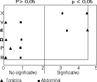
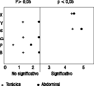
      <P>En el <I>test</I> de comparaci&oacute;n de las variables estudiadas, entre  los grupos de edades I - II y I - III en ambos segmentos de la aorta se observ&oacute;  que existe significaci&oacute;n estad&iacute;stica (p &pound; 0,0037) de las variables  estr&iacute;as adiposas (X) y total de aterosclerosis (S) (figs. 1 y 2). Sin embargo,  al comparar los grupos II y III en ambos segmentos no se aprecia significaci&oacute;n  estad&iacute;stica de variable alguna (fig. 3).     <CENTER><A HREF="/img/revistas/ibi/v17n2/f108298.gif"><IMG SRC="/img/revistas/ibi/v17n2/f108298.gif" ALT="Figura 1" VSPACE=5 BORDER=1 HEIGHT=171 WIDTH=203></A></CENTER>    
<CENTER>Figura  1.</CENTER>    <CENTER><A HREF="/img/revistas/ibi/v17n2/f208298.gif"><IMG SRC="/img/revistas/ibi/v17n2/f208298.gif" ALT="Figura 2" VSPACE=5 BORDER=1 HEIGHT=190 WIDTH=208></A></CENTER>    
<CENTER>Figura  2.</CENTER>    <CENTER><A HREF="/img/revistas/ibi/v17n2/f308298.gif"><IMG SRC="/img/revistas/ibi/v17n2/f308298.gif" ALT="Figura 3" VSPACE=5 BORDER=1 HEIGHT=188 WIDTH=215></A></CENTER>    
<CENTER>Figura  3.</CENTER>    <P>El an&aacute;lisis comparativo entre sexos no muestra significaci&oacute;n  estad&iacute;stica entre las variables estudiadas del SA (fig.4).     <CENTER><A HREF="/img/revistas/ibi/v17n2/f408299.gif"><IMG SRC="/img/revistas/ibi/v17n2/f408298.gif" ALT="Figura 4" VSPACE=5 BORDER=1 HEIGHT=173 WIDTH=193></A></CENTER>    
]]></body>
<body><![CDATA[<CENTER>Figura  4.</CENTER><H4> Discusi&oacute;n</H4>Los resultados obtenidos confirman que el  desarrollo de la lesi&oacute;n ateroscler&oacute;tica comienza en edades bien  tempranas de la vida, inici&aacute;ndose en el segmento tor&aacute;cico antes  que en el abdominal, progresando posteriormente de forma m&aacute;s acelerada  y con mayor intensidad en este &uacute;ltimo.1,3,18,20     <P>Al analizar los valores  de la media aritm&eacute;tica de las variables del SA para cada segmento de la  aorta en la muestra, se puede apreciar que son mayores en el abdominal que en  el tor&aacute;cico. Por otro lado, los valores de las estr&iacute;as adiposas  son superiores a los de las placas fibrosas, lo cual, teniendo en cuenta el grupo  de edad estudiado, apoya la hip&oacute;tesis de la transformaci&oacute;n progresiva  de las primeras en las segundas.4,20     <P>Al observar estos resultados se debe meditar  en la importancia b&aacute;sica que tiene la gen&eacute;tica en la presencia y  distribuci&oacute;n de las lesiones ateroscler&oacute;ticas, pues ya en tan poca  edad, el cuadro de distribuci&oacute;n lesional de esta enfermedad se define de  manera muy diferente y peculiar en cada individuo.25,26     <P>La significaci&oacute;n  del <I>test</I> de comparaci&oacute;n de las variables estudiadas entre los grupos  de edades I - II y I - III en los segmentos tor&aacute;cico y abdominal de la  aorta, pudiera indicar que si bien las primeras lesiones ateroscler&oacute;ticas  aparecen desde edades bien tempranas, es a partir de la segunda d&eacute;cada  que se estabiliza el desarrollo y evoluci&oacute;n de ellas. Por esta misma raz&oacute;n  al comparar los grupos II y III en ambos segmentos, no se aprecia significaci&oacute;n  estad&iacute;stica de variable alguna.     <P>El an&aacute;lisis comparativo entre  sexos no muestra significaci&oacute;n estad&iacute;stica entre las variables estudiadas  del SA, aunque los valores medios del total de aterosclerosis (S) son ligeramente  superiores en el sexo masculino.     <P>En la literatura27,28 se reporta que no existen  diferencias por sexo en relaci&oacute;n con el grado de lesi&oacute;n ateroscler&oacute;tica.  Por &uacute;ltimo se demuestra la utilidad del SA como una herramienta id&oacute;nea  para la caracterizaci&oacute;n de las lesiones ateroscler&oacute;ticas de la aorta  en edades tempranas de la vida. <H4> Conclusiones</H4><OL>     <LI> En todas las aortas  estudiadas se observ&oacute; alg&uacute;n tipo de lesi&oacute;n ateroscler&oacute;tica,  con excepci&oacute;n de las placas graves.</LI>    <LI> Las estr&iacute;as adiposas  (X) se observaron en el segmento tor&aacute;cico de la aorta desde los 5 a&ntilde;os  de edad mientras que las placas fibrosas (Y) aparecen a partir de los 16 a&ntilde;os  en el segmento abdominal.</LI>    <LI> Las lesiones ateroscler&oacute;ticas en ambos  segmentos de la aorta aumentan con el progreso de la edad, siendo m&aacute;s numerosas  en el abdominal.</LI>    <LI> El <I>test</I> de la t de Student mostr&oacute; significaci&oacute;n  estad&iacute;stica entre los grupos de edad I - II y I - III, lo que sugiere que  a partir de la segunda d&eacute;cada de la vida se estabiliza el desarrollo y  evoluci&oacute;n de las lesiones ateroscler&oacute;ticas.</LI>    ]]></body>
<body><![CDATA[<LI> El total de  aterosclerosis (S) fue ligeramente superior en el sexo masculino que en el femenino,  aunque no mostr&oacute; significaci&oacute;n estad&iacute;stica.</LI>    <LI> El sistema  aterom&eacute;trico (SA) result&oacute; una herramienta id&oacute;nea para el  estudio de las lesiones ateroscler&oacute;ticas en edades tempranas de la vida.</LI>    </OL><H4>  Summary</H4>A study was made on the atherosclerotic lesions of 50 aortas from  dead people under 35 years whose causes of death were not related to atherosclerosis.  The deceased were divided into 3 groups by age and sex. The aortas were divided  into thoracic and abdominal segments to be analyzed by the classical pathomorphological  method. The morphometric analysis was carried ot using the atherometric system.  The statistical operations were performed assisted by the commercial Number Cruncher  Statistical System software. The most important results were the following: 1)  the atherosclerotic lesions in both segments increase with rising age level and  they are more intense in the abdominal segment; 2) the fatty streak (X) were observed  in the aorta thoracic segment since 5 years of age whereas fibrous plaques (Y)  occured at 16 years of age or over but in the abdominal segment. The statistical  comparison test applied to age groups was significant in I-III groups and I-II  groups, which suggest that during the second decade of life the development and  progression of atherosclerotic lesions are stabilized. The atherometric system  was the best tool for the study of atherosclerotic lesions in early stages of  life.     <P>Subject headings: ATHEROSCLEROSIS/pathology; AORTA, ABDOMINAL/pathology;  AORTA, THORACIC/pathology; AUTOPSY. <H4> Referencias bibliogr&aacute;ficas</H4><OL>      <!-- ref --><LI> Movat HZ, Haust MD, More RH. The morphologic elemnts in the early lesions  of atherosclerosis. Am J Pathol 1959;35:93-101.</LI>    <!-- ref --><LI> Aggen DA, Solberg LA.  Variation of atherosclerosis with age. Lab Invest 1968;18:571-9.</LI>    <!-- ref --><LI> Haust  MD. The morphogenesis and fate of potential and early atherosclerotic lesions  in man. Hum Pathol 1971;2:1-29.</LI>    <!-- ref --><LI> .Atherosclerosis in childhood. Perspect  Pediatr Pathol 1978;4:155-216.</LI>    <!-- ref --><LI> Fern&aacute;ndez-Brito JE, Carlevaro P,  Bacallao J. The atherosclerosis lesion. A biometric system for its description  and characterization in any vascular sector. Acta Morphol Hung 1984;32:3-4.</LI>    <!-- ref --><LI>  .Aplication of a biometric system for characterization of atherosclerosis. Zentralbl  Allg Pathol Pathol Anat 1986;132:172-3.</LI>    <!-- ref --><LI> Fern&aacute;ndez-Brito JE, Carlevaro  P. An atherometric system to study the atherosclerotic lesions. Pathol Res Pract  1987;182:488-9.</LI>    <!-- ref --><LI> .Atherometric system: standarized methodology to study  atherosclerosis and its consequences. Gegenbaurs Morphol Jahrb 1989;135:2-12.</LI>    <!-- ref --><LI>  Fern&aacute;ndez-Britto JE, Bacallao J, Carlevaro P. Coronary atherosclerotic  lesions: Its characterization applying an atherometric system using principal  component analysis. Z Klin Med 1988;43:291-4.</LI>    <!-- ref --><LI> Fern&aacute;ndez-Britto  JE, Bacallao J, Carlevaro P. Coronary atherosclerotic lesions: Its characterization  applying an atherometric system using discriminant analysis. Zentralbl Allg Pathol  Pathol Anat 1988;134:243-9.</LI>    <!-- ref --><LI> Fern&aacute;ndez-Britto JE, Bacallao J, Carlevaro  P. Falc&oacute;n L, Campos R, Guski H. Coronary atherosclerosis in sudden death:  A comparative study applying an atherometric system. Gegenbaurs Morphol Jahrb  1989;135:183-4.</LI>    <!-- ref --><LI> Fern&aacute;ndez-Britto JE, Vikhert AM, Paramio A, Campos  R, Wong R, Madan D. Histopathological sinus node classification and its relation  to coronary atherosclerosis using an atherometric system. Zentralbl Allg Pathol  Pathol Anat 1990;136:77-86.</LI>    <!-- ref --><LI> Castillo JA, Fern&aacute;ndez-Britto JE,  Bacallao J, Campos R, Wong R, Guski H. Atherosclerosis progression related to  hypertension: a pathomorphological and morphometric study using an atherometric  system. Z Klin Med 1991;46:1417-8.</LI>    <!-- ref --><LI> Fern&aacute;ndez-Britto JE, Bacallao  J, Castillo JA, Campos R, Wong R, Guski H. Atherosclerosis progression related  to diabetes: a morphometric study using an atherometric system. Z Klin Med 1991;46:1423-6.</LI>    <!-- ref --><LI>  .Atherosclerosis in diabetes and hypertension: a comparative morphometric study  of their progression using an atherometric system. Zentralbl Pathol 1991;137:487-91.</LI>    <!-- ref --><LI>  Fern&aacute;ndez-Britto JE, Carlevaro P, Bacallao J, Koch AS, Guski H, Campos  R. Atherosclerotic lesions of the circle of Willis: Its study applying an atherometric  system using multivariate statistical techniques. Cor Vasa 1991;33(3):188-96.</LI>    <!-- ref --><LI>  Fern&aacute;ndez-Britto JE, Fuente F de la, Yero M. Coronary atherosclerosis and  hair elements contens. A correlation study of 102 autopsy subjects using the energy  disperse X-ray fluorescence analysis an atherometric system. Zentralbl Pathol  1992;138:15-20.</LI>    <!-- ref --><LI> Aizawa T. Aging and atherosclerosis. Asian Med J 1979;22:221-36.</LI>    <!-- ref --><LI>  Vikhert Am. Rhytmic structures of the aorta in children and young adults. Cor  Vasa 1988;30:298-304.</LI>    <!-- ref --><LI> Ishii T. Coronary and aortic atherosclerosis in  young men from Tokio and New Orleans. Lab Invest 1986;54:561-5.</LI>    <!-- ref --><LI> Vrimani  R. Effect of aging on aortic morphology in populations with high and low prevalence  of hypertension and atherosclerosis. Am J Pathol 1991;139:119-29.</LI>    <!-- ref --><LI> Holman  RL, Mc Gill HG, Strong SP, Geer JC. Technics for stuying the atherosclerotic lesions.  Lab Invest 1958;7:42-9.</LI>    <!-- ref --><LI> Holman RL. An index for the evaluation of arteriosclerotic  lesions in the abdominal aorta. Circulation 1960;28:1137-43.</LI>    <!-- ref --><LI> WHO. Study  group on the classification of atherosclerotic lesions. Geneva, 1958 (Technical  Report Series; 143)</LI>    <!-- ref --><LI> Berg K. Genetics of coronary heart disease and its  risk factors. Ciba Found Symp 1987;130:14-33.</LI>    <!-- ref --><LI> Hegele Ra. Gene-enviroment  interactions in atherosclerosis. Mol Cell Biocherm 1992;18:177-86.</LI>    <!-- ref --><LI> Strong  JP, Coronary and aortic atherosclerosis in New Orleans. Comparison of lesions  by age, sex and race. Lab Invest 1978;39(4):364.</LI>    <!-- ref --><LI> McGill HC, Stern Mp.  Sex and atherosclerosis. Atheroescl Rev 1979;4:157.</LI>    </OL>Recibido: 14 de febrero  de 1998. Aprobado: 24 de febrero de 1998.     <P>Dr. <I>Roberto Wong Navarro. </I>Centro  de Investigaciones y Referencia de Aterosclerosis de La Habana. Apartado 6493.  La Habana, 10600, Cuba. Fax 537-662075. Correo electr&oacute;nico <A HREF="mailto:jfbritto@infomed.sld.cu">jfbritto@infomed.sld.cu</A>       ]]></body><back>
<ref-list>
<ref id="B1">
<nlm-citation citation-type="journal">
<person-group person-group-type="author">
<name>
<surname><![CDATA[Movat]]></surname>
<given-names><![CDATA[HZ]]></given-names>
</name>
<name>
<surname><![CDATA[Haust]]></surname>
<given-names><![CDATA[MD]]></given-names>
</name>
<name>
<surname><![CDATA[More]]></surname>
<given-names><![CDATA[RH]]></given-names>
</name>
</person-group>
<article-title xml:lang="en"><![CDATA[The morphologic elemnts in the early lesions of atherosclerosis]]></article-title>
<source><![CDATA[Am J Pathol]]></source>
<year>1959</year>
<volume>35</volume>
<page-range>93-101</page-range></nlm-citation>
</ref>
<ref id="B2">
<nlm-citation citation-type="journal">
<person-group person-group-type="author">
<name>
<surname><![CDATA[Aggen]]></surname>
<given-names><![CDATA[DA]]></given-names>
</name>
<name>
<surname><![CDATA[Solberg]]></surname>
<given-names><![CDATA[LA]]></given-names>
</name>
</person-group>
<article-title xml:lang="en"><![CDATA[Variation of atherosclerosis with age]]></article-title>
<source><![CDATA[Lab Invest]]></source>
<year>1968</year>
<volume>18</volume>
<page-range>571-9</page-range></nlm-citation>
</ref>
<ref id="B3">
<nlm-citation citation-type="journal">
<person-group person-group-type="author">
<name>
<surname><![CDATA[Haust]]></surname>
<given-names><![CDATA[MD]]></given-names>
</name>
</person-group>
<article-title xml:lang="en"><![CDATA[The morphogenesis and fate of potential and early atherosclerotic lesions in man]]></article-title>
<source><![CDATA[Hum Pathol]]></source>
<year>1971</year>
<volume>2</volume>
<page-range>1-29</page-range></nlm-citation>
</ref>
<ref id="B4">
<nlm-citation citation-type="journal">
<article-title xml:lang="en"><![CDATA[Atherosclerosis in childhood]]></article-title>
<source><![CDATA[Perspect Pediatr Pathol]]></source>
<year>1978</year>
<volume>4</volume>
<page-range>155-216</page-range></nlm-citation>
</ref>
<ref id="B5">
<nlm-citation citation-type="journal">
<person-group person-group-type="author">
<name>
<surname><![CDATA[Fernández-Brito]]></surname>
<given-names><![CDATA[JE]]></given-names>
</name>
<name>
<surname><![CDATA[Carlevaro]]></surname>
<given-names><![CDATA[P]]></given-names>
</name>
<name>
<surname><![CDATA[Bacallao]]></surname>
<given-names><![CDATA[J]]></given-names>
</name>
</person-group>
<article-title xml:lang="en"><![CDATA[The atherosclerosis lesion: A biometric system for its description and characterization in any vascular sector]]></article-title>
<source><![CDATA[Acta Morphol Hung]]></source>
<year>1984</year>
<volume>32</volume>
<page-range>3-4</page-range></nlm-citation>
</ref>
<ref id="B6">
<nlm-citation citation-type="journal">
<article-title xml:lang="en"><![CDATA[Aplication of a biometric system for characterization of atherosclerosis]]></article-title>
<source><![CDATA[Zentralbl Allg Pathol Pathol Anat]]></source>
<year>1986</year>
<volume>132</volume>
<page-range>172-3</page-range></nlm-citation>
</ref>
<ref id="B7">
<nlm-citation citation-type="journal">
<person-group person-group-type="author">
<name>
<surname><![CDATA[Fernández-Brito]]></surname>
<given-names><![CDATA[JE]]></given-names>
</name>
<name>
<surname><![CDATA[Carlevaro]]></surname>
<given-names><![CDATA[P]]></given-names>
</name>
</person-group>
<article-title xml:lang="en"><![CDATA[An atherometric system to study the atherosclerotic lesions]]></article-title>
<source><![CDATA[Pathol Res Pract]]></source>
<year>1987</year>
<volume>182</volume>
<page-range>488-9</page-range></nlm-citation>
</ref>
<ref id="B8">
<nlm-citation citation-type="journal">
<article-title xml:lang="en"><![CDATA[Atherometric system: standarized methodology to study atherosclerosis and its consequences]]></article-title>
<source><![CDATA[Gegenbaurs Morphol Jahrb]]></source>
<year>1989</year>
<volume>135</volume>
<page-range>2-12</page-range></nlm-citation>
</ref>
<ref id="B9">
<nlm-citation citation-type="journal">
<person-group person-group-type="author">
<name>
<surname><![CDATA[Fernández-Britto]]></surname>
<given-names><![CDATA[JE]]></given-names>
</name>
<name>
<surname><![CDATA[Bacallao]]></surname>
<given-names><![CDATA[J]]></given-names>
</name>
<name>
<surname><![CDATA[Carlevaro]]></surname>
<given-names><![CDATA[P]]></given-names>
</name>
</person-group>
<article-title xml:lang="en"><![CDATA[Coronary atherosclerotic lesions: Its characterization applying an atherometric system using principal component analysis]]></article-title>
<source><![CDATA[Z Klin Med]]></source>
<year>1988</year>
<volume>43</volume>
<page-range>291-4</page-range></nlm-citation>
</ref>
<ref id="B10">
<nlm-citation citation-type="journal">
<person-group person-group-type="author">
<name>
<surname><![CDATA[Fernández-Britto]]></surname>
<given-names><![CDATA[JE]]></given-names>
</name>
<name>
<surname><![CDATA[Bacallao]]></surname>
<given-names><![CDATA[J]]></given-names>
</name>
<name>
<surname><![CDATA[Carlevaro]]></surname>
<given-names><![CDATA[P]]></given-names>
</name>
</person-group>
<article-title xml:lang="en"><![CDATA[Coronary atherosclerotic lesions: Its characterization applying an atherometric system using discriminant analysis]]></article-title>
<source><![CDATA[Zentralbl Allg Pathol Pathol Anat]]></source>
<year>1988</year>
<volume>134</volume>
<page-range>243-9</page-range></nlm-citation>
</ref>
<ref id="B11">
<nlm-citation citation-type="journal">
<person-group person-group-type="author">
<name>
<surname><![CDATA[Fernández-Britto]]></surname>
<given-names><![CDATA[JE]]></given-names>
</name>
<name>
<surname><![CDATA[Bacallao]]></surname>
<given-names><![CDATA[J]]></given-names>
</name>
<name>
<surname><![CDATA[Carlevaro]]></surname>
<given-names><![CDATA[P]]></given-names>
</name>
<name>
<surname><![CDATA[Falcón]]></surname>
<given-names><![CDATA[L]]></given-names>
</name>
<name>
<surname><![CDATA[Campos]]></surname>
<given-names><![CDATA[R]]></given-names>
</name>
<name>
<surname><![CDATA[Guski]]></surname>
<given-names><![CDATA[H]]></given-names>
</name>
</person-group>
<article-title xml:lang="en"><![CDATA[Coronary atherosclerosis in sudden death: A comparative study applying an atherometric system]]></article-title>
<source><![CDATA[Gegenbaurs Morphol Jahrb]]></source>
<year>1989</year>
<volume>135</volume>
<page-range>183-4</page-range></nlm-citation>
</ref>
<ref id="B12">
<nlm-citation citation-type="journal">
<person-group person-group-type="author">
<name>
<surname><![CDATA[Fernández-Britto]]></surname>
<given-names><![CDATA[JE]]></given-names>
</name>
<name>
<surname><![CDATA[Vikhert]]></surname>
<given-names><![CDATA[AM]]></given-names>
</name>
<name>
<surname><![CDATA[Paramio]]></surname>
<given-names><![CDATA[A]]></given-names>
</name>
<name>
<surname><![CDATA[Campos]]></surname>
<given-names><![CDATA[R]]></given-names>
</name>
<name>
<surname><![CDATA[Wong]]></surname>
<given-names><![CDATA[R]]></given-names>
</name>
<name>
<surname><![CDATA[Madan]]></surname>
<given-names><![CDATA[D]]></given-names>
</name>
</person-group>
<article-title xml:lang="en"><![CDATA[Histopathological sinus node classification and its relation to coronary atherosclerosis using an atherometric system]]></article-title>
<source><![CDATA[Zentralbl Allg Pathol Pathol Anat]]></source>
<year>1990</year>
<volume>136</volume>
<page-range>77-86</page-range></nlm-citation>
</ref>
<ref id="B13">
<nlm-citation citation-type="journal">
<person-group person-group-type="author">
<name>
<surname><![CDATA[Castillo]]></surname>
<given-names><![CDATA[JA]]></given-names>
</name>
<name>
<surname><![CDATA[Fernández-Britto]]></surname>
<given-names><![CDATA[JE]]></given-names>
</name>
<name>
<surname><![CDATA[Bacallao]]></surname>
<given-names><![CDATA[J]]></given-names>
</name>
<name>
<surname><![CDATA[Campos]]></surname>
<given-names><![CDATA[R]]></given-names>
</name>
<name>
<surname><![CDATA[Wong]]></surname>
<given-names><![CDATA[R]]></given-names>
</name>
<name>
<surname><![CDATA[Guski]]></surname>
<given-names><![CDATA[H]]></given-names>
</name>
</person-group>
<article-title xml:lang="en"><![CDATA[Atherosclerosis progression related to hypertension: a pathomorphological and morphometric study using an atherometric system]]></article-title>
<source><![CDATA[Z Klin Med]]></source>
<year>1991</year>
<volume>46</volume>
<page-range>1417-8</page-range></nlm-citation>
</ref>
<ref id="B14">
<nlm-citation citation-type="journal">
<person-group person-group-type="author">
<name>
<surname><![CDATA[Fernández-Britto]]></surname>
<given-names><![CDATA[JE]]></given-names>
</name>
<name>
<surname><![CDATA[Bacallao]]></surname>
<given-names><![CDATA[J]]></given-names>
</name>
<name>
<surname><![CDATA[Castillo]]></surname>
<given-names><![CDATA[JA]]></given-names>
</name>
<name>
<surname><![CDATA[Campos]]></surname>
<given-names><![CDATA[R]]></given-names>
</name>
<name>
<surname><![CDATA[Wong]]></surname>
<given-names><![CDATA[R]]></given-names>
</name>
<name>
<surname><![CDATA[Guski]]></surname>
<given-names><![CDATA[H]]></given-names>
</name>
</person-group>
<article-title xml:lang="en"><![CDATA[Atherosclerosis progression related to diabetes: a morphometric study using an atherometric system]]></article-title>
<source><![CDATA[Z Klin Med]]></source>
<year>1991</year>
<volume>46</volume>
<page-range>1423-6</page-range></nlm-citation>
</ref>
<ref id="B15">
<nlm-citation citation-type="journal">
<article-title xml:lang="en"><![CDATA[Atherosclerosis in diabetes and hypertension: a comparative morphometric study of their progression using an atherometric system]]></article-title>
<source><![CDATA[Zentralbl Pathol]]></source>
<year>1991</year>
<volume>137</volume>
<page-range>487-91</page-range></nlm-citation>
</ref>
<ref id="B16">
<nlm-citation citation-type="journal">
<person-group person-group-type="author">
<name>
<surname><![CDATA[Fernández-Britto]]></surname>
<given-names><![CDATA[JE]]></given-names>
</name>
<name>
<surname><![CDATA[Carlevaro]]></surname>
<given-names><![CDATA[P]]></given-names>
</name>
<name>
<surname><![CDATA[Bacallao]]></surname>
<given-names><![CDATA[J]]></given-names>
</name>
<name>
<surname><![CDATA[Koch]]></surname>
<given-names><![CDATA[AS]]></given-names>
</name>
<name>
<surname><![CDATA[Guski]]></surname>
<given-names><![CDATA[H]]></given-names>
</name>
<name>
<surname><![CDATA[Campos]]></surname>
<given-names><![CDATA[R]]></given-names>
</name>
</person-group>
<article-title xml:lang="en"><![CDATA[Atherosclerotic lesions of the circle of Willis: Its study applying an atherometric system using multivariate statistical techniques]]></article-title>
<source><![CDATA[Cor Vasa]]></source>
<year>1991</year>
<volume>33</volume>
<numero>3</numero>
<issue>3</issue>
<page-range>188-96</page-range></nlm-citation>
</ref>
<ref id="B17">
<nlm-citation citation-type="journal">
<person-group person-group-type="author">
<name>
<surname><![CDATA[Fernández-Britto]]></surname>
<given-names><![CDATA[JE]]></given-names>
</name>
<name>
<surname><![CDATA[Fuente]]></surname>
<given-names><![CDATA[F de la]]></given-names>
</name>
<name>
<surname><![CDATA[Yero]]></surname>
<given-names><![CDATA[M]]></given-names>
</name>
</person-group>
<article-title xml:lang="en"><![CDATA[Coronary atherosclerosis and hair elements contens: A correlation study of 102 autopsy subjects using the energy disperse X-ray fluorescence analysis an atherometric system]]></article-title>
<source><![CDATA[Zentralbl Pathol]]></source>
<year>1992</year>
<volume>138</volume>
<page-range>15-20</page-range></nlm-citation>
</ref>
<ref id="B18">
<nlm-citation citation-type="journal">
<person-group person-group-type="author">
<name>
<surname><![CDATA[Aizawa]]></surname>
<given-names><![CDATA[T]]></given-names>
</name>
</person-group>
<article-title xml:lang="en"><![CDATA[Aging and atherosclerosis]]></article-title>
<source><![CDATA[Asian Med J]]></source>
<year>1979</year>
<volume>22</volume>
<page-range>221-36</page-range></nlm-citation>
</ref>
<ref id="B19">
<nlm-citation citation-type="journal">
<person-group person-group-type="author">
<name>
<surname><![CDATA[Vikhert]]></surname>
<given-names><![CDATA[Am]]></given-names>
</name>
</person-group>
<article-title xml:lang="en"><![CDATA[Rhytmic structures of the aorta in children and young adults]]></article-title>
<source><![CDATA[Cor Vasa]]></source>
<year>1988</year>
<volume>30</volume>
<page-range>298-304</page-range></nlm-citation>
</ref>
<ref id="B20">
<nlm-citation citation-type="journal">
<person-group person-group-type="author">
<name>
<surname><![CDATA[Ishii]]></surname>
<given-names><![CDATA[T]]></given-names>
</name>
</person-group>
<article-title xml:lang="en"><![CDATA[Coronary and aortic atherosclerosis in young men from Tokio and New Orleans]]></article-title>
<source><![CDATA[Lab Invest]]></source>
<year>1986</year>
<volume>54</volume>
<page-range>561-5</page-range></nlm-citation>
</ref>
<ref id="B21">
<nlm-citation citation-type="journal">
<person-group person-group-type="author">
<name>
<surname><![CDATA[Vrimani]]></surname>
<given-names><![CDATA[R]]></given-names>
</name>
</person-group>
<article-title xml:lang="en"><![CDATA[Effect of aging on aortic morphology in populations with high and low prevalence of hypertension and atherosclerosis]]></article-title>
<source><![CDATA[Am J Pathol]]></source>
<year>1991</year>
<volume>139</volume>
<page-range>119-29</page-range></nlm-citation>
</ref>
<ref id="B22">
<nlm-citation citation-type="journal">
<person-group person-group-type="author">
<name>
<surname><![CDATA[Holman]]></surname>
<given-names><![CDATA[RL]]></given-names>
</name>
<name>
<surname><![CDATA[Mc Gill]]></surname>
<given-names><![CDATA[HG]]></given-names>
</name>
<name>
<surname><![CDATA[Strong]]></surname>
<given-names><![CDATA[SP]]></given-names>
</name>
<name>
<surname><![CDATA[Geer]]></surname>
<given-names><![CDATA[JC]]></given-names>
</name>
</person-group>
<article-title xml:lang="en"><![CDATA[Technics for stuying the atherosclerotic lesions]]></article-title>
<source><![CDATA[Lab Invest]]></source>
<year>1958</year>
<volume>7</volume>
<page-range>42-9</page-range></nlm-citation>
</ref>
<ref id="B23">
<nlm-citation citation-type="journal">
<person-group person-group-type="author">
<name>
<surname><![CDATA[Holman]]></surname>
<given-names><![CDATA[RL]]></given-names>
</name>
</person-group>
<article-title xml:lang="en"><![CDATA[An index for the evaluation of arteriosclerotic lesions in the abdominal aorta]]></article-title>
<source><![CDATA[Circulation]]></source>
<year>1960</year>
<volume>28</volume>
<page-range>1137-43</page-range></nlm-citation>
</ref>
<ref id="B24">
<nlm-citation citation-type="">
<collab>WHO</collab>
<source><![CDATA[Study group on the classification of atherosclerotic lesions]]></source>
<year>1958</year>
<publisher-loc><![CDATA[Geneva ]]></publisher-loc>
</nlm-citation>
</ref>
<ref id="B25">
<nlm-citation citation-type="journal">
<person-group person-group-type="author">
<name>
<surname><![CDATA[Berg]]></surname>
<given-names><![CDATA[K]]></given-names>
</name>
</person-group>
<article-title xml:lang="en"><![CDATA[Genetics of coronary heart disease and its risk factors]]></article-title>
<source><![CDATA[Ciba Found Symp]]></source>
<year>1987</year>
<volume>130</volume>
<page-range>14-33</page-range></nlm-citation>
</ref>
<ref id="B26">
<nlm-citation citation-type="journal">
<person-group person-group-type="author">
<name>
<surname><![CDATA[Hegele]]></surname>
<given-names><![CDATA[Ra]]></given-names>
</name>
</person-group>
<article-title xml:lang="en"><![CDATA[Gene-enviroment interactions in atherosclerosis]]></article-title>
<source><![CDATA[Mol Cell Biocherm]]></source>
<year>1992</year>
<volume>18</volume>
<page-range>177-86</page-range></nlm-citation>
</ref>
<ref id="B27">
<nlm-citation citation-type="journal">
<person-group person-group-type="author">
<name>
<surname><![CDATA[Strong]]></surname>
<given-names><![CDATA[JP]]></given-names>
</name>
</person-group>
<article-title xml:lang="en"><![CDATA[Coronary and aortic atherosclerosis in New Orleans: Comparison of lesions by age, sex and race]]></article-title>
<source><![CDATA[Lab Invest]]></source>
<year>1978</year>
<volume>39</volume>
<numero>4</numero>
<issue>4</issue>
<page-range>364</page-range></nlm-citation>
</ref>
<ref id="B28">
<nlm-citation citation-type="journal">
<person-group person-group-type="author">
<name>
<surname><![CDATA[McGill]]></surname>
<given-names><![CDATA[HC]]></given-names>
</name>
<name>
<surname><![CDATA[Stern]]></surname>
<given-names><![CDATA[Mp]]></given-names>
</name>
</person-group>
<article-title xml:lang="en"><![CDATA[Sex and atherosclerosis]]></article-title>
<source><![CDATA[Atheroescl Rev]]></source>
<year>1979</year>
<volume>4</volume>
<page-range>157</page-range></nlm-citation>
</ref>
</ref-list>
</back>
</article>
