<?xml version="1.0" encoding="ISO-8859-1"?><article xmlns:mml="http://www.w3.org/1998/Math/MathML" xmlns:xlink="http://www.w3.org/1999/xlink" xmlns:xsi="http://www.w3.org/2001/XMLSchema-instance">
<front>
<journal-meta>
<journal-id>0864-0300</journal-id>
<journal-title><![CDATA[Revista Cubana de Investigaciones Biomédicas]]></journal-title>
<abbrev-journal-title><![CDATA[Rev Cubana Invest Bioméd]]></abbrev-journal-title>
<issn>0864-0300</issn>
<publisher>
<publisher-name><![CDATA[ECIMED]]></publisher-name>
</publisher>
</journal-meta>
<article-meta>
<article-id>S0864-03002006000200003</article-id>
<title-group>
<article-title xml:lang="es"><![CDATA[Evisceración con doble cobertura escleral]]></article-title>
<article-title xml:lang="en"><![CDATA[Evisceration with doble scleral coverage]]></article-title>
</title-group>
<contrib-group>
<contrib contrib-type="author">
<name>
<surname><![CDATA[Manteiga Rodríguez]]></surname>
<given-names><![CDATA[Mary Brenda]]></given-names>
</name>
<xref ref-type="aff" rid="A01"/>
</contrib>
<contrib contrib-type="author">
<name>
<surname><![CDATA[Falcón Caballero]]></surname>
<given-names><![CDATA[Judith]]></given-names>
</name>
</contrib>
<contrib contrib-type="author">
<name>
<surname><![CDATA[Montesino Álvarez]]></surname>
<given-names><![CDATA[Isis]]></given-names>
</name>
</contrib>
<contrib contrib-type="author">
<name>
<surname><![CDATA[Soto Rodríguez]]></surname>
<given-names><![CDATA[Gladys]]></given-names>
</name>
</contrib>
</contrib-group>
<aff id="A01">
<institution><![CDATA[,Centro de Investigaciones Médico-Quirúrgicas  ]]></institution>
<addr-line><![CDATA[Ciudad de La Habana ]]></addr-line>
<country>Cuba.</country>
</aff>
<pub-date pub-type="pub">
<day>00</day>
<month>06</month>
<year>2006</year>
</pub-date>
<pub-date pub-type="epub">
<day>00</day>
<month>06</month>
<year>2006</year>
</pub-date>
<volume>25</volume>
<numero>2</numero>
<fpage>0</fpage>
<lpage>0</lpage>
<copyright-statement/>
<copyright-year/>
<self-uri xlink:href="http://scielo.sld.cu/scielo.php?script=sci_arttext&amp;pid=S0864-03002006000200003&amp;lng=en&amp;nrm=iso"></self-uri><self-uri xlink:href="http://scielo.sld.cu/scielo.php?script=sci_abstract&amp;pid=S0864-03002006000200003&amp;lng=en&amp;nrm=iso"></self-uri><self-uri xlink:href="http://scielo.sld.cu/scielo.php?script=sci_pdf&amp;pid=S0864-03002006000200003&amp;lng=en&amp;nrm=iso"></self-uri><abstract abstract-type="short" xml:lang="es"><p><![CDATA[Se incluyeron en el estudio 35 pacientes con una edad comprendida entre 13 y 78 años. La pérdida del globo ocular es traumatizante para el paciente y constituye un fracaso para el oftalmólogo. Tras la evisceración, el implante de hidroxiapatita con la colocación de una prótesis ocular, aporta beneficios estéticos para el paciente. En todos ellos, se practicó la cuadrisección con doble cobertura escleral, que es una de las técnicas quirúrgicas de mayor difusión en la actualidad, con un tiempo de seguimiento de 2 a 30 meses, permitiendo colocar implantes independientemente del tamaño de la cavidad y disminuyendo el riesgo de exposición de estos.]]></p></abstract>
<abstract abstract-type="short" xml:lang="en"><p><![CDATA[35 patients aged 13-78 were included in the study. The loss of the eye globe is traumatizing for the patient and it is considered a failure for the ophthalmologist. After evisceration, the hydroxyapatite implant with the placement of an ocular prosthesis reports aesthetic benefits for the patient. All of them underwent cuadrisection with doble scleral visceration, one of the most spread surgical techniques at present, with a follow-up from 2 to 30 months. It allows to place implants independently of the size of the cavity and to reduce their exposure risk.]]></p></abstract>
<kwd-group>
<kwd lng="es"><![CDATA[Evisceración]]></kwd>
<kwd lng="es"><![CDATA[implante de hidroxiapatita y cobertura escleral.]]></kwd>
<kwd lng="en"><![CDATA[Evisceration]]></kwd>
<kwd lng="en"><![CDATA[hydroxyapatite implant and scleral coverage]]></kwd>
</kwd-group>
</article-meta>
</front><body><![CDATA[ <p class=Normal><span lang=ES style='mso-bidi-font-size:11.0pt'>Centro de Investigaciones      Médico-Quirúrgicas</span></p>   <h2 class=Normal><span class=SpellE><span lang=ES style='mso-bidi-font-size: 11.0pt'>Evisceración</span></span><span lang=ES style='mso-bidi-font-size:11.0pt'>      con doble cobertura <span class=SpellE>escleral</span></span></h2>       <p class=Normal><span lang=ES style='mso-bidi-font-size:11.0pt'><i>Dra. <span class=SpellE>Mary</span> Brenda <span class=SpellE>Manteiga</span> Rodríguez,      Dra. Judith Falcón Caballero, Dra. <span class=SpellE>Isis</span> Montesino      Álvarez y Dra. <span class=SpellE>Gladys</span> Soto Rodríguez</i></span></p>   <h4 class=Normal><span lang=ES style='mso-bidi-font-size:11.0pt'>Resumen</span></h4>       <p class=Normal><span lang=ES style='mso-bidi-font-size:11.0pt'>Se incluyeron      en el estudio 35 pacientes con una edad comprendida entre 13 y 78 años. La      pérdida del globo ocular es <span class=SpellE>traumatizante</span> para el      paciente y constituye un fracaso para el oftalmólogo. Tras la <span class=SpellE>evisceración</span>, el implante de <span class=SpellE>hidroxiapatita</span>      con la colocación de una prótesis ocular, aporta beneficios estéticos para      el paciente. En todos ellos, se practicó la <span class=SpellE>cuadrisección</span>      con doble cobertura <span class=SpellE>escleral</span>, que es una de las      técnicas quirúrgicas de mayor difusión en la actualidad, con un tiempo de      seguimiento de 2 a 30 meses, permitiendo colocar implantes independientemente      del tamaño de la cavidad y disminuyendo el riesgo de exposición de estos.      </span></p>       <p class=Normal><span lang=ES style='mso-bidi-font-size:11.0pt'><i>Palabras      clave</i>: <span class=SpellE>Evisceración</span>, implante de <span class=SpellE>hidroxiapatita</span>      y cobertura <span class=SpellE>escleral</span>.</span></p>       <p class=Normal>&nbsp;</p>       <p class=Normal><span lang=ES style='mso-bidi-font-size:11.0pt'>La pérdida del      globo ocular producida por traumas, enfermedades, o como secuelas quirúrgicas      en el tratamiento de tumores y otras afecciones es un problema que afecta      a una parte considerable de la población. La restauración de estos defectos      ha merecido la atención de investigadores, médicos y especialistas dedicados      a este campo desde tiempos remotos, por lo que se ha prestado especial interés      al desarrollo tanto de <span class=SpellE>biomateriales</span> como de <span class=SpellE>procederes</span> quirúrgicos y de otro tipo que permitan la adecuada      rehabilitación.<sup>1</sup></span></p>       <p class=Normal><span lang=ES style='mso-bidi-font-size:11.0pt'>Hace más de      100 años que comenzaron a utilizarse los implantes orbitarios para tratar      de lograr una mejoría cosmética en los pacientes que perdían el globo ocular,      al restablecer en ellos el volumen de la cavidad y proporcionarles una mayor      motilidad a las prótesis; los primeros consistían en esferas huecas de cristal      y las utilizó <span class=SpellE><i style='mso-bidi-font-style:normal'>Mülles</i></span>      en 1884.<sup>2</sup></span></p>       <p class=Normal><span lang=ES style='mso-bidi-font-size:11.0pt'>Desde entonces,      la forma y su composición han variado mucho, entre los materiales utilizados      <span class=GramE>en</span> su confección se han tenido sustancias orgánicas (cartílagos,      <span class=SpellE>esclera</span>, hueso de cabeza de fémur de recién nacido)      e inorgánicas (cristal, tantalio, plástico, silicona y recientemente la <span class=SpellE>hidroxiapatita</span> y el <span class=SpellE>pórex</span>).</span></p>       <p class=Normal><span lang=ES style='mso-bidi-font-size:11.0pt'>Para conseguir      un buen resultado estético se debe colocar un implante del tamaño adecuado      para que la proyección <span class=SpellE>anteroposterior</span> del contenido      <span class=SpellE>orbitario</span> sea unos 3 <span class=SpellE>mm</span> menos que      el ojo <span class=SpellE>contralateral</span>, de manera que queden iguales      al colocar una prótesis del grosor óptimo.<sup>3</sup> Esto es difícil de      conseguir cuando la cavidad <span class=SpellE>escleral</span> del ojo eviscerado      es pequeña. Hace 2 años, en el Centro de Investigaciones Médico-Quirúrgicas      se comenzó a realizar la <span class=SpellE>evisceración</span> con implante de <span class=SpellE>hidroxiapatita</span>      mediante la técnica de <span class=SpellE>cuadrisección</span> con doble cobertura      <span class=SpellE>escleral</span>.</span></p>       <p class=Normal><span lang=ES style='mso-bidi-font-size:11.0pt'>A continuación      se describe la técnica, con la que, además de poder colocar un implante del      tamaño deseado, se cubre la parte anterior con 2 capas de <span class=SpellE>esclera</span>,      intentando disminuir aún mas las posibilidades de exposición.</span></p>   <h4 class=Normal><span lang=ES style='mso-bidi-font-size:11.0pt'>Métodos</span></h4>       ]]></body>
<body><![CDATA[<p class=Normal><span lang=ES style='mso-bidi-font-size:11.0pt'>Se realizó una      <span class=SpellE>evisceración</span> con la técnica que será descrita a continuación      y se colocó un implante de <span class=SpellE>hidroxiapatita</span> (HAP-200,      distribuidor en Cuba Centro Nacional de Investigaciones Científicas) a 35      pacientes entre enero de 2003 y julio de 2005. Se realizó un seguimiento entre      2 y 30 meses. Los implantes fueron de 17 <span class=SpellE>mm</span> en 26      pacientes (74,28 %), de 18 <span class=SpellE>mm</span> en 6 pacientes (17,14      %) y de 19 <span class=SpellE>mm</span> en 3 pacientes (8,57 %). Para calcular      el tamaño del implante se hizo una biometría del ojo <span class=SpellE>contralateral</span>,      y se colocó un implante de un diámetro 5 <span class=SpellE>mm</span> menos      que la longitud axial obtenida.</span></p>       <p class=Normal><span lang=ES style='mso-bidi-font-size:11.0pt'>
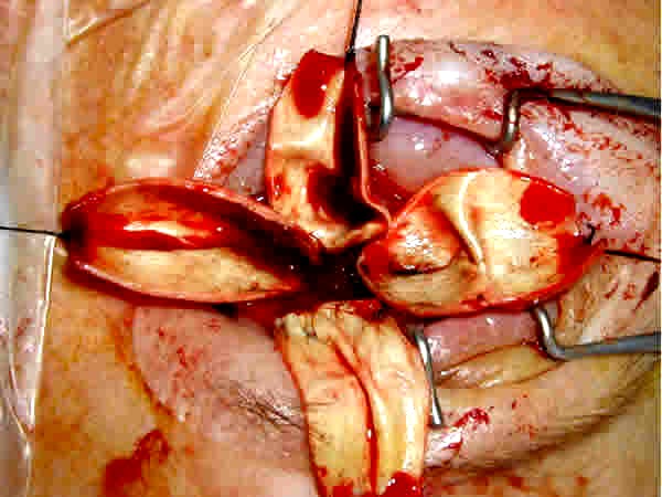
La técnica quirúrgica      se hizo mediante anestesia general (fig.).</span></p>       <p class=Normal align="center"><a href="/img/revistas/ibi/v25n2/f0103206.jpg"><img src="/img/revistas/ibi/v25n2/f0103206.jpg" width="451" height="336" border="0"></a></p>       
<p class=Normal align="center"><i>Fig.</i> T&eacute;cnica quir&uacute;rgica.</p>       <p class=Normal><span lang=ES style='mso-bidi-font-size:11.0pt'>En primer lugar,      se hace una <span class=SpellE>peritomía</span> conjuntival en limbo. Se separa      mediante disección roma la <span class=SpellE>Tennon</span> y conjuntiva del      globo ocular, llegando lo más atrás posible. Se individualizan con ganchos      de estrabismo los 4 músculos rectos, y se liberan las uniones fibrosas entre      ellos y entre cada uno y la cápsula de <span class=SpellE>Tennon</span>, para      luego poder desplazar hacia delante los fragmentos <span class=SpellE>esclerales</span>      con su músculo correspondiente sin dificultad. Se realiza una <span class=SpellE>queratectomía</span> completa, posteriormente se hacen 4 incisiones      en dirección <span class=SpellE>anteroposterior</span>, que parten del borde      anterior de la <span class=SpellE>esclera</span> justo en los vértices de      los 4 cuadrantes. Estas incisiones se unen unas con otras lo más cerca posible      del nervio óptico, de manera que se deja dividida la <span class=SpellE>esclera</span> en 4 pétalos, cada uno de los cuales lleva insertado      su músculo recto. Se introduce el implante del tamaño deseado. Se colocan      los 4 fragmentos <span class=SpellE>esclerales</span> sobre el implante, <span class=SpellE>traccionando</span> de ellos hacia delante, ayudándonos con pinzas      sin dientes que presionen el implante y eviten que se venga hacia delante.</span></p>       <p class=Normal><span lang=ES style='mso-bidi-font-size:11.0pt'>Se sutur&oacute;      con <span class=SpellE>Vicryl</span> 5/0, en primer lugar los pétalos que llevan los músculos      verticales y a continuación los que llevan los músculos horizontales, de esta      manera se produce primero una herida horizontal, sobre ella una vertical y,      por último, otra horizontal del cierre de la <span class=SpellE>Tennon</span>      y conjuntiva, disminuyendo las posibilidades de exposición. Se hace una buena      disección entre <span class=SpellE>Tennon</span> y conjuntiva, suturando siempre      los 2 planos por separado, con <span class=SpellE>Vicryl</span> 6/0 sutura      continua. Al finalizar la cirugía se coloca un conformador.</span></p>       <p class=Normal><span lang=ES style='mso-bidi-font-size:11.0pt'>Para la recolección      de la información se tomaron los datos de las historias clínicas. Como medidas      de resumen se utilizaron porcentajes.</span></p>   <h4 class=Normal><span lang=ES style='mso-bidi-font-size:11.0pt'>Resultados</span></h4>       <p class=Normal><span lang=ES style='mso-bidi-font-size:11.0pt'>En este estudio      se incluyen 35 pacientes a los que se les ha realizado una <span class=SpellE>evisceración</span>,      14 mujeres (40 %) y 21 hombres (60 %).</span></p>       <p class=Normal><span lang=ES style='mso-bidi-font-size:11.0pt'>Las indicaciones      para la cirugía fueron ojos ciegos, dolorosos o que producían alteración estética.</span></p>       <p class=Normal><span lang=ES style='mso-bidi-font-size:11.0pt'>La patología      previa que condujo a su situación actual fue:</span></p>       ]]></body>
<body><![CDATA[<p class=Normal><span lang=ES style='mso-bidi-font-size:11.0pt'>Traumatismos:      26 (74,28 %).    <br>     </span><span lang=ES style='mso-bidi-font-size:11.0pt'>Cirugía de catarata      o vítreo-retina: 3 (8,57 %).    <br>     </span><span lang=ES style='mso-bidi-font-size:11.0pt'>Glaucoma absoluto:      6 (17,14 %).</span></p>       <p class=Normal><span lang=ES style='mso-bidi-font-size:11.0pt'>Durante el seguimiento      (entre 2 y 30 meses) no ha habido ninguna complicación derivada de la cirugía.      En ningún caso hubo exposición ni desplazamiento del implante. El resultado      cosmético ha sido bueno sin <span class=SpellE>enoftalmos</span> ni hundimiento      del surco palpebral superior significativos.</span></p>   <h4 class=Normal><span lang=ES style='mso-bidi-font-size:11.0pt'>Discusión</span></h4>       <p class=Normal><span lang=ES style='mso-bidi-font-size:11.0pt'>Desde que en      1985, <span class=SpellE><i style='mso-bidi-font-style:normal'>Perry</i></span>      comenzó a utilizar <span class=SpellE>hidroxiapatita</span> como implante      que sustituye al globo ocular en <span class=SpellE>evisceraciones</span>      y enucleaciones, se han utilizado diferentes materiales<sup>4-12</sup> y diferentes      técnicas quirúrgicas encaminadas a colocar un implante que produzca pocas      complicaciones y con el que se consigan buenos resultados estéticos.</span></p>       <p class=Normal><span lang=ES style='mso-bidi-font-size:11.0pt'>Cuando se realiza      una <span class=SpellE>evisceración</span> en ojos con <span class=SpellE><i style='mso-bidi-font-style:normal'>ptisis</i></span><i style='mso-bidi-font-style:normal'> <span class=SpellE>bulbi</span></i> se ha      de modificar y ampliar la cavidad <span class=SpellE>escleral</span> disponible      porque si la prótesis colocada es muy pequeña se producirá un <span class=SpellE>enoftalmos</span>, un hundimiento del surco palpebral superior y      una ptosis palpebral por falta de apoyo, y el resultado estético no será nada      satisfactorio para el paciente.<sup>13</sup> Cuando el volumen del implante      es muy pequeño, el <span class=SpellE>protesista</span> intenta compensarlo      utilizando una prótesis más grande, pero esto produce por un lado una disminución      importante en su movilidad, y a largo plazo una pérdida del fondo de saco      inferior y una ptosis del párpado inferior debido al traumatismo producido      por el exceso de peso de esta.</span></p>       <p class=Normal><span lang=ES style='mso-bidi-font-size:11.0pt'>La complicación      más temible es la erosión de los tejidos que cubren el implante con la consiguiente      exposición de este y, si no se soluciona pronto, puede producirse una infección      que obliga a su retirada.<sup>14<span class=GramE>,15</span></sup></span></p>       <p class=Normal><span lang=ES style='mso-bidi-font-size:11.0pt'>La causa de      esta complicación, generalmente, es una cobertura del implante a tensión o      una erosión de la misma por un roce traumático.<sup>16,<span class=GramE>17</span></sup></span></p>       <p class=Normal><span lang=ES style='mso-bidi-font-size:11.0pt'>Se han descrito      diferentes técnicas de <span class=SpellE>evisceración</span> para poder colocar      prótesis grandes sin que los tejidos que cubren la prótesis estén suturados      a tensión. Se han descrito <span class=SpellE>esclerotomías</span> de descarga,      <span class=SpellE>esclerotomías</span> posteriores completas que permiten      avanzar la <span class=SpellE>esclera</span> todo lo necesario y secciones      <span class=SpellE>esclerales</span> en 2 o en 4 fragmentos<sup>,18<span class=GramE>,19</span></sup> como en este caso. Pero esta técnica quirúrgica,      igual que las anteriores, permite colocar un implante del tamaño deseado sin      tensión en los tejidos que la cubren, además, se consigue una doble cobertura      <span class=SpellE>escleral</span> del implante, de manera que es muy poco probable      que la superficie del implante llegue a erosionar por roce las 2 capas de      <span class=SpellE>esclera</span> que le cubren.<sup>20<span class=GramE>,21</span></sup></span></p>       <p class=Normal><span lang=ES style='mso-bidi-font-size:11.0pt'>Se concluye      que la <span class=SpellE>evisceración</span> con doble cobertura <span class=SpellE>escleral</span> es una técnica quirúrgica que permite colocar implantes      <span class=SpellE>biointegrables</span> del tamaño deseado, independientemente      de la cavidad <span class=SpellE>escleral</span> de que se disponga, y disminuye      las posibilidades de exposición.</span></p>   <h2 class=Normal><span lang=EN-US style='mso-bidi-font-size:11.0pt;mso-ansi-language: EN-US'>Evisceration with doble scleral coverage</span></h2>   <h4 class=Normal><span lang=EN-US style='mso-bidi-font-size:11.0pt;mso-ansi-language: EN-US'>Summary</span></h4>       ]]></body>
<body><![CDATA[<p class=Normal><span lang=EN-US style='mso-bidi-font-size:11.0pt;mso-ansi-language: EN-US'>35 patients aged 13-78 were included in the study. The loss of the eye      globe is traumatizing for the patient and it is considered a failure for the      ophthalmologist. After evisceration, the <span class=SpellE>hydroxyapatite</span>      implant with the placement of an ocular prosthesis reports aesthetic benefits      for the patient. All of them underwent <span class=SpellE>cuadrisection</span>      with <span class=SpellE>doble</span> <span class=SpellE>scleral</span> <span class=SpellE>visceration</span>, one of the most spread surgical techniques at      present, with a follow-up from </span><st1:time Minute="0" Hour="14"><span  lang=EN-US style='mso-bidi-font-size:11.0pt;mso-ansi-language:EN-US'>2</span></st1:time><span lang=EN-US style='mso-bidi-font-size:11.0pt;mso-ansi-language:EN-US'> to 30 months.<span style='mso-spacerun:yes'>       </span>It allows to place implants independently of the size of the cavity      and to reduce their exposure risk.<span style='mso-spacerun:yes'>  </span></span></p>       <p class=Normal><span lang=EN-US style='mso-bidi-font-size:11.0pt;mso-ansi-language: EN-US'><i>Key words</i>: Evisceration, <span class=SpellE>hydroxyapatite</span>      implant and <span class=SpellE>scleral</span> coverage.</span></p>   <h4 class=Normal><span lang=EN-US style='mso-bidi-font-size:11.0pt;mso-ansi-language: EN-US'>Referencias bibliográficas</span></h4>       <!-- ref --><p>1. <span lang=ES style='font-size:11.0pt; font-family:Arial'>Pérez G, González R, Acosta L, Solano M, Oliva J, Rodríguez          J. Hidroxiapatita porosa HAP-200 como bioimplante esférico integrado en          el anoftalmos quirúrgico. Rev Cubana Oftalmol 1998<span class=GramE>;11</span>(1):5-13.</span>     </div>   <!-- ref --><p><span lang=ES style='font-size:11.0pt; font-family:Arial'>2. Martínez N, Falcón I, Herrera M, Gómez C, Agramonte I, Samara          A, et al. Implantes orbitarios de HAP-200. </span><span class=SpellE><span lang=EN-US style='font-size:11.0pt;font-family:Arial; mso-ansi-language:EN-US'>Experiencia</span></span><span lang=EN-US style='font-size:11.0pt;font-family:Arial;mso-ansi-language:EN-US'> en 100 <span class=SpellE>casos</span>. Rev <span class=SpellE>Cubana</span> <span class=SpellE>Oftalmol</span> 2002<span class=GramE>;15</span>(1):10-9.</span>       </div>   <!-- ref --><p><span lang=EN-US style='font-size:11.0pt; font-family:Arial;mso-ansi-language:EN-US'>3. Jordon DR, Allen LH, Ells A, <span class=SpellE>Gilberg</span> S, <span class=SpellE>Browasteins</span> S, Munro          S. The use of <span class=SpellE>vycril</span> mesh (<span class=SpellE>poliglacten</span>     910) for implantation of <span class=SpellE>hydroxyapatite</span> orbital          implants. </span><span lang=ES style='font-size:11.0pt;font-family:Arial'>Ophthalmol          Plast Reconstr Surg 1991<span class=GramE>;11</span>(2):95-9. </span>       </div>   <!-- ref --><p><span class=GramE><span lang=EN-US style='font-size:11.0pt;font-family:Arial;mso-ansi-language:EN-US'>4. lapper</span></span><span lang=EN-US style='font-size:11.0pt;font-family:Arial;mso-ansi-language:EN-US'>     SR, </span><st1:Street><st1:address><span lang=EN-US style='font-size:11.0pt;   font-family:Arial;mso-ansi-language:EN-US'>Jordan DR</span></st1:address></st1:Street><span lang=EN-US style='font-size:11.0pt;font-family:Arial;mso-ansi-language:EN-US'>,     <span class=SpellE>Punja</span> K, Brownstein S, <span class=SpellE>Gilberg</span>    SM, <span class=SpellE>Mawn</span> LA, et al. <span class=SpellE>Hydroxyapatite</span>    implant wrapping materials: analysis of <span class=SpellE>fibrovascular</span>    <span class=SpellE>ingrowth</span> in an animal model. </span><span lang=ES style='font-size:11.0pt;font-family:Arial'>Ophthal Plast Reconstr Surg 2000<span class=GramE>;16:278</span>-85.</span>       </div>   <!-- ref --><p><span lang=EN-US style='font-size:11.0pt; font-family:Arial;mso-ansi-language:EN-US'>5. Kao LY. <span class=SpellE>Polytetrafluoroethylene</span>     as a wrapping material for a <span class=SpellE>hydroxyapatite</span> orbital          implant. </span><span lang=ES style='font-size:11.0pt;font-family:Arial'>Ophthal          Plast Reconstr Surg 2000<span class=GramE>;16:286</span>-8. </span>       </div>   <!-- ref --><p><span class=SpellE><span lang=EN-US style='font-size:11.0pt;font-family:Arial;mso-ansi-language:EN-US'>6. Kaltreider</span></span><span lang=EN-US style='font-size:11.0pt;font-family:Arial;mso-ansi-language:EN-US'>     SA. The ideal ocular prosthesis: analysis of prosthetic volume. </span><span lang=ES style='font-size:11.0pt;font-family:Arial'>Ophtal Plast Reconstr Surg          2000<span class=GramE>;16:388</span>-92. </span>       </div>   <!-- ref --><p><span lang=EN-US style='font-size:11.0pt; font-family:Arial;mso-ansi-language:EN-US'>7. Long JA, <span class=SpellE>Tann</span>     III TM, <span class=SpellE>Girkin</span> CA. Evisceration: a new technique          of trans-<span class=SpellE>scleral</span> implant placement. <span class=SpellE>Ophthal</span>     <span class=SpellE>Plast</span> <span class=SpellE>Reconsr</span> <span class=SpellE>Surg</span> 2000<span class=GramE>;16:322</span>-5. </span>       </div>   <!-- ref --><p><span lang=EN-US style='font-size:11.0pt; font-family:Arial;mso-ansi-language:EN-US'>8. Custer PL, <span class=SpellE>Reistad</span>     CE. <span class=SpellE>Enucleation</span> of blind, painful eyes. </span><span lang=ES style='font-size:11.0pt;font-family:Arial'>Ophthal Plast Reconstr Surg          2000<span class=GramE>;16:326</span>-9.</span>       </div>   <!-- ref -->9. Jordan DR</span></st1:address></st1:Street><span lang=EN-US style='font-size:11.0pt;font-family:Arial;mso-ansi-language:EN-US'>,     <span class=SpellE>Klapper</span> SR. A new titanium peg system for <span class=SpellE>hydroxyapatite</span> o orbital implants. </span><span lang=ES style='font-size:11.0pt;font-family:Arial'>Ophthal Plast Reconstr Surg 2000<span class=GramE>;16:380</span>-7. </span>       </div>   </st1:Street>    <!-- ref --><p><span lang=EN-US style='font-size:11.0pt;font-family:Arial;mso-ansi-language:EN-US'>10. Hsu          WC, Green JP, <span class=SpellE>Spilker</span> MH, Rubin PA. Primary placement          of a titanium motility post in a porous polyethylene orbital implant: animal          model with quantitative assessment of <span class=SpellE>fibrovascular</span>     <span class=SpellE>ingrowth</span> and vascular density. </span><span lang=ES style='font-size:11.0pt;font-family:Arial'>Ophthal Plast Reconstr Surg 2000<span class=GramE>;16:370</span>-9. </span>       </div>   <!-- ref --><p><span class=SpellE><span lang=EN-US style='font-size:11.0pt;font-family:Arial; mso-ansi-language:EN-US'>11. Choi</span></span><span lang=EN-US style='font-size: 11.0pt;font-family:Arial;mso-ansi-language:EN-US'> JC, Iwamoto MA, <span class=SpellE>Bstandig</span> S, Rubin PA, Shore JW. <span class=SpellE>Medpor</span>     Motility Coupling Post: a rabbit model. </span><span lang=ES style='font-size: 11.0pt;font-family:Arial'>Ophthal Plast Reconstr Surg 1999<span class=GramE>;15:190</span>-201.</span>       </div>   <!-- ref --><p><span lang=EN-US style='font-size:11.0pt;font-family:Arial;mso-ansi-language:EN-US'>12. Mules          PH. Evisceration of the globe with artificial vitreous. <span class=SpellE>Tr</span>     </span><st1:place><st1:PlaceName><span class=SpellE><span lang=EN-US   style='font-size:11.0pt;font-family:Arial;mso-ansi-language:EN-US'>Ophth</span></span></st1:PlaceName></st1:place> <st1:place><st1:PlaceName><span lang=EN-US style='font-size:11.0pt;font-family:   Arial;mso-ansi-language:EN-US'>Soc</span></st1:PlaceName><span lang=EN-US  style='font-size:11.0pt;font-family:Arial;mso-ansi-language:EN-US'> </span><st1:PlaceName><span   lang=EN-US style='font-size:11.0pt;font-family:Arial;mso-ansi-language:EN-US'>U</span></st1:PlaceName><span  lang=EN-US style='font-size:11.0pt;font-family:Arial;mso-ansi-language:EN-US'>     </span><st1:PlaceType><span lang=EN-US style='font-size:11.0pt;font-family:   Arial;mso-ansi-language:EN-US'>Kingdom</span></st1:PlaceType></st1:place><span class="Estilo1"> </span><span lang=EN-US style='font-size:11.0pt;font-family:Arial;mso-ansi-language:EN-US'>1885<span class=GramE>;5:200</span>-5.</span>     </div>   <!-- ref --><p>13. <span lang=EN-US style='font-size:11.0pt;font-family:Arial;mso-ansi-language:EN-US'>Button            R. A buried type of integrated orbital implant. Am J <span class=SpellE>Ophthalmol</span>     1956<span class=GramE>;41</span>(2):313-5. </span>       </div>   <!-- ref --><p>14. <span class=SpellE><span lang=EN-US style='font-size:11.0pt;font-family:Arial; mso-ansi-language:EN-US'>Soll</span></span><span lang=EN-US style='font-size: 11.0pt;font-family:Arial;mso-ansi-language:EN-US'> DB. A new type of evisceration            implant. </span><span lang=ES style='font-size:11.0pt;font-family: Arial'>Am J Ophthalmol 1971<span class=GramE>;71</span>(3):763.</span>         </div>   <!-- ref --><p> </span></span></span>15.        <span class=SpellE><span class=GramE><span lang=EN-US style='font-size:11.0pt; font-family:Arial;mso-ansi-language:EN-US'>llis</span></span></span><span lang=EN-US style='font-size:11.0pt;font-family:Arial;mso-ansi-language:EN-US'>        OH, Levy R. A new magnetic orbital implant. Am Arch <span class=SpellE>Ophthalmol</span>        1956<span class=GramE>;56</span>(3):352-60.</span><span lang=ES style='font-size:11.0pt;font-family:Arial'></span>       </div>   <!-- ref --><p>16. <span lang=ES style='font-size:11.0pt;font-family:Arial'>Prizvbyla VA, Piana FG la.            </span><span lang=EN-US style='font-size:11.0pt;font-family:Arial;mso-ansi-language:EN-US'>Complications            associated with use of tantalum mesh covered implants. </span><span lang=ES style='font-size:11.0pt;font-family:Arial'>Ophthalmology 1982<span class=GramE>;89:121</span>-3.            </span>         </div>   <!-- ref --><p><span class=SpellE><span lang=EN-US style='font-size:11.0pt;font-family:Arial; mso-ansi-language:EN-US'>17. Oonin</span></span><span lang=EN-US style='font-size: 11.0pt;font-family:Arial;mso-ansi-language:EN-US'> JF, Henderson JW, <span class=SpellE>Grindlay</span> JH. Evisceration of the eye with implantation of          polyvinyl sponge. </span><span lang=ES style='font-size:11.0pt;font-family: Arial'>Am Arch Ophthalmol 1955<span class=GramE>;54</span>(3):373-80. </span>       </div>   <!-- ref --><p><span lang=EN-US style='font-size:11.0pt;font-family:Arial;mso-ansi-language:EN-US'>18. Perry          CA. Advances in <span class=SpellE>enucleation</span>. Ophthalmic plastic          and reconstructive surgery. </span><span lang=ES style='font-size:11.0pt; font-family:Arial'>Ophthalmol Clin North Am 1991<span class=GramE>;4</span>(1):173-82.          </span>       </div>   <!-- ref --><p><span class=SpellE><span lang=EN-US style='font-size:11.0pt;font-family:Arial; mso-ansi-language:EN-US'>19. Kostick</span></span><span lang=EN-US style='font-size:11.0pt;font-family:Arial;mso-ansi-language:EN-US'> DA, <span class=SpellE>Linberg</span> JV. Evisceration with <span class=SpellE>hydroxyapatite</span>     <span class=SpellE>implant.Surgical</span> <span class=SpellE>tecnique</span>     and review of 31 case reports. </span><span lang=ES style='font-size:11.0pt; font-family:Arial'>Ophthalmology 1995<span class=GramE>;102:1542</span>-8. </span>       </div>   <!-- ref --><p><span lang=EN-US style='font-size:11.0pt;font-family:Arial;mso-ansi-language:EN-US'>20. Yang          JG, <span class=SpellE>Khwarg</span> SI, Wee WR, Kim DM, Lee JH. <span class=SpellE>Hydroxyapatite</span> implantation with <span class=SpellE>scleral</span>     <span class=SpellE>quadrisection</span> after evisceration. </span><span lang=ES style='font-size:11.0pt;font-family:Arial'>Ophthalmic Surg Lasers 1997<span class=GramE>;28:915</span>-9. </span>       </div>   <!-- ref --><p><span lang=ES style='font-size:11.0pt;font-family:Arial'>21. Toledano N, Prada C. Manejo          de la cavidad anoftálmica. Madrid:Digi-Art; 2002. p.31-42.</span>       </div>   <p class=Normal><span lang=ES>Recibido: 28 <span class=SpellE>de octubre</span>      de 2005. Aprobado: 23 de marzo de 2006.    <br>     </span><span lang=ES>Dra. <span class=SpellE><i style='mso-bidi-font-style: normal'>Mary</i></span><i style='mso-bidi-font-style:normal'> Brenda <span class=SpellE>Manteiga</span> Rodríguez</i>. Centro de Investigaciones Médico-Quirúrgicas.      Ave 216 y 11-B, Siboney, municipio Playa, Ciudad de La <span class=SpellE>Habana<span class=GramE>,Cuba</span></span>. <span class=SpellE>Teléf</span>.:2715022</span></p>      ]]></body><back>
<ref-list>
<ref id="B1">
<label>1</label><nlm-citation citation-type="journal">
<person-group person-group-type="author">
<name>
<surname><![CDATA[Pérez]]></surname>
<given-names><![CDATA[G]]></given-names>
</name>
<name>
<surname><![CDATA[González]]></surname>
<given-names><![CDATA[R]]></given-names>
</name>
<name>
<surname><![CDATA[Acosta]]></surname>
<given-names><![CDATA[L]]></given-names>
</name>
<name>
<surname><![CDATA[Solano]]></surname>
<given-names><![CDATA[M]]></given-names>
</name>
<name>
<surname><![CDATA[Oliva]]></surname>
<given-names><![CDATA[J]]></given-names>
</name>
<name>
<surname><![CDATA[Rodríguez]]></surname>
<given-names><![CDATA[J]]></given-names>
</name>
</person-group>
<article-title xml:lang="es"><![CDATA[Hidroxiapatita porosa HAP-200 como bioimplante esférico integrado en el anoftalmos quirúrgico]]></article-title>
<source><![CDATA[Rev Cubana Oftalmol]]></source>
<year>1998</year>
<volume>11</volume>
<numero>1</numero>
<issue>1</issue>
<page-range>5-13</page-range></nlm-citation>
</ref>
<ref id="B2">
<label>2</label><nlm-citation citation-type="journal">
<person-group person-group-type="author">
<name>
<surname><![CDATA[Martínez]]></surname>
<given-names><![CDATA[N]]></given-names>
</name>
<name>
<surname><![CDATA[Falcón]]></surname>
<given-names><![CDATA[I]]></given-names>
</name>
<name>
<surname><![CDATA[Herrera]]></surname>
<given-names><![CDATA[M]]></given-names>
</name>
<name>
<surname><![CDATA[Gómez]]></surname>
<given-names><![CDATA[C]]></given-names>
</name>
<name>
<surname><![CDATA[Agramonte]]></surname>
<given-names><![CDATA[I]]></given-names>
</name>
<name>
<surname><![CDATA[Samara]]></surname>
<given-names><![CDATA[A]]></given-names>
</name>
</person-group>
<article-title xml:lang="es"><![CDATA[Implantes orbitarios de HAP-200: Experiencia en 100 casos]]></article-title>
<source><![CDATA[Rev Cubana Oftalmol]]></source>
<year>2002</year>
<volume>15</volume>
<numero>1</numero>
<issue>1</issue>
<page-range>10-9</page-range></nlm-citation>
</ref>
<ref id="B3">
<label>3</label><nlm-citation citation-type="journal">
<person-group person-group-type="author">
<name>
<surname><![CDATA[Jordon]]></surname>
<given-names><![CDATA[DR]]></given-names>
</name>
<name>
<surname><![CDATA[Allen]]></surname>
<given-names><![CDATA[LH]]></given-names>
</name>
<name>
<surname><![CDATA[Ells]]></surname>
<given-names><![CDATA[A]]></given-names>
</name>
<name>
<surname><![CDATA[Gilberg]]></surname>
<given-names><![CDATA[S]]></given-names>
</name>
<name>
<surname><![CDATA[Browasteins]]></surname>
<given-names><![CDATA[S]]></given-names>
</name>
<name>
<surname><![CDATA[Munro]]></surname>
<given-names><![CDATA[S]]></given-names>
</name>
</person-group>
<article-title xml:lang="en"><![CDATA[The use of vycril mesh (poliglacten 910) for implantation of hydroxyapatite orbital implants]]></article-title>
<source><![CDATA[Ophthalmol Plast Reconstr Surg]]></source>
<year>1991</year>
<volume>11</volume>
<numero>2</numero>
<issue>2</issue>
<page-range>95-9</page-range></nlm-citation>
</ref>
<ref id="B4">
<label>4</label><nlm-citation citation-type="journal">
<person-group person-group-type="author">
<name>
<surname><![CDATA[lapper]]></surname>
<given-names><![CDATA[SR]]></given-names>
</name>
<name>
<surname><![CDATA[Jordan]]></surname>
<given-names><![CDATA[DR]]></given-names>
</name>
<name>
<surname><![CDATA[Punja]]></surname>
<given-names><![CDATA[K]]></given-names>
</name>
<name>
<surname><![CDATA[Brownstein]]></surname>
<given-names><![CDATA[S]]></given-names>
</name>
<name>
<surname><![CDATA[Gilberg]]></surname>
<given-names><![CDATA[SM]]></given-names>
</name>
<name>
<surname><![CDATA[Mawn]]></surname>
<given-names><![CDATA[LA]]></given-names>
</name>
</person-group>
<article-title xml:lang="en"><![CDATA[Hydroxyapatite implant wrapping materials: analysis of fibrovascular ingrowth in an animal model]]></article-title>
<source><![CDATA[Ophthal Plast Reconstr Surg]]></source>
<year>2000</year>
<volume>16</volume>
<page-range>278-85</page-range></nlm-citation>
</ref>
<ref id="B5">
<label>5</label><nlm-citation citation-type="journal">
<person-group person-group-type="author">
<name>
<surname><![CDATA[Kao]]></surname>
<given-names><![CDATA[LY]]></given-names>
</name>
</person-group>
<article-title xml:lang="en"><![CDATA[Polytetrafluoroethylene as a wrapping material for a hydroxyapatite orbital implant]]></article-title>
<source><![CDATA[Ophthal Plast Reconstr Surg]]></source>
<year>2000</year>
<volume>16</volume>
<page-range>286-8</page-range></nlm-citation>
</ref>
<ref id="B6">
<label>6</label><nlm-citation citation-type="journal">
<person-group person-group-type="author">
<name>
<surname><![CDATA[Kaltreider]]></surname>
<given-names><![CDATA[SA]]></given-names>
</name>
</person-group>
<article-title xml:lang="en"><![CDATA[The ideal ocular prosthesis: analysis of prosthetic volume]]></article-title>
<source><![CDATA[Ophtal Plast Reconstr Surg]]></source>
<year>2000</year>
<volume>16</volume>
<page-range>388-92</page-range></nlm-citation>
</ref>
<ref id="B7">
<label>7</label><nlm-citation citation-type="journal">
<person-group person-group-type="author">
<name>
<surname><![CDATA[Long]]></surname>
<given-names><![CDATA[JA]]></given-names>
</name>
<name>
<surname><![CDATA[Tann III]]></surname>
<given-names><![CDATA[TM]]></given-names>
</name>
<name>
<surname><![CDATA[Girkin]]></surname>
<given-names><![CDATA[CA]]></given-names>
</name>
</person-group>
<article-title xml:lang="en"><![CDATA[Evisceration: a new technique of trans-scleral implant placement]]></article-title>
<source><![CDATA[Ophthal Plast Reconsr Surg]]></source>
<year>2000</year>
<volume>16</volume>
<page-range>322-5</page-range></nlm-citation>
</ref>
<ref id="B8">
<label>8</label><nlm-citation citation-type="journal">
<person-group person-group-type="author">
<name>
<surname><![CDATA[Custer]]></surname>
<given-names><![CDATA[PL]]></given-names>
</name>
<name>
<surname><![CDATA[Reistad]]></surname>
<given-names><![CDATA[CE]]></given-names>
</name>
</person-group>
<article-title xml:lang="en"><![CDATA[Enucleation of blind, painful eyes]]></article-title>
<source><![CDATA[Ophthal Plast Reconstr Surg]]></source>
<year>2000</year>
<volume>16</volume>
<page-range>326-9</page-range></nlm-citation>
</ref>
<ref id="B9">
<label>9</label><nlm-citation citation-type="journal">
<person-group person-group-type="author">
<name>
<surname><![CDATA[Jordan]]></surname>
<given-names><![CDATA[DR]]></given-names>
</name>
<name>
<surname><![CDATA[Klapper]]></surname>
<given-names><![CDATA[SR]]></given-names>
</name>
</person-group>
<article-title xml:lang="en"><![CDATA[A new titanium peg system for hydroxyapatite o orbital implants]]></article-title>
<source><![CDATA[Ophthal Plast Reconstr Surg]]></source>
<year>2000</year>
<volume>16</volume>
<page-range>380-7</page-range></nlm-citation>
</ref>
<ref id="B10">
<label>10</label><nlm-citation citation-type="journal">
<person-group person-group-type="author">
<name>
<surname><![CDATA[Hsu]]></surname>
<given-names><![CDATA[WC]]></given-names>
</name>
<name>
<surname><![CDATA[Green]]></surname>
<given-names><![CDATA[JP]]></given-names>
</name>
<name>
<surname><![CDATA[Spilker]]></surname>
<given-names><![CDATA[MH]]></given-names>
</name>
<name>
<surname><![CDATA[Rubin]]></surname>
<given-names><![CDATA[PA]]></given-names>
</name>
</person-group>
<article-title xml:lang="en"><![CDATA[Primary placement of a titanium motility post in a porous polyethylene orbital implant: animal model with quantitative assessment of fibrovascular ingrowth and vascular density]]></article-title>
<source><![CDATA[Ophthal Plast Reconstr Surg]]></source>
<year>2000</year>
<volume>16</volume>
<page-range>370-9</page-range></nlm-citation>
</ref>
<ref id="B11">
<label>11</label><nlm-citation citation-type="journal">
<person-group person-group-type="author">
<name>
<surname><![CDATA[Choi]]></surname>
<given-names><![CDATA[JC]]></given-names>
</name>
<name>
<surname><![CDATA[Iwamoto]]></surname>
<given-names><![CDATA[MA]]></given-names>
</name>
<name>
<surname><![CDATA[Bstandig]]></surname>
<given-names><![CDATA[S]]></given-names>
</name>
<name>
<surname><![CDATA[Rubin]]></surname>
<given-names><![CDATA[PA]]></given-names>
</name>
<name>
<surname><![CDATA[Shore]]></surname>
<given-names><![CDATA[JW]]></given-names>
</name>
</person-group>
<article-title xml:lang="en"><![CDATA[Medpor Motility Coupling Post: a rabbit model]]></article-title>
<source><![CDATA[Ophthal Plast Reconstr Surg]]></source>
<year>1999</year>
<volume>15</volume>
<page-range>190-201</page-range></nlm-citation>
</ref>
<ref id="B12">
<label>12</label><nlm-citation citation-type="journal">
<person-group person-group-type="author">
<name>
<surname><![CDATA[Mules]]></surname>
<given-names><![CDATA[PH]]></given-names>
</name>
</person-group>
<article-title xml:lang="en"><![CDATA[Evisceration of the globe with artificial vitreous]]></article-title>
<source><![CDATA[Tr Ophth Soc U Kingdom]]></source>
<year>1885</year>
<volume>5</volume>
<page-range>200-5</page-range></nlm-citation>
</ref>
<ref id="B13">
<label>13</label><nlm-citation citation-type="journal">
<person-group person-group-type="author">
<name>
<surname><![CDATA[Button]]></surname>
<given-names><![CDATA[R]]></given-names>
</name>
</person-group>
<article-title xml:lang="en"><![CDATA[A buried type of integrated orbital implant]]></article-title>
<source><![CDATA[Am J Ophthalmol]]></source>
<year>1956</year>
<volume>41</volume>
<numero>2</numero>
<issue>2</issue>
<page-range>313-5</page-range></nlm-citation>
</ref>
<ref id="B14">
<label>14</label><nlm-citation citation-type="journal">
<person-group person-group-type="author">
<name>
<surname><![CDATA[Soll]]></surname>
<given-names><![CDATA[DB]]></given-names>
</name>
</person-group>
<article-title xml:lang="en"><![CDATA[A new type of evisceration implant]]></article-title>
<source><![CDATA[Am J Ophthalmol]]></source>
<year>1971</year>
<volume>71</volume>
<numero>3</numero>
<issue>3</issue>
<page-range>763</page-range></nlm-citation>
</ref>
<ref id="B15">
<label>1 5</label><nlm-citation citation-type="journal">
<person-group person-group-type="author">
<name>
<surname><![CDATA[llis]]></surname>
<given-names><![CDATA[OH]]></given-names>
</name>
<name>
<surname><![CDATA[Levy]]></surname>
<given-names><![CDATA[R]]></given-names>
</name>
</person-group>
<article-title xml:lang="en"><![CDATA[A new magnetic orbital impla]]></article-title>
<source><![CDATA[t. Am Arch]]></source>
<year>1956</year>
<volume>56;</volume>
<numero>3</numero>
<issue>3</issue>
<page-range>):352- 60</page-range></nlm-citation>
</ref>
<ref id="B16">
<label>16</label><nlm-citation citation-type="journal">
<person-group person-group-type="author">
<name>
<surname><![CDATA[Prizvbyla]]></surname>
<given-names><![CDATA[VA]]></given-names>
</name>
<name>
<surname><![CDATA[Piana FG]]></surname>
<given-names><![CDATA[la]]></given-names>
</name>
</person-group>
<article-title xml:lang="en"><![CDATA[Complications associated with use of tantalum mesh covered implants]]></article-title>
<source><![CDATA[Ophthalmology]]></source>
<year>1982</year>
<volume>89</volume>
<page-range>121-3</page-range></nlm-citation>
</ref>
<ref id="B17">
<label>17</label><nlm-citation citation-type="journal">
<person-group person-group-type="author">
<name>
<surname><![CDATA[Oonin]]></surname>
<given-names><![CDATA[JF]]></given-names>
</name>
<name>
<surname><![CDATA[Henderson]]></surname>
<given-names><![CDATA[JW]]></given-names>
</name>
<name>
<surname><![CDATA[Grindlay]]></surname>
<given-names><![CDATA[JH]]></given-names>
</name>
</person-group>
<article-title xml:lang="en"><![CDATA[Evisceration of the eye with implantation of polyvinyl sponge]]></article-title>
<source><![CDATA[Am Arch Ophthalmol]]></source>
<year>1955</year>
<volume>54</volume>
<numero>3</numero>
<issue>3</issue>
<page-range>373-80</page-range></nlm-citation>
</ref>
<ref id="B18">
<label>18</label><nlm-citation citation-type="journal">
<person-group person-group-type="author">
<name>
<surname><![CDATA[Perry]]></surname>
<given-names><![CDATA[CA]]></given-names>
</name>
</person-group>
<article-title xml:lang="en"><![CDATA[Advances in enucleation: Ophthalmic plastic and reconstructive surgery]]></article-title>
<source><![CDATA[Ophthalmol Clin North Am]]></source>
<year>1991</year>
<volume>4</volume>
<numero>1</numero>
<issue>1</issue>
<page-range>173-82</page-range></nlm-citation>
</ref>
<ref id="B19">
<label>19</label><nlm-citation citation-type="journal">
<person-group person-group-type="author">
<name>
<surname><![CDATA[Kostick]]></surname>
<given-names><![CDATA[DA]]></given-names>
</name>
<name>
<surname><![CDATA[Linberg]]></surname>
<given-names><![CDATA[JV]]></given-names>
</name>
</person-group>
<article-title xml:lang="en"><![CDATA[Evisceration with hydroxyapatite implant: Surgical tecnique and review of 31 case reports]]></article-title>
<source><![CDATA[Ophthalmology]]></source>
<year>1995</year>
<volume>102</volume>
<page-range>1542-8</page-range></nlm-citation>
</ref>
<ref id="B20">
<label>20</label><nlm-citation citation-type="journal">
<person-group person-group-type="author">
<name>
<surname><![CDATA[Yang]]></surname>
<given-names><![CDATA[JG]]></given-names>
</name>
<name>
<surname><![CDATA[Khwarg]]></surname>
<given-names><![CDATA[SI]]></given-names>
</name>
<name>
<surname><![CDATA[Wee]]></surname>
<given-names><![CDATA[WR]]></given-names>
</name>
<name>
<surname><![CDATA[Kim]]></surname>
<given-names><![CDATA[DM]]></given-names>
</name>
<name>
<surname><![CDATA[Lee]]></surname>
<given-names><![CDATA[JH]]></given-names>
</name>
</person-group>
<article-title xml:lang="en"><![CDATA[Hydroxyapatite implantation with scleral quadrisection after evisceration]]></article-title>
<source><![CDATA[Ophthalmic Surg Lasers]]></source>
<year>1997</year>
<volume>28</volume>
<page-range>915-9</page-range></nlm-citation>
</ref>
<ref id="B21">
<label>21</label><nlm-citation citation-type="book">
<person-group person-group-type="author">
<name>
<surname><![CDATA[Toledano]]></surname>
<given-names><![CDATA[N]]></given-names>
</name>
<name>
<surname><![CDATA[Prada]]></surname>
<given-names><![CDATA[C]]></given-names>
</name>
</person-group>
<source><![CDATA[Manejo de la cavidad anoftálmica]]></source>
<year>2002</year>
<page-range>31-42</page-range><publisher-loc><![CDATA[Madrid ]]></publisher-loc>
<publisher-name><![CDATA[Digi-Art]]></publisher-name>
</nlm-citation>
</ref>
</ref-list>
</back>
</article>
