<?xml version="1.0" encoding="ISO-8859-1"?><article xmlns:mml="http://www.w3.org/1998/Math/MathML" xmlns:xlink="http://www.w3.org/1999/xlink" xmlns:xsi="http://www.w3.org/2001/XMLSchema-instance">
<front>
<journal-meta>
<journal-id>0864-084X</journal-id>
<journal-title><![CDATA[Nucleus]]></journal-title>
<abbrev-journal-title><![CDATA[Nucleus]]></abbrev-journal-title>
<issn>0864-084X</issn>
<publisher>
<publisher-name><![CDATA[CUBAENERGIA]]></publisher-name>
</publisher>
</journal-meta>
<article-meta>
<article-id>S0864-084X2012000100002</article-id>
<title-group>
<article-title xml:lang="en"><![CDATA[Pet-Compton system. Comparative evaluation with PET system using Monte Carlo simulation]]></article-title>
<article-title xml:lang="es"><![CDATA[Sistema Pet-compton. Evaluación comparativa con el sistema PET usando la simulación por Monte Carlo]]></article-title>
</title-group>
<contrib-group>
<contrib contrib-type="author">
<name>
<surname><![CDATA[Díaz García]]></surname>
<given-names><![CDATA[Angelina]]></given-names>
</name>
<xref ref-type="aff" rid="A01"/>
</contrib>
<contrib contrib-type="author">
<name>
<surname><![CDATA[Rubio Rodríguez]]></surname>
<given-names><![CDATA[Juan A]]></given-names>
</name>
<xref ref-type="aff" rid="A02"/>
</contrib>
<contrib contrib-type="author">
<name>
<surname><![CDATA[Pérez Morales]]></surname>
<given-names><![CDATA[José M]]></given-names>
</name>
<xref ref-type="aff" rid="A02"/>
</contrib>
<contrib contrib-type="author">
<name>
<surname><![CDATA[Arce Dubois]]></surname>
<given-names><![CDATA[Pedro]]></given-names>
</name>
<xref ref-type="aff" rid="A02"/>
</contrib>
<contrib contrib-type="author">
<name>
<surname><![CDATA[Vela Morales]]></surname>
<given-names><![CDATA[Oscar]]></given-names>
</name>
<xref ref-type="aff" rid="A02"/>
</contrib>
<contrib contrib-type="author">
<name>
<surname><![CDATA[Arista Romeu]]></surname>
<given-names><![CDATA[Eduardo]]></given-names>
</name>
<xref ref-type="aff" rid="A01"/>
</contrib>
<contrib contrib-type="author">
<name>
<surname><![CDATA[Willmott Zappacosta]]></surname>
<given-names><![CDATA[Carlos]]></given-names>
</name>
<xref ref-type="aff" rid="A02"/>
</contrib>
<contrib contrib-type="author">
<name>
<surname><![CDATA[Abreu Alfonso]]></surname>
<given-names><![CDATA[Yamiel]]></given-names>
</name>
<xref ref-type="aff" rid="A01"/>
</contrib>
<contrib contrib-type="author">
<name>
<surname><![CDATA[Leyva Fabelo]]></surname>
<given-names><![CDATA[Antonio]]></given-names>
</name>
<xref ref-type="aff" rid="A01"/>
</contrib>
<contrib contrib-type="author">
<name>
<surname><![CDATA[Piñera Hernández]]></surname>
<given-names><![CDATA[Ibrahin]]></given-names>
</name>
<xref ref-type="aff" rid="A01"/>
</contrib>
<contrib contrib-type="author">
<name>
<surname><![CDATA[Bolaños Pérez]]></surname>
<given-names><![CDATA[Lourdes]]></given-names>
</name>
<xref ref-type="aff" rid="A01"/>
</contrib>
</contrib-group>
<aff id="A01">
<institution><![CDATA[,Centro de Aplicaciones Tecnológicas y Desarrollo Nuclear (CEADEN)  ]]></institution>
<addr-line><![CDATA[ ]]></addr-line>
<country>Cuba</country>
</aff>
<aff id="A02">
<institution><![CDATA[,Centro de Investigaciones Energéticas, Mediombientales y Tecnológicas (CIEMAT)  ]]></institution>
<addr-line><![CDATA[ ]]></addr-line>
<country>España</country>
</aff>
<pub-date pub-type="pub">
<day>00</day>
<month>06</month>
<year>2012</year>
</pub-date>
<pub-date pub-type="epub">
<day>00</day>
<month>06</month>
<year>2012</year>
</pub-date>
<numero>51</numero>
<fpage>6</fpage>
<lpage>13</lpage>
<copyright-statement/>
<copyright-year/>
<self-uri xlink:href="http://scielo.sld.cu/scielo.php?script=sci_arttext&amp;pid=S0864-084X2012000100002&amp;lng=en&amp;nrm=iso"></self-uri><self-uri xlink:href="http://scielo.sld.cu/scielo.php?script=sci_abstract&amp;pid=S0864-084X2012000100002&amp;lng=en&amp;nrm=iso"></self-uri><self-uri xlink:href="http://scielo.sld.cu/scielo.php?script=sci_pdf&amp;pid=S0864-084X2012000100002&amp;lng=en&amp;nrm=iso"></self-uri><abstract abstract-type="short" xml:lang="en"><p><![CDATA[Positron Emission Tomography (PET) in small animals has actually achieved spatial resolution round about 1 mm and currently there are under study different approaches to improve this spatial resolution. One of them combines PET technology with Compton Cameras. This paper presents the idea of the so called "PET-Compton" systems and has included comparative evaluation of spatial resolution and global efficiency in both PET and PET-Compton system by means of Monte Carlo simulations using Geant4 code. Simulation was done on a PET-Compton system made-up of LYSO-LuYAP scintillating detectors of particular small animal PET scanner named "Clear-PET"and for Compton detectors based on CdZnTe semiconductor. A group of radionuclides that emits a positron (<img width=17 height=13 id="_x0000_i1140" src="http://img/revistas/nuc/n51/img/e01025112.jpg" alt="http://img/revistas/nuc/n51/img/e01025112.jpg">) and <img width=14 height=19 id="_x0000_i1139" src="http://img/revistas/nuc/n51/img/e02025112.jpg" alt="http://img/revistas/nuc/n51/img/e02025112.jpg">quantum almost simultaneously and fulfills some selection criteria for their possible use in PET-Compton systems for medical and biological applications were studied under simulation conditions. By means of analytical reconstruction using SSRB (Single Slide Rebinning) method were obtained superior spatial resolution in PET-Compton system for all tested radionuclides (reaching sub-millimeter values of for <img width=28 height=14 id="_x0000_i1138" src="http://img/revistas/nuc/n51/img/e04025112.jpg" alt="http://img/revistas/nuc/n51/img/e04025112.jpg">source). However this analysis done by simulation have shown limited global efficiency values in "PET-Compton" system (in the order of <img width=49 height=15 id="_x0000_i1137" src="http://img/revistas/nuc/n51/img/e03025112.jpg" alt="http://img/revistas/nuc/n51/img/e03025112.jpg">%) instead of values around <img width=34 height=12 id="_x0000_i1136" src="http://img/revistas/nuc/n51/img/e05025112.jpg" alt="http://img/revistas/nuc/n51/img/e05025112.jpg">% that have been achieved in PET system.]]></p></abstract>
<abstract abstract-type="short" xml:lang="es"><p><![CDATA[En la actualidad la tomografía por emisión de positrones (PET en pequeños animales ha alcanzado valores de resolución espacial cercanos a mm y en estos momentos se encuentran bajo estudio diferentes aproximaciones para mejorar dicha resolución espacial. Una de ellas combina la tecnología PET con las cámaras Compton. Este trabajo presenta la idea del denominado Sistema "PET-Compton" e incluye una evaluación comparativa de la resolución espacial y la eficiencia global de los sistemas PET y PET-Compton por medio de la simulación por Monte Carlo, utilizando el código Geant4. La simulación fue realizada en un sistema PET-Compton compuesto por detectores centellantes de LYSO-LUYAP de un específico y pequeño escáner PET denominado "Clear-PET" y para detectores Compton en base al semiconductor CdZnTe. Se estudiaron bajo las condiciones de simulación un grupo de radionúclidos que emiten un positrón (<img width=17 height=13 id="_x0000_i1135" src="http://img/revistas/nuc/n51/img/e01025112.jpg" alt="http://img/revistas/nuc/n51/img/e01025112.jpg">) y un cuanto gamma casi simultáneamente y cumplen ciertos criterios de selección para su posible utilización en aplicaciones médicas y biomédicas de los sistemas PET-Compton. Por medio de la reconstrucción analítica, empleando el método de reordenamiento de cortes simples (SSRB) se obtuvo una resolución espacial superior para el sistema PET-Compton en todos los radionúclidos de prueba, que alcanzó valores por debajo del milímetro para la fuente de <img width=28 height=14 id="_x0000_i1134" src="http://img/revistas/nuc/n51/img/e04025112.jpg" alt="http://img/revistas/nuc/n51/img/e04025112.jpg">. Sin embargo, el análisis realizado por medio de la simulación demostró valores limitados de eficiencia global para el sistema PET-Compton (del orden de <img width=49 height=15 id="_x0000_i1133" src="http://img/revistas/nuc/n51/img/e03025112.jpg" alt="http://img/revistas/nuc/n51/img/e03025112.jpg">) en contraposición a los valores cercanos a <img width=34 height=12 id="_x0000_i1132" src="../img/e05025112.jpg">que se alcanzaron para el sistema PET.]]></p></abstract>
<kwd-group>
<kwd lng="en"><![CDATA[compton effect]]></kwd>
<kwd lng="en"><![CDATA[computerized simulation]]></kwd>
<kwd lng="en"><![CDATA[Monte Carlo method]]></kwd>
<kwd lng="en"><![CDATA[positron computed tomography]]></kwd>
<kwd lng="en"><![CDATA[spatial resolution, G codes]]></kwd>
<kwd lng="es"><![CDATA[efecto compton]]></kwd>
<kwd lng="es"><![CDATA[simulación computarizada]]></kwd>
<kwd lng="es"><![CDATA[método de Monte Carlo]]></kwd>
<kwd lng="es"><![CDATA[tomografía computarizada con positrón]]></kwd>
<kwd lng="es"><![CDATA[resolución espacial]]></kwd>
<kwd lng="es"><![CDATA[código G]]></kwd>
</kwd-group>
</article-meta>
</front><body><![CDATA[ <p align="right"><font face="Verdana, Arial, Helvetica, sans-serif" size="2"><b>CIENCIAS  NUCLEARES</b></font></p>    <p align="left">&nbsp;</p>    <p align="left"><font size="2" face="Verdana, Arial, Helvetica, sans-serif"><strong><font size="4">Pet-Compton  system. Comparative evaluation with PET system using Monte Carlo simulation</font></strong></font></p>    <p align="left">&nbsp;</p>    <p align="left"><font size="2" face="Verdana, Arial, Helvetica, sans-serif"><strong><font size="3">Sistema  Pet-compton. Evaluaci&oacute;n comparativa con el sistema PET usando la simulaci&oacute;n  por Monte Carlo</font></strong></font></p>    <p>&nbsp;</p>    <p>&nbsp;</p>    <p><font size="2" face="Verdana, Arial, Helvetica, sans-serif">Angelina  D&iacute;az Garc&iacute;a1*, Juan A. Rubio Rodr&iacute;guez2, Jos&eacute; M. P&eacute;rez  Morales2, Pedro Arce Dubois2, Oscar Vela Morales2, Eduardo Arista Romeu1, Carlos  Willmott Zappacosta2, Yamiel Abreu Alfonso1, Antonio Leyva Fabelo1, Ibrahin Pi&ntilde;era  Hern&aacute;ndez1, Lourdes Bola&ntilde;os P&eacute;rez1 </font></p>    <p><font size="2" face="Verdana, Arial, Helvetica, sans-serif">    <br>  1Centro de Aplicaciones Tecnol&oacute;gicas y Desarrollo Nuclear (CEADEN), Cuba    ]]></body>
<body><![CDATA[<br>  2Centro de Investigaciones Energ&eacute;ticas, Mediombientales y Tecnol&oacute;gicas  (CIEMAT), Espa&ntilde;a</font></p>    <P><font size="2" face="Verdana, Arial, Helvetica, sans-serif"><A HREF="mailto:angelina@ceaden.edu.cu">angelina@ceaden.edu.cu</A></font>  </P>    <P>&nbsp; </P><hr>     <p><font face="Verdana, Arial, Helvetica, sans-serif" size="2"><b>ABSTRACT</b></font></p>    <p><font size="2" face="Verdana, Arial, Helvetica, sans-serif">Positron  Emission Tomography (PET) in small animals has actually achieved spatial resolution  round about 1 mm and currently there are under study different approaches to improve  this spatial resolution. One of them combines PET technology with Compton Cameras.  This paper presents the idea of the so called &quot;PET-Compton&quot; systems  and has included comparative evaluation of spatial resolution and global efficiency  in both PET and PET-Compton system by means of Monte Carlo simulations using Geant4  code. Simulation was done on a PET-Compton system made-up of LYSO-LuYAP scintillating  detectors of particular small animal PET scanner named &quot;Clear-PET&quot;</font><font size="2" face="Verdana, Arial, Helvetica, sans-serif">  and for Compton detectors based on CdZnTe semiconductor. A group of radionuclides  that emits a positron <em>(<img src="/img/revistas/nuc/n51/e01025112.jpg" width="17" height="13">)</em>  and <img src="/img/revistas/nuc/n51/e02025112.jpg" width="14" height="19">  quantum almost simultaneously and fulfills some selection criteria for their possible  use in PET-Compton systems for medical and biological applications were studied  under simulation conditions. By means of analytical reconstruction using <em>SSRB  </em>(Single Slide Rebinning) method were obtained superior spatial resolution  in PET-Compton system for all tested radionuclides (reaching sub-millimeter values  of for <img src="/img/revistas/nuc/n51/e04025112.jpg" width="28" height="14">  source). However this analysis done by simulation have shown limited global efficiency  values in &quot;PET-Compton&quot; system (in the order of <img src="/img/revistas/nuc/n51/e03025112.jpg" width="49" height="15">%)  instead of values around <em> <img src="/img/revistas/nuc/n51/e05025112.jpg" width="34" height="12">%</em>  that have been achieved in PET system.</font></p>    
<p><font size="2" face="Verdana, Arial, Helvetica, sans-serif"><strong>Palabras  claves:</strong> compton effect, computerized simulation, Monte Carlo method,  positron computed tomography, spatial resolution, G codes.</font></p><hr>     <p><font face="Verdana, Arial, Helvetica, sans-serif" size="2"><b>RESUMEN</b></font></p>    <p><font size="2" face="Verdana, Arial, Helvetica, sans-serif">En  la actualidad la tomograf&iacute;a por emisi&oacute;n de positrones (PET en peque&ntilde;os  animales ha alcanzado valores de resoluci&oacute;n espacial cercanos a mm y en  estos momentos se encuentran bajo estudio diferentes aproximaciones para mejorar  dicha resoluci&oacute;n espacial. Una de ellas combina la tecnolog&iacute;a PET  con las c&aacute;maras Compton. Este trabajo presenta la idea del denominado Sistema  &quot;PET-Compton&quot; e incluye una evaluaci&oacute;n comparativa de la resoluci&oacute;n  espacial y la eficiencia global de los sistemas PET y PET-Compton por medio de  la simulaci&oacute;n por Monte Carlo, utilizando el c&oacute;digo Geant4. La simulaci&oacute;n  fue realizada en un sistema PET-Compton compuesto por detectores centellantes  de LYSO-LUYAP de un espec&iacute;fico y peque&ntilde;o esc&aacute;ner PET denominado  &quot;Clear-PET&quot; y para detectores Compton en base al semiconductor CdZnTe.  Se estudiaron bajo las condiciones de simulaci&oacute;n un grupo de radion&uacute;clidos  que emiten un positr&oacute;n (<img src="/img/revistas/nuc/n51/e01025112.jpg" width="17" height="13">)  y un cuanto gamma casi simult&aacute;neamente y cumplen ciertos criterios de selecci&oacute;n  para su posible utilizaci&oacute;n en aplicaciones m&eacute;dicas y biom&eacute;dicas  de los sistemas PET-Compton. Por medio de la reconstrucci&oacute;n anal&iacute;tica,  empleando el m&eacute;todo de&nbsp; reordenamiento de cortes simples (SSRB) se  obtuvo una resoluci&oacute;n espacial superior para el sistema PET-Compton en  todos los radion&uacute;clidos de prueba, que alcanz&oacute; valores por debajo  del mil&iacute;metro para la fuente de <img src="/img/revistas/nuc/n51/e04025112.jpg" width="28" height="14">.  Sin embargo, el an&aacute;lisis realizado por medio de la simulaci&oacute;n demostr&oacute;  valores limitados de eficiencia global para el sistema PET-Compton (del orden  de <img src="/img/revistas/nuc/n51/e03025112.jpg" width="49" height="15">)  en contraposici&oacute;n a los valores cercanos a<em> <img src="../img/e05025112.jpg" width="34" height="12"></em>que  se alcanzaron para el sistema PET.</font></p>    
<p><font size="2" face="Verdana, Arial, Helvetica, sans-serif"><strong>Key  words:</strong> efecto compton, simulaci&oacute;n computarizada, m&eacute;todo  de Monte Carlo, tomograf&iacute;a computarizada con positr&oacute;n, resoluci&oacute;n  espacial, c&oacute;digo G.</font></p><hr> <h1>&nbsp;</h1>    <p>&nbsp;</p>    ]]></body>
<body><![CDATA[<p><font size="2" face="Verdana, Arial, Helvetica, sans-serif"><strong><FONT SIZE="3">INTRODUCTION</FONT></strong></font></p>    <p><font size="2" face="Verdana, Arial, Helvetica, sans-serif">In  order to improve spatial resolution in medical imaging systems preserving its  sensitivity, it was consider the possibility of modifying PET scanner&nbsp; in  a way that it will be capable not only to detect two <img src="/img/revistas/nuc/n51/e02025112.jpg" width="12" height="15">quanta  from the annihilation of a<em> <img src="/img/revistas/nuc/n51/e01025112.jpg" width="17" height="13"></em>in  coincidence, but also at least one additional <img src="/img/revistas/nuc/n51/e02025112.jpg" width="14" height="14"><em>  </em>quantum [1]. If it is possibly to determine the direction of <img src="/img/revistas/nuc/n51/e02025112.jpg" width="13" height="14">  quantum that has not come from positron annihilation just one point in the space  could be obtained. Moreover, knowledge of the direction of non-positron annihilation  generated <img src="/img/revistas/nuc/n51/e02025112.jpg" width="14" height="14">  quantum may be relevant for the reconstruction process presumably improving PET's  spatial resolution. The aim of this work is to assess, via Monte Carlo simulation,  the possibility of obtaining better spatial resolution than that which actually  characterizes small animals PET scanners (close to 1 mm) by means of more precise  location of the radionuclide emission point and at the same time simplifying reconstruction  process. Technology setup &nbsp;that supports this idea is a combination of two  imaging techniques that benefit from the superiority of electronic collimation  in terms of better sensitivity: Positron Emission Tomography [2] and detection  system that use Compton scattering (Compton cameras) [3]. Both systems will be  used as a single imaging system, hereinafter &quot;PET-Compton&quot;. &nbsp;</font></p><h1><font size="2" face="Verdana, Arial, Helvetica, sans-serif">&quot;PET-Compton&quot;:  PET + Compton </font></h1>    
<p><font size="2" face="Verdana, Arial, Helvetica, sans-serif">PET  operation principle is based on the detection in coincidence of two <img src="/img/revistas/nuc/n51/e02025112.jpg" width="12" height="17">  quanta of 511 keV emitted simultaneously in the opposite directions (at angle  180&deg;); because of the annihilation of<em> <img src="/img/revistas/nuc/n51/e01025112.jpg" width="17" height="13"></em>  when it interact with the electrons of the medium. You can then locate the positron-emitting  tracer at some point on the line joining the detectors that record these simultaneous  events. The line is called line of response (LoR). It is important to emphasize  the necessity for accumulating a large number of LoRs to know more accurately  the emitting source position using image reconstruction algorithms.</font></p>    
<p><font size="2" face="Verdana, Arial, Helvetica, sans-serif">
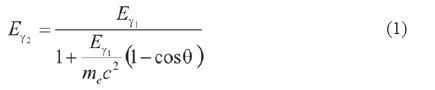
Imaging  systems using the Compton scattering can also determine the position and the source  activity. Events can be selected by energy through the sum of the energies deposited  in two detectors placed at some distance in the <img src="/img/revistas/nuc/n51/e02025112.jpg" width="13" height="14">-emitting  radionuclide field of view. First detector can measure the kinetic energy deposited  by the incident <img src="/img/revistas/nuc/n51/e02025112.jpg" width="14" height="14">  quantum <img src="/img/revistas/nuc/n51/e06025112.jpg" width="20" height="20">,  its position and interaction time. A resulted <img src="/img/revistas/nuc/n51/e02025112.jpg" width="14" height="14">  quantum suffers Compton scattering and hits second detector where its energy <img src="/img/revistas/nuc/n51/e07025112.jpg" width="18" height="23">  is absorbed and recorded time of interaction and position of incidence. Having  time information from both detectors coincidence is registered. Measurements of  the position of interaction in each detector determine the direction of scattered  <img src="/img/revistas/nuc/n51/e02025112.jpg" width="15" height="17"> quantum  and having the magnitude of the energy deposited in the first detector the scattering  angle is calculated using <a href="#e08025112">formula</a> from Compton kinematics.</font></p>    
<p><img src="/img/revistas/nuc/n51/e08025112.jpg" width="380" height="71"><a name="e08025112"></a></p>    
<p><font size="2" face="Verdana, Arial, Helvetica, sans-serif">For  each event the emission is confined to the surface of a known cone angle (<img src="/img/revistas/nuc/n51/e09025112.jpg" width="12" height="17">),  whose apex is determined by the interaction point in the first detector and its  axis is defined by the line joining points of interaction in both detectors. The  sum of the kinetic energy of the electron and the scattered <img src="/img/revistas/nuc/n51/e02025112.jpg" width="14" height="14">  quantum is equal to the incident <img src="/img/revistas/nuc/n51/e02025112.jpg" width="14" height="14">quantum  energy. The surface of each cone is a measure of the location of the activity  and is obtained through online registration. Three-dimensional distribution of  activity is obtained by reconstruction from a large number of cones using appropriate  algorithms.</font></p>    
<p><font size="2" face="Verdana, Arial, Helvetica, sans-serif">  Both PET and Compton systems allow reaching more sensitivity than mechanically  collimated Single Photon Emission Tomography systems, but still have limitations  to achieve sub millimeter spatial resolution. Also, the complex process of image  reconstruction requires a large number of lines of response (LoRs) in the case  of PET and cone of response (CoRs) in Compton scattering. </font></p><h2><font size="2" face="Verdana, Arial, Helvetica, sans-serif">LoR-PET  and CoR-Compton intersection</font></h2>    <p><font size="2" face="Verdana, Arial, Helvetica, sans-serif">As  explained above the idea is to add a Compton imaging detector to a PET system  &nbsp;with the intention that not only have to be detected two 511 keV <img src="/img/revistas/nuc/n51/e02025112.jpg" width="12" height="15">  quanta in coincidence from the annihilation of positron but at least as well another  additional <img src="/img/revistas/nuc/n51/e02025112.jpg" width="14" height="14">  quantum with energy close to 1 MeV. The principle of calculating the emission  area is based on the estimation of the intersection of the LoR-PET with CoR-Compton  as shown in <a href="#f01025112">figure 1</a>.</font></p>    
<p ALIGN="CENTER"><font size="2" face="Verdana, Arial, Helvetica, sans-serif"><img src="/img/revistas/nuc/n51/f01025112.jpg" width="311" height="183"><a name="f01025112"></a>    
<br>  </font></p>    ]]></body>
<body><![CDATA[<p><font size="2" face="Verdana, Arial, Helvetica, sans-serif">The  proposal is that using the<em> <img src="/img/revistas/nuc/n51/e21025112.jpg" width="32" height="17"></em>coincidences  obtained with specific radionuclides that emit a positron and a <img src="/img/revistas/nuc/n51/e02025112.jpg" width="12" height="15">  quantum practically at the same time the location of the emitter source could  be measured &ldquo;in three dimensions&rdquo;. </font></p>    
<p><font size="2" face="Verdana, Arial, Helvetica, sans-serif">Simulated  &quot;PET-Compton&quot; system consists of two detector rings: smaller inner diameter  ring as first (front) Compton scattering detector and an external ring with dual  function, second (rear) Compton detector and PET detector. To achieve optimum  performance of this system detectors placed in both rings must have the best possible  parameters in terms of spatial, temporal and energy resolution. In theory each  ring can separately form LoR-PET but here just the LoRs formed in the outer ring  will be considered.</font></p>    <p><font size="2" face="Verdana, Arial, Helvetica, sans-serif">  
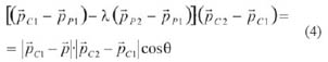
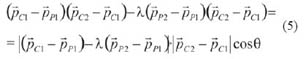
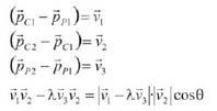
In the absence of errors is analytically possible to reconstruct the emission  point P (<a href="#f02025112">figure 2)</a> using the points of interaction in  PET <img src="/img/revistas/nuc/n51/e10025112.jpg" width="47" height="18"><em>,</em>  the interaction points of the Compton events <img src="/img/revistas/nuc/n51/e11025112.jpg" width="52" height="15">  and the Compton angle<img src="/img/revistas/nuc/n51/e09025112.jpg" width="12" height="17">.</font></p>    
<p ALIGN="CENTER"><img src="/img/revistas/nuc/n51/f02025112.jpg" width="310" height="170"><a name="f02025112"></a></p>    
<p><font size="2" face="Verdana, Arial, Helvetica, sans-serif">The  searched <a href="#e13025112">point P</a> is located in a straight line connecting  <img src="/img/revistas/nuc/n51/e36025112.jpg" width="20" height="20">, <img src="/img/revistas/nuc/n51/e37025112.jpg" width="23" height="20">  and is defined by the parameter <em><img src="/img/revistas/nuc/n51/e12025112.jpg" width="13" height="19"></em>,</font></p>    
<p><img src="/img/revistas/nuc/n51/e13025112.jpg" width="303" height="37"><a name="e13025112"></a></p>    
<p><font size="2" face="Verdana, Arial, Helvetica, sans-serif">Based  on geometrical considerations the following <a href="#e14025112">expression is  obtained</a>:</font></p>    <p><img src="/img/revistas/nuc/n51/e14025112.jpg" width="307" height="32"><a name="e14025112"></a></p>    
<p><font size="2" face="Verdana, Arial, Helvetica, sans-serif">And  <a href="#e15025112">substituing</a> <a href="#e13025112">(2)</a> in <a href="#e14025112">(3)</a></font>:  </p>    <p><img src="/img/revistas/nuc/n51/e15025112.jpg" width="302" height="57"><a name="e15025112"></a>  </p>    
]]></body>
<body><![CDATA[<p><font size="2" face="Verdana, Arial, Helvetica, sans-serif"><a href="#e16025112">This  provides</a>:</font></p>    <p><img src="/img/revistas/nuc/n51/e16025112.jpg" width="306" height="59"><a name="e16025112"></a></p>    
<p><font size="2" face="Verdana, Arial, Helvetica, sans-serif">Introducing  the <a href="#e17025112">following definitions</a>:</font></p>    <p><img src="/img/revistas/nuc/n51/e17025112.jpg" width="198" height="101"><a name="e17025112"></a></p>    
<p><font size="2" face="Verdana, Arial, Helvetica, sans-serif">Making  <img src="/img/revistas/nuc/n51/e18025112.jpg" width="71" height="25">and  rising to square both <a href="#e19025112">expressions</a>:</font></p>    
<p><img src="/img/revistas/nuc/n51/e19025112.jpg" width="299" height="32"><a name="e19025112"></a></p>    
<p><font size="2" face="Verdana, Arial, Helvetica, sans-serif">Thus  developing the <a href="#e20025112">square binomial</a>:</font></p>    <p><img src="/img/revistas/nuc/n51/e20025112.jpg" width="313" height="171"><a name="e20025112"></a></p>    
<p><font size="2" face="Verdana, Arial, Helvetica, sans-serif">The  above expression is a <a href="#e23025112">quadratic equation</a> in <em><img src="/img/revistas/nuc/n51/e12025112.jpg" width="13" height="19">:</em></font></p>    
<p><img src="/img/revistas/nuc/n51/e23025112.jpg" width="114" height="26"><a name="e23025112"></a></p>    
]]></body>
<body><![CDATA[<p><font size="2"><a href="#e22025112"><font face="Verdana, Arial, Helvetica, sans-serif">Where</font></a></font></p>    <p><img src="/img/revistas/nuc/n51/e22025112.jpg" width="333" height="47"><a name="e22025112"></a></p>    
<p><font size="2" face="Verdana, Arial, Helvetica, sans-serif">The  solutions to this <a href="#e22025112">equation</a> can be:</font></p><ul>     <li><font size="2" face="Verdana, Arial, Helvetica, sans-serif">  Discriminant &lt;0, there is no real solution, the equation can not be solved  and consequently there is not intersection.</font></li>    <li><font size="2" face="Verdana, Arial, Helvetica, sans-serif">  Discriminant <em>= </em>0, there is one solution and the line is tangential to  the cone.</font></li>    <li><font size="2" face="Verdana, Arial, Helvetica, sans-serif">  Discriminant <em>&gt; </em>0, the line cuts the cone at two points. It is possible  to identify the origin of the source through statistics accumulated by the real  point source emission. </font></li>    </ul>    <p><font size="2" face="Verdana, Arial, Helvetica, sans-serif">Described  algorithm has been incorporated into the simulation program. Thus the position  of the emitting source is directly reconstructed in three dimensions, event by  event, allowing further examinations at low count rate and to monitor kinetics  of the injected radiopharmaceutical.&nbsp;&nbsp;&nbsp; </font></p><h2><font size="2" face="Verdana, Arial, Helvetica, sans-serif">Selection  of radionuclides</font></h2>    <p><font size="2" face="Verdana, Arial, Helvetica, sans-serif">The  suggested PET-Compton system assumes the finding of a positron-emitting radionuclide  with additional gamma radiation <img src="/img/revistas/nuc/n51/e21025112.jpg" width="32" height="17">.  Besides, herein selection was based on two more criteria:</font> </p><ul>     
<li><font size="2" face="Verdana, Arial, Helvetica, sans-serif">  Physical-nuclear characteristics and production methods. </font></li>    ]]></body>
<body><![CDATA[<li><font size="2" face="Verdana, Arial, Helvetica, sans-serif">  Chemical, radiochemical, biological behavior and history of use in biology and  medicine of radioactive compounds. </font></li>    </ul>    <p><font size="2" face="Verdana, Arial, Helvetica, sans-serif">  Taking into account also detection properties of the current systems selection  has been finally limited to those radionuclides with the following properties:  1) Energy of <em><img src="/img/revistas/nuc/n51/e01025112.jpg" width="17" height="13"></em>  decay as low as possible, to avoid uncertainty product of the distance traveled  by the <em><img src="/img/revistas/nuc/n51/e01025112.jpg" width="17" height="13"></em>  before annihilation 2) non-positron <img src="/img/revistas/nuc/n51/e02025112.jpg" width="14" height="17">  quantum have to be issued with high emission rate almost simultaneously with the  <em><img src="/img/revistas/nuc/n51/e01025112.jpg" width="17" height="13"></em>  so that the coincidence time is within the typical detector time resolution which  is in the order of 500 psec, 2) <img src="/img/revistas/nuc/n51/e02025112.jpg" width="14" height="17">  quantum should preferably be emitted alone to avoid background noise due to spurious  signals in the detector and 3) its energy should be greater than energy of <img src="/img/revistas/nuc/n51/e02025112.jpg" width="14" height="16">  quanta from the positron annihilation and around 1 MeV, this fact will allow better  Compton scattering angular resolution and consequently superior &quot;PET-Compton&quot;  spatial resolution at the emission point 4) Radionuclide half-life have to be  short to avoid excessive exposure to radiation but long enough to ensure the quality  of the image. 5) Daughter radionuclide must be stable, to prevent further emissions.</font></p>    
<p><font size="2" face="Verdana, Arial, Helvetica, sans-serif">  From a detailed analysis of the radionuclides that meet these parameters and including  also the interest on their current or previous use in biomedical research or medical  applications for present simulation of &quot;PET-Compton&quot; system were selected  <img src="/img/revistas/nuc/n51/e24025112.jpg" width="26" height="13"> [4]  , <img src="/img/revistas/nuc/n51/e25025112.jpg" width="21" height="14">  [5] and <img src="/img/revistas/nuc/n51/e04025112.jpg" width="28" height="14">  [6] radionuclide. <img src="/img/revistas/nuc/n51/e04025112.jpg" width="28" height="14">  has been included despite its long half-life (2.6 years), which not allow it use  in diagnostic imaging, because it has similar <em><img src="/img/revistas/nuc/n51/e01025112.jpg" width="17" height="13"></em>and  <img src="/img/revistas/nuc/n51/e02025112.jpg" width="12" height="15"> quantum  energy emission range to other two selected radionuclides and also <img src="/img/revistas/nuc/n51/e04025112.jpg" width="28" height="14">  patterns available sources can be used in further experimental studies. Main characteristics  of these radioactive sources are shown in <a href="#t01025112">table 1</a> and  <a href="#t02025112">2</a>.</font></p>    
<p ALIGN="CENTER"><img src="/img/revistas/nuc/n51/t01025112.jpg" width="354" height="155"><a name="t01025112"></a></p>    
<p ALIGN="CENTER"><img src="/img/revistas/nuc/n51/t02025112.jpg" width="354" height="145"><a name="t02025112"></a></p>    
<p>&nbsp;</p>    <p><font size="2" face="Verdana, Arial, Helvetica, sans-serif"><strong>MATERIALS  AND METHODS&nbsp;&nbsp;&nbsp;&nbsp;&nbsp;&nbsp;&nbsp;&nbsp;&nbsp;&nbsp;&nbsp;&nbsp;&nbsp;&nbsp;&nbsp;&nbsp;&nbsp;&nbsp;&nbsp;&nbsp;&nbsp;&nbsp;&nbsp;&nbsp;&nbsp;&nbsp;&nbsp;&nbsp;&nbsp;&nbsp;&nbsp;&nbsp;&nbsp;&nbsp;&nbsp;&nbsp;&nbsp;&nbsp;&nbsp;&nbsp;&nbsp;&nbsp;&nbsp;&nbsp;&nbsp;&nbsp;&nbsp;&nbsp;&nbsp;&nbsp;&nbsp;&nbsp;&nbsp;&nbsp;&nbsp;&nbsp;&nbsp;&nbsp;&nbsp;&nbsp;&nbsp;&nbsp;&nbsp;&nbsp;&nbsp;&nbsp;&nbsp;&nbsp;&nbsp;  </strong></font></p><h1><font size="2" face="Verdana, Arial, Helvetica, sans-serif">Monte  Carlo simulation for &ldquo;PET-Compton&rdquo; - &quot;ClearPET-CZT&quot; system</font></h1>    <p><font size="2" face="Verdana, Arial, Helvetica, sans-serif">For  assessing &ldquo;PET-Compton&rdquo; possibility of obtaining better spatial resolution  than PET there were simulated both systems &nbsp;Geant4 code by means of GAMOS  framework [7].</font></p>    <p><font size="2" face="Verdana, Arial, Helvetica, sans-serif">  For physical processes GAMOS uses the extension to low energies (up to 250 eV)  of electromagnetic interactions. In the case of <em><img src="/img/revistas/nuc/n51/e01025112.jpg" width="17" height="13"></em>  includes the process of annihilation with the electrons of the medium, according  to their energy. For electrons and <img src="/img/revistas/nuc/n51/e02025112.jpg" width="14" height="14">  quanta takes into account the relevant electromagnetic processes in the energy  interval of interest, in this case, Photoelectric, Compton scattering and Raleigh  effects for <img src="/img/revistas/nuc/n51/e02025112.jpg" width="12" height="15"><em>  </em>quanta and bremsstrahlung and ionization for electrons [8].</font></p>    
]]></body>
<body><![CDATA[<p><font size="2" face="Verdana, Arial, Helvetica, sans-serif">  Detailed analysis of data was made incorporating to GAMOS the mathematical algorithm  of LoR-PET CoR-Compton intersection described in section 2.1. &quot;PET-Compton&quot;  results were compared with the results obtained in PET system for the same activity  and volume of radiation sources. For reconstruction in PET Single Slice ReBinning  (SSRB) analytical method was used.</font></p><h2><font size="2" face="Verdana, Arial, Helvetica, sans-serif">&nbsp;PET-Compton  configuration</font></h2>    <p><font size="2" face="Verdana, Arial, Helvetica, sans-serif">Simulated  &quot;PET-Compton&quot; system consists in two concentric detector rings. The  outer ring is a PET for small animals named &quot;ClearPET&quot; with high spatial  resolution (1.25 mm nominal) that uses scintillators detectors of LYSO-LuYAP [9].  This detector ring operates as positron annihilation detector and also as a second  Compton scattering detector. The inner ring covers the entire &quot;ClearPET&quot;  field of view and it is formed from an array of CdZnTe (CZT) pixelated semiconductor  detectors as a front Compton scattering detector. Because it is very important  to know as accurate as possible the position of incidence and energy deposited  by <img src="../img/e02025112.jpg" width="14" height="15"> quantum in Compton  scattering it has pixels size 300 X 300<em> </em>&micro;m and high energy resolution  (<em>~</em>1,5%). Two implemented in practice &quot;Clear-PET&quot; detector ring  diameters (<em>295</em> and <em>135</em> <em>mm</em>) were evaluated. Perspective  view of the simulated geometries is shown in <a href="#f03025112">figure 3</a>.&nbsp;</font></p>    <p ALIGN="CENTER"><img src="/img/revistas/nuc/n51/f03025112.jpg" width="354" height="201"><a name="f03025112"></a></p>    
<p><font size="2" face="Verdana, Arial, Helvetica, sans-serif">Real  &quot;ClearPET&quot; parameters obtained from the producer engineering drawings  were introduced in the simulation. They are the following:&nbsp;&nbsp; </font></p><ul>      <li><font size="2" face="Verdana, Arial, Helvetica, sans-serif"> Diameter: 295  mm and 135 mm. </font></li>    <li><font size="2" face="Verdana, Arial, Helvetica, sans-serif">  Axial field of view: 110 mm. </font></li>    <li><font size="2" face="Verdana, Arial, Helvetica, sans-serif">  20 modules (4 rings with 8x8 matrixes each coupled to photomultiplier) placed  radial. </font></li>    <li><font size="2" face="Verdana, Arial, Helvetica, sans-serif">  Detectors: Arrays of 8 x 8 dual-layer scintillating crystals 2 x 2 x 20 <img src="/img/revistas/nuc/n51/e26025112.jpg" width="34" height="15">  each (10 mm thick LYSO: Ce and 10 mm thick LuYAP: Ce), <em><img src="/img/revistas/nuc/n51/e27025112.jpg" width="44" height="19"></em>  reflector. </font></li>    
<li><font size="2" face="Verdana, Arial, Helvetica, sans-serif">  Measurement units: 5120 (8 x 8 x 4 x 20). </font></li>    <li><font size="2" face="Verdana, Arial, Helvetica, sans-serif">  Scintillating crystals units: 10240 (5120 x 2). </font></li>    ]]></body>
<body><![CDATA[<li><font size="2" face="Verdana, Arial, Helvetica, sans-serif">  Z axis rotation: 0.1&deg;, each 2778 events.     <br> </font></li>    </ul>    <p><font size="2" face="Verdana, Arial, Helvetica, sans-serif">Also  the axial modules displacement of 7 mm has been introduced in the simulation and  only active radiation detectors and<em> <img src="/img/revistas/nuc/n51/e27025112.jpg" width="44" height="19"></em>  reflectors were considered.</font></p>    
<p><font size="2" face="Verdana, Arial, Helvetica, sans-serif">CZT  detectors parameters used for simulations were the following: </font></p><ul>      <li><font size="2" face="Verdana, Arial, Helvetica, sans-serif"> Ring diameter:  50 mm. </font></li>    <li><font size="2" face="Verdana, Arial, Helvetica, sans-serif">  5 rings of 15 detectors (10 x 10 x 5 <img src="/img/revistas/nuc/n51/e26025112.jpg" width="34" height="15">).  </font></li>    
<li><font size="2" face="Verdana, Arial, Helvetica, sans-serif"> Detectors  - Arrays of 32 x 32 pixels (300 x 300) &micro;m x 5mm. </font></li>    <li><font size="2" face="Verdana, Arial, Helvetica, sans-serif">  Measurement units 75 (5 x 15). </font></li>    <li><font size="2" face="Verdana, Arial, Helvetica, sans-serif">  CZT Units -76 800 pixels (32 x 32 x 15 x 5).    ]]></body>
<body><![CDATA[<br> </font></li>    </ul>    <p><font size="2" face="Verdana, Arial, Helvetica, sans-serif">Details  of CZT geometry is shown in <a href="#f04025112">figure 4</a>. and detection system  characteristics introduced in the simulation are shown in <a href="#t03025112">table  3</a>. </font></p>    <p ALIGN="CENTER"><img src="/img/revistas/nuc/n51/f04025112.jpg" width="357" height="158"><a name="f04025112"></a></p>    
<p ALIGN="CENTER"><img src="/img/revistas/nuc/n51/t03025112.jpg" width="355" height="154"><a name="t03025112"></a></p>    
<p><font size="2" face="Verdana, Arial, Helvetica, sans-serif">  For the simulation there were used points and spherical (1 mm) <img src="/img/revistas/nuc/n51/e24025112.jpg" width="26" height="17">,  <img src="/img/revistas/nuc/n51/e25025112.jpg" width="21" height="14"> and  <img src="/img/revistas/nuc/n51/e04025112.jpg" width="28" height="14"> sources  placed in a 4 cm diameter water phantom. In order to visualize the spatial resolution  limit simulation was done also for the case of two radioactive sources located  at a distance between them slightly larger than positron average range (<img src="/img/revistas/nuc/n51/e24025112.jpg" width="26" height="13">-2,5  mm, <img src="/img/revistas/nuc/n51/e25025112.jpg" width="21" height="14">-1,7  mm, <img src="/img/revistas/nuc/n51/e04025112.jpg" width="28" height="14">-0,7  mm). We selected a value of source activity of 10 mCi (370 MBq) and the total  number of decays analyzed is 370x<img src="/img/revistas/nuc/n51/e28025112.jpg" width="20" height="13">,  i.e. the output values correspond to 1 second measurement. The selection of the  number of decays analyzed was determined by high consumption of time and computing  resources.</font></p>    
<p>&nbsp;</p>    <p><font size="2" face="Verdana, Arial, Helvetica, sans-serif"><strong><FONT SIZE="3">RESULTS</FONT></strong></font></p>    <p><font size="2" face="Verdana, Arial, Helvetica, sans-serif">Comparison  was made for two of the basic parameters that characterize medical imaging systems:  spatial resolution (FWHM) and global efficiency. Spatial resolution was determined  as the full width half maximum of the Point Spread Function (PSF) peak of radionuclide  emission. For &quot;PET-Compton&quot; was obtained by the statistical accumulation  of points from LoR-PET CoR-Compton intersection and from the reconstructed events  in PET. Global efficiency was determined as the ratio between the number of &quot;PET-Compton&quot;  or PET events produced by simulation and the number of total events launched.  <a href="#f05025112">Figure 5</a> shows one and two spherical sources placed in  water phantom inside the CZT detector ring. </font></p>    <p ALIGN="CENTER"><img src="/img/revistas/nuc/n51/f05025112.jpg" width="353" height="184"><a name="f05025112"></a></p>    
]]></body>
<body><![CDATA[<p><font size="2" face="Verdana, Arial, Helvetica, sans-serif"><a href="#f06025112">Figure  6</a> shows the magnified view of positron tracking (dashed blue), PET positron  annihilation lines (green) and electron scattering (red lines) in water phantom  for two point sources of a) <img src="/img/revistas/nuc/n51/e24025112.jpg" width="26" height="13">,  b) <img src="/img/revistas/nuc/n51/e25025112.jpg" width="21" height="14">  and c) <img src="/img/revistas/nuc/n51/e04025112.jpg" width="28" height="14">.  The difference of the positron range for each radionuclide is simply recognized.  </font></p>    
<p ALIGN="CENTER"><img src="/img/revistas/nuc/n51/f06025112.jpg" width="355" height="199"><a name="f06025112"></a></p>    
<p><font size="2" face="Verdana, Arial, Helvetica, sans-serif"><strong>Spatial  resolution (FWHM)</strong></font></p>    <p><font size="2" face="Verdana, Arial, Helvetica, sans-serif">  Spatial resolution values were calculated using AMIDE (Medical Image Data Examiner)  [10] software, a tool for the analysis of multidimensional medical data. PET and  PET-Compton FWHM values for both ClearPET diameters are plotted in <a href="#f07025112">figure  7</a>. </font></p>    <p ALIGN="CENTER"><img src="/img/revistas/nuc/n51/f07025112.jpg" width="357" height="309"><a name="f07025112"></a></p>    
<p><font size="2" face="Verdana, Arial, Helvetica, sans-serif">For  better interpretation the obtained results are shown in <a href="#t04025112">table  4</a> where we can corroborate that in all cases PET-Compton spatial resolution  is improved in comparison with spatial resolution obtained for PET only systems  using the same radionuclides and simulations parameters. </font></p>    <p ALIGN="CENTER"><a></a><img src="/img/revistas/nuc/n51/t04025112.jpg" width="307" height="143"><a name="t04025112"></a></p>    
<p><font size="2" face="Verdana, Arial, Helvetica, sans-serif">  Best values of spatial resolution are obtained in PET-Compton settings with greater  distance between the ClearPET and CZT rings (maximum &quot;ClearPET&quot; diameter)  and it went below 1 mm for <img src="/img/revistas/nuc/n51/e04025112.jpg" width="28" height="14">  due to its lower <em><img src="/img/revistas/nuc/n51/e01025112.jpg" width="17" height="13"></em>  range. </font></p>    
<p><font size="2" face="Verdana, Arial, Helvetica, sans-serif">In  order to see the difference, <a href="#f08025112">figure 8</a> shows the obtained  results in AMIDE's visualization screens for PET-Compton (top) and PET (bottom),  under the same conditions for simulated <img src="/img/revistas/nuc/n51/e24025112.jpg" width="26" height="13">  and <img src="/img/revistas/nuc/n51/e25025112.jpg" width="21" height="14">  radionuclides.</font></p>    
<p ALIGN="CENTER"><font size="2" face="Verdana, Arial, Helvetica, sans-serif"><img src="/img/revistas/nuc/n51/f08025112.jpg" width="354" height="255"><a name="f08025112"></a>      
]]></body>
<body><![CDATA[<br> </font></p>    <p><font size="2" face="Verdana, Arial, Helvetica, sans-serif">
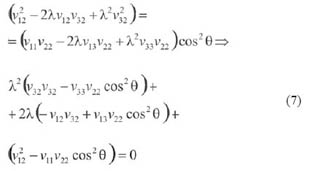
In  fact for PET-Compton system there is a background of less intense spots corresponding  to the second point of intersection LoR-PET CoR-Compton (<a href="#e22025112">see  equation 8</a>). However, because its position is random while the accumulation  in the real point of emission is high the effect on the screen has been removed  rising the lower visualization threshold. </font></p>    <p><font size="2" face="Verdana, Arial, Helvetica, sans-serif">Rendering  view of two <img src="/img/revistas/nuc/n51/e04025112.jpg" width="28" height="14">  point sources in PET-Compton system with better performance (maximum &quot;PET&quot;  diameter) placed at 0,7 mm is shown in <a href="#f09025112">Figure 9a</a>). For  comparison, in <a href="#f09025112">Figure 9b</a>) is shown the same view of these  two sources in PET system with better resolution (minimum &quot;ClearPET&quot;  diameter). Just in the first case two point sources separation can be insinuated.  </font></p>    
<p ALIGN="CENTER"><font size="2" face="Verdana, Arial, Helvetica, sans-serif"><img src="/img/revistas/nuc/n51/f09025112.jpg" width="356" height="236"><a name="f09025112"></a></font></p>    
<p><font size="2" face="Verdana, Arial, Helvetica, sans-serif">  FWHM simulation analysis indicates that this &quot;PET-Compton&quot; configuration  evidences a possible area of radionuclide emissions much narrower and thus, improved  spatial resolution than PET. </font></p>    <p><font size="2" face="Verdana, Arial, Helvetica, sans-serif"><strong>Efficiency  analysis</strong></font></p>    <p><font size="2" face="Verdana, Arial, Helvetica, sans-serif">In  order to estimate global efficiency in &quot;PET-Compton&quot; system and compare  it with the efficiency obtained in PET the ratio among simulated events (PET,  &quot;PET-Compton&quot;) and total launched events under the same simulation conditions  was calculated. </font></p>    <p><font size="2" face="Verdana, Arial, Helvetica, sans-serif">Graphics  of global system efficiency for the events of each type are shown in <a href="#f10025112">Figure  10</a>. For better identification of the results in graphics <a href="#f10025112">b</a>)  and <a href="#f10025112">c</a>) global efficiency values in % of the three radionuclides  have been joined in dotted lines (maximum &quot;ClearPET&quot; diameter) and solid  lines (minimum &quot;ClearPET&quot; diameter). </font></p>    <p ALIGN="CENTER"><img src="/img/revistas/nuc/n51/f10025112.jpg" width="430" height="992"><a name="f10025112"></a></p>    
<p><font size="2" face="Verdana, Arial, Helvetica, sans-serif">The  events called <img src="/img/revistas/nuc/n51/e29025112.jpg" width="92" height="17">  are those in which all <em><img src="/img/revistas/nuc/n51/e02025112.jpg" width="12" height="14"></em>  quanta (<em>2<img src="/img/revistas/nuc/n51/e02025112.jpg" width="14" height="19"></em>-PET  and <em>1<img src="/img/revistas/nuc/n51/e02025112.jpg" width="12" height="18"></em>-Compton)  had come up from the same radioactive decay and in PET-Comptonvalid is added the  fact that LoR-PET CoR-Compton intersection point is close to the radionuclide  point of emission (<img src="/img/revistas/nuc/n51/e30025112.jpg" width="13" height="17">  1 mm).</font></p>    
]]></body>
<body><![CDATA[<p><font size="2" face="Verdana, Arial, Helvetica, sans-serif">  As can be seen efficiency varies over a wide range depending on the type of interaction  that occurs within the field of view. <a href="#f10025112">Figure 10a)</a> shows  PET global efficiency which is about 0.5% (4000-5000 coincidences in 1 million  events). <a href="#f10025112">Figure 10b)</a> shows total PET-Compton global efficiency  that is of the order of <img src="/img/revistas/nuc/n51/e31025112.jpg" width="31" height="13">  for the minimum &quot;ClearPET&quot; diameter and <img src="/img/revistas/nuc/n51/e32025112.jpg" width="36" height="14">  for the maximum &quot;ClearPET&quot; diameter. &nbsp;<a href="#f10025112">Figure  10c)</a> shows the global efficiency of &quot;valid&quot; PET-Compton events where  all <em><img src="../img/e02025112.jpg" width="12" height="14"></em> quanta are  from the same decay event and obtained by simulation intersection point are close  to the radionuclide emission point. PET-Compton &quot;valid&quot; global efficiency  is in the order of <img src="/img/revistas/nuc/n51/e33025112.jpg" width="35" height="12">  for the &quot;ClearPET&quot; minimum diameter and <img src="/img/revistas/nuc/n51/e34025112.jpg" width="38" height="15">  for the maximum diameter. Besides, it can be observed that in all analyzed cases  there is an increased efficiency for <img src="/img/revistas/nuc/n51/e25025112.jpg" width="21" height="14">  radionuclide, because it has two non-positron <em><img src="/img/revistas/nuc/n51/e02025112.jpg" width="12" height="14"></em>  quanta in cascade that have been analyzed as one event. This is acceptable taking  into consideration that in current detector systems it is not possible practically  to separate this type of events occurring so close in time. </font></p>    
<p><font size="2" face="Verdana, Arial, Helvetica, sans-serif">  This &quot;PET-Compton&quot; system analysis done by simulation shows limited  global efficiency values, particularly when the results are restricted to &quot;valid&quot;.  </font></p>    <p><font size="2" face="Verdana, Arial, Helvetica, sans-serif"> However  it is necessary to consider also the fact that due to the large amount of computing  resources and time required by the simulation program total number of decays analyzed  for each configuration is just 370 x <img src="/img/revistas/nuc/n51/e28025112.jpg" width="20" height="13">,  hence the output values correspond to 1 second measurement while reasonable measurement  time in these techniques are from 15 minutes to 1 hour approximately. The increase  in the number of events detected in &quot;valid&quot; PET-Compton, that is possible  to achieve increasing simulation time should improve signal to noise ratio and  consequently spatial resolution of the system.</font></p>    
<p>&nbsp;</p>    <p><font size="2" face="Verdana, Arial, Helvetica, sans-serif"><b><FONT SIZE="3">CONCLUSIONS</FONT></b></font></p>    <p><font size="2" face="Verdana, Arial, Helvetica, sans-serif">Mathematical  modeling has demonstrated that the image using &quot;three <em><img src="/img/revistas/nuc/n51/e02025112.jpg" width="12" height="14">  </em>quanta&quot; is expected sharper and could permit more accurate diagnosis.  However, the fact that it is necessary to register 4 coincidence events (two corresponding  to positron annihilation and two from Compton scattering) significantly limits  the global detection efficiency and can affect the spatial resolution for the  same statistical data. Thus further development of this method should include  the optimization of the factors that could increase global &quot;PET-Compton&quot;  efficiency, more specifically, geometric efficiency of the designed system (larger  distance between detectors rings and other detection configurations with more  efficiency for Compton scattering) is suggested.</font></p>    
<p><font size="2" face="Verdana, Arial, Helvetica, sans-serif">  Finally would be interesting to note that the simulated system design also allows  it use in both, PET and &quot;three <em><img src="/img/revistas/nuc/n51/e02025112.jpg" width="12" height="14"></em>  quanta&quot; mode. They are complementary and the second mode could be used as  a higher resolution &quot;lens&quot; when it is required in PET image providing  an additional option, certainly for molecular imaging and quite possibly for clinical  diagnosis.</font></p>    
<p>&nbsp;</p>    <p><font size="2" face="Verdana, Arial, Helvetica, sans-serif"><strong><FONT SIZE="3">ACKNOWLEDGEMENTS</FONT></strong></font></p>    <p><font size="2" face="Verdana, Arial, Helvetica, sans-serif">This  work was done under the ITACA* project and we would like to thanks CIEMAT for  the financial and technological support.</font></p>    ]]></body>
<body><![CDATA[<p>&nbsp;</p>    <p><font size="2" face="Verdana, Arial, Helvetica, sans-serif"><b><FONT SIZE="3">REFERENCES</FONT></b></font></p>    <p>&nbsp;</p>    <!-- ref --><p><font size="2" face="Verdana, Arial, Helvetica, sans-serif">1.  HART H. High-Resolution Radioisotopic Imaging System. Patent No. US 4833327. 1989.    <!-- ref --><br>  2. BAILEY DL, TOWNSEND DW, VALK PE, MAISEY MN. Positron emission tomography: basic  sciences. London: Springer-Verlag, 2005.    <!-- ref --><br> 3. SINGH M. An electronically collimates  gamma camera for single photon emission computed tomography. Part I: Theoretical  considerations and design criteria. Med. Phys. 1983; 10(4): 421-427.    <!-- ref --><br>   4. GRIGNON C, et. al. Nuclear Medical Imaging using beta + gamma coincidences    from 44Sc radio-nuclide with liquid xenon as detetcion medium. Nucl. Instr.    and Meth. in Phys. Res. 2007; A571: 142-145.    <!-- ref --><br>    5. &nbsp;IWATA R, IWAI  K, IDO T, KIMURA S. Preparation of no-carrier-added 48 V(IV) and 48V(V) for biological  tracer use. J Radioanal. Nucl Chem. 1989; 134(2): 303-309.    <!-- ref --><br> 6. DAHL LK, SMILAY  MG, SILVER L, SPRARAGEN S. Evidence for a Prolonged Biological Half-Life of Na22  in Patients with Hypertension. Circ. Res. 1962; 10: 313-320.    <!-- ref --><br> 7. ARCE P,&nbsp;  RATO P, LAGARES JI. GAMOS an easy and flexible framework for GEANT4 simulations.  2008 Nuclear Science Symposium. Medical Imaging Conference and 16th Room Temperature  Semiconductor Detector Workshop. Dresde, Germany, 19-25 October 2008.    <br> 8. FIRESTONE  RB, EKSTROM L. Table of radioactive isotopes LBNL Isotopes Project&mdash;LUNDS  Universitet. Version 2.1 January 2004. <A HREF="http://ie.lbl.gov/toi/" TARGET="_blank">http://ie.lbl.gov/toi/</A>.    <!-- ref --><br>  9.&nbsp; SEMPERE P, ROLDAN E, CHEREUL O, et. al. Raytest ClearPETTM, a new generation  of small animal PET scanner. Nucl. Instr.and Meth. 2007; A571: 498-501.    <!-- ref --><br> 10.  LOENING A, GAMBHIR S. AMIDE: A Free Software Tool for Multimodality Medical Image  Analysis. Mol. Imaging. 2003; 2(3): 131-137.    </font></p>    <p>&nbsp;</p>    <p>&nbsp;</p>     <p><font size="2" face="Verdana, Arial, Helvetica, sans-serif"><strong>Recibido:    </strong>18 de octubre de 2012    <br>   <strong>Aceptado:</strong> 10 de mayo de 2012</font></p>      ]]></body><back>
<ref-list>
<ref id="B1">
<label>1</label><nlm-citation citation-type="">
<person-group person-group-type="author">
<name>
<surname><![CDATA[HART]]></surname>
<given-names><![CDATA[H]]></given-names>
</name>
</person-group>
<source><![CDATA[High-Resolution Radioisotopic Imaging System: Patent No. US 4833327]]></source>
<year>1989</year>
</nlm-citation>
</ref>
<ref id="B2">
<label>2</label><nlm-citation citation-type="book">
<person-group person-group-type="author">
<name>
<surname><![CDATA[BAILEY]]></surname>
<given-names><![CDATA[DL]]></given-names>
</name>
<name>
<surname><![CDATA[TOWNSEND]]></surname>
<given-names><![CDATA[DW]]></given-names>
</name>
<name>
<surname><![CDATA[VALK]]></surname>
<given-names><![CDATA[PE]]></given-names>
</name>
<name>
<surname><![CDATA[MAISEY]]></surname>
<given-names><![CDATA[MN]]></given-names>
</name>
</person-group>
<source><![CDATA[Positron emission tomography: basic sciences]]></source>
<year>2005</year>
<publisher-loc><![CDATA[London ]]></publisher-loc>
<publisher-name><![CDATA[Springer-Verlag]]></publisher-name>
</nlm-citation>
</ref>
<ref id="B3">
<label>3</label><nlm-citation citation-type="journal">
<person-group person-group-type="author">
<name>
<surname><![CDATA[SINGH]]></surname>
<given-names><![CDATA[M]]></given-names>
</name>
</person-group>
<article-title xml:lang="en"><![CDATA[An electronically collimates gamma camera for single photon emission computed tomography. Part I: Theoretical considerations and design criteria]]></article-title>
<source><![CDATA[Med. Phys]]></source>
<year>1983</year>
<volume>10</volume>
<numero>(4)</numero>
<issue>(4)</issue>
<page-range>421-427</page-range></nlm-citation>
</ref>
<ref id="B4">
<label>4</label><nlm-citation citation-type="journal">
<person-group person-group-type="author">
<name>
<surname><![CDATA[GRIGNON]]></surname>
<given-names><![CDATA[C]]></given-names>
</name>
</person-group>
<article-title xml:lang="en"><![CDATA[Nuclear Medical Imaging using beta + gamma coincidences from 44Sc radio-nuclide with liquid xenon as detetcion medium]]></article-title>
<source><![CDATA[Nucl. Instr. and Meth. in Phys. Res]]></source>
<year>2007</year>
<volume>A571</volume>
<page-range>142-145</page-range></nlm-citation>
</ref>
<ref id="B5">
<label>5</label><nlm-citation citation-type="journal">
<person-group person-group-type="author">
<name>
<surname><![CDATA[IWATA]]></surname>
<given-names><![CDATA[R]]></given-names>
</name>
<name>
<surname><![CDATA[IWAI]]></surname>
<given-names><![CDATA[K]]></given-names>
</name>
<name>
<surname><![CDATA[IDO]]></surname>
<given-names><![CDATA[T]]></given-names>
</name>
<name>
<surname><![CDATA[KIMURA]]></surname>
<given-names><![CDATA[S]]></given-names>
</name>
</person-group>
<article-title xml:lang="en"><![CDATA[Preparation of no-carrier-added 48 V(IV) and 48V(V) for biological tracer use]]></article-title>
<source><![CDATA[J Radioanal. Nucl Chem]]></source>
<year>1989</year>
<volume>134</volume>
<numero>(2)</numero>
<issue>(2)</issue>
<page-range>303-309</page-range></nlm-citation>
</ref>
<ref id="B6">
<label>6</label><nlm-citation citation-type="journal">
<person-group person-group-type="author">
<name>
<surname><![CDATA[DAHL]]></surname>
<given-names><![CDATA[LK]]></given-names>
</name>
<name>
<surname><![CDATA[SMILAY]]></surname>
<given-names><![CDATA[MG]]></given-names>
</name>
<name>
<surname><![CDATA[SILVER]]></surname>
<given-names><![CDATA[L]]></given-names>
</name>
<name>
<surname><![CDATA[SPRARAGEN]]></surname>
<given-names><![CDATA[S]]></given-names>
</name>
</person-group>
<article-title xml:lang="en"><![CDATA[Evidence for a Prolonged Biological Half-Life of Na22 in Patients with Hypertension]]></article-title>
<source><![CDATA[Circ. Res]]></source>
<year>1962</year>
<volume>10</volume>
<page-range>313-320</page-range></nlm-citation>
</ref>
<ref id="B7">
<label>7</label><nlm-citation citation-type="confpro">
<person-group person-group-type="author">
<name>
<surname><![CDATA[ARCE]]></surname>
<given-names><![CDATA[P]]></given-names>
</name>
<name>
<surname><![CDATA[RATO]]></surname>
<given-names><![CDATA[P]]></given-names>
</name>
<name>
<surname><![CDATA[LAGARES]]></surname>
<given-names><![CDATA[JI]]></given-names>
</name>
</person-group>
<source><![CDATA[GAMOS an easy and flexible framework for GEANT4 simulations]]></source>
<year></year>
<conf-name><![CDATA[ 2008 Nuclear Science Symposium. Medical Imaging Conference and 16th Room Temperature Semiconductor Detector Workshop]]></conf-name>
<conf-date>19-25 October 2008</conf-date>
<conf-loc>Dresde </conf-loc>
</nlm-citation>
</ref>
<ref id="B8">
<label>8</label><nlm-citation citation-type="">
<person-group person-group-type="author">
<name>
<surname><![CDATA[FIRESTONE]]></surname>
<given-names><![CDATA[RB]]></given-names>
</name>
<name>
<surname><![CDATA[EKSTROM]]></surname>
<given-names><![CDATA[L]]></given-names>
</name>
</person-group>
<source><![CDATA[of radioactive isotopes LBNL Isotopes Project-LUNDS Universitet. Version 2.1]]></source>
<year>Janu</year>
<month>ar</month>
<day>y </day>
</nlm-citation>
</ref>
<ref id="B9">
<label>9</label><nlm-citation citation-type="journal">
<person-group person-group-type="author">
<name>
<surname><![CDATA[SEMPERE]]></surname>
<given-names><![CDATA[P]]></given-names>
</name>
<name>
<surname><![CDATA[ROLDAN]]></surname>
<given-names><![CDATA[E]]></given-names>
</name>
<name>
<surname><![CDATA[CHEREUL]]></surname>
<given-names><![CDATA[O]]></given-names>
</name>
</person-group>
<article-title xml:lang="en"><![CDATA[Raytest ClearPETTM, a new generation of small animal PET scanner]]></article-title>
<source><![CDATA[Nucl. Instr.and Meth]]></source>
<year>2007</year>
<volume>A571</volume>
<page-range>498-501</page-range></nlm-citation>
</ref>
<ref id="B10">
<label>10</label><nlm-citation citation-type="journal">
<person-group person-group-type="author">
<name>
<surname><![CDATA[LOENING]]></surname>
<given-names><![CDATA[A]]></given-names>
</name>
<name>
<surname><![CDATA[GAMBHIR]]></surname>
<given-names><![CDATA[S]]></given-names>
</name>
</person-group>
<article-title xml:lang="en"><![CDATA[AMIDE: A Free Software Tool for Multimodality Medical Image Analysis]]></article-title>
<source><![CDATA[Mol. Imaging]]></source>
<year>2003</year>
<volume>2</volume>
<numero>(3)</numero>
<issue>(3)</issue>
<page-range>131-137</page-range></nlm-citation>
</ref>
</ref-list>
</back>
</article>
