<?xml version="1.0" encoding="ISO-8859-1"?><article xmlns:mml="http://www.w3.org/1998/Math/MathML" xmlns:xlink="http://www.w3.org/1999/xlink" xmlns:xsi="http://www.w3.org/2001/XMLSchema-instance">
<front>
<journal-meta>
<journal-id>0864-215X</journal-id>
<journal-title><![CDATA[Revista Cubana de Ortopedia y Traumatología]]></journal-title>
<abbrev-journal-title><![CDATA[Rev Cubana Ortop Traumatol]]></abbrev-journal-title>
<issn>0864-215X</issn>
<publisher>
<publisher-name><![CDATA[Editorial Ciencias Médicas]]></publisher-name>
</publisher>
</journal-meta>
<article-meta>
<article-id>S0864-215X2009000200003</article-id>
<title-group>
<article-title xml:lang="es"><![CDATA[Artrodesis lumbosacra mediante el sistema TRAUSON]]></article-title>
<article-title xml:lang="en"><![CDATA[Lumbosacral arthrodesis by TRAUSON system]]></article-title>
<article-title xml:lang="fr"><![CDATA[Arthrodèse lombo-sacrée par système TRAUSON]]></article-title>
</title-group>
<contrib-group>
<contrib contrib-type="author">
<name>
<surname><![CDATA[Santos Coto]]></surname>
<given-names><![CDATA[Carlos]]></given-names>
</name>
<xref ref-type="aff" rid="A01"/>
</contrib>
<contrib contrib-type="author">
<name>
<surname><![CDATA[Rivas Hernández]]></surname>
<given-names><![CDATA[Rafael]]></given-names>
</name>
<xref ref-type="aff" rid="A01"/>
</contrib>
<contrib contrib-type="author">
<name>
<surname><![CDATA[Fleites Marrero]]></surname>
<given-names><![CDATA[Ernesto]]></given-names>
</name>
<xref ref-type="aff" rid="A01"/>
</contrib>
</contrib-group>
<aff id="A01">
<institution><![CDATA[,Servicio de Columna Vertebral Hospital Ortopédico Docente Fructuoso Rodríguez ]]></institution>
<addr-line><![CDATA[La Habana ]]></addr-line>
<country>Cuba</country>
</aff>
<pub-date pub-type="pub">
<day>00</day>
<month>12</month>
<year>2009</year>
</pub-date>
<pub-date pub-type="epub">
<day>00</day>
<month>12</month>
<year>2009</year>
</pub-date>
<volume>23</volume>
<numero>2</numero>
<fpage>0</fpage>
<lpage>0</lpage>
<copyright-statement/>
<copyright-year/>
<self-uri xlink:href="http://scielo.sld.cu/scielo.php?script=sci_arttext&amp;pid=S0864-215X2009000200003&amp;lng=en&amp;nrm=iso"></self-uri><self-uri xlink:href="http://scielo.sld.cu/scielo.php?script=sci_abstract&amp;pid=S0864-215X2009000200003&amp;lng=en&amp;nrm=iso"></self-uri><self-uri xlink:href="http://scielo.sld.cu/scielo.php?script=sci_pdf&amp;pid=S0864-215X2009000200003&amp;lng=en&amp;nrm=iso"></self-uri><abstract abstract-type="short" xml:lang="es"><p><![CDATA[INTRODUCCIÓN. En 1891 se colocó por vez primera una sutura de alambre de plata para la estabilización y fusión de la columna toracolumbar y desde entonces se han desarrollado múltiples técnicas para este propósito. En el 2008 se comenzó a utilizar en el Hospital Ortopédico «Fructuoso Rodríguez» el sistema TRAUSON, de procedencia china. El objetivo del presente artículo fue mostrar los resultados obtenidos con dicha técnica para el tratamiento de 6 pacientes con afección de la columna lumbosacra. MÉTODOS. Se revisaron las historias clínicas y estudios imagenológicos de pacientes con fusión lumbosacra en los que se utilizó el sistema transpedicular TRAUSON, entre abril del 2008 y enero del 2009. En este período se realizaron 18 instrumentaciones, 6 de las cuales incluían el segmento L5-S1 que es el grupo control del presente trabajo. Se correlacionaron el cuadro clínico pre y posoperatorio, y los estudios imagenológicos disponibles. Se analizaron los datos siguientes: sexo, edad, patología de base, tiempo quirúrgico y grado de satisfacción según los escalas EVA y de Oswestry. RESULTADOS. En el 100 % de los casos la evolución fue buena si se tiene en cuenta que se superaron los síntomas iniciales que motivaron la cirugía. Cinco pacientes presentaron dolor residual en la articulación sacroilíaca, que mejoró entre el segundo y tercer mes con tratamiento fisioterapéutico. No se utilizaron infiltraciones u otros métodos invasivos. Entre las complicaciones posquirúrgicas se registraron 2 casos de reacción al material de sutura, y no hubo ni sepsis ni dehiscencia de la herida quirúrgica. CONCLUSIONES. El sistema TRAUSON permite una fijación estable útil para el tratamiento de patologías en esta zona y permite también una movilización precoz de los pacientes. Es altamente aceptado por éstos.]]></p></abstract>
<abstract abstract-type="short" xml:lang="en"><p><![CDATA[INTRODUCTION: In 1981 for the first time a silver wire suture was placed for stabilization and fusion of thoracolumbar spine and from then multiple techniques have been developed to this end. At 2008 the TRAUSON system was used for the first time in &#8220;Fructuoso Rodríguez&#8221; Orthopedics Hospital originating in China. The aim of present article was to shows the results obtained with this technique for treatment of 6 patients presenting with lumbosacral spine affection. METHODS: We made a review of medical records and imaging studies from patients with lumbosacral fusion using the TRAUSON system from April, 2008 to 2009. During this period 18 instrumentations were performed, where six included the L5-S1 segment (control group). Preoperative and postoperative clinical picture were correlated with the available imaging studies. Following data were analyzed: sex, age, baseline pathology, surgical time and satisfaction level according the EVA and Oswestry scales. RESULTS: In the 100% of cases the course was good if we considered that first symptoms causing surgery were overcome. Five patients had residual pain in sacroiliac joint improving between the second and the third month with physiotherapy. Neither infiltrations nor other invasive methods were used. Among the postsurgical complications were two cases of reaction to suture material without sepsis and surgical wound dehiscence. CONCLUSIONS: TRAUSON system allows a stable fixation useful for treatment of pathologies in this zone and also an early mobilization of patients. This system is very well accepted by patients.]]></p></abstract>
<abstract abstract-type="short" xml:lang="fr"><p><![CDATA[INTRODUCTION. En 1891, la suture avec fil en argent a été utilisée pour la première fois dans la stabilisation et la fusion du rachis thoraco-lombaire, et depuis lors de multiples techniques ont été développées à ce fin. En 2008, le système TRAUSON, d'origine chinoise, a commencé à être utilisé à l'Hópital d'orthopédie &#8220;Fructuoso Rodríguez&#8221;. Le but de cet article est de montrer les résultats obtenus avec cette dite technique dans le traitement de 6 patients avec une affection du rachis lombo-sacré. MÉTHODES. Tous les dossiers médicaux et les examens d'imagerie des patients atteints d'une fusion lombo-sacrée ont été examinés à l'aide du système transpédiculaire TRAUSON entre avril 2008 et janvier 2009. Dans cette période, on a réalisé 18 instrumentations, dont 6 ont inclus le segment L5-S1, étant le groupe-contrôle de cette étude. Les tableaux cliniques pré- et postopératoire et les examens d'imagerie disponibles ont été comparés. Les variables telles que sexe, âge, pathologie de base, temps chirurgical et grade de satisfaction, d'après les scales EVA et d'Oswestry, ont été aussi analysées. RÉSULTATS. Dans 100% des cas, l'évolution a été satisfaisante si on tient compte de l'amélioration des premiers symptômes causant la chirurgie. Cinq patients ont subi une douleur résiduelle au niveau de l'articulation sacro-ilîaque qui s'est soulagée en deux et trois mois avec kinésithérapie. On n'a pas utilisé d'infiltrations ni d'autres méthodes invasives. Parmi les complications postchirurgicales, on a trouvé deux cas de réactions au matériel de suture, mais il n'y a pas eu d'infection ni de déhiscence dans la blessure chirurgicale. CONCLUSIONS. Le système TRAUSON permet une fixation stable dans le traitement des pathologies de cette région anatomique, et permet aussi une mobilisation précoce des patients. Il est hautement accepté par eux.]]></p></abstract>
<kwd-group>
<kwd lng="es"><![CDATA[Artrodesis lumbosacra]]></kwd>
<kwd lng="es"><![CDATA[fusión posterior]]></kwd>
<kwd lng="es"><![CDATA[sistema TRAUSON]]></kwd>
<kwd lng="en"><![CDATA[Lumbosacral arthrodesis]]></kwd>
<kwd lng="en"><![CDATA[posterior fusion]]></kwd>
<kwd lng="en"><![CDATA[TRAUSON system]]></kwd>
<kwd lng="fr"><![CDATA[Arthrodèse lombo-sacrée]]></kwd>
<kwd lng="fr"><![CDATA[fusion postérieure]]></kwd>
<kwd lng="fr"><![CDATA[système TRAUSON]]></kwd>
</kwd-group>
</article-meta>
</front><body><![CDATA[ <div align="right"><font size="2" face="Verdana, Arial, Helvetica, sans-serif">    <B>ORIGINALES</B></font></div>     <P ALIGN="left"><font size="2"><b><font size="4" face="Verdana, Arial, Helvetica, sans-serif">    <br>       <br>       <br>       <br>   Artrodesis lumbosacra mediante el sistema TRAUSON   </font>   </b> </font>     <P ALIGN="left"><font size="2" face="Verdana, Arial, Helvetica, sans-serif"><strong><font size="3">Lumbosacral    arthrodesis by TRAUSON system</font></strong></font>      <P ALIGN="left"><font size="3" face="Verdana, Arial, Helvetica, sans-serif"><strong>Arthrod&egrave;se lombo-sacr&eacute;e  par syst&egrave;me TRAUSON</strong></font><font size="2" face="Verdana, Arial, Helvetica, sans-serif"><strong></strong></font>     <P ALIGN="left">    <br>         ]]></body>
<body><![CDATA[<br>         <br>         <br>   <font size="2"><b><font face="Verdana, Arial, Helvetica, sans-serif">Carlos Santos Coto,<SUP>I</SUP> Rafael Rivas    Hern&aacute;ndez,<SUP>II</SUP> Ernesto Fleites Marrero <SUP>III</SUP></font></b></font>      <P ALIGN="left"><font size="2" face="Verdana, Arial, Helvetica, sans-serif"><SUP>I</SUP> Especialista de I Grado en Ortopedia y Traumatolog&iacute;a. Instructor. Servicio de Columna    Vertebral. Hospital Ortop&eacute;dico Docente &#171;Fructuoso Rodr&iacute;guez&#187;. La Habana, Cuba. </font>       <br>   <font size="2" face="Verdana, Arial, Helvetica, sans-serif"><SUP>II</SUP> Especialista de I Grado en Ortopedia y Traumatolog&iacute;a. Instructor. Servicio de Columna    Vertebral. Hospital Ortop&eacute;dico Docente &#171;Fructuoso Rodr&iacute;guez&#187;. La Habana, Cuba. </font>       <br>   <font size="2" face="Verdana, Arial, Helvetica, sans-serif"><SUP>III</SUP> Especialista de I Grado en Ortopedia y Traumatolog&iacute;a. Instructor. Servicio de Columna    Vertebral. Hospital Ortop&eacute;dico Docente &#171;Fructuoso Rodr&iacute;guez&#187;. La Habana, Cuba. </font>     <P ALIGN="left">    <br>         <br>         <br>         ]]></body>
<body><![CDATA[<br> <hr size="1" noshade>       <P ALIGN="left"><font size="2" face="Verdana, Arial, Helvetica, sans-serif"><strong>RESUMEN</strong></font>     <P ALIGN="left"><strong><font size="2" face="Verdana, Arial, Helvetica, sans-serif">INTRODUCCI&Oacute;N.</font></strong><font size="2" face="Verdana, Arial, Helvetica, sans-serif"> En 1891 se coloc&oacute; por vez primera una sutura de alambre de plata para    la estabilizaci&oacute;n y fusi&oacute;n de la columna toracolumbar y desde entonces se han desarrollado    m&uacute;ltiples t&eacute;cnicas para este prop&oacute;sito. En el 2008 se comenz&oacute; a utilizar en el Hospital Ortop&eacute;dico    &#171;Fructuoso Rodr&iacute;guez&#187; el sistema TRAUSON, de procedencia china. El objetivo del presente art&iacute;culo fue    mostrar los resultados obtenidos con dicha t&eacute;cnica para el tratamiento de 6 pacientes con afecci&oacute;n de    la columna lumbosacra. </font>    <br>   <font size="2" face="Verdana, Arial, Helvetica, sans-serif"><B>M&Eacute;TODOS</B>. Se revisaron las historias cl&iacute;nicas y estudios imagenol&oacute;gicos de pacientes con    fusi&oacute;n lumbosacra en los que se utiliz&oacute; el sistema transpedicular TRAUSON, entre abril del 2008 y enero    del 2009. En este per&iacute;odo se realizaron 18 instrumentaciones, 6 de las cuales inclu&iacute;an el segmento    L5-S1 que es el grupo control del presente trabajo. Se correlacionaron el cuadro cl&iacute;nico pre y    posoperatorio, y los estudios imagenol&oacute;gicos disponibles. Se analizaron los datos siguientes: sexo, edad,    patolog&iacute;a de base, tiempo quir&uacute;rgico y grado de satisfacci&oacute;n seg&uacute;n los escalas EVA y de Oswestry. </font>       <br>   <font size="2" face="Verdana, Arial, Helvetica, sans-serif"><B>RESULTADOS</B>. En el 100 % de los casos la evoluci&oacute;n fue buena si se tiene en cuenta que se    superaron los s&iacute;ntomas iniciales que motivaron la cirug&iacute;a. Cinco pacientes presentaron dolor residual en    la articulaci&oacute;n sacroil&iacute;aca, que mejor&oacute; entre el segundo y tercer mes con tratamiento    fisioterap&eacute;utico. No se utilizaron infiltraciones u otros m&eacute;todos invasivos. Entre las complicaciones posquir&uacute;rgicas se   registraron 2 casos de reacci&oacute;n al material de sutura, y no hubo ni sepsis ni dehiscencia de la    herida quir&uacute;rgica. </font>       <br>   <font size="2" face="Verdana, Arial, Helvetica, sans-serif"><B>CONCLUSIONES</B>. El sistema TRAUSON permite una fijaci&oacute;n estable &uacute;til para el tratamiento    de patolog&iacute;as en esta zona y permite tambi&eacute;n una movilizaci&oacute;n precoz de los pacientes. Es    altamente aceptado por &eacute;stos. </font>     <P ALIGN="left"><font size="2" face="Verdana, Arial, Helvetica, sans-serif"><B>Palabras clave</B>: Artrodesis lumbosacra, fusi&oacute;n posterior, sistema TRAUSON. </font> <hr size="1" noshade>     <P ALIGN="left"><font size="2" face="Verdana, Arial, Helvetica, sans-serif"><strong>ABSTRACT</strong></font>     <P ALIGN="left"><font size="2" face="Verdana, Arial, Helvetica, sans-serif"><strong>INTRODUCTION</strong>: In 1981 for  the first time a silver wire suture was placed for stabilization and fusion of  thoracolumbar spine and from then multiple techniques have been developed to  this end. At 2008 the TRAUSON system was used for the first time in &ldquo;Fructuoso  Rodr&iacute;guez&rdquo; Orthopedics Hospital originating in China. The aim of present  article was to shows the results obtained with this technique for treatment of  6 patients presenting with lumbosacral spine affection.    <br>     <strong>METHODS</strong>: We made a review of medical  records and imaging studies from patients with lumbosacral fusion using the  TRAUSON system from April, 2008 to 2009. During this period 18 instrumentations  were performed, where six included the L5-S1 segment (control group).  Preoperative and postoperative clinical picture were correlated with the  available imaging studies. Following data were analyzed: sex, age, baseline  pathology, surgical time and satisfaction level according the EVA and Oswestry  scales.    ]]></body>
<body><![CDATA[<br>     <strong>RESULTS</strong>: In the 100% of cases the  course was good if we considered that first symptoms causing surgery were  overcome. Five patients had residual pain in sacroiliac joint improving between  the second and the third month with physiotherapy. Neither infiltrations nor  other invasive methods were used. Among the postsurgical complications were two  cases of reaction to suture material without sepsis and surgical wound dehiscence.    <br>     <strong>CONCLUSIONS</strong>: TRAUSON system  allows a stable fixation useful for treatment of pathologies in this zone and  also an early mobilization of patients. This system is very well accepted by  patients.</font>     <P ALIGN="left"><font size="2" face="Verdana, Arial, Helvetica, sans-serif"><strong>Key words</strong>:  Lumbosacral arthrodesis, posterior fusion, TRAUSON system. </font> <hr size="1" noshade>     <P ALIGN="left"><font size="2" face="Verdana, Arial, Helvetica, sans-serif"><strong>R&Eacute;SUM&Eacute;</strong></font>     <P ALIGN="left"><font size="2" face="Verdana, Arial, Helvetica, sans-serif"><strong>INTRODUCTION</strong>. En 1891, la suture avec fil en argent a &eacute;t&eacute; utilis&eacute;e pour la  premi&egrave;re fois dans la stabilisation et la fusion du rachis thoraco-lombaire, et  depuis lors de multiples techniques ont &eacute;t&eacute;&nbsp;  d&eacute;velopp&eacute;es &agrave; ce fin. En 2008, le syst&egrave;me TRAUSON, d'origine chinoise, a  commenc&eacute; &agrave; &ecirc;tre utilis&eacute; &agrave; l'H&oacute;pital d'orthop&eacute;die &ldquo;Fructuoso Rodr&iacute;guez&rdquo;. Le but  de cet article est de montrer les r&eacute;sultats obtenus avec cette dite technique  dans le traitement de 6 patients avec une affection du rachis lombo-sacr&eacute;. </font>    <br> <font size="2" face="Verdana, Arial, Helvetica, sans-serif"><strong>M&Eacute;THODES</strong>. Tous les dossiers m&eacute;dicaux et les examens d'imagerie des  patients atteints d'une fusion lombo-sacr&eacute;e ont &eacute;t&eacute; examin&eacute;s &agrave; l'aide du  syst&egrave;me transp&eacute;diculaire TRAUSON entre avril 2008 et janvier 2009. Dans cette p&eacute;riode,  on a r&eacute;alis&eacute; 18 instrumentations, dont 6 ont inclus le segment L5-S1, &eacute;tant le  groupe-contr&ocirc;le de cette &eacute;tude. Les tableaux cliniques pr&eacute;- et postop&eacute;ratoire  et les examens d'imagerie disponibles ont &eacute;t&eacute; compar&eacute;s. Les variables telles  que sexe, &acirc;ge, pathologie de base, temps chirurgical et grade de satisfaction,  d'apr&egrave;s les scales EVA et d'Oswestry, ont &eacute;t&eacute; aussi analys&eacute;es. </font>    <br>   <font size="2" face="Verdana, Arial, Helvetica, sans-serif"><strong>R&Eacute;SULTATS</strong>. Dans 100% des cas, l'&eacute;volution a &eacute;t&eacute; satisfaisante si on tient  compte de l'am&eacute;lioration des premiers sympt&ocirc;mes causant la chirurgie. Cinq  patients ont subi une douleur r&eacute;siduelle au niveau de l'articulation  sacro-il&icirc;aque qui s'est soulag&eacute;e en deux et trois mois avec kin&eacute;sith&eacute;rapie. On  n'a pas utilis&eacute; d'infiltrations ni d'autres m&eacute;thodes invasives. Parmi les  complications postchirurgicales, on a trouv&eacute; deux cas de r&eacute;actions au mat&eacute;riel  de suture, mais il n'y a pas eu d'infection ni de d&eacute;hiscence dans la blessure  chirurgicale. </font>         <br> <font size="2" face="Verdana, Arial, Helvetica, sans-serif"><strong>CONCLUSIONS</strong>. Le syst&egrave;me TRAUSON permet une fixation stable dans le traitement  des pathologies de cette r&eacute;gion anatomique, et permet aussi une mobilisation  pr&eacute;coce des patients. Il est hautement accept&eacute; par eux. </font>     <P ALIGN="left"><font size="2" face="Verdana, Arial, Helvetica, sans-serif"><strong>Mots cl&eacute;s</strong>: Arthrod&egrave;se lombo-sacr&eacute;e, fusion post&eacute;rieure, syst&egrave;me TRAUSON. </font> <hr size="1" noshade>     <P ALIGN="left">    ]]></body>
<body><![CDATA[<br>         <br>         <br>         <br>   <font size="3" face="Verdana, Arial, Helvetica, sans-serif"><B>INTRODUCCI&Oacute;N</B></font>      <P ALIGN="left"><font size="2" face="Verdana, Arial, Helvetica, sans-serif">Ya desde el lejano 1891 cuando Hadra de Galveston colocara una sutura de alambre de plata en 8    en torno a ap&oacute;fisis espinosas para la estabilizaci&oacute;n y fusi&oacute;n en la columna    toracolumbar,<SUP>1 </SUP>se han desarrollado para este prop&oacute;sito m&uacute;ltiples t&eacute;cnicas, que comprenden desde las no    instrumentadas basadas en el aporte de injerto, hasta las instrumentadas con diferentes sistemas de placas y    osteos&iacute;ntesis transfacetarias, con moderna instrumentaci&oacute;n, tanto por v&iacute;a anterior como por v&iacute;a posterior,    con tornillos transpediculares.<SUP>2</SUP> </font>     <P ALIGN="left"><font size="2" face="Verdana, Arial, Helvetica, sans-serif">En 1953 Cloward report&oacute; por primera vez la fusi&oacute;n intervertebral de la columna lumbosacra,    utilizando un abordaje posterior.<SUP>3</SUP> Posteriormente fue Boucher en 1959 quien introdujo la fijaci&oacute;n con    tornillos transpediculares, en tanto que la t&eacute;cnica fue popularizada por Roy Camille en la d&eacute;cada    1960-1970.<SUP>4,5</SUP> En 1984 Cloward, bas&aacute;ndose en su experiencia de 45 a&ntilde;os, propone evitar la utilizaci&oacute;n    de procedimientos aislados como la laminectom&iacute;a, la nucleotom&iacute;a, etc., por su nula    utilidad.<SUP>6,7</SUP> Se producir&aacute; en adelante un r&aacute;pido desarrollo en el dise&ntilde;o de sistemas de instrumentaci&oacute;n. </font>     <P ALIGN="left"><font size="2" face="Verdana, Arial, Helvetica, sans-serif">
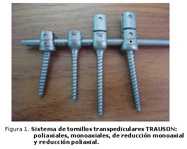
En    el 2002 se incorporan en Cuba estos modernos m&eacute;todos con la adquisici&oacute;n    del instrumental SOCON y SPINE SYSTEM para la Cl&iacute;nica &#171;Cira Garc&iacute;a&#187;;    el Complejo Cient&iacute;fico Internacional &#171;Frank Pa&iacute;s&#187; adquiri&oacute;    el sistema TSRH, y fueron estos los primeros utilizados en el pa&iacute;s.<SUP>8</SUP>    Posteriormente se introdujo esta t&eacute;cnica en otras unidades del Sistema    Nacional de Salud y a partir de abril del 2008 es que llega &eacute;sta al Hospital    Ortop&eacute;dico Docente &#171;Fructuoso Rodr&iacute;guez&#187;, con el sistema    TRAUSON, de procedencia china. La <a href="#fig1">figura 1</a> muestra distintos    modelos de tornillos transpediculares. </font>      <P ALIGN="center"><a name="fig1"></a><img src="/img/revistas/ort/v23n2/f0103209.jpg" width="374" height="298">      
<P ALIGN="left"><font size="2" face="Verdana, Arial, Helvetica, sans-serif"><B>    <br>           ]]></body>
<body><![CDATA[<br>       <font size="3">M&Eacute;TODOS</font></B> </font>      <P ALIGN="left"><font size="2" face="Verdana, Arial, Helvetica, sans-serif">Se revisaron las historias cl&iacute;nicas y estudios imagenol&oacute;gicos en el Servicio de Columna, T&oacute;rax    y Pelvis del Hospital Ortop&eacute;dico &#171;Fructuoso Rodr&iacute;guez&#187;, de pacientes con fusi&oacute;n lumbosacra en    los que se utiliz&oacute; el sistema transpedicular TRAUSON, en el per&iacute;odo comprendido entre abril del    2008 y enero del 2009.
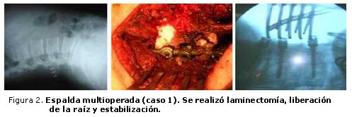
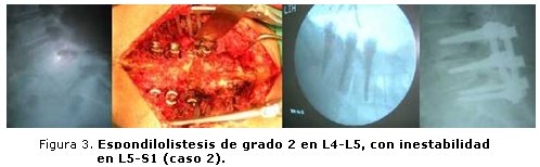
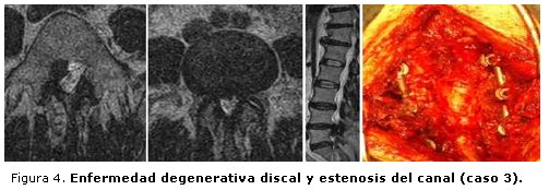
 </font>     <P ALIGN="left"><font size="2" face="Verdana, Arial, Helvetica, sans-serif">En    este per&iacute;odo se realizaron 18 instrumentaciones y 6 de ellas inclu&iacute;an    el segmento L5-S1 (<a href="#fig2">figuras 2</a>, <a href="#fig3">3</a> y <a href="#fig4">4</a>),    que es el grupo control del presente trabajo. Se correlacion&oacute; el cuadro    cl&iacute;nico pre y posoperatorio, as&iacute; como los estudios de imagen disponibles.    Se tomaron como base los datos siguientes: sexo, edad, patolog&iacute;a de base,    tiempo quir&uacute;rgico y grado de satisfacci&oacute;n seg&uacute;n las escalas    EVA y Oswestry utilizados. </font>      <P ALIGN="center"><a name="fig2"></a><img src="/img/revistas/ort/v23n2/f0203209.jpg" width="497" height="166">      
<P ALIGN="center"><a name="fig3"></a><img src="/img/revistas/ort/v23n2/f0303209.jpg" width="498" height="154">      
<P ALIGN="center"><a name="fig4"></a><img src="/img/revistas/ort/v23n2/f0403209.jpg" width="503" height="174">      
<P ALIGN="left">     <P ALIGN="left">    <br>         <br>   <font size="3" face="Verdana, Arial, Helvetica, sans-serif"><B>RESULTADOS</B></font>     ]]></body>
<body><![CDATA[<P ALIGN="left"><font size="2" face="Verdana, Arial, Helvetica, sans-serif">De los 6 pacientes operados con el sistema TRAUSON de fijaci&oacute;n transpedicular, 2 son    hombres (33&#160;%) y 4 mujeres (67 %), con edades comprendidas entre los 46 a 63 a&ntilde;os, para una edad media    de 55 a&ntilde;os. </font>     <P ALIGN="left"><font size="2" face="Verdana, Arial, Helvetica, sans-serif">Seg&uacute;n patolog&iacute;as el grupo se comport&oacute; de la manera siguiente: </font> <ul>       <li><font size="2" face="Verdana, Arial, Helvetica, sans-serif" align="left">     Espondilolistesis: 3 pacientes; 50 %. </font>  </li>       <li><font size="2" face="Verdana, Arial, Helvetica, sans-serif" align="left">     Estenosis del canal: 2 pacientes; 34 %. </font>  </li>       <li><font size="2" face="Verdana, Arial, Helvetica, sans-serif" align="left">     Espalda multioperada: 1 paciente; 16 %. </font>  </li>     </ul>     <P ALIGN="left"><font size="2" face="Verdana, Arial, Helvetica, sans-serif">El tiempo quir&uacute;rgico oscil&oacute; entre 180 y 420 min, con un tiempo medio de 300 min. </font>     <P ALIGN="left"><font size="2" face="Verdana, Arial, Helvetica, sans-serif">Los segmentos incluidos fueron: </font> <ul>       <li><font size="2" face="Verdana, Arial, Helvetica, sans-serif" align="left">     L5-S1: 2 pacientes; 33 %. </font>  </li>       <li><font size="2" face="Verdana, Arial, Helvetica, sans-serif" align="left">     L4-S1: 4 pacientes; 67 %. </font>  </li>       ]]></body>
<body><![CDATA[<li><font size="2" face="Verdana, Arial, Helvetica, sans-serif" align="left">     L3-S1: ning&uacute;n paciente; 0 %. </font>  </li>     </ul>     <P ALIGN="left"><font size="2" face="Verdana, Arial, Helvetica, sans-serif">Los s&iacute;ntomas preoperatorios m&aacute;s frecuentes fueron dolor radicular (5 pacientes; 93 %);    parestesias en miembros inferiores (4 pacientes; 67 %); claudicaci&oacute;n neur&oacute;gena (2 pacientes; 33 %) y    disminuci&oacute;n de la fuerza muscular (4 pacientes; 67 %). </font>     <P ALIGN="left"><font size="2" face="Verdana, Arial, Helvetica, sans-serif">El    grado de satisfacci&oacute;n de los pacientes fue valorado mediante dos escalas:    Escala Visual An&aacute;loga (EVA) y la escala de Oswestry; ambas se compararon    seg&uacute;n los valores obtenidos en la encuesta pre y posoperatoria (<a href="#tab">tabla</a>).    </font>      <P ALIGN="center"><font size="2" face="Verdana, Arial, Helvetica, sans-serif"><a name="tab"></a>Tabla.    <B>Comparaci&oacute;n de los valores de las escalas EVA y de Oswestry</B> </font>      <div align="left">   <table border="1" align="center" cellpadding="0" cellspacing="3">     <tr>       <td width="167" valign="top">    <p align="center"><font size="2" face="Verdana, Arial, Helvetica, sans-serif">Escalas </font></p></td>       <td width="167" valign="top">    <p align="center"><font size="2" face="Verdana, Arial, Helvetica, sans-serif">Valores preoperatorios </font></p></td>       <td width="166" valign="top">    <p align="center"><font size="2" face="Verdana, Arial, Helvetica, sans-serif">Valores posoperatorios </font></p></td>     </tr>     <tr>       <td width="167" valign="top">    <p><font size="2" face="Verdana, Arial, Helvetica, sans-serif">Oswestry    lumbar </font></p></td>       <td width="167" valign="top">    ]]></body>
<body><![CDATA[<p align="center"><font size="2" face="Verdana, Arial, Helvetica, sans-serif">70,8 % </font></p></td>       <td width="166" valign="top">    <p align="center"><font size="2" face="Verdana, Arial, Helvetica, sans-serif">22,3 % </font></p></td>     </tr>     <tr>       <td width="167" valign="top">    <p><font size="2" face="Verdana, Arial, Helvetica, sans-serif">EVA    lumbar </font></p></td>       <td width="167" valign="top">    <p align="center"><font size="2" face="Verdana, Arial, Helvetica, sans-serif">8 </font></p></td>       <td width="166" valign="top">    <p align="center"><font size="2" face="Verdana, Arial, Helvetica, sans-serif">2 </font></p></td>     </tr>   </table> </div>     <P ALIGN="left">    <br>   <font size="2" face="Verdana, Arial, Helvetica, sans-serif">En el 100 % de los casos la sintomatolog&iacute;a que motiv&oacute; la cirug&iacute;a desapareci&oacute;. En 5 de los    pacientes (93 %) se present&oacute; un dolor residual en la articulaci&oacute;n sacroil&iacute;aca, que mejor&oacute; entre el segundo    y tercer mes despu&eacute;s de la operaci&oacute;n, con tratamiento fisioterap&eacute;utico. No se utilizaron infiltraciones    u otros m&eacute;todos invasivos. </font>      <P ALIGN="left"><font size="2" face="Verdana, Arial, Helvetica, sans-serif">Como complicaciones posquir&uacute;rgicas se present&oacute; la reacci&oacute;n al material de sutura en 2    pacientes, para un 33 %, y no hubo casos de sepsis ni dehiscencias de la herida quir&uacute;rgica. </font>     <P ALIGN="left"><font size="2" face="Verdana, Arial, Helvetica, sans-serif"><B>    <br>           ]]></body>
<body><![CDATA[<br>       <font size="3">DISCUSI&Oacute;N</font></B> </font>     <P ALIGN="left"><font size="2" face="Verdana, Arial, Helvetica, sans-serif">La inestabilidad cl&iacute;nica seg&uacute;n <I>Mc Cormick</I> se define como la p&eacute;rdida o franca reducci&oacute;n de la    integridad mec&aacute;nica de un segmento vertebral m&oacute;vil que se manifiesta por dolor local o s&iacute;ntomas    radiculares. Las condiciones cl&iacute;nicas en las cuales estos se presentan    son:<SUP>9</SUP> </font> <ul>       <li><font size="2" face="Verdana, Arial, Helvetica, sans-serif" align="left">     Inestabilidad iatrog&eacute;nica. </font>  </li>       <li><font size="2" face="Verdana, Arial, Helvetica, sans-serif" align="left">     Espondilitis degenerativa. </font>  </li>       <li><font size="2" face="Verdana, Arial, Helvetica, sans-serif" align="left">     Trauma. </font>  </li>       <li><font size="2" face="Verdana, Arial, Helvetica, sans-serif" align="left">     Enfermedad discal. </font>  </li>       <li><font size="2" face="Verdana, Arial, Helvetica, sans-serif" align="left">     Enfermedad articular degenerativa. </font>  </li>     </ul>     <P ALIGN="left"><font size="2" face="Verdana, Arial, Helvetica, sans-serif">Es el segmento lumbosacro y en especial la charnela lumbosacra (L5-S1), por su rango de    movimiento <FONT COLOR="#000000">de      60&#186; de flexi&oacute;n y 35&#186; de        extensi&oacute;n,<SUP>10</SUP> con la compresi&oacute;n axial a la cual est&aacute; sometida, es la causa    por la cual este segmento se vea frecuentemente comprometido por las patolog&iacute;as antes descritas. <I>Evans</I> en 1984 propone la fusi&oacute;n lumbar como mecanismo para mantener la estabilidad biomec&aacute;nica de    la columna </FONT>vertebral y de este modo prevenir los efectos irritativos por la movilidad del    segmento afectado.<SUP>11</SUP> </font>     <P ALIGN="left"><font size="2" face="Verdana, Arial, Helvetica, sans-serif">Estas t&eacute;cnicas por sus buenos resultados han resistido el paso del tiempo y son hasta el presente    el m&eacute;todo de referencia para el tratamiento de este segmento, ante lesiones que generan inestabilidad    en &eacute;l, a pesar de contar ya con sistemas din&aacute;micos que se acercan m&aacute;s a lo que fisiol&oacute;gicamente    ser&iacute;a una columna estable. </font>     ]]></body>
<body><![CDATA[<P ALIGN="left"><font size="2" face="Verdana, Arial, Helvetica, sans-serif">En s&iacute;ntesis, el sistema TRAUSON de fijaci&oacute;n transpedicular es eficaz y seguro para los    segmentos inferiores de la columna vertebral. Permite una movilizaci&oacute;n temprana de los pacientes, con un    m&iacute;nimo de s&iacute;ntomas y una frecuencia baja de complicaciones posoperatorias. En los casos aqu&iacute;    presentados no fue necesaria la utilizaci&oacute;n de cors&eacute; de yeso y los pacientes manifestaron un alto grado de    satisfacci&oacute;n con la cirug&iacute;a. </font>     <P ALIGN="left"><font size="2" face="Verdana, Arial, Helvetica, sans-serif"><B>    <br>           <br>       <font size="3">REFERENCIAS BIBLIOGR&Aacute;FICAS</font></B> </font>     <!-- ref --><P ALIGN="left"><font size="2" face="Verdana, Arial, Helvetica, sans-serif">1.     Edmonton AS, Crenshaw AH. Campbell: Cirug&iacute;a Ortop&eacute;dica. 6ta edici&oacute;n. T-4 Madrid:    Editorial M&eacute;dica Panamericana; 1994. </font>    <!-- ref --><P ALIGN="left"><font size="2" face="Verdana, Arial, Helvetica, sans-serif">2.     Cabello Gallardo J, Roca Burniol J, Iborra Gonz&aacute;lez M, V&aacute;zquez Solano JJ.    An&aacute;lisis biomec&aacute;nico de la doble fijaci&oacute;n h&iacute;brida en la columna lumbar. Trauma Fund    MAPFRE. 2008;19(2):74-82. </font>    <!-- ref --><P ALIGN="left"><font size="2" face="Verdana, Arial, Helvetica, sans-serif">3.     Cloward RB. The treatment of ruptured lumbar intervertebral disc by vertebral    interbody fusion. Neurosurgery. 1953;10:154-68. </font>    <!-- ref --><P ALIGN="left"><font size="2" face="Verdana, Arial, Helvetica, sans-serif">4.     Boucher HH. A method of spinal fusion. J  Bone Joint Surg. 1959;41B:248. </font>    <!-- ref --><P ALIGN="left"><font size="2" face="Verdana, Arial, Helvetica, sans-serif">5.     Floman Y, Argenson C. Thoracolumbar spine Fractures. New York: Raven Press, Ltd.; 1993. </font>    <!-- ref --><P ALIGN="left"><font size="2" face="Verdana, Arial, Helvetica, sans-serif">6.     Altamirano Alcocer C, Rodr&iacute;guez D&iacute;as D. Artrodesis lumbosacra mediante sistema    SOCON experiencia en el centro m&eacute;dico nacional 20 de Noviembre ISSSTE. Cir Ciruj. 1998;66:45-8. </font>    <!-- ref --><P ALIGN="left"><font size="2" face="Verdana, Arial, Helvetica, sans-serif">7.     Cloward RB. Posterior lumbar interbody fusion update. Clin Orth. 1985;193:16-9. </font>    <!-- ref --><P ALIGN="left"><font size="2" face="Verdana, Arial, Helvetica, sans-serif">8.     Rubinos RR, Fleites ME, Campos F R, Rodr&iacute;guez-Triana OJ. Adecuaci&oacute;n del    dispositivo sublaminar de Luque combin&aacute;ndolo con otros modernos 3D. XVI Foro de Ciencia y    T&eacute;cnica. Cl&iacute;nica Central Cira Garc&iacute;a; junio de 2006. </font>    <!-- ref --><P ALIGN="left"><font size="2" face="Verdana, Arial, Helvetica, sans-serif">9.     Mc Cormick PC. Indication and technique of Lumbar spine fusion in neurological surgery.    In: Youmans JR (ed): Youmans' Neurological Surgery.    Vol 3. Philadelphia: WB Saunders Co., 1982. </font>    <!-- ref --><P ALIGN="left"><font size="2" face="Verdana, Arial, Helvetica, sans-serif">10.     Rull Bartomeu M, Miralles RC. Dolor de espalda. Diagn&oacute;stico. Enfoque general del    tratamiento. Prevenci&oacute;n. Escuela de columna. En: Torres LM. Medicina del Dolor. Barcelona: Masson    S.A.; 1997. </font>    <!-- ref --><P ALIGN="left"><font size="2" face="Verdana, Arial, Helvetica, sans-serif">11.     Evans J, Geng B. Biomechanics of lumbar fusion. Clin Orth. 1985;193:38-46. </font>    <P ALIGN="left">    <br>         <br>         <br>         <br>   <font size="2" face="Verdana, Arial, Helvetica, sans-serif">Recibido: 9 de junio de 2009.    ]]></body>
<body><![CDATA[<br> </font><font size="2" face="Verdana, Arial, Helvetica, sans-serif">Aprobado: 5 de julio de 2009.</font>     <P ALIGN="left">    <br>         <br>         <br>   <font size="2" face="Verdana, Arial, Helvetica, sans-serif"><I>Carlos Santos Coto</I>. Servicio de Columna Vertebral. Hospital Ortop&eacute;dico Docente    &#171;Fructuoso Rodr&iacute;guez&#187;. Avenida de los Presidentes y calle 29, El Vedado. La Habana, Cuba. </font>         <br>   <font size="2" face="Verdana, Arial, Helvetica, sans-serif">Correo electr&oacute;nico: <a href="mailto:carlossantos@infomed.sld.cu">carlossantos@infomed.sld.cu</a></font>      ]]></body><back>
<ref-list>
<ref id="B1">
<label>1</label><nlm-citation citation-type="book">
<person-group person-group-type="author">
<name>
<surname><![CDATA[Edmonton]]></surname>
<given-names><![CDATA[AS]]></given-names>
</name>
<name>
<surname><![CDATA[Crenshaw]]></surname>
<given-names><![CDATA[AH.]]></given-names>
</name>
</person-group>
<source><![CDATA[Campbell:: Cirugía Ortopédica.]]></source>
<year>1994</year>
<edition>6ta edición</edition>
<publisher-loc><![CDATA[Madrid ]]></publisher-loc>
<publisher-name><![CDATA[Editorial Médica Panamericana]]></publisher-name>
</nlm-citation>
</ref>
<ref id="B2">
<label>2</label><nlm-citation citation-type="journal">
<person-group person-group-type="author">
<name>
<surname><![CDATA[Cabello Gallardo]]></surname>
<given-names><![CDATA[J]]></given-names>
</name>
<name>
<surname><![CDATA[Roca Burniol]]></surname>
<given-names><![CDATA[J]]></given-names>
</name>
<name>
<surname><![CDATA[Iborra González]]></surname>
<given-names><![CDATA[M]]></given-names>
</name>
<name>
<surname><![CDATA[Vázquez Solano]]></surname>
<given-names><![CDATA[JJ]]></given-names>
</name>
</person-group>
<article-title xml:lang="es"><![CDATA[Análisis biomecánico de la doble fijación híbrida en la columna lumbar]]></article-title>
<source><![CDATA[Trauma Fund MAPFRE.]]></source>
<year>2008</year>
<volume>19</volume>
<numero>2</numero>
<issue>2</issue>
<page-range>74-82</page-range></nlm-citation>
</ref>
<ref id="B3">
<label>3</label><nlm-citation citation-type="journal">
<person-group person-group-type="author">
<name>
<surname><![CDATA[Cloward]]></surname>
<given-names><![CDATA[RB]]></given-names>
</name>
</person-group>
<article-title xml:lang="en"><![CDATA[The treatment of ruptured lumbar intervertebral disc by vertebral interbody fusion]]></article-title>
<source><![CDATA[Neurosurgery.]]></source>
<year>1953</year>
<volume>10</volume>
<page-range>154-68</page-range></nlm-citation>
</ref>
<ref id="B4">
<label>4</label><nlm-citation citation-type="journal">
<person-group person-group-type="author">
<name>
<surname><![CDATA[Boucher]]></surname>
<given-names><![CDATA[HH]]></given-names>
</name>
</person-group>
<article-title xml:lang="en"><![CDATA[A method of spinal fusion]]></article-title>
<source><![CDATA[J Bone Joint Surg.]]></source>
<year>1959</year>
<volume>41B</volume>
<page-range>248</page-range></nlm-citation>
</ref>
<ref id="B5">
<label>5</label><nlm-citation citation-type="book">
<person-group person-group-type="author">
<name>
<surname><![CDATA[Floman]]></surname>
<given-names><![CDATA[Y]]></given-names>
</name>
<name>
<surname><![CDATA[Argenson]]></surname>
<given-names><![CDATA[C.]]></given-names>
</name>
</person-group>
<source><![CDATA[Thoracolumbar spine Fractures.]]></source>
<year>1993</year>
<publisher-loc><![CDATA[New York ]]></publisher-loc>
<publisher-name><![CDATA[Raven Press, Ltd]]></publisher-name>
</nlm-citation>
</ref>
<ref id="B6">
<label>6</label><nlm-citation citation-type="journal">
<person-group person-group-type="author">
<name>
<surname><![CDATA[Altamirano Alcocer]]></surname>
<given-names><![CDATA[C]]></given-names>
</name>
<name>
<surname><![CDATA[Rodríguez Días]]></surname>
<given-names><![CDATA[D]]></given-names>
</name>
</person-group>
<article-title xml:lang="es"><![CDATA[Artrodesis lumbosacra mediante sistema SOCON experiencia en el centro médico nacional 20 de Noviembre ISSSTE]]></article-title>
<source><![CDATA[Cir Ciruj.]]></source>
<year>1998</year>
<volume>66</volume>
<page-range>45-8</page-range></nlm-citation>
</ref>
<ref id="B7">
<label>7</label><nlm-citation citation-type="journal">
<person-group person-group-type="author">
<name>
<surname><![CDATA[Cloward]]></surname>
<given-names><![CDATA[RB]]></given-names>
</name>
</person-group>
<article-title xml:lang="en"><![CDATA[Posterior lumbar interbody fusion update]]></article-title>
<source><![CDATA[Clin Orth.]]></source>
<year>1985</year>
<volume>193</volume>
<page-range>16-9</page-range></nlm-citation>
</ref>
<ref id="B8">
<label>8</label><nlm-citation citation-type="confpro">
<person-group person-group-type="author">
<name>
<surname><![CDATA[Rubinos]]></surname>
<given-names><![CDATA[RR]]></given-names>
</name>
<name>
<surname><![CDATA[Fleites]]></surname>
<given-names><![CDATA[ME]]></given-names>
</name>
<name>
<surname><![CDATA[Campos]]></surname>
<given-names><![CDATA[F R]]></given-names>
</name>
<name>
<surname><![CDATA[Rodríguez-Triana]]></surname>
<given-names><![CDATA[OJ.]]></given-names>
</name>
</person-group>
<source><![CDATA[Adecuación del dispositivo sublaminar de Luque combinándolo con otros modernos 3D.]]></source>
<year></year>
<conf-name><![CDATA[XVI Foro de Ciencia y Técnica.]]></conf-name>
<conf-date>junio de 2006</conf-date>
<conf-loc> </conf-loc>
</nlm-citation>
</ref>
<ref id="B9">
<label>9</label><nlm-citation citation-type="book">
<person-group person-group-type="author">
<name>
<surname><![CDATA[Mc Cormick]]></surname>
<given-names><![CDATA[PC.]]></given-names>
</name>
</person-group>
<article-title xml:lang="en"><![CDATA[Indication and technique of Lumbar spine fusion in neurological surgery.]]></article-title>
<person-group person-group-type="editor">
<name>
<surname><![CDATA[Youmans]]></surname>
<given-names><![CDATA[JR]]></given-names>
</name>
</person-group>
<source><![CDATA[Youmans' Neurological Surgery.]]></source>
<year>1982</year>
<publisher-loc><![CDATA[Philadelphia ]]></publisher-loc>
<publisher-name><![CDATA[WB Saunders Co]]></publisher-name>
</nlm-citation>
</ref>
<ref id="B10">
<label>10</label><nlm-citation citation-type="book">
<person-group person-group-type="author">
<name>
<surname><![CDATA[Rull Bartomeu]]></surname>
<given-names><![CDATA[M]]></given-names>
</name>
<name>
<surname><![CDATA[Miralles]]></surname>
<given-names><![CDATA[RC.]]></given-names>
</name>
</person-group>
<article-title xml:lang="es"><![CDATA[Dolor de espalda.: Diagnóstico. Enfoque general del tratamiento. Prevención. Escuela de columna.]]></article-title>
<person-group person-group-type="editor">
<name>
<surname><![CDATA[Torres]]></surname>
<given-names><![CDATA[LM.]]></given-names>
</name>
</person-group>
<source><![CDATA[Medicina del Dolor.]]></source>
<year>1997</year>
<publisher-loc><![CDATA[Barcelona ]]></publisher-loc>
<publisher-name><![CDATA[Masson S.A]]></publisher-name>
</nlm-citation>
</ref>
<ref id="B11">
<label>11</label><nlm-citation citation-type="journal">
<person-group person-group-type="author">
<name>
<surname><![CDATA[Evans]]></surname>
<given-names><![CDATA[J]]></given-names>
</name>
<name>
<surname><![CDATA[Geng]]></surname>
<given-names><![CDATA[B]]></given-names>
</name>
</person-group>
<article-title xml:lang="en"><![CDATA[Biomechanics of lumbar fusion]]></article-title>
<source><![CDATA[Clin Orth.]]></source>
<year>1985</year>
<volume>193</volume>
<page-range>38-46</page-range></nlm-citation>
</ref>
</ref-list>
</back>
</article>
