<?xml version="1.0" encoding="ISO-8859-1"?><article xmlns:mml="http://www.w3.org/1998/Math/MathML" xmlns:xlink="http://www.w3.org/1999/xlink" xmlns:xsi="http://www.w3.org/2001/XMLSchema-instance">
<front>
<journal-meta>
<journal-id>0864-2176</journal-id>
<journal-title><![CDATA[Revista Cubana de Oftalmología]]></journal-title>
<abbrev-journal-title><![CDATA[Rev Cubana Oftalmol]]></abbrev-journal-title>
<issn>0864-2176</issn>
<publisher>
<publisher-name><![CDATA[Editorial Ciencias Médicas]]></publisher-name>
</publisher>
</journal-meta>
<article-meta>
<article-id>S0864-21762014000300015</article-id>
<title-group>
<article-title xml:lang="es"><![CDATA[Hipertensión ocular secundaria a endoqueratoplastia]]></article-title>
<article-title xml:lang="en"><![CDATA[Ocular hypertension secondary to endokeratoplasty]]></article-title>
</title-group>
<contrib-group>
<contrib contrib-type="author">
<name>
<surname><![CDATA[Guerra Almaguer]]></surname>
<given-names><![CDATA[Michel]]></given-names>
</name>
<xref ref-type="aff" rid="A01"/>
</contrib>
<contrib contrib-type="author">
<name>
<surname><![CDATA[Cárdenas Díaz]]></surname>
<given-names><![CDATA[Taimí]]></given-names>
</name>
<xref ref-type="aff" rid="A01"/>
</contrib>
<contrib contrib-type="author">
<name>
<surname><![CDATA[Miranda Hernández]]></surname>
<given-names><![CDATA[Iramis]]></given-names>
</name>
<xref ref-type="aff" rid="A01"/>
</contrib>
<contrib contrib-type="author">
<name>
<surname><![CDATA[Piloto Díaz]]></surname>
<given-names><![CDATA[Ibraín]]></given-names>
</name>
<xref ref-type="aff" rid="A01"/>
</contrib>
<contrib contrib-type="author">
<name>
<surname><![CDATA[Hernández López]]></surname>
<given-names><![CDATA[Iván]]></given-names>
</name>
<xref ref-type="aff" rid="A01"/>
</contrib>
<contrib contrib-type="author">
<name>
<surname><![CDATA[Fernández Argones]]></surname>
<given-names><![CDATA[Liamet]]></given-names>
</name>
<xref ref-type="aff" rid="A01"/>
</contrib>
</contrib-group>
<aff id="A01">
<institution><![CDATA[,Instituto Cubano de Oftalmología Ramón Pando Ferrer  ]]></institution>
<addr-line><![CDATA[La Habana ]]></addr-line>
<country>Cuba</country>
</aff>
<pub-date pub-type="pub">
<day>00</day>
<month>09</month>
<year>2014</year>
</pub-date>
<pub-date pub-type="epub">
<day>00</day>
<month>09</month>
<year>2014</year>
</pub-date>
<volume>27</volume>
<numero>3</numero>
<fpage>490</fpage>
<lpage>496</lpage>
<copyright-statement/>
<copyright-year/>
<self-uri xlink:href="http://scielo.sld.cu/scielo.php?script=sci_arttext&amp;pid=S0864-21762014000300015&amp;lng=en&amp;nrm=iso"></self-uri><self-uri xlink:href="http://scielo.sld.cu/scielo.php?script=sci_abstract&amp;pid=S0864-21762014000300015&amp;lng=en&amp;nrm=iso"></self-uri><self-uri xlink:href="http://scielo.sld.cu/scielo.php?script=sci_pdf&amp;pid=S0864-21762014000300015&amp;lng=en&amp;nrm=iso"></self-uri><abstract abstract-type="short" xml:lang="es"><p><![CDATA[La queratoplastia endotelial no está exenta de complicaciones y hasta el momento ha demostrado ser un tratamiento efectivo para la disfunción endotelial. Se presenta una paciente femenina, blanca, de 76 años de edad, con antecedente patológico personal de hipertensión arterial controlada. Se le realizó queratoplastia endotelial con pelado de la descemet asistida con láser de excímero. Se obtuvo buena transparencia corneal y recuperación visual en ojo derecho. Al mes siguiente acudió a consulta y refirió dolor ocular intenso. En el examen oftalmológico presentaba edema palpebral, inyección cilioconjuntival, edema corneal ligero, cámara anterior muy estrecha y contacto iridocorneal en periferia temporal con cifras de presión ocular aumentada.]]></p></abstract>
<abstract abstract-type="short" xml:lang="en"><p><![CDATA[Endothelial keratoplasty is not complication-free and it has so far proved to be an effective treatment for endothelial dysfunction. This is a Caucasian female patient aged 76 years, who has personal pathological history of controlled blood hypertension. She underwent Descemet stripping with Excimer laser endothelial keratoplasty. Good corneal transparency and visual recovery in her right eye were achieved after surgery. One month later, she went to the ophthalmologist´s again and complained about intense ocular pain. The eye examination yielded palpebral edema, cilioconjunctival injection, mild corneal edema, very narrow anterior chamber, iridocorneal contact in temporal periphery and high ocular pressure figures.]]></p></abstract>
<kwd-group>
<kwd lng="es"><![CDATA[queratoplastia endotelial]]></kwd>
<kwd lng="es"><![CDATA[hipertensión ocular secundaria]]></kwd>
<kwd lng="en"><![CDATA[endothelial keratoplasty]]></kwd>
<kwd lng="en"><![CDATA[secondary ocular hypertension]]></kwd>
</kwd-group>
</article-meta>
</front><body><![CDATA[ <p   align="right" ><font size="2" color="#000000" face="Verdana, Arial, Helvetica, sans-serif"><B>PRESENTACI&Oacute;N    DE CASO </b></font></p >     <p>&nbsp;</p> <FONT size="+1" color="#000000">      <p   align="justify" ><b><font color="#323232" face="Verdana, Arial, Helvetica, sans-serif" size="4">Hipertensi&oacute;n    ocular secundaria a endoqueratoplastia</font></b></p > <B>     <p   align="justify" >&nbsp;</p > </B>      <p   align="justify" ><b><font face="Verdana, Arial, Helvetica, sans-serif" size="3">Ocular hypertension    secondary to endokeratoplasty</font></b></p > </font>      <p>&nbsp;</p>     <p>&nbsp;</p> <FONT size="+1" color="#000000">     <DIV class="Part"   >        <p><font face="Verdana, Arial, Helvetica, sans-serif" size="2">Dr. Michel Guerra      Almaguer, Dra. Taim&iacute; C&aacute;rdenas D&iacute;az, Dra Iramis Miranda      Hern&aacute;ndez, Dr. Ibra&iacute;n Piloto D&iacute;az, Dr. Iv&aacute;n Hern&aacute;ndez      L&oacute;pez, Dra. Liamet Fern&aacute;ndez Argones </font></p>   <FONT color="#323232"><B></b></font>        <p   align="justify" ><font color="#323232" face="Verdana, Arial, Helvetica, sans-serif" size="2">Instituto      Cubano de Oftalmolog&iacute;a &quot;Ram&oacute;n Pando Ferrer&quot;. La Habana,      Cuba. </font></p >       ]]></body>
<body><![CDATA[<p   align="justify" >&nbsp;</p >       <p   align="justify" >&nbsp;</p >   <hr>       <br> </DIV > <FONT color="#323232">      <DIV class="Part"   >        <p   align="justify" ><font color="#000000" face="Verdana, Arial, Helvetica, sans-serif" size="2"><B>RESUMEN</b></font></p >       <p   align="justify" ><font face="Verdana, Arial, Helvetica, sans-serif" size="2">La queratoplastia      endotelial no est&aacute; exenta de complicaciones y hasta el momento ha demostrado      ser un tratamiento efectivo para la disfunci&oacute;n endotelial. Se presenta      una paciente femenina, blanca, de 76 a&ntilde;os de edad, con antecedente      <FONT color="#323232">patol&oacute;gico personal de hipertensi&oacute;n arterial      controlada<FONT color="#000000">. Se le realiz&oacute; queratoplastia endotelial      con pelado de la descemet asistida con l&aacute;ser de exc&iacute;mero. Se      obtuvo buena transparencia corneal y recuperaci&oacute;n visual en ojo derecho.      Al mes siguiente acudi&oacute; a consulta y refiri&oacute; dolor ocular intenso.      <FONT color="#323232">En el examen oftalmol&oacute;gico presentaba edema palpebral,      inyecci&oacute;n cilioconjuntival, edema corneal ligero<FONT color="#000000">,      c&aacute;mara anterior muy estrecha y contacto iridocorneal en periferia temporal      con cifras de presi&oacute;n ocular aumentada. </font><font size="+1" color="#000000"><font color="#323232"><font color="#000000"><font color="#323232"><font color="#000000"><font color="#323232"><font color="#000000"><font size="+1"><font size="+1"><font color="#211E1E"><font color="#000000"><font size="+1"><font size="+1"><font size="+1"><font size="+1"><font size="+1"><font size="+1"><font color="#323232"><font color="#000000"><font color="#323232"><font color="#000000"><font color="#323232"><font size="+1"><font size="+1"><font color="#000000"><font color="#323232"><font color="#000000"><font color="#323232"><font color="#143779"><font color="#323232"><font color="#000000"><font color="#323232"><font color="#143779"><font color="#323232"><font color="#143779"><font color="#323232"><font color="#000000"><font color="#211E1E"><font size="+1"><font size="+1"><font size="+1"><font size="+1"><font size="+1"><font size="+1"><font size="+1"><font size="+1"><font size="+1"><font size="+1"><font size="+1"><font size="+1"><font size="+1"><font size="+1"><font size="+1"><font size="+1"><font size="+1"><font size="+1"><font size="+1"></font></font></font></font></font></font></font></font></font></font></font></font></font></font></font></font></font></font></font></font></font></font></font></font></font></font></font></font></font></font></font></font></font></font></font></font></font></font></font></font></font></font></font></font></font></font></font></font></font></font></font></font></font></font></font></font></font></font></font></font></font></p >   <FONT color="#000000"><FONT color="#323232"><FONT color="#000000"><FONT color="#323232"><FONT color="#000000">        <p   align="justify" ><font face="Verdana, Arial, Helvetica, sans-serif" size="2"><B>Palabras clave</B>:      queratoplastia endotelial, hipertensi&oacute;n ocular secundaria. </font></p >   </font></font></font></font></font>    <hr>   <FONT color="#000000"><FONT color="#323232"><FONT color="#000000"><FONT color="#323232"><FONT color="#000000">        <p   align="justify" ><b><font size="2" face="Verdana, Arial, Helvetica, sans-serif">ABSTRACT</font></b></p >       <p   align="justify" ><font face="Verdana, Arial, Helvetica, sans-serif" size="2">Endothelial keratoplasty      is not complication-free and it has so far proved to be an effective treatment      for endothelial dysfunction. This is a Caucasian female patient aged 76 years,      who has personal pathological history of controlled blood hypertension. She      underwent Descemet stripping with Excimer laser endothelial keratoplasty.      Good corneal transparency and visual recovery in her right eye were achieved      after surgery. One month later, she went to the ophthalmologist&acute;s again      and complained about intense ocular pain. The eye examination yielded palpebral      edema, cilioconjunctival injection, mild corneal edema, very narrow anterior      chamber, iridocorneal contact in temporal periphery and high ocular pressure      figures.    <br>         ]]></body>
<body><![CDATA[<br>     <b>Key words:</b> endothelial keratoplasty, secondary ocular hypertension.</font></p >   </font></font></font></font></font>    <hr>       <p>&nbsp;</p>       <p>&nbsp;</p> </DIV > <FONT color="#000000"><FONT color="#323232"><FONT color="#000000"><FONT color="#323232"><FONT color="#000000">     <DIV class="Part"   >     <p   align="justify" ><font face="Verdana, Arial, Helvetica, sans-serif" size="2"><B><font size="3">INTRODUCCI&Oacute;N      </font> </b></font></p >       <p   align="justify" ><font face="Verdana, Arial, Helvetica, sans-serif" size="2">El trasplante corneal      lamelar ha constituido sin duda una t&eacute;cnica m&aacute;s conservadora      que la queratoplastia penetrante, al permitir sustituir la porci&oacute;n      anterior enferma de la c&oacute;rnea del receptor y respetar las capas sanas      posteriores. La queratoplastia lamelar posterior consiste en la extirpaci&oacute;n      del endotelio enfermo, membrana de descement (MD) y una capa final del estroma      posterior del receptor. La evoluci&oacute;n de esta t&eacute;cnica ha dado      lugar a distintas modificaciones y denominaciones seg&uacute;n los diferentes      autores.<Sup>1,2 </Sup></font></p >       <p><font size="2" face="Verdana, Arial, Helvetica, sans-serif"><I>Melles </I>y      otros en el a&ntilde;o 2004 pusieron en pr&aacute;ctica el &quot;pelado&quot;      de la membrana de descement del receptor basado en <FONT color="#211E1E">retirar      la capa endotelial y su MD del receptor<FONT color="#000000">. Esto mejor&oacute;      la adherencia del tejido donante a la c&oacute;rnea receptora al conseguir      una superficie m&aacute;s homog&eacute;nea y optimiz&oacute; la interfase      donante-receptor; a esta maniobra se le conoce como descemetorrexis.<Sup>3</Sup>      A&ntilde;os m&aacute;s tarde <I>Price </I>continu&oacute; aplicando esta t&eacute;cnica      y nombr&oacute; la cirug&iacute;a como queratoplastia endotelial con pelado      de la descemet (DSEK: <I>descemet stripping endothelial keratoplasty</I>),      que consiste en la inserci&oacute;n de un disco posterior donante plegado      en combinaci&oacute;n con una descemetorrexis.<Sup>1,4 </Sup></font></font></font></p>   <FONT size="+1"><FONT size="+1"><FONT color="#211E1E"><FONT color="#000000"><FONT size="+1"><FONT size="+1"><FONT size="+1">        <p   align="justify" ><font size="2" face="Verdana, Arial, Helvetica, sans-serif">Las t&eacute;cnicas      de obtenci&oacute;n del injerto se realizan mediante la disecci&oacute;n manual      del disco donante. Cuando se prepara el bot&oacute;n donante con un microquer&aacute;tomo      se denomina queratoplastia endotelial automatizada con pelado de la descemet      (DSEK). Capote realiza otra modificaci&oacute;n a la t&eacute;cnica al usar      el l&aacute;ser de exc&iacute;mero para tallar el disco donante y la llama      queratoplastia endotelial con pelado de la descemet asistida con l&aacute;ser      de exc&iacute;mero (EL DSEK: <I>Excimer Laser Assisted Descemet Stripping      Endothelial Keratoplasty</I>).<Sup>5 </Sup></font></p >       <p   align="justify" >&nbsp;</p >   </font></font></font></font></font></font></font></DIV > <FONT size="+1"><FONT size="+1"><FONT color="#211E1E"><FONT color="#000000"><FONT size="+1"><FONT size="+1"><FONT size="+1"><FONT size="+1"><FONT size="+1">      <DIV class="Part"   >        ]]></body>
<body><![CDATA[<p   align="justify" ><font size="2" face="Verdana, Arial, Helvetica, sans-serif"><B><font size="3">PRESENTACI&Oacute;N      DEL CASO </font></b></font></p >       <p><font face="Verdana, Arial, Helvetica, sans-serif" size="2"><FONT color="#323232">Paciente      femenina, de 76 a&ntilde;os de edad, con antecedentes patol&oacute;gico personal      de hipertensi&oacute;n arterial controlada con hidroclorotiazida (25 mg) 1      tableta al d&iacute;a. En su historia oftalmol&oacute;gica refiere cirug&iacute;a      de catarata de ambos ojos hace dos a&ntilde;os. Posterior al a&ntilde;o de      operada del ojo derecho desarroll&oacute; una queratopat&iacute;a bullosa      pseudof&aacute;quica, con edema corneal ligero y bulas epiteliales y presencia      de <FONT color="#000000">opacidad corneal paracentral cicatrizal secundaria      a la<FONT color="#323232"> queratopat&iacute;a<FONT color="#000000"> bullosa,      que no interfer&iacute;a con el eje visual<FONT color="#323232">, por lo cual      se le realiz&oacute; una endoqueratoplastia asistida con Exc&iacute;mer l&aacute;ser      (EL-DSEK) en el ojo derecho, con resultados visuales satisfactorios. La c&oacute;rnea      donante (3,300 c&eacute;l/mm<Sup>2</Sup>) fue tallada en una c&aacute;mara      anterior artificial (Coronet, NetWork Medical Products Ltd., Ripon, Inglaterra)      utilizando el l&aacute;ser de exc&iacute;mero (Esiris, Schwind eye-tech solutions      Gmbh, Kleinostheim, Alemania) en el modo de queratectom&iacute;a fototerap&eacute;utica      bajo monitoreo paquim&eacute;trico para dejar un lecho de 100 a 150 micras.      Se cort&oacute; desde el lado endotelial con ponche de 8,5 mm. Con el uso      de anestesia t&oacute;pica (clorhidrato de tetraca&iacute;na 0,5 %) se hizo      el pelado del complejo descemet-endotelio a un di&aacute;metro de 8,0 mm.      Se introdujo el injerto por incisi&oacute;n corneal de 4,0 mm, se centr&oacute;      y fij&oacute; con burbuja grande de aire en c&aacute;mara anterior.<FONT color="#000000">      A las 24 horas y a la semana la c&oacute;rnea presentaba mejor transparencia,      con disco bien centrado y la presi&oacute;n intraocular se hab&iacute;a mantenido      en valores normales (12-14 mmHg). Alcanz&oacute;<FONT color="#323232"> 0,4      de agudeza visual no corregida; c&aacute;mara formada y lente intraocular      en saco capsular.
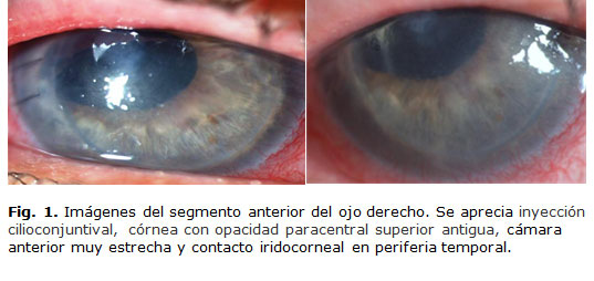
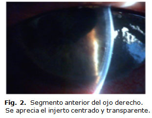
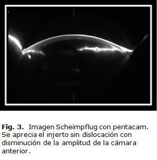
 </font></font></font></font></font></font></font></font></p>   <FONT size="+1"><FONT color="#323232"><FONT color="#000000"><FONT color="#323232"><FONT color="#000000"><FONT color="#323232"><FONT size="+1"><FONT size="+1"><FONT color="#000000"><FONT color="#323232">        <p   align="justify" ><font face="Verdana, Arial, Helvetica, sans-serif" size="2">Al mes de la cirug&iacute;a      acudi&oacute; a la consulta por presentar dolor ocular intenso del ojo derecho.      En el examen oftalmol&oacute;gico presentaba agudeza visual sin correcci&oacute;n      (AVSC) de 0,05 en el OD y de 0,7 en el OI, que no mejoro con correcci&oacute;n.      En la biomicroscopia se observ&oacute; edema palpebral, inyecci&oacute;n cilioconjuntival,      c&oacute;rnea con opacidad paracentral superior antigua,<FONT color="#000000">      c&aacute;mara anterior muy estrecha y contacto iridocorneal en periferia temporal<FONT color="#323232">      (<FONT color="#143779"><a href="#f1">Fig. 1</a><FONT color="#323232">), <FONT color="#000000">      con cifras de presi&oacute;n ocular de (5012 mmHg). El injerto se manten&iacute;a      centrado, transparente y sin dislocaci&oacute;n <FONT color="#323232">(<FONT color="#143779"><a href="#f2">Fig.      2</a> y<a href="#f3"> 3</a><FONT color="#323232">). </font></font></font></font></font></font></font></font></font></p >       <p   align="justify" >&nbsp;</p >   </font></font></font></font></font></font></font></font></font></font></DIV > </font></font></font></font></font></font></font></font></font></font></font></font></font></font></font></font>     <blockquote><FONT size="+1" color="#000000"><FONT color="#323232"><FONT color="#000000"><FONT color="#323232"><FONT color="#000000"><FONT color="#323232"><FONT color="#000000"><FONT size="+1"><FONT size="+1"><FONT color="#211E1E"><FONT color="#000000"><FONT size="+1"><FONT size="+1"><FONT size="+1"><FONT size="+1"><FONT size="+1">       <DIV class="Part"   >         <div align="center">            <p><FONT size="+1"><FONT color="#323232"><FONT color="#000000"><FONT color="#323232"><FONT color="#000000"><FONT color="#323232"><FONT size="+1"><FONT size="+1"><FONT color="#000000"><FONT color="#323232"><img src="/img/revistas/oft/v27n3/f0115314.jpg" width="537" height="254"><a name="f1"></a></font></font></font></font></font></font></font></font></font></font></p>           <p>&nbsp;</p>           <p><font size="+1" color="#000000"><font color="#323232"><font color="#000000"><font color="#323232"><font color="#000000"><font color="#323232"><font color="#000000"><font size="+1"><font size="+1"><font color="#211E1E"><font color="#000000"><font size="+1"><font size="+1"><font size="+1"><font size="+1"><font size="+1"><img src="/img/revistas/oft/v27n3/f0215314.jpg" width="311" height="241"><a name="f2"></a></font></font></font></font></font></font></font></font></font></font></font></font></font></font></font></font></p>           ]]></body>
<body><![CDATA[<p>&nbsp;</p>           <p><img src="/img/revistas/oft/v27n3/f0315314.jpg" width="306" height="312"><a name="f3"></a></p>           <p>&nbsp;</p>       <FONT size="+1"><FONT color="#323232"><FONT color="#000000"><FONT color="#323232"><FONT color="#000000"><FONT color="#323232"><FONT size="+1"><FONT size="+1"><FONT color="#000000"><FONT color="#323232"><FONT color="#000000"><FONT color="#323232"><FONT color="#143779"><FONT color="#323232"><FONT color="#000000"><FONT color="#323232"><FONT color="#143779"><FONT color="#323232">        </font></font></font></font></font></font></font></font></font></font></font></font></font></font></font></font></font></font></div>   </DIV >   </font></font></font></font></font></font></font></font></font></font></font></font></font></font></font></font></blockquote> <FONT size="+1" color="#000000"><FONT color="#323232"><FONT color="#000000"><FONT color="#323232"><FONT color="#000000"><FONT color="#323232"><FONT color="#000000"><FONT size="+1"><FONT size="+1"><FONT color="#211E1E"><FONT color="#000000"><FONT size="+1"><FONT size="+1"><FONT size="+1"><FONT size="+1"><FONT size="+1">      <DIV class="Part"   ><FONT size="+1"><FONT color="#323232"><FONT color="#000000"><FONT color="#323232"><FONT color="#000000"><FONT color="#323232"><FONT size="+1"><FONT size="+1"><FONT color="#000000"><FONT color="#323232"><FONT color="#000000"><FONT color="#323232"><FONT color="#143779"><FONT color="#323232"><FONT color="#000000"><FONT color="#323232"><FONT color="#143779"><FONT color="#323232">        <p   align="justify" ><font face="Verdana, Arial, Helvetica, sans-serif" size="2">
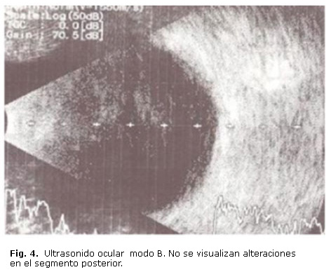
No se constataron      alteraciones en la oftalmoscopia del segmento posterior ni en ultrasonido      ocular (<FONT color="#143779"><a href="#f4">Fig. 4</a><FONT color="#323232">).      El ojo izquierdo sin modificaciones visuales con una longitud axial de 22,2      mm y c&aacute;mara anterior de 3,16. No presentaba alteraciones en el fondo      de ojo. </font></font></font></p >       <p   align="justify" >&nbsp;</p >   </font></font></font></font></font></font></font></font></font></font></font></font></font></font></font></font></font></font></DIV > </font></font></font></font></font></font></font></font></font></font></font></font></font></font></font></font>     <blockquote><FONT size="+1" color="#000000"><FONT color="#323232"><FONT color="#000000"><FONT color="#323232"><FONT color="#000000"><FONT color="#323232"><FONT color="#000000"><FONT size="+1"><FONT size="+1"><FONT color="#211E1E"><FONT color="#000000"><FONT size="+1"><FONT size="+1"><FONT size="+1"><FONT size="+1"><FONT size="+1">       <DIV class="Part"   >         <div align="center"><FONT size="+1"><FONT color="#323232"><FONT color="#000000"><FONT color="#323232"><FONT color="#000000"><FONT color="#323232"><FONT size="+1"><FONT size="+1"><FONT color="#000000"><FONT color="#323232"><FONT color="#000000"><FONT color="#323232"><FONT color="#143779"><FONT color="#323232"><FONT color="#000000"><FONT color="#323232"><FONT color="#143779"><FONT color="#323232"><img src="/img/revistas/oft/v27n3/f0415314.jpg" width="472" height="407"><a name="f4"></a></font></font></font></font></font></font></font></font></font></font></font></font></font></font></font></font></font></font></div>   </DIV >   </font></font></font></font></font></font></font></font></font></font></font></font></font></font></font></font></blockquote> <FONT size="+1" color="#000000"><FONT color="#323232"><FONT color="#000000"><FONT color="#323232"><FONT color="#000000"><FONT color="#323232"><FONT color="#000000"><FONT size="+1"><FONT size="+1"><FONT color="#211E1E"><FONT color="#000000"><FONT size="+1"><FONT size="+1"><FONT size="+1"><FONT size="+1"><FONT size="+1">      <DIV class="Part"   ><FONT size="+1"><FONT color="#323232"><FONT color="#000000"><FONT color="#323232"><FONT color="#000000"><FONT color="#323232"><FONT size="+1"><FONT size="+1"><FONT color="#000000"><FONT color="#323232"><FONT color="#000000"><FONT color="#323232"><FONT color="#143779"><FONT color="#323232"><FONT color="#000000"><FONT color="#323232"><FONT color="#143779"><FONT color="#323232">        ]]></body>
<body><![CDATA[<p   align="justify" >&nbsp;</p >       <p   align="justify" ><font face="Verdana, Arial, Helvetica, sans-serif" size="2">A pesar del tratamiento      t&oacute;pico (timolol 0,5 % y dorzolamida 1 gota cada 12 horas) y sist&eacute;mico      (acetazolamida 250 mg 1 tableta cada 8 horas y manitol 20 %-250 mL cada 12      horas), no se logr&oacute; un control de la presi&oacute;n intraocular (PIO),      por lo que se decidi&oacute; realizar implante valvular (Ahmed). La PIO posterior      a la colocaci&oacute;n del dispositivo de drenaje manten&iacute;a valores      elevados y finalmente se recurri&oacute; a la ciclofotocoagulaci&oacute;n      en el ojo derecho. </font></p >       <p   align="justify" >&nbsp;</p >   <FONT color="#143779"><FONT color="#323232"></font></font></font></font></font></font></font></font></font></font></font></font></font></font></font></font></font></font></font></font></DIV > <FONT size="+1"><FONT color="#323232"><FONT color="#000000"><FONT color="#323232"><FONT color="#000000"><FONT color="#323232"><FONT size="+1"><FONT size="+1"><FONT color="#000000"><FONT color="#323232"><FONT color="#000000"><FONT color="#323232"><FONT color="#143779"><FONT color="#323232"><FONT color="#000000"><FONT color="#323232"><FONT color="#143779"><FONT color="#323232"><FONT color="#143779"><FONT color="#323232">      <DIV class="Part"   >        <p   align="justify" ><font color="#000000" face="Verdana, Arial, Helvetica, sans-serif" size="2"><B><font size="3">DISCUSI&Oacute;N      </font> </b></font></p >       <p><font face="Verdana, Arial, Helvetica, sans-serif" size="2"><FONT color="#211E1E">La      sustituci&oacute;n del endotelio corneal en entidades como la distrofia endotelial      de Fuchs y la queratopat&iacute;a bullosa pseudof&aacute;quica ha constituido      en los &uacute;ltimos a&ntilde;os una opci&oacute;n terap&eacute;utica efectiva,      ya que al evitar un remplazo de todas sus capas se reduce la aparici&oacute;n      de complicaciones transquir&uacute;rgica como la hemorragia supracoroidea      y, a su vez, se logra una recuperaci&oacute;n visual temprana en el paciente.      </font></font></p>   <FONT color="#000000"><FONT color="#211E1E">        <p   align="justify" ><font face="Verdana, Arial, Helvetica, sans-serif" size="2">La elevaci&oacute;n      de la presi&oacute;n intraocular es un evento frecuente despu&eacute;s de      la queratoplastia penetrante. Varios autores definen la hipertensi&oacute;n      ocular posqueratoplastia como la elevaci&oacute;n de la PIO posoperatoria,      sin evidencias de cambios en la cabeza del nervio &oacute;ptico y el campo      visual. Una vez presentes estos cambios se define el diagn&oacute;stico de      glaucoma,<Sup>6-8 </Sup>que constituye una de las causas m&aacute;s comunes      de p&eacute;rdida visual irreversible en los trasplantados y la segunda causa      de fallo del injerto despu&eacute;s del rechazo. Se ha descrito comportamiento      bif&aacute;sico de la PIO que ocurre entre el 2do. y el 3er. d&iacute;a y      en la 2da. o 3ra. semana, aunque este no es un patr&oacute;n de presentaci&oacute;n      estable. El uso prolongado de esteroides t&oacute;picos previo a la queratoplastia      parece estar muy relacionado con el aumento de la PIO. Establecer un diagn&oacute;stico      oportuno de glaucoma es determinante para conservar la transparencia del injerto      y la funci&oacute;n del nervio &oacute;ptico.<Sup>9 </Sup></font></p >   <FONT size="+1"><FONT size="+1"><FONT size="+1">        <p   align="justify" ><font size="2" face="Verdana, Arial, Helvetica, sans-serif">En pacientes con      glaucoma prexistente o cirug&iacute;a previa, y cuando se combina la cirug&iacute;a      con trabeculectom&iacute;a o implante valvular, se prefiere la queratoplastia      endotelial. Estudios relacionados con esta t&eacute;cnica han demostrado su      efectividad y han encontrado como la complicaci&oacute;n m&aacute;s frecuente      la dislocaci&oacute;n del disco.<Sup>10</Sup> Se ha publicado que existe una      alta variaci&oacute;n que oscila nada menos entre el 0 y el 82 % con una media      de dislocaci&oacute;n del 14,5 %. Por ejemplo, en el <I>Bascom Palmer Eye      Institute </I>(EE.UU.), encontraron un 23 % de dislocaci&oacute;n en una serie      bien documentada de 118 casos,<Sup>9</Sup> mientras que <I>Terry</I> reporta      &uacute;nicamente un 1,8 % de dislocaci&oacute;n en 225 ojos intervenidos      mediante triple procedimiento (DSAEK+facoemulsificaci&oacute;n+LIO) y un 4      % en 90 pacientes operados &uacute;nicamente mediante DSAEK.<Sup>11-13</Sup>      La dislocaci&oacute;n del disco corneal ocurre por la p&eacute;rdida de adherencia      del lent&iacute;culo del donante al estroma del receptor. La separaci&oacute;n      del disco donante puede verse simplemente como l&iacute;quido en la interfase      o bien como una dislocaci&oacute;n completa en la c&aacute;mara anterior,      donde la realizaci&oacute;n de la tomograf&iacute;a de coherencia &oacute;ptica      (OCT) es de gran utilidad, sobre todo si el edema corneal es severo.<Sup>10      </Sup></font></p >   <FONT size="+1"><FONT size="+1"><FONT size="+1"><FONT size="+1"><FONT size="+1"><FONT size="+1"><FONT size="+1"><FONT size="+1">        <p   align="justify" ><font size="2" face="Verdana, Arial, Helvetica, sans-serif">El s&iacute;ndrome      Urrets-Zavalia se describe con mayor frecuencia posterior a las queratoplastias      penetrantes en el queratocono y con menos frecuencia en la cirug&iacute;a      lamelar. Se presenta despu&eacute;s de una cirug&iacute;a sin complicaciones      con midriasis irreversible, atrofia de iris y glaucoma temprano y tard&iacute;o.<Sup>9      </Sup></font></p >   <FONT size="+1"><FONT size="+1">        <p   align="justify" ><font size="2" face="Verdana, Arial, Helvetica, sans-serif">Con este tipo de      t&eacute;cnica de queratoplastia endotelial se deben individualizar los pacientes,      como es el caso de c&aacute;maras anteriores muy estrechas o sinequias perif&eacute;ricas      anteriores, donde se aconseja disminuir el tama&ntilde;o del disco donante      para, de esta forma, evitar su cercan&iacute;a excesiva y el contacto o cierre      del &aacute;ngulo.<Sup>9</Sup> La opci&oacute;n de recurrir al uso de dispositivos      de drenajes hace que tengamos en cuenta un factor determinante en la supervivencia      del injerto, que es la colocaci&oacute;n del tubo. <I>Arroyave</I> y otros<Sup>14      </Sup>demostraron en su estudio que aquellos implantados en la cavidad v&iacute;trea      ten&iacute;an una mayor supervivencia al no tener un contacto directo con      el endotelio corneal, criterio que valoramos para la colocaci&oacute;n del      implante valvular en esta paciente. </font></p >   <FONT size="+1"><FONT size="+1"><FONT size="+1"><FONT size="+1"><FONT size="+1">        ]]></body>
<body><![CDATA[<p   align="justify" ><font face="Verdana, Arial, Helvetica, sans-serif" size="2">La queratoplastia      endotelial con pelado de la descemet asistida con l&aacute;ser de exc&iacute;mero      provee discos de alta calidad en cuanto a homogeneidad de la superficie y      espesor final preciso y regular seg&uacute;n lo deseado. No solo se aplica      en casos de integridad anat&oacute;mica del segmento anterior, sino tambi&eacute;n      en casos con dif&iacute;cil visualizaci&oacute;n, desorganizaci&oacute;n y      situaciones complejas. Estudios destacan su uso en el s&iacute;ndrome iridocorneoendotelial      mediante ELDSEK, y el logro de un buen resultado morfol&oacute;gico y una      significativa ganancia visual.<Sup>15 </Sup></font></p >   <FONT size="+1">        <p   align="justify" ><font size="2" face="Verdana, Arial, Helvetica, sans-serif">La queratoplastia      endotelial constituye una novedosa t&eacute;cnica quir&uacute;rgica que permite      mejorar la calidad de vida de nuestros pacientes. </font></p >       <p   align="justify" >&nbsp;</p >   </font></font></font></font></font></font></font></font></font></font></font></font></font></font></font></font></font></font></font></font></font></DIV > <FONT color="#000000"><FONT color="#211E1E"><FONT size="+1"><FONT size="+1"><FONT size="+1"><FONT size="+1"><FONT size="+1"><FONT size="+1"><FONT size="+1"><FONT size="+1"><FONT size="+1"><FONT size="+1"><FONT size="+1"><FONT size="+1"><FONT size="+1"><FONT size="+1"><FONT size="+1"><FONT size="+1"><FONT size="+1"><FONT size="+1"><FONT size="+1"><FONT size="+1">      <DIV class="Part"   >        <p   align="justify" ><font color="#000000" face="Verdana, Arial, Helvetica, sans-serif" size="2"><B><font size="3">REFERENCIAS      BIBLIOGR&Aacute;FICAS </font></b></font><FONT color="#000000"> </font></p > </DIV > </font></font></font></font></font></font></font></font></font></font></font></font></font></font></font></font></font></font></font></font></font></font></font></font></font></font></font></font></font></font></font></font></font></font></font></font></font></font></font></font></font></font></font></font></font></font></font></font></font></font></font></font></font></font></font></font></font></font><FONT size="+1" color="#000000"><FONT color="#323232"><FONT color="#000000"><FONT color="#323232"><FONT color="#000000"><FONT color="#323232"><FONT color="#000000"><FONT size="+1"><FONT size="+1"><FONT color="#211E1E"><FONT color="#000000"><FONT size="+1"><FONT size="+1"><FONT size="+1"><FONT size="+1"><FONT size="+1"><FONT size="+1"><FONT color="#323232"><FONT color="#000000"><FONT color="#323232"><FONT color="#000000"><FONT color="#323232"><FONT size="+1"><FONT size="+1"><FONT color="#000000"><FONT color="#323232"><FONT color="#000000"><FONT color="#323232"><FONT color="#143779"><FONT color="#323232"><FONT color="#000000"><FONT color="#323232"><FONT color="#143779"><FONT color="#323232"><FONT color="#143779"><FONT color="#323232"><FONT color="#000000"><FONT color="#211E1E"><FONT size="+1"><FONT size="+1"><FONT size="+1"><FONT size="+1"><FONT size="+1"><FONT size="+1"><FONT size="+1"><FONT size="+1"><FONT size="+1"><FONT size="+1"><FONT size="+1"><FONT size="+1"><FONT size="+1"><FONT size="+1"><FONT size="+1"><FONT size="+1"><FONT size="+1"><FONT size="+1"><FONT size="+1"><FONT size="+1">     <!-- ref --><DIV class="Part"   ><FONT color="#000000"><font face="Verdana, Arial, Helvetica, sans-serif" size="2">    1. </font><font size="+1" color="#000000"><font color="#323232"><font color="#000000"><font color="#323232"><font color="#000000"><font color="#323232"><font color="#000000"><font size="+1"><font size="+1"><font color="#211E1E"><font color="#000000"><font size="+1"><font size="+1"><font size="+1"><font size="+1"><font size="+1"><font size="+1"><font color="#323232"><font color="#000000"><font color="#323232"><font color="#000000"><font color="#323232"><font size="+1"><font size="+1"><font color="#000000"><font color="#323232"><font color="#000000"><font color="#323232"><font color="#143779"><font color="#323232"><font color="#000000"><font color="#323232"><font color="#143779"><font color="#323232"><font color="#143779"><font color="#323232"><font color="#000000"><font color="#211E1E"><font size="+1"><font size="+1"><font size="+1"><font size="+1"><font size="+1"><font size="+1"><font size="+1"><font size="+1"><font size="+1"><font size="+1"><font size="+1"><font size="+1"><font size="+1"><font size="+1"><font size="+1"><font size="+1"><font size="+1"><font size="+1"><font size="+1"><font size="+1"><font color="#000000"><font face="Verdana, Arial, Helvetica, sans-serif" size="2">Busin    M. </font></font></font></font></font></font></font></font></font></font></font></font></font></font></font></font></font></font></font></font></font></font></font></font></font></font></font></font></font></font></font></font></font></font></font></font></font></font></font></font></font></font></font></font></font></font></font></font></font></font></font></font></font></font></font></font></font></font></font></font><font size="+1" color="#000000"><font color="#323232"><font color="#000000"><font color="#323232"><font color="#000000"><font color="#323232"><font color="#000000"><font size="+1"><font size="+1"><font color="#211E1E"><font color="#000000"><font size="+1"><font size="+1"><font size="+1"><font size="+1"><font size="+1"><font size="+1"><font color="#323232"><font color="#000000"><font color="#323232"><font color="#000000"><font color="#323232"><font size="+1"><font size="+1"><font color="#000000"><font color="#323232"><font color="#000000"><font color="#323232"><font color="#143779"><font color="#323232"><font color="#000000"><font color="#323232"><font color="#143779"><font color="#323232"><font color="#143779"><font color="#323232"><font color="#000000"><font color="#211E1E"><font size="+1"><font size="+1"><font size="+1"><font size="+1"><font size="+1"><font size="+1"><font size="+1"><font size="+1"><font size="+1"><font size="+1"><font size="+1"><font size="+1"><font size="+1"><font size="+1"><font size="+1"><font size="+1"><font size="+1"><font size="+1"><font size="+1"><font size="+1"><font color="#000000"><font face="Verdana, Arial, Helvetica, sans-serif" size="2">DMEK    latest in selective tissue corneal transplantation surgery. EE.UU. Ocular Surgery    News. 2008 [citado marzo de 2014]: [aprox 11 p.]. Disponible en: </font><font size="+1" color="#000000"><font color="#323232"><font color="#000000"><font color="#323232"><font color="#000000"><font color="#323232"><font color="#000000"><font size="+1"><font size="+1"><font color="#211E1E"><font color="#000000"><font size="+1"><font size="+1"><font size="+1"><font size="+1"><font size="+1"><font size="+1"><font color="#323232"><font color="#000000"><font color="#323232"><font color="#000000"><font color="#323232"><font size="+1"><font size="+1"><font color="#000000"><font color="#323232"><font color="#000000"><font color="#323232"><font color="#143779"><font color="#323232"><font color="#000000"><font color="#323232"><font color="#143779"><font color="#323232"><font color="#143779"><font color="#323232"><font color="#000000"><font color="#211E1E"><font size="+1"><font size="+1"><font size="+1"><font size="+1"><font size="+1"><font size="+1"><font size="+1"><font size="+1"><font size="+1"><font size="+1"><font size="+1"><font size="+1"><font size="+1"><font size="+1"><font size="+1"><font size="+1"><font size="+1"><font size="+1"><font size="+1"><font size="+1"><font color="#000000"><font color="#143779" face="Verdana, Arial, Helvetica, sans-serif" size="2"><a href="http://www.healio.com/ophthalmology/cornea-externaldisease/news/print/ocular-surgery-news/%7B32e7c112-e605-47c19ba0-6644dc82d316%7D/dmek-latest-in-selective-tissue-cornealtransplantation-surgery" target="_blank">http://www.healio.com/ophthalmology/cornea-externaldisease/news/print/ocular-surgery-news/%7B32e7c112-e605-47c19ba0-6644dc82d316%7D/dmek-latest-in-selective-tissue-cornealtransplantation-surgery</a>    </font></font></font></font></font></font></font></font></font></font></font></font></font></font></font></font></font></font></font></font></font></font></font></font></font></font></font></font></font></font></font></font></font></font></font></font></font></font></font></font></font></font></font></font></font></font></font></font></font></font></font></font></font></font></font></font></font></font></font></font></font></font></font></font></font></font></font></font></font></font></font></font></font></font></font></font></font></font></font></font></font></font></font></font></font></font></font></font></font></font></font></font></font></font></font></font></font></font></font></font></font></font></font></font></font></font></font></font></font></font></font></font></font></font></font></font></font></font></font></font></DIV > </font></font></font></font></font></font></font></font></font></font></font></font></font></font></font></font></font></font></font></font></font></font></font></font></font></font></font></font></font></font></font></font></font></font></font></font></font></font></font></font></font></font></font></font></font></font></font></font></font></font></font></font></font></font></font></font></font></font>     <blockquote><FONT size="+1" color="#000000"><FONT color="#323232"><FONT color="#000000"><FONT color="#323232"><FONT color="#000000"><FONT color="#323232"><FONT color="#000000"><FONT size="+1"><FONT size="+1"><FONT color="#211E1E"><FONT color="#000000"><FONT size="+1"><FONT size="+1"><FONT size="+1"><FONT size="+1"><FONT size="+1"><FONT size="+1"><FONT color="#323232"><FONT color="#000000"><FONT color="#323232"><FONT color="#000000"><FONT color="#323232"><FONT size="+1"><FONT size="+1"><FONT color="#000000"><FONT color="#323232"><FONT color="#000000"><FONT color="#323232"><FONT color="#143779"><FONT color="#323232"><FONT color="#000000"><FONT color="#323232"><FONT color="#143779"><FONT color="#323232"><FONT color="#143779"><FONT color="#323232"><FONT color="#000000"><FONT color="#211E1E"><FONT size="+1"><FONT size="+1"><FONT size="+1"><FONT size="+1"><FONT size="+1"><FONT size="+1"><FONT size="+1"><FONT size="+1"><FONT size="+1"><FONT size="+1"><FONT size="+1"><FONT size="+1"><FONT size="+1"><FONT size="+1"><FONT size="+1"><FONT size="+1"><FONT size="+1"><FONT size="+1"><FONT size="+1"><FONT size="+1">       <DIV class="Part"   ><FONT color="#000000"></font></DIV >   </font></font></font></font></font></font></font></font></font></font></font></font></font></font></font></font></font></font></font></font></font></font></font></font></font></font></font></font></font></font></font></font></font></font></font></font></font></font></font></font></font></font></font></font></font></font></font></font></font></font></font></font></font></font></font></font></font></font></blockquote> <FONT size="+1" color="#000000"><FONT color="#323232"><FONT color="#000000"><FONT color="#323232"><FONT color="#000000"><FONT color="#323232"><FONT color="#000000"><FONT size="+1"><FONT size="+1"><FONT color="#211E1E"><FONT color="#000000"><FONT size="+1"><FONT size="+1"><FONT size="+1"><FONT size="+1"><FONT size="+1"><FONT size="+1"><FONT color="#323232"><FONT color="#000000"><FONT color="#323232"><FONT color="#000000"><FONT color="#323232"><FONT size="+1"><FONT size="+1"><FONT color="#000000"><FONT color="#323232"><FONT color="#000000"><FONT color="#323232"><FONT color="#143779"><FONT color="#323232"><FONT color="#000000"><FONT color="#323232"><FONT color="#143779"><FONT color="#323232"><FONT color="#143779"><FONT color="#323232"><FONT color="#000000"><FONT color="#211E1E"><FONT size="+1"><FONT size="+1"><FONT size="+1"><FONT size="+1"><FONT size="+1"><FONT size="+1"><FONT size="+1"><FONT size="+1"><FONT size="+1"><FONT size="+1"><FONT size="+1"><FONT size="+1"><FONT size="+1"><FONT size="+1"><FONT size="+1"><FONT size="+1"><FONT size="+1"><FONT size="+1"><FONT size="+1"><FONT size="+1">      <DIV class="Part"   ></DIV >     <DIV class="Part"   ></DIV >     ]]></body>
<body><![CDATA[<!-- ref --><DIV class="Part"   ><FONT color="#000000"><font face="Verdana, Arial, Helvetica, sans-serif" size="2">2.    </font><font size="+1" color="#000000"><font color="#323232"><font color="#000000"><font color="#323232"><font color="#000000"><font color="#323232"><font color="#000000"><font size="+1"><font size="+1"><font color="#211E1E"><font color="#000000"><font size="+1"><font size="+1"><font size="+1"><font size="+1"><font size="+1"><font size="+1"><font color="#323232"><font color="#000000"><font color="#323232"><font color="#000000"><font color="#323232"><font size="+1"><font size="+1"><font color="#000000"><font color="#323232"><font color="#000000"><font color="#323232"><font color="#143779"><font color="#323232"><font color="#000000"><font color="#323232"><font color="#143779"><font color="#323232"><font color="#143779"><font color="#323232"><font color="#000000"><font color="#211E1E"><font size="+1"><font size="+1"><font size="+1"><font size="+1"><font size="+1"><font size="+1"><font size="+1"><font size="+1"><font size="+1"><font size="+1"><font size="+1"><font size="+1"><font size="+1"><font size="+1"><font size="+1"><font size="+1"><font size="+1"><font size="+1"><font size="+1"><font size="+1"><font color="#000000"><font color="#143779"><font size="+1" color="#000000"><font color="#323232"><font color="#000000"><font color="#323232"><font color="#000000"><font color="#323232"><font color="#000000"><font size="+1"><font size="+1"><font color="#211E1E"><font color="#000000"><font size="+1"><font size="+1"><font size="+1"><font size="+1"><font size="+1"><font size="+1"><font color="#323232"><font color="#000000"><font color="#323232"><font color="#000000"><font color="#323232"><font size="+1"><font size="+1"><font color="#000000"><font color="#323232"><font color="#000000"><font color="#323232"><font color="#143779"><font color="#323232"><font color="#000000"><font color="#323232"><font color="#143779"><font color="#323232"><font color="#143779"><font color="#323232"><font color="#000000"><font color="#211E1E"><font size="+1"><font size="+1"><font size="+1"><font size="+1"><font size="+1"><font size="+1"><font size="+1"><font size="+1"><font size="+1"><font size="+1"><font size="+1"><font size="+1"><font size="+1"><font size="+1"><font size="+1"><font size="+1"><font size="+1"><font size="+1"><font size="+1"><font size="+1"><font color="#000000"><font color="#000000" size="+1"><font face="Verdana, Arial, Helvetica, sans-serif" size="2">R&iacute;os    Torres M,</font></font></font></font></font></font></font></font></font></font></font></font></font></font></font></font></font></font></font></font></font></font></font></font></font></font></font></font></font></font></font></font></font></font></font></font></font></font></font></font></font></font></font></font></font></font></font></font></font></font></font></font></font></font></font></font></font></font></font></font></font>    <font color="#000000" face="Verdana, Arial, Helvetica, sans-serif" size="2">Capote    Cabrera A, Hern&aacute;ndez Silva JR, Egu&iacute;a Mart&iacute;nez F, Padilla    Gonz&aacute;lez C. Criterios y tendencias actuales. La Habana: Editorial Ciencias    M&eacute;dicas; 2009.     </font></font></font></font></font></font></font></font></font></font></font></font></font></font></font></font></font></font></font></font></font></font></font></font></font></font></font></font></font></font></font></font></font></font></font></font></font></font></font></font></font></font></font></font></font></font></font></font></font></font></font></font></font></font></font></font></font></font></font></font></font></font></DIV >     <DIV class="Part"   ><FONT color="#000000">   <OL   type="1" >         <div class="Part"   ></div >         <div class="Part"   ></div >         <div class="Part"   ></div >   </OL >   </font></DIV >      <DIV class="Part"   ></DIV >     <!-- ref --><DIV class="Part"   ><FONT color="#000000"><FONT color="#143779"><FONT color="#000000"><font face="Verdana, Arial, Helvetica, sans-serif" size="2">3.    Melles GR, Wijdh RH, Nieuwendaal CP. A technique to excise the descemet membrane    from a recipient cornea (descemetorhexis). Cornea. 2004;23(3):286-8.    <br>       <!-- ref --><br>   4. </font><font size="+1" color="#000000"><font color="#323232"><font color="#000000"><font color="#323232"><font color="#000000"><font color="#323232"><font color="#000000"><font size="+1"><font size="+1"><font color="#211E1E"><font color="#000000"><font size="+1"><font size="+1"><font size="+1"><font size="+1"><font size="+1"><font size="+1"><font color="#323232"><font color="#000000"><font color="#323232"><font color="#000000"><font color="#323232"><font size="+1"><font size="+1"><font color="#000000"><font color="#323232"><font color="#000000"><font color="#323232"><font color="#143779"><font color="#323232"><font color="#000000"><font color="#323232"><font color="#143779"><font color="#323232"><font color="#143779"><font color="#323232"><font color="#000000"><font color="#211E1E"><font size="+1"><font size="+1"><font size="+1"><font size="+1"><font size="+1"><font size="+1"><font size="+1"><font size="+1"><font size="+1"><font size="+1"><font size="+1"><font size="+1"><font size="+1"><font size="+1"><font size="+1"><font size="+1"><font size="+1"><font size="+1"><font size="+1"><font size="+1"><font color="#000000"><font color="#143779"><font color="#000000"><font size="+1" color="#000000"><font color="#323232"><font color="#000000"><font color="#323232"><font color="#000000"><font color="#323232"><font color="#000000"><font size="+1"><font size="+1"><font color="#211E1E"><font color="#000000"><font size="+1"><font size="+1"><font size="+1"><font size="+1"><font size="+1"><font size="+1"><font color="#323232"><font color="#000000"><font color="#323232"><font color="#000000"><font color="#323232"><font size="+1"><font size="+1"><font color="#000000"><font color="#323232"><font color="#000000"><font color="#323232"><font color="#143779"><font color="#323232"><font color="#000000"><font color="#323232"><font color="#143779"><font color="#323232"><font color="#143779"><font color="#323232"><font color="#000000"><font color="#211E1E"><font size="+1"><font size="+1"><font size="+1"><font size="+1"><font size="+1"><font size="+1"><font size="+1"><font size="+1"><font size="+1"><font size="+1"><font size="+1"><font size="+1"><font size="+1"><font size="+1"><font size="+1"><font size="+1"><font size="+1"><font size="+1"><font size="+1"><font size="+1"><font color="#000000"><font color="#143779"><font color="#000000"><font color="#211E1E" face="Verdana, Arial, Helvetica, sans-serif" size="2">Price    FW Jr, </font></font></font></font></font></font></font></font></font></font></font></font></font></font></font></font></font></font></font></font></font></font></font></font></font></font></font></font></font></font></font></font></font></font></font></font></font></font></font></font></font></font></font></font></font></font></font></font></font></font></font></font></font></font></font></font></font></font></font></font></font></font><font color="#211E1E" face="Verdana, Arial, Helvetica, sans-serif" size="2">Price    MO. Descemet's stripping with endothelial keratoplasty in 200 eyes: early challenges    and techniques to enhance donor adherence. J Cataract Refract Surg. 2006;32(3):    411-8.    <br>       <br>   </font></font></font></font></font></font></font></font></font></font></font></font></font></font></font></font></font></font></font></font></font></font></font></font></font></font></font></font></font></font></font></font></font></font></font></font></font></font></font></font></font></font></font></font></font></font></font></font></font></font></font></font></font></font></font></font></font></font></font></font></font></font></font></font></font>        <div class="Part"   ></div >       <div class="Part"   ></div >       <!-- ref --><div class="Part"   ><font color="#000000" size="+1"><font face="Verdana, Arial, Helvetica, sans-serif" size="2">5.      Capote A, R&iacute;os M, C&aacute;rdenas T. Excimer Laser Descemet Striping      Endothelial Keratoplasty. En: Surgical Tecniques for corneal Trasplantation.      Jaypee-Highlights. Medical Publishers Inc; 2010. p. 157-65.    <br>         <br>     </font></font></div > </DIV >     <DIV class="Part"   ></DIV >      <DIV class="Part"   ></DIV >      <DIV class="Part"   >        ]]></body>
<body><![CDATA[<!-- ref --><p><FONT color="#143779"><FONT color="#000000"><FONT color="#211E1E"><FONT color="#000000"><font face="Verdana, Arial, Helvetica, sans-serif" size="2">6.      Rojas E, M&eacute;ndez AM, Gonz&aacute;lez J, Casanueva HC, Alberro M. Hipertensi&oacute;n      ocular postqueratoplastia. Rev Mex Oftalmol. 2010; 84(1):30-3.    </font></font></font></font></font></p>       <!-- ref --><p><FONT color="#143779"><FONT color="#000000"><FONT color="#211E1E"><FONT color="#000000"><font face="Verdana, Arial, Helvetica, sans-serif" size="2">7.      Al-Swailem SA, Edward DP. Glaucoma and Corneal Transplant Procedures. 2012[citado      18 de marzo de 2014];[aprox 2 p.] Disponible en: <FONT color="#4E82BC"><a href="http://www.hindawi.com/journals/joph/2012/576394" target="_blank">http://www.hindawi.com/journals/joph/2012/576394</a><FONT color="#000000">/      </font></font></font></font></font></font></font><!-- ref --><DIV class="Part"   ><FONT color="#000000"><FONT color="#143779"><FONT color="#000000"><FONT color="#211E1E"><FONT color="#000000"><FONT color="#4E82BC"><FONT color="#000000"><font face="Verdana, Arial, Helvetica, sans-serif" size="2">8.    Egu&iacute;a Mart&iacute;nez F, R&iacute;os Torres M, Capote Cabrera A. Manual    de diagn&oacute;stico y tratamiento en oftalmolog&iacute;a. La Habana: Editorial    Ciencias M&eacute;dicas; 2009.    <br>       <!-- ref --><br>       9. Argones Fern&aacute;ndez L, Piloto D&iacute;az I, Randulfe Dom&iacute;nguez    M. Glaucoma temas quir&uacute;rgicos. La Habana: Editorial Ciencias M&eacute;dicas;    2013.    <br>       <br>   </font></font></font></font></font></font></font></font></DIV >      <DIV class="Part"   ></DIV >     <DIV class="Part"   ><FONT color="#000000"><FONT color="#143779"><FONT color="#000000"><FONT color="#211E1E"><FONT color="#000000"><FONT color="#4E82BC"><FONT color="#000000"><font size="+1" color="#000000"><font color="#323232"><font color="#000000"><font color="#323232"><font color="#000000"><font color="#323232"><font color="#000000"><font size="+1"><font size="+1"><font color="#211E1E"><font color="#000000"><font size="+1"><font size="+1"><font size="+1"><font size="+1"><font size="+1"><font size="+1"><font color="#323232"><font color="#000000"><font color="#323232"><font color="#000000"><font color="#323232"><font size="+1"><font size="+1"><font color="#000000"><font color="#323232"><font color="#000000"><font color="#323232"><font color="#143779"><font color="#323232"><font color="#000000"><font color="#323232"><font color="#143779"><font color="#323232"><font color="#143779"><font color="#323232"><font color="#000000"><font color="#211E1E"><font size="+1"><font size="+1"><font size="+1"><font size="+1"><font size="+1"><font size="+1"><font size="+1"><font size="+1"><font size="+1"><font size="+1"><font size="+1"><font size="+1"><font size="+1"><font size="+1"><font size="+1"><font size="+1"><font size="+1"><font size="+1"><font size="+1"><font size="+1"><font color="#000000"><font color="#143779"><font color="#000000"><font color="#211E1E"><font color="#000000"><font color="#4E82BC"><font color="#000000"><font face="Verdana, Arial, Helvetica, sans-serif" size="2">10.    </font></font></font></font></font></font></font></font></font></font></font></font></font></font></font></font></font></font></font></font></font></font></font></font></font></font></font></font></font></font></font></font></font></font></font></font></font></font></font></font></font></font></font></font></font></font></font></font></font></font></font></font></font></font></font></font></font></font></font></font></font></font></font></font></font></font><font face="Verdana, Arial, Helvetica, sans-serif" size="2">    Villarubia A, Palac&iacute;na E, Ar&aacute;nguez C, Solanab J, Garc&iacute;a-Alonso    CR. Complicaciones tras queratoplastia endotelial: tres a&ntilde;os de experiencia.    Arch Soc Esp Oftalmol. 2011[citado marzo de 2014];86(6):[aprox p.]. Disponible    en: </font></font></font></font></font></font></font></font></DIV >     ]]></body>
<body><![CDATA[<DIV class="Part"   ></DIV >     <DIV class="Part"   ><FONT color="#000000"><FONT color="#143779"><FONT color="#000000"><FONT color="#211E1E"><FONT color="#000000"><FONT color="#4E82BC"><FONT color="#000000">        <p   align="justify" ><font color="#4E82BC" face="Verdana, Arial, Helvetica, sans-serif" size="2"><a href="http://scielo.isciii.es/scielo.php?pid=S036566912011000600003&script=sci_arttext" target="_blank">http://scielo.isciii.es/scielo.php?pid=S036566912011000600003&amp;script=sci_arttext</a>      </font></p >   </font></font></font></font></font></font></font></DIV >     <!-- ref --><DIV class="Part"   ><FONT color="#000000"><FONT color="#143779"><FONT color="#000000"><FONT color="#211E1E"><FONT color="#000000"><FONT color="#4E82BC"><FONT color="#000000"><FONT color="#4E82BC"><FONT size="+1" color="#4E82BC"><font color="#000000" face="Verdana, Arial, Helvetica, sans-serif" size="2">    11. Terry MA, Shamie N, Chen ES, Phillips PM, Shah AK, Hoar KL, et al. Endothelial    keratoplasty for Fuchs' dystrophy with cataract: complications and clinical    results with the new triple procedure. Ophthalmology. 2009;116(4):631-9.    <br>       <br>   </font></font></font></font></font></font></font></font></font></font></DIV >     <!-- ref --><DIV class="Part"   ><FONT color="#000000"><FONT color="#143779"><FONT color="#000000"><FONT color="#211E1E"><FONT color="#000000"><FONT color="#4E82BC"><FONT color="#000000"><FONT color="#4E82BC"><FONT size="+1" color="#4E82BC"><FONT color="#000000"><font face="Verdana, Arial, Helvetica, sans-serif" size="2">    12. Wylegala E, Tarnawska D. Management of pseudophakic bullou<FONT color="#000000">s    keratopathy by combined Descemet-stripping endothelial keratoplasty and intraocular    lens exchange. J Cataract Refract Surg. 2008;34(10):1708-14.    <br>       <!-- ref --><br>   </font></font></font></font></font></font></font></font></font></font></font></font><FONT color="#000000"><FONT color="#143779"><FONT color="#000000"><FONT color="#211E1E"><FONT color="#000000"><FONT color="#4E82BC"><FONT color="#000000"><FONT color="#4E82BC"><FONT size="+1" color="#4E82BC"><FONT color="#000000"><FONT color="#000000"><font face="Verdana, Arial, Helvetica, sans-serif" size="2">    13. Bahar I, Kaiserman I, Sansanayudh W, Levinger E, Rootman DS. Busin guide    <I>vs</I>. forceps for the insertion of the donor lenticule in descemet stripping    automated endothelial keratoplasty. Am J Ophthalmol. 2009;147(2):220-6.    <br>       ]]></body>
<body><![CDATA[<!-- ref --><br>   14. </font></font></font></font></font></font></font></font></font></font></font></font><FONT color="#000000"><FONT color="#143779"><FONT color="#000000"><FONT color="#211E1E"><FONT color="#000000"><FONT color="#4E82BC"><FONT color="#000000"><FONT color="#4E82BC"><FONT size="+1" color="#4E82BC"><FONT color="#000000"><FONT color="#000000"><font face="Verdana, Arial, Helvetica, sans-serif" size="2">Arroyave    CP, Scott IU, Fantes FE, Feuer WJ, Murray TG. Corneal graft survival and intraocular    pressure control after penetrating keratoplasty and glaucoma drainage device    implantation. Ophthalmology. 2001;108(11):1978-85.    <br>       <br>   </font></font></font></font></font></font></font></font></font></font></font></font></DIV > </font></font></font></font></font></font></font></font></font></font></font></font></font></font></font></font></font></font></font></font></font></font></font></font></font></font></font></font></font></font></font></font></font></font></font></font></font></font></font></font></font></font></font></font></font></font></font></font></font></font></font></font></font></font></font></font></font></font><FONT size="+1" color="#000000"><FONT color="#323232"><FONT color="#000000"><FONT color="#323232"><FONT color="#000000"><FONT color="#323232"><FONT color="#000000"><FONT size="+1"><FONT size="+1"><FONT color="#211E1E"><FONT color="#000000"><FONT size="+1"><FONT size="+1"><FONT size="+1"><FONT size="+1"><FONT size="+1"><FONT size="+1"><FONT color="#323232"><FONT color="#000000"><FONT color="#323232"><FONT color="#000000"><FONT color="#323232"><FONT size="+1"><FONT size="+1"><FONT color="#000000"><FONT color="#323232"><FONT color="#000000"><FONT color="#323232"><FONT color="#143779"><FONT color="#323232"><FONT color="#000000"><FONT color="#323232"><FONT color="#143779"><FONT color="#323232"><FONT color="#143779"><FONT color="#323232"><FONT color="#000000"><FONT color="#211E1E"><FONT size="+1"><FONT size="+1"><FONT size="+1"><FONT size="+1"><FONT size="+1"><FONT size="+1"><FONT size="+1"><FONT size="+1"><FONT size="+1"><FONT size="+1"><FONT size="+1"><FONT size="+1"><FONT size="+1"><FONT size="+1"><FONT size="+1"><FONT size="+1"><FONT size="+1"><FONT size="+1"><FONT size="+1"><FONT size="+1">      <DIV class="Part"   ></DIV >     <DIV class="Part"   ><FONT color="#000000"><FONT color="#143779"><FONT color="#000000"><FONT color="#211E1E"><FONT color="#000000"><FONT color="#4E82BC"><FONT color="#000000"><FONT color="#4E82BC"><FONT size="+1" color="#4E82BC"><FONT color="#000000"><FONT color="#000000"><font face="Verdana, Arial, Helvetica, sans-serif" size="2">    15. Capote A, C&aacute;rdenas T, Cruz D. Queratoplastia endotelial con pelado    de la descemet asistida con l&aacute;ser de exc&iacute;mero en el s&iacute;ndrome    iridocorneoendotelial. Arch Soc Esp Oftalmol. 2012[citado marzo de 2013];88(8):[aprox    4 p.]. Disponible en: </font></font></font></font></font></font></font></font></font></font></font></font></DIV >     <DIV class="Part"   ></DIV >     <DIV class="Part"   ><FONT color="#000000"><FONT color="#143779"><FONT color="#000000"><FONT color="#211E1E"><FONT color="#000000"><FONT color="#4E82BC"><FONT color="#000000"><FONT color="#4E82BC"><FONT size="+1" color="#4E82BC"><FONT color="#000000"><FONT color="#000000">        <p   align="justify" ><font color="#4E82BC" face="Verdana, Arial, Helvetica, sans-serif" size="2"><a href="http://www.sciencedirect.com/science/article/pii/S0365669112001840" target="_blank">http://www.sciencedirect.com/science/article/pii/S0365669112001840</a>      </font></p >       <p   align="justify" >&nbsp;</p >       <p   align="justify" >&nbsp;</p >       ]]></body>
<body><![CDATA[<p   align="justify" ><font color="#000000" face="Verdana, Arial, Helvetica, sans-serif" size="2">Recibido:      4 de abril de 2014.    <br>     Aprobado: 18 de junio de 2014.</font></p >       <p   align="justify" >&nbsp;</p >       <p   align="justify" >&nbsp;</p >   <FONT color="#4E82BC"><FONT color="#000000">        <p   align="justify" ><font color="#323232" face="Verdana, Arial, Helvetica, sans-serif" size="2">Dr.      <I>Michel Guerra Almaguer</I>.<FONT color="#000000"> Instituto Cubano de Oftalmolog&iacute;a      &quot;Ram&oacute;n Pando Ferrer&quot;. Ave. 76 No. 3104 entre 31 y 41 Marianao,      La Habana, Cuba. Correo electr&oacute;nico: </font></font></p >   </font></font></font></font></font></font></font></font></font></font></font></font></font></DIV > </font></font></font></font></font></font></font></font></font></font></font></font></font></font></font></font></font></font></font></font></font></font></font></font></font></font></font></font></font></font></font></font></font></font></font></font></font></font></font></font></font></font></font></font></font></font></font></font></font></font></font></font></font></font></font></font></font></font>       ]]></body><back>
<ref-list>
<ref id="B1">
<label>1</label><nlm-citation citation-type="journal">
<person-group person-group-type="author">
<name>
<surname><![CDATA[Busin]]></surname>
<given-names><![CDATA[M]]></given-names>
</name>
</person-group>
<article-title xml:lang="en"><![CDATA[DMEK latest in selective tissue corneal transplantation surgery: EE.UU]]></article-title>
<source><![CDATA[Ocular Surgery News]]></source>
<year>2008</year>
<page-range>11</page-range></nlm-citation>
</ref>
<ref id="B2">
<label>2</label><nlm-citation citation-type="book">
<person-group person-group-type="author">
<name>
<surname><![CDATA[Ríos Torres]]></surname>
<given-names><![CDATA[M]]></given-names>
</name>
<name>
<surname><![CDATA[Capote Cabrera]]></surname>
<given-names><![CDATA[A]]></given-names>
</name>
<name>
<surname><![CDATA[Hernández Silva]]></surname>
<given-names><![CDATA[JR]]></given-names>
</name>
<name>
<surname><![CDATA[Eguía Martínez]]></surname>
<given-names><![CDATA[F]]></given-names>
</name>
<name>
<surname><![CDATA[Padilla González]]></surname>
<given-names><![CDATA[C]]></given-names>
</name>
</person-group>
<source><![CDATA[Criterios y tendencias actuales]]></source>
<year>2009</year>
<publisher-loc><![CDATA[La Habana ]]></publisher-loc>
<publisher-name><![CDATA[Editorial Ciencias Médicas]]></publisher-name>
</nlm-citation>
</ref>
<ref id="B3">
<label>3</label><nlm-citation citation-type="journal">
<person-group person-group-type="author">
<name>
<surname><![CDATA[Melles]]></surname>
<given-names><![CDATA[GR]]></given-names>
</name>
<name>
<surname><![CDATA[Wijdh]]></surname>
<given-names><![CDATA[RH]]></given-names>
</name>
<name>
<surname><![CDATA[Nieuwendaal]]></surname>
<given-names><![CDATA[CP]]></given-names>
</name>
</person-group>
<article-title xml:lang="en"><![CDATA[A technique to excise the descemet membrane from a recipient cornea (descemetorhexis)]]></article-title>
<source><![CDATA[Cornea]]></source>
<year>2004</year>
<volume>23</volume>
<numero>3</numero>
<issue>3</issue>
<page-range>286-8</page-range></nlm-citation>
</ref>
<ref id="B4">
<label>4</label><nlm-citation citation-type="journal">
<person-group person-group-type="author">
<name>
<surname><![CDATA[Price FW Jr]]></surname>
</name>
<name>
<surname><![CDATA[Price]]></surname>
<given-names><![CDATA[MO]]></given-names>
</name>
</person-group>
<article-title xml:lang="en"><![CDATA[Descemet's stripping with endothelial keratoplasty in 200 eyes: early challenges and techniques to enhance donor adherence]]></article-title>
<source><![CDATA[J Cataract Refract Surg]]></source>
<year>2006</year>
<volume>32</volume>
<numero>3</numero>
<issue>3</issue>
<page-range>411-8</page-range></nlm-citation>
</ref>
<ref id="B5">
<label>5</label><nlm-citation citation-type="book">
<person-group person-group-type="author">
<name>
<surname><![CDATA[Capote]]></surname>
<given-names><![CDATA[A]]></given-names>
</name>
<name>
<surname><![CDATA[Ríos]]></surname>
<given-names><![CDATA[M]]></given-names>
</name>
<name>
<surname><![CDATA[Cárdenas]]></surname>
<given-names><![CDATA[T]]></given-names>
</name>
</person-group>
<article-title xml:lang="en"><![CDATA[Excimer Laser Descemet Striping Endothelial Keratoplasty]]></article-title>
<source><![CDATA[Surgical Tecniques for corneal Trasplantation]]></source>
<year>2010</year>
<page-range>157-65</page-range><publisher-loc><![CDATA[Jaypee-Highlights ]]></publisher-loc>
<publisher-name><![CDATA[Medical Publishers Inc]]></publisher-name>
</nlm-citation>
</ref>
<ref id="B6">
<label>6</label><nlm-citation citation-type="journal">
<person-group person-group-type="author">
<name>
<surname><![CDATA[Rojas]]></surname>
<given-names><![CDATA[E]]></given-names>
</name>
<name>
<surname><![CDATA[Méndez]]></surname>
<given-names><![CDATA[AM]]></given-names>
</name>
<name>
<surname><![CDATA[González]]></surname>
<given-names><![CDATA[J]]></given-names>
</name>
<name>
<surname><![CDATA[Casanueva]]></surname>
<given-names><![CDATA[HC]]></given-names>
</name>
<name>
<surname><![CDATA[Alberro]]></surname>
<given-names><![CDATA[M]]></given-names>
</name>
</person-group>
<article-title xml:lang="en"><![CDATA[Hipertensión ocular postqueratoplastia]]></article-title>
<source><![CDATA[Rev Mex Oftalmol]]></source>
<year>2010</year>
<volume>84</volume>
<numero>1</numero>
<issue>1</issue>
<page-range>30-3</page-range></nlm-citation>
</ref>
<ref id="B7">
<label>7</label><nlm-citation citation-type="">
<person-group person-group-type="author">
<name>
<surname><![CDATA[Al-Swailem]]></surname>
<given-names><![CDATA[SA]]></given-names>
</name>
<name>
<surname><![CDATA[Edward]]></surname>
<given-names><![CDATA[DP]]></given-names>
</name>
</person-group>
<source><![CDATA[Glaucoma and Corneal Transplant Procedures]]></source>
<year>2012</year>
<page-range>2</page-range></nlm-citation>
</ref>
<ref id="B8">
<label>8</label><nlm-citation citation-type="book">
<person-group person-group-type="author">
<name>
<surname><![CDATA[Eguía Martínez]]></surname>
<given-names><![CDATA[F]]></given-names>
</name>
<name>
<surname><![CDATA[Ríos Torres]]></surname>
<given-names><![CDATA[M]]></given-names>
</name>
<name>
<surname><![CDATA[Capote Cabrera]]></surname>
<given-names><![CDATA[A]]></given-names>
</name>
</person-group>
<source><![CDATA[Manual de diagnóstico y tratamiento en oftalmología]]></source>
<year>2009</year>
<publisher-loc><![CDATA[La Habana ]]></publisher-loc>
<publisher-name><![CDATA[Editorial Ciencias Médicas]]></publisher-name>
</nlm-citation>
</ref>
<ref id="B9">
<label>9</label><nlm-citation citation-type="book">
<person-group person-group-type="author">
<name>
<surname><![CDATA[Argones Fernández]]></surname>
<given-names><![CDATA[L]]></given-names>
</name>
<name>
<surname><![CDATA[Piloto Díaz]]></surname>
<given-names><![CDATA[I]]></given-names>
</name>
<name>
<surname><![CDATA[Randulfe Domínguez]]></surname>
<given-names><![CDATA[M]]></given-names>
</name>
</person-group>
<source><![CDATA[Glaucoma temas quirúrgicos]]></source>
<year>2013</year>
<publisher-loc><![CDATA[La Habana ]]></publisher-loc>
<publisher-name><![CDATA[Editorial Ciencias Médicas]]></publisher-name>
</nlm-citation>
</ref>
<ref id="B10">
<label>10</label><nlm-citation citation-type="journal">
<person-group person-group-type="author">
<name>
<surname><![CDATA[Villarubia]]></surname>
<given-names><![CDATA[A]]></given-names>
</name>
<name>
<surname><![CDATA[Palacína]]></surname>
<given-names><![CDATA[E]]></given-names>
</name>
<name>
<surname><![CDATA[Aránguez]]></surname>
<given-names><![CDATA[C]]></given-names>
</name>
<name>
<surname><![CDATA[Solanab]]></surname>
<given-names><![CDATA[J]]></given-names>
</name>
<name>
<surname><![CDATA[García-Alonso]]></surname>
<given-names><![CDATA[CR]]></given-names>
</name>
</person-group>
<article-title xml:lang="es"><![CDATA[Complicaciones tras queratoplastia endotelial: tres años de experiencia]]></article-title>
<source><![CDATA[Arch Soc Esp Oftalmol]]></source>
<year>2011</year>
</nlm-citation>
</ref>
<ref id="B11">
<label>11</label><nlm-citation citation-type="journal">
<person-group person-group-type="author">
<name>
<surname><![CDATA[Terry]]></surname>
<given-names><![CDATA[MA]]></given-names>
</name>
<name>
<surname><![CDATA[Shamie]]></surname>
<given-names><![CDATA[N]]></given-names>
</name>
<name>
<surname><![CDATA[Chen]]></surname>
<given-names><![CDATA[ES]]></given-names>
</name>
<name>
<surname><![CDATA[Phillips]]></surname>
<given-names><![CDATA[PM]]></given-names>
</name>
<name>
<surname><![CDATA[Shah]]></surname>
<given-names><![CDATA[AK]]></given-names>
</name>
<name>
<surname><![CDATA[Hoar]]></surname>
<given-names><![CDATA[KL]]></given-names>
</name>
</person-group>
<article-title xml:lang="en"><![CDATA[Endothelial keratoplasty for Fuchs' dystrophy with cataract: complications and clinical results with the new triple procedure]]></article-title>
<source><![CDATA[Ophthalmology]]></source>
<year>2009</year>
<volume>116</volume>
<numero>4</numero>
<issue>4</issue>
<page-range>631-9</page-range></nlm-citation>
</ref>
<ref id="B12">
<label>12</label><nlm-citation citation-type="journal">
<person-group person-group-type="author">
<name>
<surname><![CDATA[Wylegala]]></surname>
<given-names><![CDATA[E]]></given-names>
</name>
<name>
<surname><![CDATA[Tarnawska]]></surname>
<given-names><![CDATA[D]]></given-names>
</name>
</person-group>
<article-title xml:lang="en"><![CDATA[Management of pseudophakic bullous keratopathy by combined Descemet-stripping endothelial keratoplasty and intraocular lens exchange]]></article-title>
<source><![CDATA[J Cataract Refract Surg]]></source>
<year>2008</year>
<volume>34</volume>
<numero>10</numero>
<issue>10</issue>
<page-range>1708-14</page-range></nlm-citation>
</ref>
<ref id="B13">
<label>13</label><nlm-citation citation-type="journal">
<person-group person-group-type="author">
<name>
<surname><![CDATA[Bahar]]></surname>
<given-names><![CDATA[I]]></given-names>
</name>
<name>
<surname><![CDATA[Kaiserman]]></surname>
<given-names><![CDATA[I]]></given-names>
</name>
<name>
<surname><![CDATA[Sansanayudh]]></surname>
<given-names><![CDATA[W]]></given-names>
</name>
<name>
<surname><![CDATA[Levinger]]></surname>
<given-names><![CDATA[E]]></given-names>
</name>
<name>
<surname><![CDATA[Rootman]]></surname>
<given-names><![CDATA[DS]]></given-names>
</name>
</person-group>
<article-title xml:lang="en"><![CDATA[Busin guide vs forceps for the insertion of the donor lenticule in descemet stripping automated endothelial keratoplasty]]></article-title>
<source><![CDATA[Am J Ophthalmol]]></source>
<year>2009</year>
<volume>147</volume>
<numero>2</numero>
<issue>2</issue>
<page-range>220-6</page-range></nlm-citation>
</ref>
<ref id="B14">
<label>14</label><nlm-citation citation-type="journal">
<person-group person-group-type="author">
<name>
<surname><![CDATA[Arroyave]]></surname>
<given-names><![CDATA[CP]]></given-names>
</name>
<name>
<surname><![CDATA[Scott]]></surname>
<given-names><![CDATA[IU]]></given-names>
</name>
<name>
<surname><![CDATA[Fantes]]></surname>
<given-names><![CDATA[FE]]></given-names>
</name>
<name>
<surname><![CDATA[Feuer]]></surname>
<given-names><![CDATA[WJ]]></given-names>
</name>
<name>
<surname><![CDATA[Murray]]></surname>
<given-names><![CDATA[TG]]></given-names>
</name>
</person-group>
<article-title xml:lang="en"><![CDATA[Corneal graft survival and intraocular pressure control after penetrating keratoplasty and glaucoma drainage device implantation]]></article-title>
<source><![CDATA[Ophthalmology]]></source>
<year>2001</year>
<volume>108</volume>
<numero>11</numero>
<issue>11</issue>
<page-range>1978-85</page-range></nlm-citation>
</ref>
<ref id="B15">
<label>15</label><nlm-citation citation-type="journal">
<person-group person-group-type="author">
<name>
<surname><![CDATA[Capote]]></surname>
<given-names><![CDATA[A]]></given-names>
</name>
<name>
<surname><![CDATA[Cárdenas]]></surname>
<given-names><![CDATA[T]]></given-names>
</name>
<name>
<surname><![CDATA[Cruz]]></surname>
<given-names><![CDATA[D]]></given-names>
</name>
</person-group>
<article-title xml:lang="es"><![CDATA[Queratoplastia endotelial con pelado de la descemet asistida con láser de excímero en el síndrome iridocorneoendotelial]]></article-title>
<source><![CDATA[Arch Soc Esp Oftalmol]]></source>
<year>2012</year>
<volume>88</volume>
<numero>8</numero>
<issue>8</issue>
<page-range>4</page-range></nlm-citation>
</ref>
</ref-list>
</back>
</article>
