Etiology of tree tomato (Solanum betaceum CAV.) diseases
Etiología de enfermedades del cultivo de tomate de árbol (Solanum betaceum CAV.)
Joaquín Guillermo Ramírez-Gil, Alejandro Gil-Aguirre, Juan Gonzalo Morales-Osorio*
Laboratorio de Fitotecnia Tropical, Departamento de Ciencias Agronómicas, Facultad de Ciencias Agrarias, Universidad Nacional de Colombia sede Medellín, Calle 59 A N 63-20, Núcleo El Volador, bloque 11, oficina 101, Medellín, Colombia.
]]>
ABSTRACT
The present study had as objectives the identification of the causal agents associated with different pathologies in tree tomato (Solanum betaceumCav.) and the estimation of the disease prevalence in commercial lots located in tree tomato-producing regions of Antioquia, Colombia. Surveys were performed in the three main growing areas of the department of Antioquia, Colombia: the North, the East and the Southwest. Symptoms were recorded and tissue samples taken for pathogen isolation. Tree tomato seedlings were inoculated with not previously identified diseases, and the pathogen postulates of Koch were confirmed. Prevalence of each disease was determined for the time period tested of two years. Results suggested that the major pathogens for the tree tomato crop under field conditions in Antioquia during the period evaluated were Phytophthora infestans sensu lato, Alternaria sp., Xanthomonas sp., Colletotrichum sp., Spongospora subterranea, Verticillium sp., Clavibacter sp., Meloidogyne sp., and viral species classified into six genera: Potyvirus, Cucumovirus, Tospovirus, Tobamovirus, Potexvirus, and Polerovirus. The low percentage of relative identity of the sequences with known viruses merits further research. The diseases identified in the two years of sampling corresponded to those that developed under the specific conditions present such as the cultivated area, weather, and agronomical management for this period of time; therefore, the disease prevalence may change each growing season according to how these parameters vary. Clavibacter sp., S. subterranea and Verticillium sp., were reported as new pathogens for this crop. An accurate and timely diagnosis followed by a prompt and appropriate disease management will contribute to tree tomato production.
Key words: fungal, bacterial and viral pathogens, tamarillo.
RESUMEN
Este estudio tuvo como objetivos identificar los agentes causales asociados con las diferentes patologías en el cultivo de tomate de árbol (Solanum betaceum Cav.) y determinar la prevalencia de las enfermedades en lotes comerciales localizados en las regiones productoras de tomate de árbol en Antioquia, Colombia. Se realizaron monitoreos en las tres principales regiones productoras del departamento de Antioquia, Colombia: el Norte, el Oriente y el Suroeste. Se registraron los síntomas y se muestrearon tejidos para el aislamiento de los patógenos. Para las enfermedades no registradas previamente, se inocularon plántulas de tomate de árbol y se confirmaron los postulados de Koch. Se determinó la prevalencia de cada enfermedad para el periodo de dos años evaluado. Los resultados sugieren que los principales patógenos en Antioquia en condiciones de campo, en el cultivo de tomate de árbol para el periodo evaluado, fueron Phytophthora infestans sensu lato, Alternaria sp., Xanthomonas sp., Colletotrichum sp., Spongospora subterranea, Verticillium sp., Clavibacter sp., Meloidogyne sp. y especies de virus clasificadas en seis géneros: Potyvirus, Cucumovirus, Tospovirus, Tobamovirus, Potexvirus y Polerovirus. El bajo porcentaje de identidad relativa de las secuencias con los virus conocidos amerita una investigación posterior. Las enfermedades identificadas durante los dos años de muestreo corresponden a aquellas que se desarrollaron bajo las condiciones específicas presentes como son el área cultivada, el clima y el manejo agronómico para este periodo de tiempo; por lo tanto, la prevalencia de las enfermedades puede cambiar cada periodo de siembra de acuerdo al modo en que varíen esos parámetros. Clavibacter sp., S. subterranea y Verticillium sp. se informan como nuevos patógenos para este cultivo. El diagnóstico preciso y oportuno, seguido de un manejo oportuno y apropiado de la enfermedad, ayudará a la producción del cultivo de tomate de árbol.
Palabras clave: patógenos fungosos, bacterianos y virales, tamarillo.
INTRODUCTION
]]> Tree tomato, or tamarillo (Solanum betaceum Cav.), is a fruit native to the South American Andes, where it is widely cultivated. This crop is currently grown on a global scale in numerous countries of Central and South America, Asia, Africa and Oceania. Great efforts have been made to develop this crop in New Zealand, Ecuador and Colombia, and these countries have increased its production and exportation in recent years (1,2,3). Tree tomato has great prospects for being grown as a productive alternative in moderately cold climate zones; its nutritional qualities and flavor make it attractive for fresh consumption and the agroindustrial processing for the purposes of both exportation and domestic consumption.In Colombia, cultivation of tree tomato (S. betaceum) has shown a sustained increase, with 8862 hectares planted in 2014. This area was distributed across 19 departments, of which Antioquia was the largest producer (4). Despite the importance of this crop, there are major problems with its production. The most notable of these are the use of low-quality planting materials; the indiscriminate use of agrochemical products including insecticides, fungicides and fertilizers; inappropriate post-harvest handling; weak and discontinuous professional technical assistance; small number of varieties; an emerging marketing system; price fluctuation; inadequate crop planning; and a high prevalence of pests and diseases. In addition to these problems, no efficient and timely phytosanitary diagnostic system exists (5,6,7).
Diseases of tree tomato are caused by various pathogenic agents, including fungi, bacteria, viruses, nematodes and oomycetes. Historically, anthracnose on the fruit, caused by Colletotrichum gloeosporioides (Penz.) Penz & Sacc, and Colletotrichum acutatum Simmonds, and the root-knot nematodes have been considered the most important diseases in tree tomato (8,9). However, in recent years, the prevalence and severity of other phytopathological problems has notably increased, such as viral diseases caused by the viruses: Potato virus Y (PVY, Potyvirus), Tamarillo leaf malformation virus (TaLMV, Potyvirus), Tamarillo mosaic virus (TaMV, Potyvirus), Cucumber mosaic virus, (CMV, Cucumovirus), Potato aucuba mosaic virus (PAMV, Potexvirus), Alfalfa mosaic virus (AMV, Alfamovirus), Tomato spotted wilt virus (TSWV, Tospovirus), Tomato mosaic virus (ToMV, Tobamovirus), Potato leafroll virus (PLRV, Polerovirus), Tomato ringspot virus (ToRSV, Nepovirus), Potato aucuba mosaic virus (PAMV, Potexvirus), Potato virus A (PVA, Potyvirus), Potato virus V (PVV, Potyvirus), and Peru tomato mosaic virus (PTV, Potyvirus), have been informed as inducing symptoms on tree tomato (3,7).
Other phytosanitary problem recently informed is the late blight disease, for which the oomycete Phytophthora infestans (Mont.) de Bary was identified as the causal agent. Later, a new species named P. andina was informed causing late blight, but it has been the subject of an intense debate (10,11,12). For this reason, this causal agent is referred to as P. infestans sensu lato in the literature (10,12).
There is practically no information available in Colombia about the abiotic agents that cause pathologies like scarring (13). There has been little monitoring, identification, diagnosis, management, or research carried out regarding biotic problems caused by other pathogens including Alternaria sp., Ralstonia sp., Xanthomonas sp., Oidium sp., and others. The biology of the plant-pathogen interaction, the incidence, prevalence and distribution, the severity, and the appropriate conditions for managing these problems in the crop are unknown essential aspects for mangement of these pathogens management. Additionally, pathologies present in foreign countries and not identified in Colombia, like that caused by Candidatus Liberibacter solanacearum, represents a potential quarantine risk (14).
The present study had as objectives the identification of the causal agents associated with different pathologies in the tree tomato crop and the estimation of the disease prevalence in commercial lots located in tree tomato-producing regions of Antioquia, Colombia.
MATERIALS AND METHODS
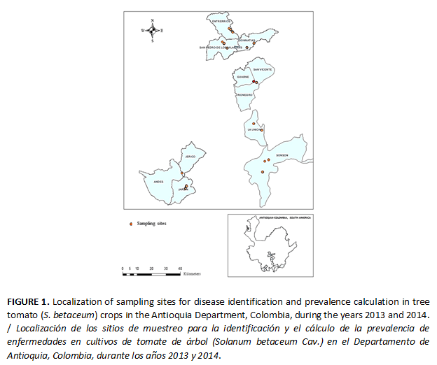
The present study was carried out in eight municipalities of the department of Antioquia, Colombia, with a history of producing tree tomato, during 2013 and 2014. The lots evaluated were located in the Northern Highlands region (Don Matías, San Pedro, Entrerríos), Eastern Antioquia (San Vicente, La Unión and Sonsón), and Southwestern Antioquia (Andes and Jardín). Three plots were selected in each of these municipalities (Figure 1).
The regions evaluated were located in the Andean mountains above 1800 masl and corresponded to life zones humid, very humid and pluvial, premontane and low montane forests (Sensu Holdridge), characterized for mild temperatures, high precipitation and high relative humidity most of the year round, which are conditions that favored plant pathogen development.
]]> Identification of the causal agentsIn the regions of study, tissue samples (roots, stems, leaves, fruits and flower buds) with any symptomatology, regardless of whether or not it had been reported previously, were collected (7,8,9,10). The symptoms were recorded, tissues collected, and microorganisms isolated. The tissues were stored in hermetic bags prior to laboratory processing.
A portion of the samples was taken to humidity chambers, and the others were processed in accordance with the methods described by Ramírez et al. (15): rye agar (RA) (Difco, USA) for Phytophthora infestans sensu lato; acidified potato dextrose agar plus lactic acid (PDA-A) (Difco, USA) and vegetable juice agar (V8-A) (Difco, USA) supplemented with streptomycin (100 µg/ml) for fungi; and nutrient agar (NA) (Difco, USA) supplemented with Benomyl® fungicide (50 µg/ml), yeast, dextrose, calcium carbonate (YDC) (Difco, USA) (16) supplemented with Benomyl® fungicide (50 µg/ml), and the semi-selective culture medium Kelman's TZC Agar (K-TZC-A) (17) as a medium for bacterial growth.
Afterwards, the samples were incubated at 28°C for 15 days, with the exception of the rye agar, which was incubated at 16°C with a photoperiod of 12 hours of light and 12 hours of darkness. For the nematode analysis, roots were taken from the field, washed with water, air-dried, and used to make histological mountings on microscope glass slides.
Sporulating tissue samples and isolates taken from the culture media were used to create microassemblies, which were observed under a light microscope with DIC (Differential Interference Contrast, Nikon Eclipse Ni) for identification at the genus level by using the keys from the manual by Barnett and Hunter (18) for fungi, and the manual by Erwin y Ribeiro (19) for Phytophthora spp. Meanwhile, bacteria were identified through routine biochemical tests, including: Gram staining; hydrolysis of mucoid colonies on YDC at 30 °C; colony color on YDC; oxidase; and urease, based on the instructions of Shaad (16).
For nematodes identification, roots with galls or knots from plants showing symptoms were collected. Galls or knots were dissected under a stereomicroscope with a sterile scalpel blade. Perineal patterns were performed from the females recovered and then fixed and mounted permanently on a glass slide. Structures were visualized under light microscopy (Kikon Eclipse E-200). Taxonomic identification of nematodes was performed using the keys provided by Mai and Mullin (20).
For virus identification, samples of symptomatic tissues were collected and sent to the AGDIA, Inc. diagnostic center (United States). The procedure at AGDIA Company included an initial serological identification for the virus genera Begomovirus, Bromoviridade, Carlavirus, Closteroviridae, Comovirus, Curtovirus, Ilarvirus, Nepovirus, Potexvirus, Potyviridade, Tobamovirus, Tobravirus, Tombusvirus and Tospovirus, by the ELISA test, followed by PCR amplification using species-specific primers, sequencing and sequence comparison analysis for determining the identity values with the closest known virus.
For each pathology found, the corresponding description of the associated symptomatology was recorded, and for those that had not been previously reported, the Koch's postulates were performed as follows: Plants of Red and Yellow cultivars were grown in pots containing sterile soil (autoclaved at 15 psi and 121°C for two cycles under net house conditions until they showed five completely expanded leaves. Standard fertilization, watering and other nursery practices were applied during seedling development.
Verticillium sp.: An isolate was grown on PDA as described. Two hundred milliliters of sterile distilled water were added per plate and mixed thoroughly. The resulting suspension was adjusted to a concentration of 105 conidia per ml-1 using a Neubauer chamber. The inoculum was applied homogeneously over the plant roots. The microorganism was re-isolated from plants showing symptoms.
Clavibacter sp.: A pure colony was grown on AN (Merck) media as described. The colony was inoculated in nutritive broth (Merck) under constant shaking at 150 rpm for 48 h at 25°C. Bacterial suspension was adjusted to a concentration of 106 UFC ml-1 in 50 ml of final volume, with sterile distilled water measured by spectrophotometry at 640 nm. The bacterial suspension was inoculated into the stems and leaves of tomato tree plants. The bacteria were re-isolated from plants showing symptoms.
]]> Spongospora subterranea : Tree tomato plants were grown in soil infested with sporosori (spore balls) of S. subterranea as described previously (21,22). Plants were kept under net house conditions and the onset of symptoms was recorded daily. Sporosori and plasmodium presence were identified in the roots by staining as described previously (21,22).Determination of the prevalence of the different pathologies during the two year period tested
In each municipality of sampling, three plots planted with tree tomato in different stages of development were examined. Within each lot, 30 plants were selected at random. The data obtained in the previously described analyses were used to determine the prevalence of each disease, calculated as the number of diseased plants for a given pathology divided by the total number of plants evaluated.
Data analysis
The prevalence percentage was calculated for each lot, and the value obtained for prevalence in each lot was used to determine the mean and standard deviation in each region for the data regarding each disease. A general mean was then calculated for each disease and for all of the sampling zones.
RESULTS AND DISCUSSION
Pathogen recognition and description of the corresponding symptomatology
Late blight or black pest
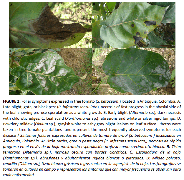
The presence of this disease in the field was characterized by irregular spots on the leaves, which initially have a greasy appearance and later become necrotic and invade a large portion of the tissue (Figure 2A). In humid conditions, profuse sporulation was observed on the surface of infected tissues as a white growth at the leading edge of lesions, usually on the abaxial side of the leaves (Figure 2A). On rye agar medium, the mycelium presented slow, hyaline, and aerial growth. Under the light microscope, we observed a non-partitioned mycelium with branched sporangiophores of indeterminate growth. Semi-papillate (papillae flattened) and caduceus sporangia exhibiting lemon-like shape with short pedicels.
]]> Based on symptomatology observed and morphological characteristics of the microorganism, this pathology was associated with the presence of P. infestans sensu lato (10,11,12,19). This microorganism was clearly differentiated from the other reported foliar potato pathogen P. nicotianae because it had prominently papillate and noncaducous sporangia and, at least when infecting potato, no visible white sporulation was observed with P. nicotianae (23).Early blight, sudden leaf burn, or target spots
This disease was associated with semicircular intense dark brown spots of variable sizes, with chlorotic edges (Figure 2B). In the majority of these situations, this damage occurred in the leaf margins; in advanced stages, the spots coalesced, giving rise to leaf drop. The colony on PDA exhibited slow growth and a grayish to dark brown color. Short, simple and erect conidiophores were observed bearing single or branched chains of large, elongated, dark and pear-shaped conidia with both transverse and longitudinal septa. The symptomatology and morphological identification corresponded to Alternaria sp. (18,24).
Leaf scald or silver spots
The initial symptoms were small irregular lesions, which could appear on any part of the leaf. In advanced stages, this disease was characterized by the presence of abrasions and white or silver rigid bumps on the leaves (Figure 2C). Over time, these symptoms became more pronounced and could lead to leaf drop. On the YDC culture medium, yellow, circular colonies with a convex surface, rounded border and mucoid consistency were observed. The biochemical tests corresponded to a Gram-negative bacteria type, yellow or orange colonies on YDC, negative oxidase, mucoid hydrolysis on YDC at 30°C, and negative urease. The test results and symptoms observed were consistent with the bacteria Xanthomonas sp. (16).
Mildew or powdery mildew
This disease was characterized by the presence of a grayish white to ashy gray blight that primarily affected the leaf surfaces (Figure 2D). In severe infections, it also appeared on the underside of the leaves, where irregularly-shaped, dark necrotic lesions were also observed. In advanced stages, these symptoms were also observed in the peduncles causing leaf drop. Hyaline, elliptical conidia with thin walls that emerged in a chain from short, non-branched conidiophores were observed under the light microscope. The symptoms and morphological structures of the microorganism corresponded to Oidium sp. (18).
Viral complex
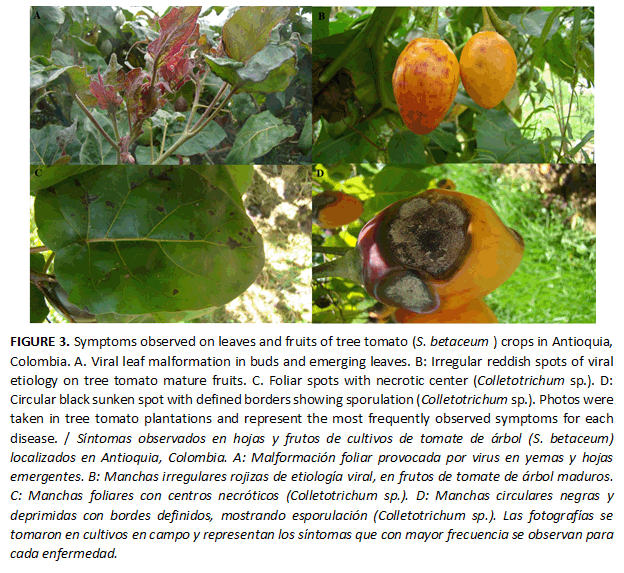
The symptoms associated with plants with the viral complex were very diverse, with the most noteworthy including mosaics, blisters, vein thickening, and leaf deformation, especially in tender buds and new leaves (Figure 3A). Irregular reddish spots of variable intensity and pulp hardening in the mature fruits were also observed (Figure 3B). In advanced stages, the plants exhibited a general state of decay.
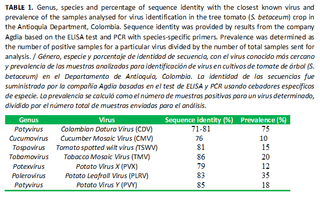
Results from the Agdia diagnostic services based on the ELISA test and nucleic acid sequence identity informed a close relationship of the samples with seven virus classified into the genera Potyvirus, Cucumovirus, Tospovirus, Tobamovirus, Potexvirus, and Polerovirus (Table 1). The samples evaluated showed values of sequence identity from 71%, with Colombian Datura Virus (CDV), to 86%, with Tobacco Mosaic Virus (TMV). The most prevalent (75%) was the group with 71-81% of sequence identity with the Colombian Datura Virus (CDV). The least prevalent (10%) was the group showing 76% of sequence identity with the Cucumber Mosaic Virus (CMV) (Table 1).
]]> Usually, for disease diagnosis purposes, PCR with species-specific primers and sequence similarity analysis with known isolates of a virus is sufficient and more sensitive than serological techniques (25). Even more, for some virus species a threshold for the sequence identity value has been established, as for example the species classified within the genus Potyvirus (76%) (26). However, accurate thresholds for all viral plant pathogens are not available and sequence identity criteria is under permanent discussion as more sequences from all the world are publicly available (27). For this reason, caution should be exercised when using sequence identity as the sole diagnostic procedure to identify or confirm a virus species, and it is always desirable to confirm or complement sequence identity with another test such as ELISA.Results in the present work showed sequence identity between 71 to 86% suggesting high intraspecific variability with presence of lineages or even unknown or closely related viral species to known viruses. For example, the Tamarillo Leaf Malformation Virus (TaLMV), a closely relative species of the Colombian Datura Virus (CDV) was recently proposed as a new species involved in the tree tomato viral complex (3).
A group of samples exhibited sequence identity of 86% with the Tobacco Mosaic Virus (TMV). TMV was reported previously in Ecuador by using the ELISA test but no sequences are available of virus isolates infecting S. betaceum(28). ToMV has frequently been informed as one of the species of the viral complex causing disease symptoms on tree tomato (3,7). TMV and ToMV are classified into the genus Tobamovirus and are closely related species with sequence identity about 80% (Calculated from sequences obtained from http://www.dpvweb.net/seqs/plantviruses.php, consulted 17 November of 2016, using the DnaSP v5 software). At least 14 different virus species have been reported causing symptoms on tree tomato, some of them in mixed infections and some of them closely related, making research, diagnosis and management of viral diseases, in particular the viral complex, a difficult challenge.
It is expected that with new information coming in the next years using cutting edge technologies, as for example the complete genome sequencing, accurate and fast virus identification will contribute to a better disease management.
Fruit rot, necrotic spots and dieback (anthracnose complex)
Rapidly growing greasy black foliar spots with a necrotic center coalescing in advanced stages were observed (Figure 3C). In all stages of fruit development, these initially appeared as circular black sunken spots with defined borders (Figure 3D). When the attack occurred in early stages, it led to fruit mummification. In some cases, a pink or salmon color appeared on the lesion, corresponding to sporulation. Bud rot leading to dieback was also observed. The colonies on PDA showed rapid growth with great color variation that ranged from white and gray to orange. The conidia exhibited variable shapes, which could be cylindrical, oval, or ellipsoid-fusiform. In the majority of them, one of the sides was tapered and the other rounded. The symptoms observed in the field and the morphological characteristics of the associated isolate showed the fungus Colletotrichum sp. as the causative agent of this disease (8,18).
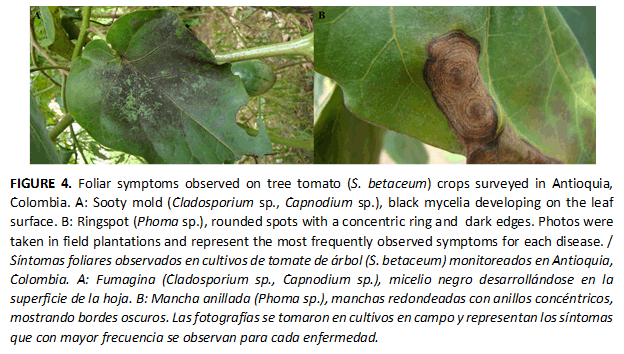
Sooty mold
This disease was characterized by the presence of a black mycelial layer on the surface of the leaves, on which chlorosis sometimes appeared (Figure 4A). These symptoms may appear on the stem and fruits and with time can cause defoliation. Rigid conidiophores were observed to have a series of olivaceous, easily disintegrable, uni- or bicellular conidia at the apex. These structures are characteristic of Cladosporium sp. (18). Additionally, rounded, cylindrical, emergent and nearly pedunculated fructiferous bodies of a dark brown color, characteristic to Capnodium sp. were observed (18).
Ringspot
This pathology was characterized by the presence of rounded spots with a light brown concentric ring in the center and margins with a darker tone (Figure 4B). In advanced stages of the disease, the internal tissue collapsed causing a perforation in the affected leaves. On PDA culture medium, the mycelium was aerial, cottony, and dark brown, with a slightly lighter center. In the micro-assemblies, globular pycnidia with a well-differentiated ostiole and thin yellow walls containing cylindrical non-septate conidia were obsrved. The symptomatology found and the morphological characteristics of the microorganism indicated that this pathology was associated with Phoma sp. (18).
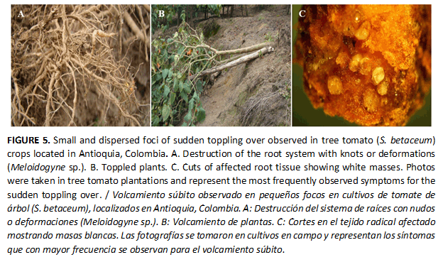
]]> Sudden toppling or generalized yellowingThe presence of this disease is characterized by leaf yellowing, stunted growth, and in advanced stages, plant death, usually localized in dispersed small foci inside the crop lot. It was observed an extensive destruction of the root system, with knots or deformations, which sometimes caused the plants to topple over, mainly because of the wind (Figure 5A and 5B). In cuts made to affected root tissues, white females and egg masses of root knot nematodes were observed (Figure 5C). Under the microscope, female nematodes with morphological characteristics coinciding with Meloidogyne sp. were observed (9,20).
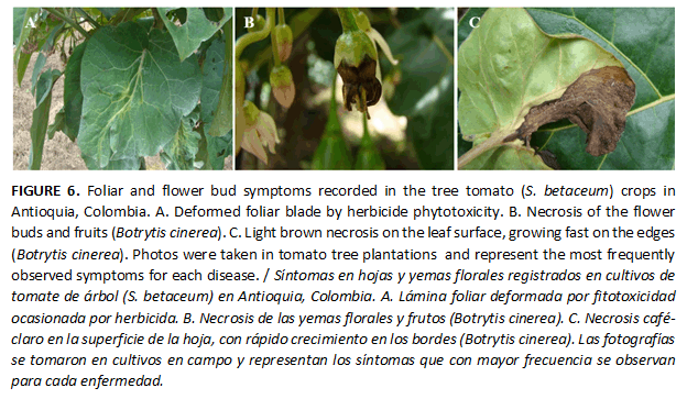
Phytotoxicity
Symptoms characteristic of herbicide phytotoxicity, including the growth of deformed tissue followed by necrosis, were found to be the most frequent pathology associated with abiotic causes (Figure 6A).
Necrotic spots or leaf blight
This disease was characterized by necrosis and subsequent detachment of flower buds and fruits (Figure 6B). A light brown, moist-looking lesion that grew rapidly on the margins appeared on the leaf surfaces. (Figure 6C). In times of high humidity, reproductive structures were observed on the lesions. On PDA medium, the mycelium was gray, and, under the microscope, the structures observed were long branched conidiophores with thickened or globular vesicles on their terminal part and clusters of ovoid or spherical conidia found on the surface. The observed symptomatology and morphology of the microorganism corresponded to the fungus Botrytis cinerea(18).
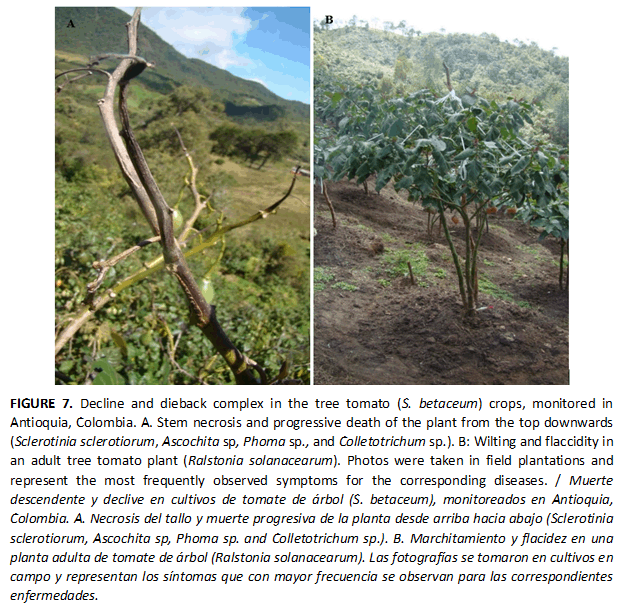
Decline and dieback complex
The presence of this disease was characterized by wilting, leaf drop and stem necrosis, a symptomatology that in advanced stages caused the progressive death of the plant from the top downwards (Figure 7A). This pathology was observed primarily in adult plantations, although it was also seen on young plants with a poor agricultural management. The isolates obtained were associated with the previously reported microorganisms: Sclerotinia sclerotiorum, Ascochita sp., Phoma sp., and Colletotrichum sp.(18).
Wilt
Widespread wilting and flaccidity (Figure 7B), which was accentuated when the ambient temperature was higher, was identified. In advanced stages, the symptoms described were accompanied by leaf yellowing, defoliation, and general plant deterioration. In the macroscopic analysis of the tissue, an obstruction of the vascular bundles, characterized by a dark brown coloration on the stem, was observed. From the diseased tissue, red-colored Gram-negative bacterial colonies surrounded by a white halo were isolated on TZC medium. The Hydrolysis on YDC at 30°C, the absence of yellow or orange colonies on YDC culture medium, and the positive oxidase and urease tests, taken together, indicated the isolation of R. solanacearum (16,17).
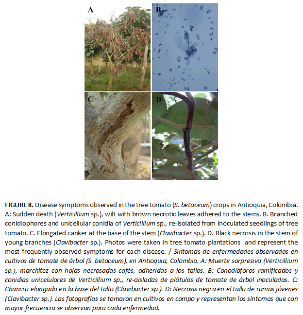
]]> Reporting new pathologies under field conditionsSudden death
This pathology was characterized by a widespread wilt, with the leaves subsequently acquiring a brown tone and remaining adhered to the stems (Figure 8A). This was followed by the death of the plant (Figure 8A). The macroscopic examination of the stem tissue showed the presence of a brown coloration in the vascular bundles. A hyaline, floccus, partitioned and thin mycelium grew on the PDA culture medium.
The microassemblies showed branched conidiophores in whorled bundles with unicellular, hyaline and ovoid conidia at the apex (Figure 8B). The symptomatology was similar to the wilt reported in avocado (15) and the results of the tests indicated that this disease was caused by the fungus Verticillium sp. (15,18). The pathogenicity tests reproduced the symptoms and the same microorganism was re-isolated, which suggested that it was the causal agent of sudden death in tree tomato (Figure 8B).
Stem canker, split stem and branch rot
On young branches and at the base of the stem, a semi-moist rot of an intense black color was observed. On the stem, elongated cankers were found (Figures 8C and 8D). A bacterium was consistently isolated from the diseased tissue; the test performed showed Gram-positive colonies, hydrolysis on YDC at 30°C, and yellow or orange colonies on YDC. They were negative for oxidase and urease. The results were consistent with a species belonging to the Clavibacter genus (16). When the isolate was re-inoculated into tree tomato seedlings, stem rot symptoms similar to those recorded were observed, and the same bacterium was re-isolated from the diseased tissues, confirming it as the causal agent.
Leaf curl
This pathology was characterized by leaf yellowing, wilt and curl, in addition to a stunted growth (Figure 9A). Root galls were also found associated with this symptomatology; these appeared as a row of irregular spheres with light coloration that eventually increased slightly in size, turned brown, and disintegrated into powder (Figure 9B). Roots collected in the fields, stained and observed under the light microscope, revealed yellow-brown sporosori (Figure 9C) with a polyhedral or slightly ellipsoidal shape and zoosporangia (Figure 9D), consistent with the plant pathogen Spongospora subterranea (21,22).
Tree tomato seedlings grown on soil inoculated with sporosori (spore balls) as described in materials and methods, showed similar symptoms to those observed under field conditions. Plasmodium (Figure 9E) and sporosori (Figure 9F) were identified on the stained roots from the plant seedlings grown in soi inoculated with sporosori, being confirmed the pathogenicity of these structures.
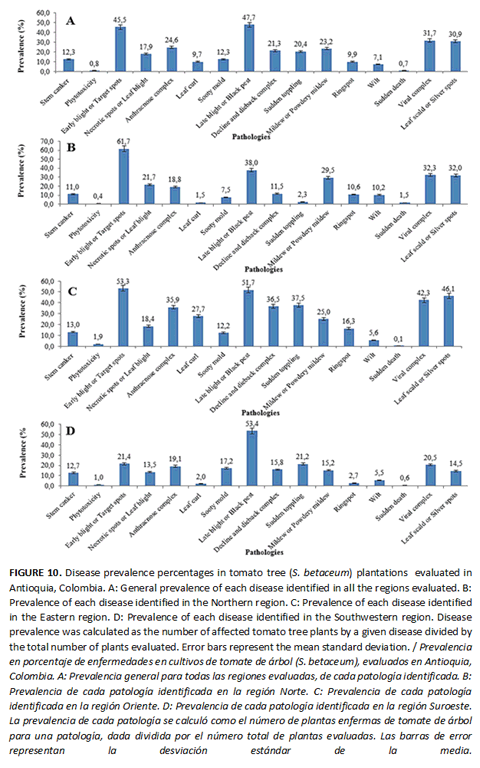
Prevalence of pathologies in tree tomato in Antioquia
]]> A number of parameters, such as the agronomical management, the sources and amount of inoculum, and the climatic conditions, determined the prevalence observed for each disease. The recorded pathologies corresponded to those expressed under the conditions prevailing during the two years of the study, which were usually of high precipitation and relative humidity most of the year round. The absence of others diseases was likely due to the absence of the environmental conditions required by their causal agents to develop, or maybe being present the causal agent, the disease did not show high prevalence values because the climatic conditions were not the optimal for pathogen development, or control measures were applied promptly and correctly in the fields sampled.The presence of 16 diseases was defined in this study (Figure 10A); of these, 13 presented a prevalence percentage higher than 9.5%. This signified a high pressure on the crop. The prevalence of the microorganisms identified varied by sampling region (Figures 10B, 10C and 10D).
Blight caused by P. infestans sensu lato was the disease with the highest prevalence in Antioquia, with values even higher than 53% in the Southwestern region, and it was therefore the most important causal agent for this crop (10,11). Anthracnose (Colletotrichum sp.), previously considered by different authors the most limiting disease in tree tomato (6,8), was in the fifth place, after the diseases caused by Xanthomonas sp., the different viruses, and Alternaria sp. (Figures 10B, 10C and 10D). Even more. The Meloidogyne sp. complex, also considered to be of importance (9), occupied the eighth spot in terms of prevalence, with a level of 20.4%.
The results of this study show that the diseases in tree tomato crops are caused by a wide range of causal agents. For this reason, timely and precise monitoring and diagnosis of pathologies should be considered fundamental for planning effective and efficient control strategies.
The prevalence values for late blight, and the known severity of this disease when the environmental conditions are favorable for P. infestans sensu lato, suggest that this disease has the potential to become the most limiting disease for tree tomato crops, as it is already for various Solanaceae crops on a global scale. This pathogen is found associated with numerous hosts in the Andean region, including potato, tomato, lulo, cape gooseberry, cucumber, and others. These crops are cultivated together with tree tomato, thus making it more difficult to control the pathogen (10,11,12).
The disease caused by Alternaria sp. was another of increasing importance found in this study. It had the second-highest prevalence, both in general (Figure 10A) and in each of the regions evaluated (Figures 10B, 10C and 10D). This microorganism has been reported as a pathogen of great economic significance in tomato (Solanum lycopersicum) and potato (Solanum tuberosum) because of its high aggressiveness and the major financial losses it can cause. Additionally, it can survive under adverse conditions(24).
The disease with the third-highest prevalence was caused by the viral complex was associated with the presence of six distinct genera. The viral complex is of difficult management because 14 viruses have been identified causing symptoms on tree tomato. In this regard, Jaramillo et al. (7), proposed this disease to be considered among the most limiting since, in its advanced stages, crop eradication is required, there is no chemical treatment available, and the current varieties are susceptible. In the present work, the strains showed low sequence identity suggesting a high variability or possibly new viral species that require further research. Different virus species implies that epidemiological factors such as virus particle transmission, strain virulence, vectors and other aspects of virus-host biology, must be studied in detail for each virus in order to implement adequate integrated management strategies for this viral complex.
Meanwhile, leaf scald, mildew, the dieback complex and necrotic spots had prevalence values higher than 15% (Figures 10A, 10B, 10C and 10D), meaning they are significant in the crop. The prevalence found in the crops for the majority of the diseases was indicative of poor phytosanitary management practices, misdiagnosis, or resistant strains of the pathogens.
Stem cankers, not previously reported in tree tomato, were associated with the presence of the bacterium Clavibacter sp. In tomato, Clavibacter michiganensis, subspecies michiganensis, causes bacterial canker (29). S. subterránea has been reported as a pathogen of different species within the family Solanaceae under greenhouse conditions (21,22). The presence of this disease causes substantial losses in the potato crop (S. tuberosum) and it has the characteristic that its inoculum increases cycle after cycle (21,22). In regard to the visual identification of this disease, it can be confused with problems caused by nematodes classified in the genus Meloidogyne. A disease that is similar to the sudden death caused by Verticillium sp. has been reported in avocado in the region of study (15). Worldwide, this causal agent has been associated with species belonging to the Solanaceae family, such as tomato and potato (30).
The majority of the diseases identified in this study had been previously reported (3,5,6,7,8,9,10,11,22). However, new diseases, such as those caused by S. subterranea, Verticillium sp. and Clavibacter sp., were reported. Moreover, the variation between the prevalence observed and that previously published for each disease indicated variation in the factors favoring disease development such as the weather, agronomical management and inoculum. For this reason, permanent surveillance must be established to prevent severe epidemics in this crop. Traditionally, diseases have been managed in this crop almost exclusively through the scheduled spraying of chemical products, which represents 27% of production costs. Usually, these applications are not based on a timely and accurate diagnosis, which reduces the competitiveness and sustainability of the crop. Additionally, the indiscriminate application of pesticides causes the environmental pollution and health problems for producers and consumers, and is a strong selection factor for pathogens, generating populations that are resistant to the molecules. The results described in this study will help lay the foundations for appropriate diagnosis and management of the pathogens associated with this crop.
]]>CONCLUSION
Pathologies associated to tree tomato are complex and require permanent monitoring to apply appropriate and timely integrated disease management programs.
AKNOWLEDGMENTS
The present research was funded by Universidad Nacional de Colombia sede Medellín, under the project: "Alternativas de manejo de la gota del tomate de árbol (Solanum betaceum Cav), mediante el uso de inductores de resistencia y caracterización del organismo causal (Phytophthora infestans sensu lato)", "grant: Resolución A-0891 de 2012 Mayo 18.
1. Acosta-Quezada P, Riofrío-Cuenca P, Rojas J, Vilanova S, Plazas M, Prohens J. Phenological growth stages of tree tomato (Solanum betaceum Cav.), an emerging fruit crop, according to the basic and extended BBCH scales. Scientia Horticulturae. 2016; 199 (16): 216-223.
]]>2. Bioversity International, Departamento de Ciencias Agropecuarias y de Alimentos, and COMAV. Descriptors for tree tomato (Solanum betaceum Cav.) and wild relatives. Bioversity International, Rome, Italy; Departamento de Ciencias Agropecuarias y de Alimentos (UTPL), Loja, Ecuador; Instituto de Conservacion y Mejora de la Agrodiversidad Valenciana, Valencia, Spain. 2013. 67p.
3. Gutiérrez P, Alzate J, Marín M. Genome sequence of a virus isolate from tamarillo (Solanum betaceum) in Colombia: evidence for a new potyvirus. Archives of Virology. 2015; 160:557-560.
4. Agronet. Sistema de estadística agropecuaria: área, producción y rendimiento para tomate de árbol. 2014. (Consultado 07-11-2016). Disponible: www.agronet.gov.co/agronetweb1/Estad%C3%ADsticas.aspx.
5. Hierro A, Guerra S, Padilla F, Arroyo C, Soria N, et al. Assessing the Morphological Variations on the pollen grains of Solanum betaceum caused by chemical, biological and ecological pesticides. Biol Med (Aligarh). 2016; 8: 286. doi: 10.4172/0974-8369.1000286.
6. Luengas-Gómez C, Roa-Vásquez M, Orrego-Vásquez J. Evaluation of a prebiotic and potassium for the control of anthracnose in the tree tomato. Agronomía Colombiana. 2012; 30(2): 230-235.
]]>7. Jaramillo M, Álvarez J, Marín M. Características de los virus asociados a la virosis del tomate de árbol (Solanum betaceum) en Colombia. Revista Lasallista de Investigación. 2012; 9 (1): 115-127.
8. Pardo-De la Hoz C, Calderón C, Rincón A, Cárdenas M, Danies G, López-Kleine L, Restrepo S, Jiménez P. Species from the Colletotrichum acutatum, Colletotrichum boninense and Colletotrichum gloeosporioides species complexes associated with tree tomato and mango crops in Colombia. Plant Pathology. 2016; 65: 227–237. doi:10.1111/ppa.12410.
9. Mosquera-Espinosa A. Plant parasitic nematodes associated with Cyphomandra betacea (Cav.) Sendtn., Solanum quitoense Lam. and Daucus carota L. in Boyacá, Colombia. Acta Agronómica. 2016; 65 (1): 87-9. doi: http://dx.doi.org/10.15446/acag.v65n1.45180.
10. Forbes G, Morales J, Restrepo S, Pérez W, Gamboa S, Ruiz R, Cedeño L, Fermin G, Adriana B, Andreu B, Ivette Acuña I, Oliva R. Phytophthora infestans and Phytophthora andina on Solanaceous Hosts in South America. En: Phytophthora: A Global Perspective (ed. Lamour K). CAB International, CABI Wallingford, UK. 2013.
11. Oliva RF, Kroon L, Chacón G, Flier W, Ristaino J, Forbes G. Phytophthora andina sp. nov., a newly identified heterothallic pathogen of solanaceous hosts in the Andean highlands. Plant Pathology. 2010; 59: 613–625.
12. Castaño J, Ramirez J, Patiño L, Morales J. Alternativa para el manejo de Phytophthora infestans (Mont.) de Bary en Solanum betaceum Cav. mediante inductores de resistencia. Rev.Protección Veg. 2015; 30 (3): 204-212.
]]>13. Rheinländer P, Jamieson L, Fullerton R, Manning M, Meier X. Scarring in tamarillo fruit (Solanum betaceum). New Zealand Plant Protection, 2009; 62: 315-320.
14. Liefting L, Weir B, Pennycook S, Clover G. 'Candidatus Liberibacter solanacearum', associated with plants in the family Solanaceae. International Journal of Systematic and Evolutionary Microbiology. 2009; 59, 2274-2276.
15. Ramírez J, Castañeda D, Morales J. Estudios etiológicos de la marchitez del aguacate en Antioquia-Colombia. Revista Ceres, 2014; 61(1): 050-061.
16. Shaad N. Laboratory guide for identification of plant pathogenic bacteria. Departament of Plant Pathology. University of Georgia. 2001. 398 p.
17. Burgos C, Silva B, Salazar M, Morales J. Diversidad genética de aislados de Ralstonia solanacearum procedentes de tres regiones de Colombia. Rev. Protección Veg. 2015; 30 (3): 213-224.
]]>18. Barnett H, Hunter B. Illustrated genera of imperfect fungi. Third edition. Burgess Publishing Company. Minnesota, E.U. 1972. 241 p.
19. Erwin D, Ribeiro O. Phytophthora Diseases Worldwide. The American Phytophathological Society, St. Paul, Minnesota, E.U. 1996. 562 p.
20. Mai W, Mullin P. Plant-parasitic nematodes- a pictorial key to genera. Cornell University Press, Ithaca, NY, USA. 1996. 277 p.
21. Soler J, Benavidez W, Gilchrist E, Morales J, Pérez J. Evaluation of an in vitro system for the study of Spongospora subterranea f. sp. subterranea in potato roots (Solanum tuberosum subsp. andigena L.) Var. Diacol Capiro. Revista de la Facultad de Ciencias. 2012; 1(2): 34-46.
22. Bastidas N, Morales J, Gonzales E, Gutierrez P, Marin M. Detección y cuantificación de Spongospora subterranea f. sp. subterranea en plantas señuelo y cultivos de papa en Colombia mediante qPCR. Acta Biológica Colombiana. 2013; 18 (1): 121 -136.
]]>23. Taylor R, Pasche J, Gallup C, Shew H, Gudmestad N. A foliar blight and tuber rot of potato caused by Phytophthora nicotianae: New occurrences and characterization of isolates. Plant Disease. 2008; 92: 492-503.
24. Upadhyay P, Rai A, Kumar R, Singh M, Sinha B. Microarray analyses during early stage of the tomato/Alternaria solani interaction. Genomics Data. 2015; 6: 170–172.
25. Schneider W, Sherman D, Stone A, Damsteegt V, Frederick R. Specific detection and quantification of Plum pox virus by real-time fluorescent reverse transcription-PCR. Journal of Virological Methods. 2004; 120: 97-105.
26. Adams M, Antoniw J, Fauquet C. Molecular criteria for genus and species discrimination within the family Potyviridae. Archives of Virology. 2005; 150: 459-479. doi:10.1007/s00705-004-0440-6.
27. Duffy S, Seah Y. 98% identical, 100% wrong: per cent nucleotide identity can lead plant virus epidemiology astray. Philosophical Trans. Royal Society B. 2010; 365: 1891-1897. doi:10.1098/rstb.2010.0056.
28. Ochoa L, Insuasti A. Etiología de las enfermedades virales del tomate de árbol en Ecuador. Instituto Nacional de Investigaciones Agropecuarias, Quito (Ecuador). En: Informe Técnico Anual - INIAP (Ecuador). Est. Exp. Santa Catalina. Departamento Nacional de Protección Vegetal, estudios agronómicos, fitopatológicos y entomológicos de frutales nativos andinos. INIAP, Quito, Ecuador. 2005. pp. 1-4.
29. European and Mediterranean Plant Protection Organization. PM 7/42 (2) Clavibacter michiganensis subsp. michiganensis Bulletin. OEPP/EPPO Bulletin. 2013; 43 (1): 46-67.
30. Uribe P, Jansky S, Halterman D. Two CAPS markers predict Verticillium wilt resistance in wild Solanum species. Molecular Breeding. 2014; 33 (2): 465-476.
Recibido: 22/4/2016
Aceptado: 27/10/2016
]]>
* Autor para correspondencia: Juan Gonzalo Morales-Osorio. E-mail: jgmoraleso@unal.edu.co
]]>