<?xml version="1.0" encoding="ISO-8859-1"?><article xmlns:mml="http://www.w3.org/1998/Math/MathML" xmlns:xlink="http://www.w3.org/1999/xlink" xmlns:xsi="http://www.w3.org/2001/XMLSchema-instance">
<front>
<journal-meta>
<journal-id>1027-2852</journal-id>
<journal-title><![CDATA[Biotecnología Aplicada]]></journal-title>
<abbrev-journal-title><![CDATA[Biotecnol Apl]]></abbrev-journal-title>
<issn>1027-2852</issn>
<publisher>
<publisher-name><![CDATA[Editorial Elfos Scientiae]]></publisher-name>
</publisher>
</journal-meta>
<article-meta>
<article-id>S1027-28522009000100008</article-id>
<title-group>
<article-title xml:lang="en"><![CDATA[Electrochemical impedance spectroscopy: An effective tool for a fast microbiological diagnosis]]></article-title>
<article-title xml:lang="es"><![CDATA[Espectroscopía de impedancia electroquímica, herramienta eficaz para el diagnóstico rápido microbiológico]]></article-title>
</title-group>
<contrib-group>
<contrib contrib-type="author">
<name>
<surname><![CDATA[Ramírez]]></surname>
<given-names><![CDATA[Nardo]]></given-names>
</name>
<xref ref-type="aff" rid="A01"/>
</contrib>
<contrib contrib-type="author">
<name>
<surname><![CDATA[Regueiro]]></surname>
<given-names><![CDATA[Angel]]></given-names>
</name>
<xref ref-type="aff" rid="A02"/>
</contrib>
<contrib contrib-type="author">
<name>
<surname><![CDATA[Arias]]></surname>
<given-names><![CDATA[Olimpia]]></given-names>
</name>
<xref ref-type="aff" rid="A03"/>
</contrib>
<contrib contrib-type="author">
<name>
<surname><![CDATA[Contreras]]></surname>
<given-names><![CDATA[Rolando]]></given-names>
</name>
<xref ref-type="aff" rid="A01"/>
</contrib>
</contrib-group>
<aff id="A01">
<institution><![CDATA[,Centro Nacional de Investigaciones Científicas  ]]></institution>
<addr-line><![CDATA[Ciudad de La Habana ]]></addr-line>
<country>Cuba</country>
</aff>
<pub-date pub-type="pub">
<day>00</day>
<month>03</month>
<year>2009</year>
</pub-date>
<pub-date pub-type="epub">
<day>00</day>
<month>03</month>
<year>2009</year>
</pub-date>
<volume>26</volume>
<numero>1</numero>
<fpage>72</fpage>
<lpage>78</lpage>
<copyright-statement/>
<copyright-year/>
<self-uri xlink:href="http://scielo.sld.cu/scielo.php?script=sci_arttext&amp;pid=S1027-28522009000100008&amp;lng=en&amp;nrm=iso"></self-uri><self-uri xlink:href="http://scielo.sld.cu/scielo.php?script=sci_abstract&amp;pid=S1027-28522009000100008&amp;lng=en&amp;nrm=iso"></self-uri><self-uri xlink:href="http://scielo.sld.cu/scielo.php?script=sci_pdf&amp;pid=S1027-28522009000100008&amp;lng=en&amp;nrm=iso"></self-uri><abstract abstract-type="short" xml:lang="en"><p><![CDATA[Dielectric spectroscopy, also called electrochemical impedance spectroscopy, is traditionally used in monitoring corrosion and electro-deposition processes in the coating and characterization assessment of many kinds of sensors and semi-conductors. Its application in biotechnology for the characterization of cell cultures has, however, been notably expanded in the last decade. As a transductional principle, impedance has been applied in the field of microbiology as a means of detecting and quantifying pathogenic bacteria. This paper reviews the state-of-the-art of Impedance Microbiology, its progress and its applications for the detection of foodborne pathogenic bacteria, including the use of interdigitated microelectrodes, the development of chip-based impedance microbiology and the integration of impedance biosensors along with other techniques such as dielectrophoresis and electropermeabilization. Reference is made to basic components, definitions and principles of this technique, as well as to the explanation of the components and principles for cell culture design and the use of equivalent circuits for the analysis of the systems based on this alternative.]]></p></abstract>
<abstract abstract-type="short" xml:lang="es"><p><![CDATA[La espectroscopia dieléctrica o espectroscopia de impedancia electroquímica es empleada tradicionalmente en el registro y estudio de los procesos de corrosión y electrodeposición, en la evaluación de recubrimientos y en la caracterización de muchos tipos de sensores y semiconductores. En la última década se han ampliado notablemente sus aplicaciones en la Biotecnología para la caracterización de células biológicas, el diagnóstico de enfermedades y la caracterización del cultivo de células. Como principio de transducción, la técnica de impedancia ha sido aplicada en el campo de la microbiología como un medio para detectar y cuantificar microorganismos patógenos. El presente trabajo revisa el estado del arte de la microbiología de impedancia, el progreso y las aplicaciones en la detección de microorganismos patógenos incluido el uso de los microelectrodos interdigitados, el desarrollo de la miniaturización de la microbiología de impedancia y la integración de los biosensores de impedancia con otras técnicas como la dielectroforesis y la electropermeabilización. Se hace referencia a conceptos básicos, definiciones y fundamentos de esta técnica, así como se abordan los componentes, principios para el diseño del medio de cultivo y uso de circuitos equivalentes para el análisis de los sistemas basados en esta alternativa.]]></p></abstract>
<kwd-group>
<kwd lng="en"><![CDATA[bacteria detection]]></kwd>
<kwd lng="en"><![CDATA[diagnosis]]></kwd>
<kwd lng="en"><![CDATA[impedance]]></kwd>
<kwd lng="en"><![CDATA[microbiology]]></kwd>
<kwd lng="es"><![CDATA[detección de bacterias]]></kwd>
<kwd lng="es"><![CDATA[diagnóstico]]></kwd>
<kwd lng="es"><![CDATA[impedancia]]></kwd>
<kwd lng="es"><![CDATA[microbiología]]></kwd>
</kwd-group>
</article-meta>
</front><body><![CDATA[ <div class=Section1>      <div>      <p align=right style='text-align:right'><b><span lang=EN-US style='font-size: 10.0pt;font-family:"Verdana","sans-serif";color:#211E1E'>FOCUS </span></b></p>      <p align=right style='text-align:right'><span lang=EN-US>&nbsp;</span></p>      <p><b><span lang=EN-US style='font-size:13.5pt;font-family:"Verdana","sans-serif"; color:#211E1E'>Electrochemical impedance spectroscopy: An effective tool for a fast microbiological diagnosis</span></b></p>      <p><span lang=EN-US>&nbsp;</span></p>      <p><b><span style='font-family:"Verdana","sans-serif";color:#211E1E'>Espectroscopía de impedancia electroquímica, herramienta eficaz para el diagnóstico rápido microbiológico</span></b><span style='font-size:11.0pt;font-family:"Arial","sans-serif"; color:red'> </span></p>      <p><span style='font-size:13.5pt;color:#211E1E'>&nbsp;</span></p>      <p><span style='font-size:13.5pt;color:#211E1E'>&nbsp;</span></p>      <p><span style='font-size:11.0pt;font-family:"Arial","sans-serif";color:blue'>&nbsp;</span><b><span style='font-size:10.0pt;font-family:"Verdana","sans-serif";color:#211E1E'>Nardo Ramírez<sup>1</sup>, Angel Regueiro<sup>2</sup>, Olimpia Arias<sup>3</sup>, Rolando Contreras<sup>1</sup></span></b></p>      ]]></body>
<body><![CDATA[<p><sup><span style='font-size:10.0pt;font-family:"Verdana","sans-serif"; color:#211E1E'>1</span></sup><span style='font-size:10.0pt;font-family:"Verdana","sans-serif"; color:#211E1E'>Dirección de Diagnóstico Microbiológico, Centro Nacional de Investigaciones Científicas, CNIC Ave. 25 esq. 158, Cubanacán, Playa, Ciudad de La Habana, Cuba <sup>    <br> 2</sup>Centro de Bioingeniería, CEBIO. Cuba <sup>    <br> 3</sup>Instituto de Ciencia y Tecnología de Materiales, Universidad de La Habana, UH. Cuba </span></p>      <p><span style='font-size:13.5pt;color:#211E1E'>&nbsp;</span></p>      <p><span style='font-size:13.5pt;color:#211E1E'>&nbsp;</span></p>      <div class=MsoNormal align=center style='text-align:center'>  <hr size=2 width="100%" align=center>  </div>      <p><b><span lang=EN-US style='font-size:10.0pt;font-family:"Verdana","sans-serif"; color:#211E1E'>ABSTRACT </span></b></p>      <p><span lang=EN-US style='font-size:10.0pt;font-family:"Verdana","sans-serif"; color:#211E1E'>Dielectric spectroscopy, also called electrochemical impedance spectroscopy, is traditionally used in monitoring corrosion and electro-deposition processes in the coating and characterization assessment of many kinds of sensors and semi-conductors. Its application in biotechnology for the characterization of cell cultures has, however, been notably expanded in the last decade. As a transductional principle, impedance has been applied in the field of microbiology as a means of detecting and quantifying pathogenic bacteria. This paper reviews the state-of-the-art of Impedance Microbiology, its progress and its applications for the detection of foodborne pathogenic bacteria, including the use of interdigitated microelectrodes, the development of chip-based impedance microbiology and the integration of impedance biosensors along with other techniques such as dielectrophoresis and electropermeabilization. Reference is made to basic components, definitions and principles of this technique, as well as to the explanation of the components and principles for cell culture design and the use of equivalent circuits for the analysis of the systems based on this alternative. </span></p>      <p class=MsoNormal><b><span lang=EN-US style='font-size:10.0pt;font-family: "Verdana","sans-serif";color:#211E1E'>Keywords:</span></b><span lang=EN-US style='font-size:10.0pt;font-family:"Verdana","sans-serif";color:#211E1E'> bacteria detection, diagnosis, impedance, microbiology.</span></p>      <div class=MsoNormal align=center style='text-align:center'>  <hr size=2 width="100%" align=center>  </div>      ]]></body>
<body><![CDATA[<p><b><span style='font-size:10.0pt;font-family:"Verdana","sans-serif"; color:#211E1E'>RESUMEN </span></b></p>      <p><span style='font-size:10.0pt;font-family:"Verdana","sans-serif";color:#211E1E'>La espectroscopia dieléctrica o espectroscopia de impedancia electroquímica es empleada tradicionalmente en el registro y estudio de los procesos de corrosión y electrodeposición, en la evaluación de recubrimientos y en la caracterización de muchos tipos de sensores y semiconductores. En la última década se han ampliado notablemente sus aplicaciones en la Biotecnología para la caracterización de células biológicas, el diagnóstico de enfermedades y la caracterización del cultivo de células. Como principio de transducción, la técnica de impedancia ha sido aplicada en el campo de la microbiología como un medio para detectar y cuantificar microorganismos patógenos. El presente trabajo revisa el estado del arte de la microbiología de impedancia, el progreso y las aplicaciones en la detección de microorganismos patógenos incluido el uso de los microelectrodos interdigitados, el desarrollo de la miniaturización de la microbiología de impedancia y la integración de los biosensores de impedancia con otras técnicas como la dielectroforesis y la electropermeabilización. Se hace referencia a conceptos básicos, definiciones y fundamentos de esta técnica, así como se abordan los componentes, principios para el diseño del medio de cultivo y uso de circuitos equivalentes para el análisis de los sistemas basados en esta alternativa. </span></p>      <p class=MsoNormal><b><span style='font-size:10.0pt;font-family:"Verdana","sans-serif"; color:#211E1E'>Palabras clave:</span></b><span style='font-size:10.0pt; font-family:"Verdana","sans-serif";color:#211E1E'> detección de bacterias, diagnóstico, impedancia, microbiología.</span><span style='font-size:11.0pt; font-family:"Arial","sans-serif";color:red'> </span></p>      <div class=MsoNormal align=center style='text-align:center'>  <hr size=2 width="100%" align=center>  </div>      <p><span lang=EN-US style='font-size:13.5pt'>&nbsp;</span></p>      <p><span lang=EN-US style='font-size:13.5pt'>&nbsp;</span></p>      <p><b><span lang=EN-US style='font-family:"Verdana","sans-serif";color:#211E1E'>INTRODUCTION </span></b></p>      <p><span lang=EN-US style='font-size:10.0pt;font-family:"Verdana","sans-serif"; color:#211E1E'>The growing use of electrochemical sensors in environmental applications, the industry and in the medical field has brought about the urging need to understand surface properties and the important analytical systems as a whole. This seems to indicate that the scientific community has chosen impedance spectroscopy to characterize a large number of electrochemical systems. The information obtained from this technique has been used to make electrochemical sensors having excellent properties (linearity, thermal and temporal stability, etc). </span></p>      <p><span lang=EN-US style='font-size:10.0pt;font-family:"Verdana","sans-serif"; color:#211E1E'>Impedance spectroscopy is a powerful tool for a fast biomolecule diagnosis and for analyses in cell cultures. Its superiority over other laboratory techniques lies in that it uses a small signal, generally in the tension mode, thus minimizing the alterations of the properties of the medium, in other words, applied stimulation does not alter the equilibrium conditions of the system. </span></p>      <p><span lang=EN-US style='font-size:10.0pt;font-family:"Verdana","sans-serif"; color:#211E1E'>The signal applied to the samples makes it possible to link the properties of the liquid or solid being studied with the variations or changes obtained in its characteristic impedance. This is due to the physical structure of the material, to the chemical processes occurring in it, or to a combination of both. Consequently, electrochemical impedance spectroscopy is a non-destructive technique providing robust measurements. </span></p>      ]]></body>
<body><![CDATA[<p><b><span lang=EN-US style='font-size:10.0pt;font-family:"Verdana","sans-serif"; color:#211E1E'>Classical impedance microbiology: Definition and basic concepts </span></b></p>      <p><span lang=EN-US style='font-size:10.0pt;font-family:"Verdana","sans-serif"; color:#211E1E'>Electric impedance as a transduction principle has been applied to a great variety of biological, physiological and medical problems (1, 2). </span></p>      <p><span lang=EN-US style='font-size:10.0pt;font-family:"Verdana","sans-serif"; color:#211E1E'>Before going into the topic, it is important to analyze some basic concepts. It is widely known that electric resistance R is the ability of an element from the circuit to resist the flow of electric current. Ohm’s Law defines the resistance as the relation between the tension and the electric current. </span></p>      <p align=center style='text-align:center'><span style='font-size:10.0pt; font-family:"Verdana","sans-serif";color:#211E1E'><img width=210 height=52 src="/img/revistas/bta/v26n1/fr0112109.gif"></span></p>      
<p><span lang=EN-US style='font-size:10.0pt;font-family:"Verdana","sans-serif"; color:#211E1E'>Where: <i>V<b> </b></i>corresponds to the value of tension, and <i>I<b> </b></i>to that of the electric current. </span></p>      <p><span lang=EN-US style='font-size:10.0pt;font-family:"Verdana","sans-serif"; color:#211E1E'>The use of equation 1 is limited only to an element of the circuit: the ideal resistor, which has several determining factors: 1) it fulfills Ohm’s Law in all levels of tension and current, 2) its resistance value is not related to frequency, and 3) signals of tension and current through a resistor are in the same phase. </span></p>      <p><span lang=EN-US style='font-size:10.0pt;font-family:"Verdana","sans-serif"; color:#211E1E'>The real world has elements with a much more complex behavior. These elements make us reject the simple concept of resistance. As a result, impedance is used instead, which is a parameter of a much more general circuit. As with resistance, impedance is a measurement of the ability of a circuit to resist the flow of the electric current. Unlike resistance, impedance is not limited by any of the above mentioned determining factors. </span></p>      <p><span lang=EN-US style='font-size:10.0pt;font-family:"Verdana","sans-serif"; color:#211E1E'>Electrochemical impedance is usually obtained by applying a potential of alternating current to an electrochemical cell and measuring the current flowing through it. The response to this sinusoidal potential of excitation is an alternating current signal that may be analyzed as the sum total of all sinusoidal functions (Fourier’s series). </span></p>      <p><span lang=EN-US style='font-size:10.0pt;font-family:"Verdana","sans-serif"; color:#211E1E'>Another concept that must be considered is that of the capacity or capacitance, expressing the ability of a capacitor to store electric charge. This property determines the relationship between the potential difference existing between the plates of the capacitor, and the electrical charge stored in it, through the following equation. </span></p>      <p align=center style='text-align:center'><span style='font-size:10.0pt; font-family:"Verdana","sans-serif";color:#211E1E'><img width=204 height=49 src="/img/revistas/bta/v26n1/fr0212109.gif"></span></p>      
]]></body>
<body><![CDATA[<p><span lang=EN-US style='font-size:10.0pt;font-family:"Verdana","sans-serif"; color:#211E1E'>Where: <i>C </i>is the capacitance, measured in farads, a relatively big unit that is usually used in submultiples such as microfarads and picofarads. <i>V </i>is the potential difference measured in volts. <i>Q </i>is the stored electrical charge measured in coulombs. </span></p>      <p><span lang=EN-US style='font-size:10.0pt;font-family:"Verdana","sans-serif"; color:#211E1E'>On the other hand, conductance is directly related to the ease offered by any material for the flow of the electric current. Conductance G<b><i>, </i></b>and resistance are inversely related. The value of the conductance of a material is described in siemens and it is identified by the letter S. One siemens equals 1/</span><span style='font-size:10.0pt;font-family:"Verdana","sans-serif"; color:#211E1E'><img width=17 height=13 src="/img/revistas/bta/v26n1/f0812109.GIF"></span><span lang=EN-US style='font-size:10.0pt;font-family:"Verdana","sans-serif"; color:#211E1E'>, or also ohm<sup>-1</sup>. </span></p>      
<p><span lang=EN-US style='font-size:10.0pt;font-family:"Verdana","sans-serif"; color:#211E1E'>An increasing application is that of automatically recording impedance in microbiology. Impedance microbiology (IM) is formed by two important techniques: Classical IM, based on the measurement of bipolar impedance or the resistance of the medium (disregarding the dispersion phenomenon) and impedance spectroscopy, also called dielectric spectroscopy basing its measurement specifically on the dispersion of the medium. </span></p>      <p><span lang=EN-US style='font-size:10.0pt;font-family:"Verdana","sans-serif"; color:#211E1E'>
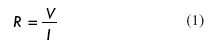
IM is the recording of the variability of impedance with time during the growth of the microorganisms in a sample. It is done by placing an arrangement of metal electrodes, submerged in an inoculated culture medium (</span><span style='font-size:10.0pt;font-family:"Verdana","sans-serif";color:#211E1E'><a href="#fig1"><span lang=EN-US>Figure 1A</span></a></span><span lang=EN-US style='font-size:10.0pt;font-family:"Verdana","sans-serif";color:#211E1E'>), thus achieving measurement between the electrodes (bipolar impedance) while the microorganisms found there grow. The technique consists of making measurements of impedance components such as: conductance, capacity, impedance module, phase angle and others through bipolar or tetra polar methods, with electrodes placed in a flask containing an inoculated culture media kept at a constant temperature. These measurements make it possible to record detect, quantify and even identify certain microorganisms found in the samples coming from the industry or from clinical practice </span></p>      <p align=center style='text-align:center'><span style='font-size:13.5pt'><img border=0 width=453 height=281 src="/img/revistas/bta/v26n1/f0112109.gif"></span><a name=fig1></a></p>      
<p><b><span lang=EN-US style='font-family:"Verdana","sans-serif";color:#211E1E'>BASES OF IMPEDANCE MICROBIOLOGY </span></b></p>      <p><span lang=EN-US style='font-size:10.0pt;font-family:"Verdana","sans-serif"; color:#211E1E'>In IM, impedance changes are typically measured by the use of a pair of electrodes placed within a growth medium or reacting solution (</span><span style='font-size:10.0pt;font-family:"Verdana","sans-serif";color:#211E1E'><a href="#fig1"><span lang=EN-US>Figure 1A</span></a></span><span lang=EN-US style='font-size:10.0pt;font-family:"Verdana","sans-serif";color:#211E1E'>). </span></p>      <p><span lang=EN-US style='font-size:10.0pt;font-family:"Verdana","sans-serif"; color:#211E1E'>The measurement can be made in two ways: directly, or indirectly. In the direct technique, a pair of metal electrodes is introduced in the medium inoculated with the bacteria we wish to measure. The metabolic products created during the growth of microorganisms modify the composition of the medium, thus changing the ionic content, which in turn produces a change in the conductivity of the culture media. These changes are recorded through time when variations in the electrode-electrolyte-sample interface are produced. These modifications are proportional to the concentration of living microorganisms that can be recorded through impedance measuring techniques. </span></p>      <p><span lang=EN-US style='font-size:10.0pt;font-family:"Verdana","sans-serif"; color:#211E1E'>
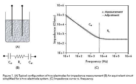
The release of the ion by the bacteria into its growth medium (</span><span style='font-size:10.0pt;font-family:"Verdana","sans-serif";color:#211E1E'><a href="#fig2"><span lang=EN-US>Figure 2</span></a></span><span lang=EN-US style='font-size:10.0pt;font-family:"Verdana","sans-serif";color:#211E1E'>) is due to two main mechanisms (3). The first mechanism is related to the energy metabolism (catabolism) in which, the bacteria consumes oxygen and carbohydrates, and produces carbon dioxide and organic acids. Some simple examples indicate that the conversion of a non-ionized glucose substrate into two molecules of lactic acid would increase culture media conductivity. Furthermore, the metabolism will take the lactic acid and three molecules of oxygen to produce carbonic acid. The smaller and most mobile bicarbonate ion is a more effective ionic conductor than the lactate ion. Hydrogen ions are almost seven times more</span><span lang=EN-US style='font-size:10.0pt;font-family: "Verdana","sans-serif"'> </span><span lang=EN-US style='font-size:10.0pt; font-family:"Verdana","sans-serif";color:#211E1E'>effective as ionic conductors than as sodium ions, all of which leads to the variation of the impedance caused by this action produced by the bacteria (2). The second mechanism is related to ion exchange through the cell membrane (active transportation). Ions such as sodium and potassium are actively transported through the ionic channels of the cell membrane (double lipid layer), which function as regulators of the membrane potential and of the osmotic pressure exerted between the inner and outer parts of cells. </span></p>      <p align=center style='text-align:center'><span style='font-size:13.5pt; color:#211E1E'><img border=0 width=562 height=423 src="/img/revistas/bta/v26n1/f0212109.gif"></span><a name=fig2></a></p>      
]]></body>
<body><![CDATA[<p><span lang=EN-US style='font-size:10.0pt;font-family:"Verdana","sans-serif"; color:#211E1E'>Although energy metabolism is the main cause of the release of ions from the cell into its environment; the ionic exchange process also offers a small contribution. What has definitely been clarified is that these ion release processes produce changes in the ionic composition of the culture medium and in its conductivity, which are the bases for the measurement of impedance changes. </span></p>      <p><span lang=EN-US style='font-size:10.0pt;font-family:"Verdana","sans-serif"; color:#211E1E'>
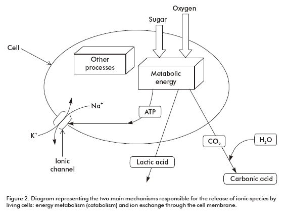
In contrast to that of the direct technique, the indirect technique does not measure the changes in impedance directly in the bacterial growth medium. The electrodes, instead of being submerged in the inoculated growth medium, they are introduced into a separate solution (usually into a potassium hydroxide solution). The gases produced by bacterial metabolism (mainly CO<sub>2</sub>) are absorbed by the potassium hydroxide solution, producing a decrease in the conductance of the alkaline solution. </span></p>      <p><span lang=EN-US style='font-size:10.0pt;font-family:"Verdana","sans-serif"; color:#211E1E'>In order to detect bacteria, impedance systems measure absolute or relative changes in conductance, capacitance or impedance at regular time intervals during bacterial growth under controlled temperature and humidity. The electric signals measured are graphically represented (amplitude at the vertical axis vs. incubation time at the horizontal axis) and impedance variability curves are generated. </span></p>      <p><span style='font-size:10.0pt;font-family:"Verdana","sans-serif";color:#211E1E'><a href="#fig3"><span lang=EN-US>Figure 3</span></a></span><span style='font-size: 10.0pt;font-family:"Verdana","sans-serif";color:#211E1E'> <span lang=EN-US>shows the typical impedance curve where it is observed that the impedance is quite stable at the initial part of the curve and then it starts decreasing. The time at which the impedance decreases and crosses a certain threshold value is defined as detection time (<i>t<sub>d</sub></i>). Generally, detection time does not appear until the number of bacteria is of about 10<sup>6</sup>-10<sup>7</sup> colony forming units (c.f.u.) per mL. When impedance finally reaches its limit, bacteria are at a high concentration of about 10<sup>8</sup> c.f.u./mL or more, and all the resources in the medium have been metabolized and converted into final products. The shape of the impedance curve corresponds well with the three typical phases of bacterial growth: Phase I, where the bacteria metabolize but do not replicate; Phase II of logarithmic or exponential growth, where the bacteria exponentially replicate, and Phase III, a steady stage, where cell concentration remains relatively constant (4). </span></span></p>      <p align=center style='text-align:center'><span style='font-size:13.5pt; color:#211E1E'><img border=0 width=348 height=330 src="/img/revistas/bta/v26n1/f0312109.gif"></span><a name=fig3></a></p>      
<p><span lang=EN-US style='font-size:10.0pt;font-family:"Verdana","sans-serif"; color:#211E1E'>A simple theoretical analysis, confirmed by experimental observation, shows that detection time (td, the time needed for impedance to cross an arbitrary threshold), is related to the initial cell concentration (<i>C</i>o) according to the model represented (5): </span></p>      <p align=center style='text-align:center'><span style='font-size:10.0pt; font-family:"Verdana","sans-serif";color:#211E1E'><img border=0 width=248 height=42 src="/img/revistas/bta/v26n1/fr0312109.gif"></span></p>      
<p><span lang=EN-US style='font-size:10.0pt;font-family:"Verdana","sans-serif"; color:#211E1E'>In this expression, a (<i>t </i>&gt; 0) and b are constants that depend on the particular type of microorganism, its growth conditions, etc. Eden et al (2) obtained experimental values of these constants: </span><span style='font-size:10.0pt;font-family:"Verdana","sans-serif";color:#211E1E'>&#945;</span><span lang=EN-US style='font-size:10.0pt;font-family:"Verdana","sans-serif"; color:#211E1E'> = 0.96 and </span><span style='font-size:10.0pt;font-family: "Verdana","sans-serif";color:#211E1E'>&#946;</span><span lang=EN-US style='font-size:10.0pt;font-family:"Verdana","sans-serif";color:#211E1E'> = 7.75, for <i>t<sub>d</sub> </i>in hours, and <i>C</i><sub>o</sub> in cfu/mL in the incubation medium. </span></p>      <p><span style='font-size:13.5pt;color:#211E1E'><a href="#fig4"><span lang=EN-US style='font-size:10.0pt;font-family:"Verdana","sans-serif"'>Figure 4</span></a></span><span lang=EN-US style='font-size:10.0pt;font-family:"Verdana","sans-serif"; color:#211E1E'> shows the representation of model (3) for the previous constant values, which indicates that detection time intervals go from 1 hr when <i>C</i>o is approximately equal to 10<sup>7</sup> c.f.u./mL, up to eight hours when <i>C</i><sub>o</sub> is of approximately 1 c.f.u./mL. Colony forming units is the minimal number of separable cells on the surface (or within it) in a semisolid agar medium giving rise to the development of a visible colony in the order of tenths of millions of progeny cells. </span></p>      <p align=center style='text-align:center'><span style='font-size:13.5pt; color:#211E1E'><img border=0 width=350 height=325 src="/img/revistas/bta/v26n1/f0412109.gif"></span><a name=fig4></a></p>      
]]></body>
<body><![CDATA[<p><b><span lang=EN-US style='font-family:"Verdana","sans-serif";color:#211E1E'>CLASSICAL IMPEDANCE MICROBIOLOGY: CULTURE MEDIUM AND <i>Salmonella</i> DETECTION </span></b></p>      <p><span lang=EN-US style='font-size:10.0pt;font-family:"Verdana","sans-serif"; color:#211E1E'>Many studies have been made to optimize the development of culture media since the direct IM technique is based on the observation of impedance changes. The principles for the design of the culture medium, which are so important for traditional microbiology, are also important for IM. Firstly, the medium must be chosen according to the bacterial growth </span></p>      <p><span lang=EN-US style='font-size:10.0pt;font-family:"Verdana","sans-serif"; color:#211E1E'>to be analyzed, which grants selectivity to impedance microbiological methods. Secondly, the formulation of the medium should be such that an optimum variability of impedance can be achieved. </span></p>      <p><i><span lang=EN-US style='font-size:10.0pt;font-family:"Verdana","sans-serif"; color:#211E1E'>Salmonella </span></i><span lang=EN-US style='font-size:10.0pt; font-family:"Verdana","sans-serif";color:#211E1E'>is the main cause of food intoxication with the highest number of cases reported. Salmonelosis is the infection produced by this bacterium. According to the Center for Disease Control (CDC), salmonelosis is responsible for approximately 1.4 million food intoxication cases more than 600 deaths in the United States each year. These infections produce direct and indirect medical expenses of a billion US dollars per year (6). Conventional microbiology methods for the detection of <i>Salmonella </i>ssp<i>. </i>require three to four days for a presumptive result, and five to seven for its confirmation. Considering the above data, the detection of <i>Salmonella </i>spp<i>. </i>has been one of the main concerns of IM studies. </span></p>      <p><b><span lang=EN-US style='font-family:"Verdana","sans-serif";color:#211E1E'>IMPEDANCE COMPONENTS</span></b><b><span lang=EN-US style='font-size:10.0pt;font-family: "Verdana","sans-serif";color:#211E1E'> </span></b></p>      <p><span lang=EN-US style='font-size:10.0pt;font-family:"Verdana","sans-serif"; color:#211E1E'>While a great part of microbiological impedance methods only measure the conductance of the medium at a fixed frequency using a pair of electrodes placed within an inoculated medium, several studies have found that total impedance during bacterial growth is composed of two elements that can be measured at different frequency intervals: one refers to the medium, which is called medium or electrolyte impedance, and the other is attributed to the electrode-electrolyte interface, called electrode or interface impedance (7). </span></p>      <p><b><span lang=EN-US style='font-family:"Verdana","sans-serif";color:#211E1E'>EQUIVALENT CIRCUIT FOR IMPEDANCE COMPONENTS</span></b><b><span lang=EN-US style='font-size:10.0pt;font-family:"Verdana","sans-serif";color:#211E1E'> </span></b></p>      <p><span lang=EN-US style='font-size:10.0pt;font-family:"Verdana","sans-serif"; color:#211E1E'>The impedance of the medium and the electrode, as well as their contributions to total impedance, depending on the frequency used, can be properly interpreted by means of an equivalent circuit of the system. </span></p>      <p><span lang=EN-US style='font-size:10.0pt;font-family:"Verdana","sans-serif"; color:#211E1E'>For these elements of the equivalent circuit to be useful, they must always be based on the electrochemical physics of the system. Basically, the impedance between two electrodes (</span><span style='font-size:10.0pt; font-family:"Verdana","sans-serif";color:#211E1E'><a href="#fig1"><span lang=EN-US>Figure 1A</span></a></span><span lang=EN-US style='font-size:10.0pt; font-family:"Verdana","sans-serif";color:#211E1E'>) may be represented by a simple circuit connected in series as shown in </span><span style='font-size: 10.0pt;font-family:"Verdana","sans-serif";color:#211E1E'><a href="#fig1"><span lang=EN-US>figure 1B</span></a></span><span lang=EN-US style='font-size:10.0pt; font-family:"Verdana","sans-serif";color:#211E1E'>, formed by the resistance of the solution between both electrodes (<i>Rs</i>) and the capacitors of the metal-sample interface (one for each electrode: <i>C<sub>dl</sub></i>). </span></p>      <p><span lang=EN-US style='font-size:10.0pt;font-family:"Verdana","sans-serif"; color:#211E1E'>Yang <i>et al</i>. in 2003 (8) demonstrated the feasibility of using an equivalent circuit to analyze the impedance detection system for bacterial growth. They showed that the impedance spectrum obtained in a growth medium with 1.1 x 10<sup>3</sup> c.f.u./mL of <i>Salmonella typhimurium </i>corresponded to the adjusted spectrum (shown in </span><span style='font-size:10.0pt; font-family:"Verdana","sans-serif";color:#211E1E'><a href="#fig1"><span lang=EN-US>figure 1C</span></a></span><span lang=EN-US style='font-size:10.0pt; font-family:"Verdana","sans-serif";color:#211E1E'>), which corroborates the validity of the equivalent circuit used to justify impedance changes in the system. </span></p>      ]]></body>
<body><![CDATA[<p><span lang=EN-US style='font-size:10.0pt;font-family:"Verdana","sans-serif"; color:#211E1E'>Based on the equivalent circuit, when a sinusoidal potential of alternating current is applied to the system, the impedance (<i>z</i>) of the section between the electrodes is a function of its resistance (<i>Rs</i>), its capacitance (<i>C<sub>dl</sub></i>), and also of the applied frequency (<i>f</i>), as expressed in equation 4: </span></p>      <p align=center style='text-align:center'><span style='font-size:10.0pt; font-family:"Verdana","sans-serif";color:#211E1E'><img border=0 width=260 height=70 src="/img/revistas/bta/v26n1/fr0412109.gif"></span></p>      
<p><span lang=EN-US style='font-size:10.0pt;font-family:"Verdana","sans-serif"; color:#211E1E'>The above model explains the impedance variability curve (</span><span style='font-size:10.0pt;font-family:"Verdana","sans-serif";color:#211E1E'><a href="#fig3"><span lang=EN-US>Figure 3</span></a></span><span lang=EN-US style='font-size:10.0pt;font-family:"Verdana","sans-serif";color:#211E1E'>), where it always decreases when the concentration of bacteria grows in the culture medium. The decrease in impedance has two causes: the decrease of <i>Rs</i>, and the increase of <i>C<sub>dl</sub></i>. It is acknowledged that bacteria metabolize uncharged large molecules producing small charged molecules, thereby decreasing the resistance of the medium (<i>R<sub>s</sub></i>). The increase of the capacitance of the electrode-sample interface is related to the change in the ionic composition of the medium in the area surrounding the metal surface, which strengthens the formation of a double layer. The value of the capacitance depends on many factors, which include the electrode potential, temperature, the ionic concentration of the medium, the types of ions, and the properties of the electrode surface (electrode rugosity, absorption, etc). In this case, the capacitance at the double layer thus formed, may be expressed as follows:
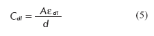
 </span></p>      <p align=center style='text-align:center'><span style='font-size:10.0pt; font-family:"Verdana","sans-serif";color:#211E1E'><img border=0 width=225 height=53 src="/img/revistas/bta/v26n1/fr0512109.gif"></span></p>      
<p><span lang=EN-US style='font-size:10.0pt;font-family:"Verdana","sans-serif"; color:#211E1E'>Where: </span><span style='font-size:10.0pt;font-family:"Verdana","sans-serif"; color:#211E1E'>&#949;</span><i><sub><span lang=EN-US style='font-size:10.0pt; font-family:"Verdana","sans-serif";color:#211E1E'>dl</span></sub></i><i><span lang=EN-US style='font-size:10.0pt;font-family:"Verdana","sans-serif"; color:#211E1E'> </span></i><span lang=EN-US style='font-size:10.0pt;font-family: "Verdana","sans-serif";color:#211E1E'>is the dielectric permittivity at the electrically charged double layer; </span><span style='font-size:10.0pt; font-family:"Verdana","sans-serif";color:#211E1E'>&#949;</span><i><sub><span lang=EN-US style='font-size:10.0pt;font-family:"Verdana","sans-serif"; color:#211E1E'>dl</span></sub></i><i><span lang=EN-US style='font-size:10.0pt; font-family:"Verdana","sans-serif";color:#211E1E'> </span></i><span lang=EN-US style='font-size:10.0pt;font-family:"Verdana","sans-serif";color:#211E1E'>= </span><span style='font-size:10.0pt;font-family:"Verdana","sans-serif";color:#211E1E'>&#949;</span><i><span style='font-size:10.0pt;font-family:"Verdana","sans-serif";color:#211E1E'> <sub><span lang=EN-US>0</span></sub><span lang=EN-US> </span></span></i><span style='font-size:10.0pt;font-family:"Verdana","sans-serif";color:#211E1E'>&#949;</span><i><sub><span lang=EN-US style='font-size:10.0pt;font-family:"Verdana","sans-serif"; color:#211E1E'>p</span></sub></i><span lang=EN-US style='font-size:10.0pt; font-family:"Verdana","sans-serif";color:#211E1E'>, </span><span style='font-size:10.0pt;font-family:"Verdana","sans-serif";color:#211E1E'>&#949;</span><i><span style='font-size:10.0pt;font-family:"Verdana","sans-serif";color:#211E1E'> <sub><span lang=EN-US>0</span></sub><span lang=EN-US> </span></span></i><span lang=EN-US style='font-size:10.0pt;font-family:"Verdana","sans-serif";color:#211E1E'>is the permittivity of the open space, and </span><span style='font-size:10.0pt; font-family:"Verdana","sans-serif";color:#211E1E'>&#949;</span><i><sub><span lang=EN-US style='font-size:10.0pt;font-family:"Verdana","sans-serif"; color:#211E1E'>p</span></sub></i><i><span lang=EN-US style='font-size:10.0pt; font-family:"Verdana","sans-serif";color:#211E1E'> </span></i><span lang=EN-US style='font-size:10.0pt;font-family:"Verdana","sans-serif";color:#211E1E'>is the effective dielectric constant of the layer separating the ionic charges of the electrodes; <i>A</i> is the electrode area, and d is the thickness of the double layer. </span></p>      <p><span lang=EN-US style='font-size:10.0pt;font-family:"Verdana","sans-serif"; color:#211E1E'>Before bacterial growth, the medium contains uncharged or weakly charged substrates such as lactose. During growth, these compounds are transformed into highly electrically charged small ions. As a result, the number of polar molecules and of charged small molecules at the double layer increases, thereby increasing the dielectric permittivity edl<i>, </i>and at the same time reducing the double layer thickness (<i>d). </i>These combined changes provoke an increase in capacitance, and as a result, the impedance decreases. </span></p>      <p><span lang=EN-US style='font-size:10.0pt;font-family:"Verdana","sans-serif"; color:#211E1E'>Expression (4) also gives the best possible explanation on the properties of the impedance measurement during bacterial growth, which also depends on the frequency. As shown in </span><span style='font-size:10.0pt; font-family:"Verdana","sans-serif";color:#211E1E'><a href="#fig1"><span lang=EN-US>figure 1C</span></a></span><span lang=EN-US style='font-size:10.0pt; font-family:"Verdana","sans-serif";color:#211E1E'>, total impedance decreases after the increase of the frequency in the low frequency interval, from 10Hz to 10 kHz, while the impedance becomes independent from the frequency at the high frequency interval (between 10 kHZ and 1 MHz). In the low frequency zone (<i>f </i>&lt; 10 kHz), capacitance of the double layer essentially offers high impedance and it turns it into the main source of the total impedance of the system, so that the resistance of the medium can be ignored. This region is defined as the capacitive region of the double layer, at which the electrode impedance can be detected (</span><span style='font-size:10.0pt;font-family:"Verdana","sans-serif"; color:#211E1E'><a href="#fig1"><span lang=EN-US>Figure 1C</span></a></span><span lang=EN-US style='font-size:10.0pt;font-family:"Verdana","sans-serif"; color:#211E1E'>, <i>Cdl </i>region). </span></p>      <p><span lang=EN-US style='font-size:10.0pt;font-family:"Verdana","sans-serif"; color:#211E1E'>On the other hand, at the high frequency interval (<i>f </i>&gt; 10 kHz) there is no substantial contribution to the double layer capacitance. Thus, the most important contribution to the total impedance of the system at high frequencies is related to the resistance of the medium, which is independent from the frequency. This region is defined as the resistive region, in which ion conduction in the medium is dominant (</span><span style='font-size:10.0pt;font-family:"Verdana","sans-serif";color:#211E1E'><a href="#fig1"><span lang=EN-US>Figure 1C</span></a></span><span lang=EN-US style='font-size:10.0pt;font-family:"Verdana","sans-serif";color:#211E1E'>, <i>R<sub>s </sub></i>region). Therefore, the changes in the double layer of the electrode, and the changes in the medium during bacterial growth can be detected by measuring impedance at different frequencies, which is the reason for the present study to develop new systems that may improve the processes of microbiological detection in relation to resolution and time taken for diagnosis. </span></p>      <p align=center style='text-align:center'><span lang=EN-US style='font-size: 13.5pt;color:#211E1E'>&nbsp;</span></p>      <p><b><span lang=EN-US style='font-family:"Verdana","sans-serif";color:#211E1E'>HISTORICAL BACKGROUND AND APPLICATIONS IN BIOTECHNOLOGY</span></b><b><span lang=EN-US style='font-size:10.0pt;font-family:"Verdana","sans-serif";color:#211E1E'> </span></b></p>      ]]></body>
<body><![CDATA[<p><span lang=EN-US style='font-size:10.0pt;font-family:"Verdana","sans-serif"; color:#211E1E'>It can be considered that the last decade of the XIX Century marks the beginning of IM. As long ago as 1890, the American researcher GN Stewart started a series of experiments introducing elements of conductance and conductivity as parameters to estimate circulation time and the volume of the heart output (9). In July 1898, Stewart made a presentation at the British Medical Association in Edinburgh titled “The changes produced by the growth of bacteria in the molecular concentration and the electrical conductivity of culture media”, which gave rise to impedance bacteriometry and was published the following year (10). The growth curves he obtained were very similar to those now obtained with the impedance systems available (</span><span style='font-size:10.0pt;font-family:"Verdana","sans-serif";color:#211E1E'><a href="#fig5"><span lang=EN-US>Figure 5</span></a></span><span lang=EN-US style='font-size:10.0pt;font-family:"Verdana","sans-serif";color:#211E1E'>), with the notable difference in the extraordinary speed of the present systems, while Steward had to work on the measurement for more than 30 days. </span></p>      <p align=center style='text-align:center'><span style='font-size:13.5pt; color:#211E1E'><img border=0 width=348 height=325 src="/img/revistas/bta/v26n1/f0512109.gif"></span><a name=fig5></a></p>      
<p><span lang=EN-US style='font-size:10.0pt;font-family:"Verdana","sans-serif"; color:#211E1E'>In 1957, Schwan publishes a very important paper on the electric properties of tissues and cells in suspension (11); but it was not until the 1970’s that IM started to expand, through the increasing number of papers published that greatly promoted it and spread it worldwide. During this period, outstanding papers such as those of Ur (12, 13) and Cady (14, 15) were published. The works of the groups of Eden and Torry in the United States set the bases for the IM, which gave way to the introduction of the Bactometer and Malthus measurement systems, respectively (2, 16). </span></p>      <p><span lang=EN-US style='font-size:10.0pt;font-family:"Verdana","sans-serif"; color:#211E1E'>From 1975 to 1999 new papers were published on this topic especially important were the contributions of Felice and Valentinuzzi (7). In that period, the papers on the practical applications of the methods were mainly concerned with food industry and dairy products, where it was used as a tool for quality control. Some of these detection and quantification applications were performed in either raw or pasteurized cow milk. Cady <i>et al. </i>(17) and Gnan-Luedecke (18), were the first to propose the use of impedance as an alternative method for plate counts. </span></p>      <p><span lang=EN-US style='font-size:10.0pt;font-family:"Verdana","sans-serif"; color:#211E1E'>Impedance was also successfully applied in the study and recording of microbial load of a wide range of food products that include: vegetables (19), cereals (20-21), sweets (22), and meat (23). Moreover, the technique was also used to identify groups of microorganisms among which coliform bacilli in meat (24), gram negative bacteria in pasteurized milk (25), and <i>Salmonella </i>(26-28) were included; as well as for the evaluation of antibiotics(29). </span></p>      <p><span lang=EN-US style='font-size:10.0pt;font-family:"Verdana","sans-serif"; color:#211E1E'>Applications for detection and quantification have also included beer (30), wine (31), fish (32), pharmaceuticals, and cosmetics (33), as well as fruit juices (34). Other applications have dealt with sewage effluents to detect coliform bacteria (35), and for the detection of urinary infections (36), or in the human blood (37). A less common application has been in the study of antibiograms (2). In this case, turbidimetry already has equipment and experience available at a commercial scale (38); but there is still no automated commercial system based on impedance measurement that is able to make antibiograms. </span></p>      <p><span lang=EN-US style='font-size:10.0pt;font-family:"Verdana","sans-serif"; color:#211E1E'>Several commercial analytical instruments are based on the classical IM principle, for instance: the Malthus System (Malthus Instruments, Crawley, West Sussex, UK), Bactometer (bioMerieux, Hazelwood, MO, USA), the rapid automated bacterial impedance technique (RABIT) (Don Whitley Scientific Ltd., Shipley, UK) and BacTrac (Sy-Lab, Purkersdorf, Austria) (29, 39- 41), respectively. </span></p>      <p><span lang=EN-US style='font-size:10.0pt;font-family:"Verdana","sans-serif"; color:#211E1E'>Impedance techniques can also be used to monitor the form of bacterial growth. In 1998, Fehlhaber and Kruger found that different species of bacteria, under different conditions, showed specific impedance growth curves (42). </span></p>      <p><span lang=EN-US style='font-size:10.0pt;font-family:"Verdana","sans-serif"; color:#211E1E'>The new interdigitated microelectrodes have radically revolutionized research on impedance spectroscopy (</span><span style='font-size:10.0pt;font-family:"Verdana","sans-serif";color:#211E1E'><a href="#fig6"><span lang=EN-US>Figure 6A</span></a></span><span lang=EN-US style='font-size:10.0pt;font-family:"Verdana","sans-serif";color:#211E1E'>). Microelectrodes have many advantages over conventional electrodes in regard to analytical measurements because of the low resistance and high signal-sound ratio, because they reach the steady state faster and because of the use of small volumes of the solutions (43). In the last decade, the microelectrodes in the form of interdigitated arrangements have been of interest in the fields of biosensors and impedimetric immunosensors (44-47). </span></p>      <p align=center style='text-align:center'><span style='font-size:13.5pt; color:#211E1E'><img border=0 width=355 height=375 src="/img/revistas/bta/v26n1/f0612109.gif"></span><a name=fig6></a></p>      
]]></body>
<body><![CDATA[<p><span lang=EN-US style='font-size:10.0pt;font-family:"Verdana","sans-serif"; color:#211E1E'>The trend in current research for the detection of pathogenic elements is focused on the production of biosensors for the measurement of spectra of impedance. These biosensors have been able to detect particular ones and the responses have been obtained much faster. </span></p>      <p><span lang=EN-US style='font-size:10.0pt;font-family:"Verdana","sans-serif"; color:#211E1E'>
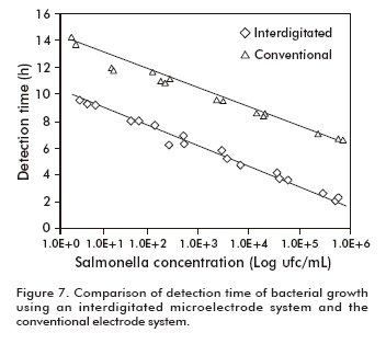
Applications including the fast determination of <i>Salmonella typhimurium </i>have shown the feasibility of using the interdigitated microelectrodes to measure impedance for the purpose of monitoring bacterial growth (48). The volume of the analyzed sample can be reduced from 10-15 mL to 1-2 mL. Furthermore, detection time for the same initial concentration of bacteria is reduced to 3 or 4 hours when compared with the use of the conventional electrode system (</span><span style='font-size:10.0pt;font-family:"Verdana","sans-serif"; color:#211E1E'><a href="#fig7"><span lang=EN-US>Figure 7</span></a></span><span lang=EN-US style='font-size:10.0pt;font-family:"Verdana","sans-serif"; color:#211E1E'>). </span></p>      <p align=center style='text-align:center'><span style='font-size:13.5pt; color:#211E1E'><img border=0 width=352 height=314 src="/img/revistas/bta/v26n1/f0712109.gif"></span><a name=fig7></a></p>      
<p><span lang=EN-US style='font-size:10.0pt;font-family:"Verdana","sans-serif"; color:#211E1E'>The miniaturization of impedance systems known as <i>biochip </i>or <i>lab-on-a-chip </i>to detect bacteria has increased the hope of reaching a faster detection of bacterial growth (</span><span style='font-size:10.0pt; font-family:"Verdana","sans-serif";color:#211E1E'><a href="#fig6"><span lang=EN-US>Figure 6B</span></a></span><span lang=EN-US style='font-size:10.0pt; font-family:"Verdana","sans-serif";color:#211E1E'>). Gomez et al were among the first to make this type of device to detect impedance changes caused by microbial metabolism. The basic idea is to confine a few living cells into a volume in an order starting from nanoliters to picoliters, so that the few living cells in a low conductivity buffer solution may be rapidly detected measuring impedance using interdigitated microelectrodes. Other studies focus the application of this new method known as IM dielectrophoresis (DEPIM), combined with electropermeabilization using a chip as the support. Dielectrophoresis (DEP) is the electrokinetic movement of dielectrically polarized particles in a “non uniform” electric field (50). Progress in the development of microelectrode arrangements has made DEP a highly useful technique for manipulating biological cells in micro-fluid devices, biochips, and biosensors. With this method 10<sup>2</sup> c.f.u./mL were detected in 3 hours. </span></p>      <p><span lang=EN-US style='font-size:10.0pt;font-family:"Verdana","sans-serif"; color:#211E1E'>Most of the IM applications have been widely reviewed by relevant researchers (54-60). </span></p>      <p><b><span lang=EN-US style='font-family:"Verdana","sans-serif";color:#211E1E'>FUTURE GOALS </span></b></p>      <p><span lang=EN-US style='font-size:10.0pt;font-family:"Verdana","sans-serif"; color:#211E1E'>Taking into account the results obtained in the detection of pathogenic microorganisms when combining impedance spectroscopy with new techniques for cell manipulation, and the use of reduced volumes, it is considered that the most important restriction for the use of this technique is that it is very young. Advances in the micro-manufacturing of devices and biochips, that can store volumes in the order of the nanoliters and picoliters where very few bacteria are confined would be possible, but their development would require at least a decade. Another problem would consist in the mass production of these devices preventing the exclusiveness of its use only for the elite. An increase in the sensitivity of this technique is feasible with the use of biosensors based on new geometric models of interdigitated electrodes, together with a change in mathematical data processing. </span></p>      <p><b><span lang=EN-US style='font-family:"Verdana","sans-serif";color:#211E1E'>CONCLUSIONS </span></b></p>      <p><span lang=EN-US style='font-size:10.0pt;font-family:"Verdana","sans-serif"; color:#211E1E'>As observed, IM and its applications in the detection of pathogenic microorganisms, together with the current use of interdigitated microelectrodes, the development of miniaturization, and the integration of biosensors with other techniques such as dielectrophoresis and electropermeabilization will surely lead to future developments. The main purpose of this integration is to increase the sensitivity of detection through the use of a reduced volume of the sample. </span></p>      <p><span lang=EN-US style='font-size:10.0pt;font-family:"Verdana","sans-serif"; color:#211E1E'>Other aspects that must be implemented are the need of redefining the theoretical bases, the development of new components, the principles for designing the culture media and the use of equivalent circuits for the analysis of impedance systems. </span></p>      ]]></body>
<body><![CDATA[<p><span lang=EN-US style='font-size:10.0pt;font-family:"Verdana","sans-serif"; color:#211E1E'>The impedance technique as a transduction principle has become a promising field for the development of rapid and effective methods to detect microbiological growth. In spite of the fact that IM was established more than 100 years ago, it is just now entering into a new stage based on miniaturized devices (nanosciences and nanoelectronics). Advances in micromanufacturing have brought about the conditions for the development of micro-devices and biochips, which have proven their efficacy in maximizing the impedance signal, increasing sensitivity and reducing time in the detection of pathogenic microorganisms. This trend confirms that among the new automated methods those of impedance microbiology are the most successful. </span></p>      <p><span lang=EN-US style='font-size:11.0pt;font-family:"Arial","sans-serif"; color:red'>&nbsp;</span><b><span style='font-family:"Verdana","sans-serif"; color:#211E1E'>REFERENCES </span></b></p>      <!-- ref --><p><span style='font-size:10.0pt;font-family:"Verdana","sans-serif";color:#211E1E'>1. Geddes</span><span style='font-size:10.0pt;font-family:"Verdana","sans-serif"'> </span><span style='font-size:10.0pt;font-family:"Verdana","sans-serif"; color:#211E1E'>LA, Baker</span><span style='font-size:10.0pt;font-family:"Verdana","sans-serif"'> </span><span style='font-size:10.0pt;font-family:"Verdana","sans-serif"; color:#211E1E'>LE. </span><span lang=EN-US style='font-size:10.0pt;font-family: "Verdana","sans-serif";color:#211E1E'>Principles of applied biomedical instrumentation. New York: John Wiley &amp; Sons. 1989</span><span lang=SV style='font-size: 10.0pt;font-family:"Verdana","sans-serif"'>.</span><span lang=SV style='font-family:"Arial","sans-serif";color:navy'> </span><!-- ref --><p><span lang=EN-US style='font-size:10.0pt;font-family:"Verdana","sans-serif"; color:#211E1E'>2. Eden G, Eden R. Enumeration of microorganisms by their dynamic AC conductance patterns. IEEE Trans Biomed Eng</span><span lang=EN-US style='font-size:11.0pt;font-family:"Arial","sans-serif";color:red'> </span><span lang=EN-US style='font-size:10.0pt;font-family:"Verdana","sans-serif"; color:#211E1E'>1984;31</span><span lang=EN-US style='font-size:10.0pt; font-family:"Arial","sans-serif"'>(</span><span lang=EN-US style='font-size: 10.0pt;font-family:"Verdana","sans-serif";color:#211E1E'>2):93-8.</span><!-- ref --><p><span lang=EN-US style='font-size:10.0pt;font-family:"Verdana","sans-serif"; color:#211E1E'>3. Owicki J, Parce J. Biosensors based on the energy metabolism of living cells: the physical chemistry and cell biology of extracellular acidification. Biosens Bioelectron</span><span lang=EN-US style='font-size: 11.0pt;font-family:"Arial","sans-serif";color:red'> </span><span lang=EN-US style='font-size:10.0pt;font-family:"Verdana","sans-serif";color:#211E1E'>1992;7:257-72.</span><!-- ref --><p><span lang=EN-US style='font-size:10.0pt;font-family:"Verdana","sans-serif"; color:#211E1E'>4. Talaro</span><span lang=EN-US style='font-size:10.0pt; font-family:"Verdana","sans-serif"'> </span><span lang=EN-US style='font-size: 10.0pt;font-family:"Verdana","sans-serif";color:#211E1E'>KP. Foundations in microbiology. 5th ed. New York: McGraw-Hill. 2005</span><span lang=SV style='font-size:10.0pt;font-family:"Verdana","sans-serif"'>.</span><span lang=SV style='font-family:"Arial","sans-serif";color:navy'> </span><!-- ref --><p><span lang=EN-US style='font-size:10.0pt;font-family:"Verdana","sans-serif"; color:#211E1E'>5. Gómez R, Bashir R, Bhunia A. Microscale electronic detection of bacterial metabolism. Sensor Actuators B</span><span lang=EN-US style='font-size:11.0pt;font-family:"Arial","sans-serif";color:red'> </span><span lang=EN-US style='font-size:10.0pt;font-family:"Verdana","sans-serif"; color:#211E1E'>2002;86:198-208.</span><!-- ref --><p><span lang=EN-US style='font-size:10.0pt;font-family:"Verdana","sans-serif"; color:#211E1E'>6. Mead PS, Slutsker L, Dietz V, McCaig LF, Bresee JS, Shapiro C, et al. Food-related illness and death in the United States. Emerg Infect Dis</span><span lang=EN-US style='font-size:11.0pt;font-family:"Arial","sans-serif";color:red'> </span><span lang=EN-US style='font-size:10.0pt;font-family:"Verdana","sans-serif"; color:#211E1E'>1999;5:607-25.</span><!-- ref --><p><span lang=EN-US style='font-size:10.0pt;font-family:"Verdana","sans-serif"; color:#211E1E'>7. Felice CL, Valentinuzzi ME. Medium and interface components in impedance microbiology. IEEE Trans Biomed Eng</span><span lang=EN-US style='font-size:11.0pt;font-family:"Arial","sans-serif";color:red'> </span><span lang=EN-US style='font-size:10.0pt;font-family:"Verdana","sans-serif"; color:#211E1E'>1999;46:483-7.</span><!-- ref --><p><span lang=EN-US style='font-size:10.0pt;font-family:"Verdana","sans-serif"; color:#211E1E'>8. Yang L, Ruan C, Li Y. Detection of viable <i>Salmonella typhimurium </i>by impedance measurement of electrode capacitance and medium resistance. Biosens Bioelectron</span><span lang=EN-US style='font-size:11.0pt; font-family:"Arial","sans-serif";color:red'> </span><span lang=EN-US style='font-size:10.0pt;font-family:"Verdana","sans-serif";color:#211E1E'>2003;19:495-502.</span><p><span lang=EN-US style='font-size:10.0pt;font-family:"Verdana","sans-serif"; color:#211E1E'>9. Stewart GN. Researches on circulation time and on the influences which affect it. IV. The output of the heart. J Physiol (London) 1897;XXII:25.</span></p>      <!-- ref --><p><span lang=EN-US style='font-size:10.0pt;font-family:"Verdana","sans-serif"; color:#211E1E'>10. Stewart GN. The changes produced by the growth of bacteria in the molecular concentration and electrical conductivity of culture media. J Exp Med</span><span lang=EN-US style='font-size:11.0pt;font-family:"Arial","sans-serif"; color:red'> </span><span lang=EN-US style='font-size:10.0pt;font-family:"Verdana","sans-serif"; color:#211E1E'>1899;4:235-43.</span><!-- ref --><p><span lang=EN-US style='font-size:10.0pt;font-family:"Verdana","sans-serif"; color:#211E1E'>11. Schwan HP. Electrical properties of tissue and cell suspensions. Adv Biol Med Phys</span><span lang=EN-US style='font-size:11.0pt; font-family:"Arial","sans-serif";color:red'> </span><span lang=EN-US style='font-size:10.0pt;font-family:"Verdana","sans-serif";color:#211E1E'>1957;5:147-209.</span><!-- ref --><p><span lang=EN-US style='font-size:10.0pt;font-family:"Verdana","sans-serif"; color:#211E1E'>12. Ur A. Determination of blood coagulation using impedance measurements. Biomed Eng</span><span lang=EN-US style='font-size:11.0pt;font-family:"Arial","sans-serif"; color:red'> </span><span lang=EN-US style='font-size:10.0pt;font-family:"Verdana","sans-serif"; color:#211E1E'>1970;5:342-5.</span><!-- ref --><p><span lang=EN-US style='font-size:10.0pt;font-family:"Verdana","sans-serif"; color:#211E1E'>13. Ur A. The changes in the electrical impedance of blood during coagulation. Nature</span><span lang=EN-US style='font-size:11.0pt; font-family:"Arial","sans-serif";color:red'> </span><span lang=EN-US style='font-size:10.0pt;font-family:"Verdana","sans-serif";color:#211E1E'>1970;226:269-70.</span><!-- ref --><p><span lang=EN-US style='font-size:10.0pt;font-family:"Verdana","sans-serif"; color:#211E1E'>14. Cady P. Instrumentation in food microbiology. Food Product Dev</span><span lang=EN-US style='font-size:11.0pt;font-family:"Arial","sans-serif"; color:red'> </span><span lang=EN-US style='font-size:10.0pt;font-family:"Verdana","sans-serif"; color:#211E1E'>1977;April:80-5.</span><p><span lang=EN-US style='font-size:10.0pt;font-family:"Verdana","sans-serif"; color:#211E1E'>15. Cady</span><span lang=EN-US style='font-family:"Arial","sans-serif"; color:blue'> </span><span lang=EN-US style='font-size:10.0pt;font-family:"Verdana","sans-serif"; color:#211E1E'>P. Progress in impedance measurements in microbiology. In: Mechanizing Microbiology: Sharp</span><span lang=EN-US style='font-family:"Arial","sans-serif"; color:blue'> </span><span lang=EN-US style='font-size:10.0pt;font-family:"Verdana","sans-serif"; color:#211E1E'>AN, Clark</span><span lang=EN-US style='font-family:"Arial","sans-serif"; color:blue'> </span><span lang=EN-US style='font-size:10.0pt;font-family:"Verdana","sans-serif"; color:#211E1E'>DS</span><span lang=EN-US style='font-family:"Arial","sans-serif"; color:gray'> </span><span lang=EN-US style='font-size:10.0pt;font-family:"Verdana","sans-serif"; color:#211E1E'>&nbsp;eds. Springfield (IL): 1978. p.</span><span lang=EN-US style='font-family:"Arial","sans-serif";color:gray'> </span><span lang=EN-US style='font-size:10.0pt;font-family:"Verdana","sans-serif";color:#211E1E'>99-239</span><span lang=SV style='font-family:"Arial","sans-serif";color:teal'>.</span><span lang=SV style='font-size:10.0pt;font-family:"Verdana","sans-serif";color:#211E1E'> </span></p>      <!-- ref --><p><span lang=EN-US style='font-size:10.0pt;font-family:"Verdana","sans-serif"; color:#211E1E'>16. Richards JCS, Jason AC, Hobbs G, Gibson DM, Christie RH. Electronic measurement of bacterial growth. J Phys E Sci Instrum</span><span lang=EN-US style='font-size:11.0pt;font-family:"Arial","sans-serif";color:red'> </span><span lang=EN-US style='font-size:10.0pt;font-family:"Verdana","sans-serif"; color:#211E1E'>1978;11:560-8.</span><!-- ref --><p><span lang=EN-US style='font-size:10.0pt;font-family:"Verdana","sans-serif"; color:#211E1E'>17. Cady P, Hardy D, Martins S, Dufour SW, Kraeger SJ. Automated impedance measurements for rapid screening of milk microbial content. J Food Prot</span><span lang=EN-US style='font-size:11.0pt;font-family:"Arial","sans-serif"; color:red'> </span><span lang=EN-US style='font-size:10.0pt;font-family:"Verdana","sans-serif"; color:#211E1E'>1978;41</span><span lang=EN-US style='font-size:10.0pt; font-family:"Arial","sans-serif"'>(</span><span lang=EN-US style='font-size: 10.0pt;font-family:"Verdana","sans-serif";color:#211E1E'>4):277-83.</span><!-- ref --><p><span lang=EN-US style='font-size:10.0pt;font-family:"Verdana","sans-serif"; color:#211E1E'>18. Gnan S, Luedecke LO. Impedance measurements in raw milk as an alternative to the standard plate count. J Food Prot</span><span lang=EN-US style='font-size:11.0pt;font-family:"Arial","sans-serif";color:red'> </span><span lang=EN-US style='font-size:10.0pt;font-family:"Verdana","sans-serif"; color:#211E1E'>1982;45</span><span lang=EN-US style='font-size:10.0pt; font-family:"Arial","sans-serif"'>(</span><span lang=EN-US style='font-size: 10.0pt;font-family:"Verdana","sans-serif";color:#211E1E'>1):4-7.</span><!-- ref --><p><span lang=EN-US style='font-size:10.0pt;font-family:"Verdana","sans-serif"; color:#211E1E'>19. Hardy</span><span lang=EN-US style='font-family:"Arial","sans-serif"; color:blue'> </span><span lang=EN-US style='font-size:10.0pt;font-family:"Verdana","sans-serif"; color:#211E1E'>D, Kraeger</span><span lang=EN-US style='font-family:"Arial","sans-serif"; color:blue'> </span><span lang=EN-US style='font-size:10.0pt;font-family:"Verdana","sans-serif"; color:#211E1E'>SJ, Dufour</span><span lang=EN-US style='font-family:"Arial","sans-serif"; color:blue'> </span><span lang=EN-US style='font-size:10.0pt;font-family:"Verdana","sans-serif"; color:#211E1E'>SW, Cady</span><span lang=EN-US style='font-family:"Arial","sans-serif"; color:blue'> </span><span lang=EN-US style='font-size:10.0pt;font-family:"Verdana","sans-serif"; color:#211E1E'>P. Rapid Detection of Microbial Contamination in Frozen Vegetables by Automated Impedance Measurements. Palo Alto (California): Bactomatic, Inc. 94303. 1977</span><span lang=SV style='font-size:10.0pt; font-family:"Verdana","sans-serif";color:windowtext'>.</span><span lang=SV style='font-size:10.0pt;font-family:"Verdana","sans-serif";color:#211E1E'> </span><!-- ref --><p><span lang=EN-US style='font-size:10.0pt;font-family:"Verdana","sans-serif"; color:#211E1E'>20. Sorrells KM. Rapid detection of bacterial content in cereal grain. J Food Prot</span><span lang=EN-US style='font-size:11.0pt;font-family: "Arial","sans-serif";color:red'> </span><span lang=EN-US style='font-size:10.0pt; font-family:"Verdana","sans-serif";color:#211E1E'>1981;44:832-4.</span><!-- ref --><p><span lang=EN-US style='font-size:10.0pt;font-family:"Verdana","sans-serif"; color:#211E1E'>21. Coppola K, Firstenberg-Eden R. Impedance based rapid method for detection of spoilage organisms in UHT low-acid foods. J Food Sci</span><span lang=EN-US style='font-size:11.0pt;font-family:"Arial","sans-serif";color:red'> </span><span lang=EN-US style='font-size:10.0pt;font-family:"Verdana","sans-serif"; color:#211E1E'>1988;53:1521-7.</span><!-- ref --><p><span lang=EN-US style='font-size:10.0pt;font-family:"Verdana","sans-serif"; color:#211E1E'>22. Davda C, Pugh SJ. An improved protocol for the detection and rapid confirmation within 48h of Salmonellas in confectionary products. Lett Appl Microbiol</span><span lang=EN-US style='font-size:11.0pt;font-family:"Arial","sans-serif"; color:red'> </span><span lang=EN-US style='font-size:10.0pt;font-family:"Verdana","sans-serif"; color:#211E1E'>1991;13:287-90.</span><!-- ref --><p><span lang=EN-US style='font-size:10.0pt;font-family:"Verdana","sans-serif"; color:#211E1E'>23. Firstenberg-Eden R. Rapid estimation of microorganisms in raw meat by impedance measurement. Food Technol</span><span lang=EN-US style='font-size:11.0pt;font-family:"Arial","sans-serif";color:red'> </span><span lang=EN-US style='font-size:10.0pt;font-family:"Verdana","sans-serif"; color:#211E1E'>1983;37:64-70.</span><!-- ref --><p><span lang=EN-US style='font-size:10.0pt;font-family:"Verdana","sans-serif"; color:#211E1E'>24. Martins SB, Selby MJ. Evaluation of a rapid method for the quantitative estimation of coliforms in meat by impedimetric procedures. Appl Environ Microbiol</span><span lang=EN-US style='font-size:11.0pt;font-family: "Arial","sans-serif";color:red'> </span><span lang=EN-US style='font-size:10.0pt; font-family:"Verdana","sans-serif";color:#211E1E'>1980;39:518-24.</span><p><span lang=EN-US style='font-size:10.0pt;font-family:"Verdana","sans-serif"; color:#211E1E'>25. Nieuwenhof FFJ, Hoolwerf JD. Detection of post-pasteurization contamination of pasteurized milk with the Bioscreen, Nederlands Instituut voor Zuivelonderzoekx, NIZO-EDE Report. 1205. Nov1986:10.</span><span lang=EN-US style='font-size:11.0pt;font-family:"Arial","sans-serif";color:blue'> </span></p>      <!-- ref --><p><span lang=EN-US style='font-size:10.0pt;font-family:"Verdana","sans-serif"; color:#211E1E'>26. Easter MC, Gibson DM. Rapid and automated detection of <i>Salmonella </i>by electrical measurement. J Hyg</span><span lang=EN-US style='font-size: 11.0pt;font-family:"Arial","sans-serif";color:red'> </span><span lang=EN-US style='font-size:10.0pt;font-family:"Verdana","sans-serif";color:#211E1E'>1985;94:245-62.</span><!-- ref --><p><span lang=EN-US style='font-size:10.0pt;font-family:"Verdana","sans-serif"; color:#211E1E'>27. Gibson MD. Some modification to the media for rapid automated detection of Salmonellas by conductance measurement. J Appl Bacteriol</span><span lang=EN-US style='font-size:11.0pt;font-family:"Arial","sans-serif";color:red'> </span><span lang=EN-US style='font-size:10.0pt;font-family:"Verdana","sans-serif"; color:#211E1E'>1987;63:299-304.</span><!-- ref --><p><span lang=EN-US style='font-size:10.0pt;font-family:"Verdana","sans-serif"; color:#211E1E'>28. Ogden ID. A conductance medium to distinguish between <i>Salmonella </i>and <i>Citrobacter </i>spp. Int J Food Microbiol</span><span lang=EN-US style='font-size:11.0pt;font-family:"Arial","sans-serif";color:red'> </span><span lang=EN-US style='font-size:10.0pt;font-family:"Verdana","sans-serif"; color:#211E1E'>1988;7:287-97.</span><!-- ref --><p><span lang=EN-US style='font-size:10.0pt;font-family:"Verdana","sans-serif"; color:#211E1E'>29. Gould IM, Jason AC, Milne K. Use of the Malthus Microbial Growth Analyser to study the post antibiotic effect of antibiotics. J Antimicrob Chemother</span><span lang=EN-US style='font-size:11.0pt;font-family: "Arial","sans-serif";color:red'> </span><span lang=EN-US style='font-size:10.0pt; font-family:"Verdana","sans-serif";color:#211E1E'>1989;24</span><span lang=EN-US style='font-size:10.0pt;font-family:"Arial","sans-serif"'>(</span><span lang=EN-US style='font-size:10.0pt;font-family:"Verdana","sans-serif"; color:#211E1E'>4):523-31.</span><!-- ref --><p><span lang=EN-US style='font-size:10.0pt;font-family:"Verdana","sans-serif"; color:#211E1E'>30. Evans HAV. A note on two uses for impedimetry in brewing microbiology. J Appl Bacteriol</span><span lang=EN-US style='font-size:11.0pt; font-family:"Arial","sans-serif";color:red'> </span><span lang=EN-US style='font-size:10.0pt;font-family:"Verdana","sans-serif";color:#211E1E'>1982;53:423-6.</span><!-- ref --><p><span lang=EN-US style='font-size:10.0pt;font-family:"Verdana","sans-serif"; color:#211E1E'>31. Henschke PA, Thomas DS. Detection of wine yeast by electronic methods. J Appl Bacteriol</span><span lang=EN-US style='font-size: 11.0pt;font-family:"Arial","sans-serif";color:red'> </span><span lang=EN-US style='font-size:10.0pt;font-family:"Verdana","sans-serif";color:#211E1E'>1988;64:123-33.</span><p><span lang=EN-US style='font-size:10.0pt;font-family:"Verdana","sans-serif"; color:#211E1E'>32. Van Spreekens KJA, Stekelenburg FK. Rapid estimation of the bacteriological quality of fresh fish by impedance measurements. Appl Microbiol Biotechnol</span><span lang=EN-US style='font-size:11.0pt;font-family:"Arial","sans-serif";color:red'> </span><span lang=EN-US style='font-size:10.0pt;font-family:"Verdana","sans-serif"; color:#211E1E'>1986;24:95-6.</span></p>      <!-- ref --><p><span lang=EN-US style='font-size:10.0pt;font-family:"Verdana","sans-serif"; color:#211E1E'>33. Connolly P, Bloomfield SF, Denyer SP. The use of impedance for preservative efficacy testing of pharmaceuticals and cosmetic products. J Appl Bacteriol</span><span lang=EN-US style='font-size:11.0pt;font-family:"Arial","sans-serif"; color:red'> </span><span lang=EN-US style='font-size:10.0pt;font-family:"Verdana","sans-serif"; color:#211E1E'>1994;76:68-74.</span><!-- ref --><p><span lang=EN-US style='font-size:10.0pt;font-family:"Verdana","sans-serif"; color:#211E1E'>34. Deak T, Beuchat LR. Comparison of conductimetric and traditional plating techniques for detecting yeasts in fruit juices. J Appl Bacteriol</span><span lang=EN-US style='font-size:11.0pt;font-family:"Arial","sans-serif"; color:red'> </span><span lang=EN-US style='font-size:10.0pt;font-family:"Verdana","sans-serif"; color:#211E1E'>1993;75:546–50.</span><!-- ref --><p><span lang=EN-US style='font-size:10.0pt;font-family:"Verdana","sans-serif"; color:#211E1E'>35. Silverman MP, Munoz EF. Automated electrical impedance technique for rapid enumeration of fecal coliforms in effluents from sewage treatment plants. Appl Environ Microbiol</span><span lang=EN-US style='font-size:11.0pt;font-family:"Arial","sans-serif";color:red'> </span><span lang=EN-US style='font-size:10.0pt;font-family:"Verdana","sans-serif"; color:#211E1E'>1979;37:521-6.</span><!-- ref --><p><span lang=EN-US style='font-size:10.0pt;font-family:"Verdana","sans-serif"; color:#211E1E'>36. Cady P, Dufour SW, Lawless P, Nunke B, Kraeger SJ. Impedimetric screening for bacteriuria. J Clin Microbiol</span><span lang=EN-US style='font-size:11.0pt;font-family:"Arial","sans-serif";color:red'> </span><span lang=EN-US style='font-size:10.0pt;font-family:"Verdana","sans-serif"; color:#211E1E'>1978;7:273-8.</span><!-- ref --><p><span lang=EN-US style='font-size:10.0pt;font-family:"Verdana","sans-serif"; color:#211E1E'>37. Buckland A, Kessock-Philip S, Bascomb S. Early detection of bacterial growth in blood culture by impedance monitoring with a Bactometer model 32. J Clin Pathol</span><span lang=EN-US style='font-size:11.0pt; font-family:"Arial","sans-serif";color:red'> </span><span lang=EN-US style='font-size:10.0pt;font-family:"Verdana","sans-serif";color:#211E1E'>1983;36:823-8.</span><p><span lang=EN-US style='font-size:10.0pt;font-family:"Verdana","sans-serif"; color:#211E1E'>38. Contreras OR, Roura G, Novo F, Hernández S, Ramírez N, Ramírez I <i>et al</i>, inventors. Equipment, kit and method for microbiological diagnosis. US patent 6537772. 2003; March:25. </span></p>      ]]></body>
<body><![CDATA[<!-- ref --><p><span lang=EN-US style='font-size:10.0pt;font-family:"Verdana","sans-serif"; color:#211E1E'>39. Bactometer</span><span lang=EN-US style='font-family:"Arial","sans-serif"; color:gray'> </span><span lang=EN-US style='font-size:10.0pt;font-family:"Verdana","sans-serif"; color:#211E1E'>2008. (</span><span style='font-size:10.0pt;font-family:"Verdana","sans-serif"'><a href="http://industry.biomerieux-usa.com/industry/food/bactometer/index.htm"><span lang=EN-US>http://industry.biomerieux-usa.com/industry/food/bactometer/index.htm</span></a></span><span lang=EN-US style='font-size:10.0pt;font-family:"Verdana","sans-serif"; color:#211E1E'>, 24 oct 2008).</span><span lang=EN-US style='font-family:"Arial","sans-serif"; color:navy'> </span><!-- ref --><p><span lang=EN-US style='font-size:10.0pt;font-family:"Verdana","sans-serif"; color:#211E1E'>40. Don Whitley Scientific 2008. (</span><span lang=EN-US style='font-size:10.0pt;font-family:"Verdana","sans-serif"'><a href="http://www.dwscientific.co.uk/">http://www.dwscientific.co.uk/</a></span><span lang=EN-US style='font-size:10.0pt;font-family:"Verdana","sans-serif"; color:#211E1E'>, 24 oct 2008). </span><!-- ref --><p><span lang=EN-US style='font-size:10.0pt;font-family:"Verdana","sans-serif"; color:#211E1E'>41. BacTrac 2008. (</span><span style='font-size:10.0pt; font-family:"Verdana","sans-serif"'><a href="http://www.sylab.com/bactrac.htm"><span lang=EN-US>http://www.sylab.com/bactrac.htm</span></a></span><span lang=EN-US style='font-size:10.0pt;font-family:"Verdana","sans-serif";color:#211E1E'>, 24 oct 2008).</span><span lang=EN-US style='font-family:"Arial","sans-serif"; color:navy'> </span><!-- ref --><p><span lang=EN-US style='font-size:10.0pt;font-family:"Verdana","sans-serif"; color:#211E1E'>42. Fehlhaber K, Kruger G. The study of <i>Salmonella enteritidis </i>growth kinetics using rapid automated bacterial impedance technique. J Appl Microbiol</span><span lang=EN-US style='font-size:11.0pt; font-family:"Arial","sans-serif";color:red'> </span><span lang=EN-US style='font-size:10.0pt;font-family:"Verdana","sans-serif";color:#211E1E'>1998;84:945-9.</span><!-- ref --><p><span lang=EN-US style='font-size:10.0pt;font-family:"Verdana","sans-serif"; color:#211E1E'>43. Stulik K, Amatore C, Holub K, Marecek V, Kutner W. Microelectrodes. Definitions, characterization, and applications. Pure Appl Chem</span><span lang=EN-US style='font-size:11.0pt;font-family:"Arial","sans-serif"; color:red'> </span><span lang=EN-US style='font-size:10.0pt;font-family:"Verdana","sans-serif"; color:#211E1E'>2000;72:1483-92.</span><p><span lang=EN-US style='font-size:10.0pt;font-family:"Verdana","sans-serif"; color:#211E1E'>44. Van Gerwen P, Laureyn W, Laureys W, Huyberechts G, Op De Beeck M, Baert K <i>et</i> <i>al. </i>Nano-scaled inter-digitated electrode arrays for biochemical sensors. Sens Actuators B</span><span lang=EN-US style='font-size:11.0pt;font-family:"Arial","sans-serif";color:red'> </span><span lang=EN-US style='font-size:10.0pt;font-family:"Verdana","sans-serif"; color:#211E1E'>1998;49:73-80.</span></p>      <!-- ref --><p><span lang=EN-US style='font-size:10.0pt;font-family:"Verdana","sans-serif"; color:#211E1E'>45. Laureyn W, Nelis D, Van Gerwen P, Baert K, Hermans L, Magnee R, et al. Nano-scaled interdigitated titanium electrodes for impedimetric biosensing. Sens Actuators B</span><span lang=EN-US style='font-size:11.0pt; font-family:"Arial","sans-serif";color:red'> </span><span lang=EN-US style='font-size:10.0pt;font-family:"Verdana","sans-serif";color:#211E1E'>2000;68:360-70.</span><!-- ref --><p><span lang=EN-US style='font-size:10.0pt;font-family:"Verdana","sans-serif"; color:#211E1E'>46. Ruan C, Yang L, Li Y. Immunobiosensor chips doe detection of <i>Escherichia coli </i>O157:H7 using electrochemical impedance spectroscopy. Anal Chem</span><span lang=EN-US style='font-size:11.0pt;font-family:"Arial","sans-serif"; color:red'> </span><span lang=EN-US style='font-size:10.0pt;font-family:"Verdana","sans-serif"; color:#211E1E'>2002;74:4814-20.</span><!-- ref --><p><span lang=EN-US style='font-size:10.0pt;font-family:"Verdana","sans-serif"; color:#211E1E'>47. Varshney M, Li Y. Interdigitated array microelectrode based impedance biosensor coupled with magnetic nanoparticle-antibody conjugates for detection of <i>Escherichia coli </i>O157:H7 in food samples. Biosens Bioelectron</span><span lang=EN-US style='font-size:11.0pt;font-family:"Arial","sans-serif"; color:red'> </span><span lang=EN-US style='font-size:10.0pt;font-family:"Verdana","sans-serif"; color:#211E1E'>2007;22:2408-14.</span><!-- ref --><p><span lang=EN-US style='font-size:10.0pt;font-family:"Verdana","sans-serif"; color:#211E1E'>48. Yang L, Li Y. Detection of viable <i>Salmonella </i>using microelectrode-based capacitance measurement coupled with immunomagnetic separation. J Microbiol Methods</span><span lang=EN-US style='font-size:11.0pt;font-family:"Arial","sans-serif"; color:red'> </span><span lang=EN-US style='font-size:10.0pt;font-family:"Verdana","sans-serif"; color:#211E1E'>2006;64:9-16.</span><!-- ref --><p><span lang=EN-US style='font-size:10.0pt;font-family:"Verdana","sans-serif"; color:#211E1E'>49. Gómez R, Bashir R, Sarikaya A, Ladisch M, Sturgis J, Robinson J, et al. Microfluidic biochip for impedance spectroscopy of biological species. Biomed Microdev</span><span lang=EN-US style='font-size: 11.0pt;font-family:"Arial","sans-serif";color:red'> </span><span lang=EN-US style='font-size:10.0pt;font-family:"Verdana","sans-serif";color:#211E1E'>2001;3:201-9.</span><!-- ref --><p><span lang=EN-US style='font-size:10.0pt;font-family:"Verdana","sans-serif"; color:#211E1E'>50. Pohl</span><span lang=EN-US style='font-family:"Arial","sans-serif"; color:blue'> </span><span lang=EN-US style='font-size:10.0pt;font-family:"Verdana","sans-serif"; color:#211E1E'>HA. Dielectrophoresis. London: Cambridge University Press. 1978.</span><span lang=EN-US style='font-family:"Arial","sans-serif";color:navy'> </span><p><span lang=EN-US style='font-size:10.0pt;font-family:"Verdana","sans-serif"; color:#211E1E'>51. Medoro</span><span lang=EN-US style='font-family:"Arial","sans-serif"; color:blue'> </span><span lang=EN-US style='font-size:10.0pt;font-family:"Verdana","sans-serif"; color:#211E1E'>G, Manaresi</span><span lang=EN-US style='font-family:"Arial","sans-serif"; color:blue'> </span><span lang=EN-US style='font-size:10.0pt;font-family:"Verdana","sans-serif"; color:#211E1E'>N, Leonardi</span><span lang=EN-US style='font-family:"Arial","sans-serif"; color:blue'> </span><span lang=EN-US style='font-size:10.0pt;font-family:"Verdana","sans-serif"; color:#211E1E'>A, Altomare</span><span lang=EN-US style='font-family:"Arial","sans-serif"; color:blue'> </span><span lang=EN-US style='font-size:10.0pt;font-family:"Verdana","sans-serif"; color:#211E1E'>L, Tartagni</span><span lang=EN-US style='font-family:"Arial","sans-serif"; color:blue'> </span><span lang=EN-US style='font-size:10.0pt;font-family:"Verdana","sans-serif"; color:#211E1E'>M, Guerrieri</span><span lang=EN-US style='font-family:"Arial","sans-serif"; color:blue'> </span><span lang=EN-US style='font-size:10.0pt;font-family:"Verdana","sans-serif"; color:#211E1E'>R. A labon- a-chip for cell detection and manipulation. Orlando (FL), USA: Sensors IEEE. 2002. p. 472-5.</span><span lang=EN-US style='font-family:"Arial","sans-serif";color:navy'> </span></p>      <!-- ref --><p><span lang=EN-US style='font-size:10.0pt;font-family:"Verdana","sans-serif"; color:#211E1E'>52. Sengupta S, Battigelli DA, Chang HC. A micro-scale multi-frequency reactance measurement technique to detect bacterial growth at low bio-particle concentrations. Lab Chip</span><span lang=EN-US style='font-size:11.0pt;font-family:"Arial","sans-serif";color:red'> </span><span lang=EN-US style='font-size:10.0pt;font-family:"Verdana","sans-serif"; color:#211E1E'>2006;6:682-92.</span><!-- ref --><p><span lang=EN-US style='font-size:10.0pt;font-family:"Verdana","sans-serif"; color:#211E1E'>53. Suehiro J, Shutou M, Hatano T, Hara M. Improvement of electric pulse shape for electropermeabilization-assisted dielectrophoretic impedance measurement for high sensitive bacteria detection. Sens Actuator B</span><span lang=EN-US style='font-size:11.0pt;font-family:"Arial","sans-serif";color:red'> </span><span lang=EN-US style='font-size:10.0pt;font-family:"Verdana","sans-serif"; color:#211E1E'>2005;109:209-15.</span><!-- ref --><p><span lang=EN-US style='font-size:10.0pt;font-family:"Verdana","sans-serif"; color:#211E1E'>54. Silley P, Forsythe S. Impedance microbiology- a rapid change for microbiologists. J &nbsp;Appl Bacteriol</span><span lang=EN-US style='font-size:11.0pt;font-family:"Arial","sans-serif";color:red'> </span><span lang=EN-US style='font-size:10.0pt;font-family:"Verdana","sans-serif"; color:#211E1E'>1996;80:233-43.</span><!-- ref --><p><span lang=EN-US style='font-size:10.0pt;font-family:"Verdana","sans-serif"; color:#211E1E'>55. Valentinuzzi</span><span lang=EN-US style='font-family:"Arial","sans-serif"; color:gray'> </span><span lang=EN-US style='font-size:10.0pt;font-family:"Verdana","sans-serif"; color:#211E1E'>ME</span><span lang=SV style='font-size:10.0pt;font-family:"Verdana","sans-serif"'>, </span><span lang=EN-US style='font-size:10.0pt;font-family:"Verdana","sans-serif"; color:#211E1E'>Morucci</span><span lang=EN-US style='font-size:10.0pt; font-family:"Verdana","sans-serif"'> </span><span lang=EN-US style='font-size: 10.0pt;font-family:"Verdana","sans-serif";color:#211E1E'>JP</span><span lang=SV style='font-size:10.0pt;font-family:"Verdana","sans-serif"'>, </span><span lang=EN-US style='font-size:10.0pt;font-family:"Verdana","sans-serif"; color:#211E1E'>Felice</span><span lang=EN-US style='font-size:10.0pt; font-family:"Verdana","sans-serif"'> </span><span lang=EN-US style='font-size: 10.0pt;font-family:"Verdana","sans-serif";color:#211E1E'>CJ. Monitoring of physiological events by impedance. Bioelectrical Impedance Techniques in Medicine. In: Bourne</span><span lang=EN-US style='font-family:"Arial","sans-serif"; color:gray'> </span><span lang=EN-US style='font-size:10.0pt;font-family:"Verdana","sans-serif"; color:#211E1E'>JR, ed. CRC Reviews in Biomedical Engineering. New York: Begell House. 1996:</span><span lang=EN-US style='font-family:"Arial","sans-serif"; color:gray'> </span><span lang=EN-US style='font-size:10.0pt;font-family:"Verdana","sans-serif"; color:#211E1E'>427-66. </span><!-- ref --><p><span lang=EN-US style='font-size:10.0pt;font-family:"Verdana","sans-serif"; color:#211E1E'>56. Wawerla M, Stolle A, Schalch B, Eisgruber H. Impedance microbiology: applications in food hygiene. J Food Prot</span><span lang=EN-US style='font-size:11.0pt;font-family:"Arial","sans-serif";color:red'> </span><span lang=EN-US style='font-size:10.0pt;font-family:"Verdana","sans-serif"; color:#211E1E'>1999;62:1488-96.</span><!-- ref --><p><span lang=EN-US style='font-size:10.0pt;font-family:"Verdana","sans-serif"; color:#211E1E'>57. Guan JG, Miao YQ, Zhanga QJ. Impedimetric biosensors. J Biosci Bioeng</span><span lang=EN-US style='font-size:11.0pt;font-family:"Arial","sans-serif"; color:red'> </span><span lang=EN-US style='font-size:10.0pt;font-family:"Verdana","sans-serif"; color:#211E1E'>2004;97</span><span lang=EN-US style='font-size:10.0pt; font-family:"Arial","sans-serif"'>(</span><span lang=EN-US style='font-size: 10.0pt;font-family:"Verdana","sans-serif";color:#211E1E'>4):219-26.</span><!-- ref --><p><span lang=EN-US style='font-size:10.0pt;font-family:"Verdana","sans-serif"; color:#211E1E'>58. K’Owino IO, Omowunmi AS. Impedance spectroscopy: a powerful tool for rapid biomolecular screening and cell culture monitoring. Electroanalysis</span><span lang=EN-US style='font-size:11.0pt;font-family: "Arial","sans-serif";color:red'> </span><span lang=EN-US style='font-size:10.0pt; font-family:"Verdana","sans-serif";color:#211E1E'>2005;17</span><span lang=EN-US style='font-size:10.0pt;font-family:"Arial","sans-serif"'>(</span><span lang=EN-US style='font-size:10.0pt;font-family:"Verdana","sans-serif"; color:#211E1E'>23):2101-13.</span><!-- ref --><p><span lang=EN-US style='font-size:10.0pt;font-family:"Verdana","sans-serif"; color:#211E1E'>59. Pejcic B, DeMarco R. Impedance spectroscopy: Over 35 years of electrochemical sensor optimization. Electrochim Acta</span><span lang=EN-US style='font-size:11.0pt;font-family:"Arial","sans-serif";color:red'> </span><span lang=EN-US style='font-size:10.0pt;font-family:"Verdana","sans-serif"; color:#211E1E'>2006;51</span><span lang=EN-US style='font-size:10.0pt; font-family:"Arial","sans-serif"'>(</span><span lang=EN-US style='font-size: 10.0pt;font-family:"Verdana","sans-serif";color:#211E1E'>28):6217-29.</span><!-- ref --><p><span lang=EN-US style='font-size:10.0pt;font-family:"Verdana","sans-serif"; color:#211E1E'>60. Yang L, Bashir R. Electrical/electrochemical impedance for rapid detection of food-borne pathogenic bacteria. Biotechnol Adv</span><span lang=EN-US style='font-size:11.0pt;font-family:"Arial","sans-serif";color:red'> </span><span lang=EN-US style='font-size:10.0pt;font-family:"Verdana","sans-serif"; color:#211E1E'>2008;26:135-50.</span><span lang=EN-US style='font-size:11.0pt; font-family:"Arial","sans-serif";color:green'> </span><p><span lang=EN-US style='font-size:13.5pt'>&nbsp;</span></p>      <p><span lang=EN-US style='font-size:10.0pt;font-family:"Verdana","sans-serif"; color:#211E1E'>Received in June, 2008.     <br> Accepted for publication in February, 2009.</span><span lang=EN-US style='font-size:11.0pt;font-family:"Arial","sans-serif";color:red'> </span></p>      <p><span style='font-size:10.0pt;font-family:"Verdana","sans-serif";color:#211E1E'>Nardo Ramírez. Centro Nacional de Investigaciones Científicas, CNIC. Ave. 25 esq. 158, Cubanacán, Playa, Ciudad de La Habana, Cuba. E-mail: <a href="mailto:nardo.ramirez@cnic.edu.cu">nardo.ramirez@cnic.edu.cu</a></span><span style='font-size:11.0pt;font-family:"Arial","sans-serif";color:#FF6600'> </span></p>      <p><span style='font-size:13.5pt'>&nbsp;</span></p>  </div>  </div>       ]]></body><back>
<ref-list>
<ref id="B1">
<label>1</label><nlm-citation citation-type="book">
<person-group person-group-type="author">
<name>
<surname><![CDATA[Geddes]]></surname>
<given-names><![CDATA[LA]]></given-names>
</name>
<name>
<surname><![CDATA[Baker]]></surname>
<given-names><![CDATA[LE]]></given-names>
</name>
</person-group>
<source><![CDATA[Principles of applied biomedical instrumentation]]></source>
<year>1989</year>
<publisher-loc><![CDATA[New York ]]></publisher-loc>
<publisher-name><![CDATA[John Wiley & Sons]]></publisher-name>
</nlm-citation>
</ref>
<ref id="B2">
<label>2</label><nlm-citation citation-type="journal">
<person-group person-group-type="author">
<name>
<surname><![CDATA[Eden]]></surname>
<given-names><![CDATA[G]]></given-names>
</name>
<name>
<surname><![CDATA[Eden]]></surname>
<given-names><![CDATA[R]]></given-names>
</name>
</person-group>
<article-title xml:lang="en"><![CDATA[Enumeration of microorganisms by their dynamic AC conductance patterns]]></article-title>
<source><![CDATA[IEEE Trans Biomed Eng]]></source>
<year>1984</year>
<volume>31</volume>
<numero>2</numero>
<issue>2</issue>
<page-range>93-8</page-range></nlm-citation>
</ref>
<ref id="B3">
<label>3</label><nlm-citation citation-type="journal">
<person-group person-group-type="author">
<name>
<surname><![CDATA[Owicki]]></surname>
<given-names><![CDATA[J]]></given-names>
</name>
<name>
<surname><![CDATA[Parce]]></surname>
<given-names><![CDATA[J]]></given-names>
</name>
</person-group>
<article-title xml:lang="en"><![CDATA[Biosensors based on the energy metabolism of living cells: the physical chemistry and cell biology of extracellular acidification]]></article-title>
<source><![CDATA[Biosens Bioelectron]]></source>
<year>1992</year>
<volume>7</volume>
<page-range>257-72</page-range></nlm-citation>
</ref>
<ref id="B4">
<label>4</label><nlm-citation citation-type="book">
<person-group person-group-type="author">
<name>
<surname><![CDATA[Talaro]]></surname>
<given-names><![CDATA[KP]]></given-names>
</name>
</person-group>
<source><![CDATA[Foundations in microbiology]]></source>
<year>2005</year>
<edition>5th</edition>
<publisher-loc><![CDATA[New York ]]></publisher-loc>
<publisher-name><![CDATA[McGraw-Hill]]></publisher-name>
</nlm-citation>
</ref>
<ref id="B5">
<label>5</label><nlm-citation citation-type="journal">
<person-group person-group-type="author">
<name>
<surname><![CDATA[Gómez]]></surname>
<given-names><![CDATA[R]]></given-names>
</name>
<name>
<surname><![CDATA[Bashir]]></surname>
<given-names><![CDATA[R]]></given-names>
</name>
<name>
<surname><![CDATA[Bhunia]]></surname>
<given-names><![CDATA[A]]></given-names>
</name>
</person-group>
<article-title xml:lang="en"><![CDATA[Microscale electronic detection of bacterial metabolism]]></article-title>
<source><![CDATA[Sensor Actuators B]]></source>
<year>2002</year>
<volume>86</volume>
<page-range>198-208</page-range></nlm-citation>
</ref>
<ref id="B6">
<label>6</label><nlm-citation citation-type="journal">
<person-group person-group-type="author">
<name>
<surname><![CDATA[Mead]]></surname>
<given-names><![CDATA[PS]]></given-names>
</name>
<name>
<surname><![CDATA[Slutsker]]></surname>
<given-names><![CDATA[L]]></given-names>
</name>
<name>
<surname><![CDATA[Dietz]]></surname>
<given-names><![CDATA[V]]></given-names>
</name>
<name>
<surname><![CDATA[McCaig]]></surname>
<given-names><![CDATA[LF]]></given-names>
</name>
<name>
<surname><![CDATA[Bresee]]></surname>
<given-names><![CDATA[JS]]></given-names>
</name>
<name>
<surname><![CDATA[Shapiro]]></surname>
<given-names><![CDATA[C]]></given-names>
</name>
</person-group>
<article-title xml:lang="en"><![CDATA[Food-related illness and death in the United States]]></article-title>
<source><![CDATA[Emerg Infect Dis]]></source>
<year>1999</year>
<volume>5</volume>
<page-range>607-25</page-range></nlm-citation>
</ref>
<ref id="B7">
<label>7</label><nlm-citation citation-type="journal">
<person-group person-group-type="author">
<name>
<surname><![CDATA[Felice]]></surname>
<given-names><![CDATA[CL]]></given-names>
</name>
<name>
<surname><![CDATA[Valentinuzzi]]></surname>
<given-names><![CDATA[ME]]></given-names>
</name>
</person-group>
<article-title xml:lang="en"><![CDATA[Medium and interface components in impedance microbiology]]></article-title>
<source><![CDATA[IEEE Trans Biomed Eng]]></source>
<year>1999</year>
<volume>46</volume>
<page-range>483-7</page-range></nlm-citation>
</ref>
<ref id="B8">
<label>8</label><nlm-citation citation-type="journal">
<person-group person-group-type="author">
<name>
<surname><![CDATA[Yang]]></surname>
<given-names><![CDATA[L]]></given-names>
</name>
<name>
<surname><![CDATA[Ruan]]></surname>
<given-names><![CDATA[C]]></given-names>
</name>
<name>
<surname><![CDATA[Li]]></surname>
<given-names><![CDATA[Y]]></given-names>
</name>
</person-group>
<article-title xml:lang="en"><![CDATA[Detection of viable Salmonella typhimurium by impedance measurement of electrode capacitance and medium resistance]]></article-title>
<source><![CDATA[Biosens Bioelectron]]></source>
<year>2003</year>
<volume>19</volume>
<page-range>495-502</page-range></nlm-citation>
</ref>
<ref id="B9">
<label>9</label><nlm-citation citation-type="journal">
<person-group person-group-type="author">
<name>
<surname><![CDATA[Stewart]]></surname>
<given-names><![CDATA[GN]]></given-names>
</name>
</person-group>
<article-title xml:lang="en"><![CDATA[Researches on circulation time and on the influences which affect it: IV. The output of the heart]]></article-title>
<source><![CDATA[J Physiol]]></source>
<year>1897</year>
<volume>XXII</volume>
<page-range>25</page-range></nlm-citation>
</ref>
<ref id="B10">
<label>10</label><nlm-citation citation-type="journal">
<person-group person-group-type="author">
<name>
<surname><![CDATA[Stewart]]></surname>
<given-names><![CDATA[GN]]></given-names>
</name>
</person-group>
<article-title xml:lang="en"><![CDATA[The changes produced by the growth of bacteria in the molecular concentration and electrical conductivity of culture media]]></article-title>
<source><![CDATA[J Exp Med]]></source>
<year>1899</year>
<volume>4</volume>
<page-range>235-43</page-range></nlm-citation>
</ref>
<ref id="B11">
<label>11</label><nlm-citation citation-type="journal">
<person-group person-group-type="author">
<name>
<surname><![CDATA[Schwan]]></surname>
<given-names><![CDATA[HP]]></given-names>
</name>
</person-group>
<article-title xml:lang="en"><![CDATA[Electrical properties of tissue and cell suspensions]]></article-title>
<source><![CDATA[Adv Biol Med Phys]]></source>
<year>1957</year>
<volume>5</volume>
<page-range>147-209</page-range></nlm-citation>
</ref>
<ref id="B12">
<label>12</label><nlm-citation citation-type="journal">
<person-group person-group-type="author">
<name>
<surname><![CDATA[Ur]]></surname>
<given-names><![CDATA[A]]></given-names>
</name>
</person-group>
<article-title xml:lang="en"><![CDATA[Determination of blood coagulation using impedance measurements]]></article-title>
<source><![CDATA[Biomed Eng]]></source>
<year>1970</year>
<volume>5</volume>
<page-range>342-5</page-range></nlm-citation>
</ref>
<ref id="B13">
<label>13</label><nlm-citation citation-type="journal">
<person-group person-group-type="author">
<name>
<surname><![CDATA[Ur]]></surname>
<given-names><![CDATA[A]]></given-names>
</name>
</person-group>
<article-title xml:lang="en"><![CDATA[The changes in the electrical impedance of blood during coagulation]]></article-title>
<source><![CDATA[Nature]]></source>
<year>1970</year>
<volume>226</volume>
<page-range>269-70</page-range></nlm-citation>
</ref>
<ref id="B14">
<label>14</label><nlm-citation citation-type="journal">
<person-group person-group-type="author">
<name>
<surname><![CDATA[Cady]]></surname>
<given-names><![CDATA[P]]></given-names>
</name>
</person-group>
<article-title xml:lang="en"><![CDATA[Instrumentation in food microbiology]]></article-title>
<source><![CDATA[Food Product Dev]]></source>
<year>1977</year>
<volume>April</volume>
<page-range>80-5</page-range></nlm-citation>
</ref>
<ref id="B15">
<label>15</label><nlm-citation citation-type="">
<person-group person-group-type="author">
<name>
<surname><![CDATA[Cady]]></surname>
<given-names><![CDATA[P]]></given-names>
</name>
</person-group>
<article-title xml:lang="en"><![CDATA[Progress in impedance measurements in microbiology]]></article-title>
<person-group person-group-type="editor">
<name>
<surname><![CDATA[Sharp]]></surname>
<given-names><![CDATA[AN]]></given-names>
</name>
<name>
<surname><![CDATA[Clark]]></surname>
<given-names><![CDATA[DS]]></given-names>
</name>
</person-group>
<source><![CDATA[Mechanizing Microbiology]]></source>
<year>1978</year>
<page-range>199-239</page-range><publisher-loc><![CDATA[Springfield ]]></publisher-loc>
</nlm-citation>
</ref>
<ref id="B16">
<label>16</label><nlm-citation citation-type="journal">
<person-group person-group-type="author">
<name>
<surname><![CDATA[Richards]]></surname>
<given-names><![CDATA[JCS]]></given-names>
</name>
<name>
<surname><![CDATA[Jason]]></surname>
<given-names><![CDATA[AC]]></given-names>
</name>
<name>
<surname><![CDATA[Hobbs]]></surname>
<given-names><![CDATA[G]]></given-names>
</name>
<name>
<surname><![CDATA[Gibson]]></surname>
<given-names><![CDATA[DM]]></given-names>
</name>
<name>
<surname><![CDATA[Christie]]></surname>
<given-names><![CDATA[RH]]></given-names>
</name>
</person-group>
<article-title xml:lang="en"><![CDATA[Electronic measurement of bacterial growth]]></article-title>
<source><![CDATA[J Phys E Sci Instrum]]></source>
<year>1978</year>
<volume>11</volume>
<page-range>560-8</page-range></nlm-citation>
</ref>
<ref id="B17">
<label>17</label><nlm-citation citation-type="journal">
<person-group person-group-type="author">
<name>
<surname><![CDATA[Cady]]></surname>
<given-names><![CDATA[P]]></given-names>
</name>
<name>
<surname><![CDATA[Hardy]]></surname>
<given-names><![CDATA[D]]></given-names>
</name>
<name>
<surname><![CDATA[Martins]]></surname>
<given-names><![CDATA[S]]></given-names>
</name>
<name>
<surname><![CDATA[Dufour]]></surname>
<given-names><![CDATA[SW]]></given-names>
</name>
<name>
<surname><![CDATA[Kraeger]]></surname>
<given-names><![CDATA[SJ]]></given-names>
</name>
</person-group>
<article-title xml:lang="en"><![CDATA[Automated impedance measurements for rapid screening of milk microbial content]]></article-title>
<source><![CDATA[J Food Prot]]></source>
<year>1978</year>
<volume>41</volume>
<numero>4</numero>
<issue>4</issue>
<page-range>277-83</page-range></nlm-citation>
</ref>
<ref id="B18">
<label>18</label><nlm-citation citation-type="journal">
<person-group person-group-type="author">
<name>
<surname><![CDATA[Gnan]]></surname>
<given-names><![CDATA[S]]></given-names>
</name>
<name>
<surname><![CDATA[Luedecke]]></surname>
<given-names><![CDATA[LO]]></given-names>
</name>
</person-group>
<article-title xml:lang="en"><![CDATA[Impedance measurements in raw milk as an alternative to the standard plate count]]></article-title>
<source><![CDATA[J Food Prot]]></source>
<year>1982</year>
<volume>45</volume>
<numero>1</numero>
<issue>1</issue>
<page-range>4-7</page-range></nlm-citation>
</ref>
<ref id="B19">
<label>19</label><nlm-citation citation-type="book">
<person-group person-group-type="author">
<name>
<surname><![CDATA[Hardy]]></surname>
<given-names><![CDATA[D]]></given-names>
</name>
<name>
<surname><![CDATA[Kraeger]]></surname>
<given-names><![CDATA[SJ]]></given-names>
</name>
<name>
<surname><![CDATA[Dufour]]></surname>
<given-names><![CDATA[SW]]></given-names>
</name>
<name>
<surname><![CDATA[Cady]]></surname>
<given-names><![CDATA[P]]></given-names>
</name>
</person-group>
<source><![CDATA[Rapid Detection of Microbial Contamination in Frozen Vegetables by Automated Impedance Measurements]]></source>
<year>1977</year>
<publisher-loc><![CDATA[Palo Alto ]]></publisher-loc>
<publisher-name><![CDATA[Bactomatic, Inc]]></publisher-name>
</nlm-citation>
</ref>
<ref id="B20">
<label>20</label><nlm-citation citation-type="journal">
<person-group person-group-type="author">
<name>
<surname><![CDATA[Sorrells]]></surname>
<given-names><![CDATA[KM]]></given-names>
</name>
</person-group>
<article-title xml:lang="en"><![CDATA[Rapid detection of bacterial content in cereal grain]]></article-title>
<source><![CDATA[J Food Prot]]></source>
<year>1981</year>
<volume>44</volume>
<page-range>832-4</page-range></nlm-citation>
</ref>
<ref id="B21">
<label>21</label><nlm-citation citation-type="journal">
<person-group person-group-type="author">
<name>
<surname><![CDATA[Coppola]]></surname>
<given-names><![CDATA[K]]></given-names>
</name>
<name>
<surname><![CDATA[Firstenberg-Eden]]></surname>
<given-names><![CDATA[R]]></given-names>
</name>
</person-group>
<article-title xml:lang="en"><![CDATA[Impedance based rapid method for detection of spoilage organisms in UHT low-acid foods]]></article-title>
<source><![CDATA[J Food Sci]]></source>
<year>1988</year>
<volume>53</volume>
<page-range>1521-7</page-range></nlm-citation>
</ref>
<ref id="B22">
<label>22</label><nlm-citation citation-type="journal">
<person-group person-group-type="author">
<name>
<surname><![CDATA[Davda]]></surname>
<given-names><![CDATA[C]]></given-names>
</name>
<name>
<surname><![CDATA[Pugh]]></surname>
<given-names><![CDATA[SJ]]></given-names>
</name>
</person-group>
<article-title xml:lang="en"><![CDATA[An improved protocol for the detection and rapid confirmation within 48h of Salmonellas in confectionary products]]></article-title>
<source><![CDATA[Lett Appl Microbiol]]></source>
<year>1991</year>
<volume>13</volume>
<page-range>287-90</page-range></nlm-citation>
</ref>
<ref id="B23">
<label>23</label><nlm-citation citation-type="journal">
<person-group person-group-type="author">
<name>
<surname><![CDATA[Firstenberg-Eden]]></surname>
<given-names><![CDATA[R]]></given-names>
</name>
</person-group>
<article-title xml:lang="en"><![CDATA[Rapid estimation of microorganisms in raw meat by impedance measurement]]></article-title>
<source><![CDATA[Food Technol]]></source>
<year>1983</year>
<volume>37</volume>
<page-range>64-70</page-range></nlm-citation>
</ref>
<ref id="B24">
<label>24</label><nlm-citation citation-type="journal">
<person-group person-group-type="author">
<name>
<surname><![CDATA[Martins]]></surname>
<given-names><![CDATA[SB]]></given-names>
</name>
<name>
<surname><![CDATA[Selby]]></surname>
<given-names><![CDATA[MJ]]></given-names>
</name>
</person-group>
<article-title xml:lang="en"><![CDATA[Evaluation of a rapid method for the quantitative estimation of coliforms in meat by impedimetric procedures]]></article-title>
<source><![CDATA[Appl Environ Microbiol]]></source>
<year>1980</year>
<volume>39</volume>
<page-range>518-24</page-range></nlm-citation>
</ref>
<ref id="B25">
<label>25</label><nlm-citation citation-type="journal">
<person-group person-group-type="author">
<name>
<surname><![CDATA[Nieuwenhof]]></surname>
<given-names><![CDATA[FFJ]]></given-names>
</name>
<name>
<surname><![CDATA[Hoolwerf]]></surname>
<given-names><![CDATA[JD]]></given-names>
</name>
</person-group>
<article-title xml:lang="en"><![CDATA[Detection of post-pasteurization contamination of pasteurized milk with the Bioscreen, Nederlands Instituut voor Zuivelonderzoek]]></article-title>
<source><![CDATA[NIZO-EDE Report]]></source>
<year>1986</year>
<volume>10</volume>
</nlm-citation>
</ref>
<ref id="B26">
<label>26</label><nlm-citation citation-type="journal">
<person-group person-group-type="author">
<name>
<surname><![CDATA[Easter]]></surname>
<given-names><![CDATA[MC]]></given-names>
</name>
<name>
<surname><![CDATA[Gibson]]></surname>
<given-names><![CDATA[DM]]></given-names>
</name>
</person-group>
<article-title xml:lang="en"><![CDATA[Rapid and automated detection of Salmonella by electrical measurement]]></article-title>
<source><![CDATA[J Hyg]]></source>
<year>1985</year>
<volume>94</volume>
<page-range>245-62</page-range></nlm-citation>
</ref>
<ref id="B27">
<label>27</label><nlm-citation citation-type="journal">
<person-group person-group-type="author">
<name>
<surname><![CDATA[Gibson]]></surname>
<given-names><![CDATA[MD]]></given-names>
</name>
</person-group>
<article-title xml:lang="en"><![CDATA[Some modification to the media for rapid automated detection of Salmonellas by conductance measurement]]></article-title>
<source><![CDATA[J Appl Bacteriol]]></source>
<year>1987</year>
<volume>63</volume>
<page-range>299-304</page-range></nlm-citation>
</ref>
<ref id="B28">
<label>28</label><nlm-citation citation-type="journal">
<person-group person-group-type="author">
<name>
<surname><![CDATA[Ogden]]></surname>
<given-names><![CDATA[ID]]></given-names>
</name>
</person-group>
<article-title xml:lang="en"><![CDATA[A conductance medium to distinguish between Salmonella and Citrobacter spp]]></article-title>
<source><![CDATA[Int J Food Microbiol]]></source>
<year>1988</year>
<volume>7</volume>
<page-range>287-97</page-range></nlm-citation>
</ref>
<ref id="B29">
<label>29</label><nlm-citation citation-type="journal">
<person-group person-group-type="author">
<name>
<surname><![CDATA[Gould]]></surname>
<given-names><![CDATA[IM]]></given-names>
</name>
<name>
<surname><![CDATA[Jason]]></surname>
<given-names><![CDATA[AC]]></given-names>
</name>
<name>
<surname><![CDATA[Milne]]></surname>
<given-names><![CDATA[K]]></given-names>
</name>
</person-group>
<article-title xml:lang="en"><![CDATA[Use of the Malthus Microbial Growth Analyser to study the post antibiotic effect of antibiotics]]></article-title>
<source><![CDATA[J Antimicrob Chemother]]></source>
<year>1989</year>
<volume>24</volume>
<numero>4</numero>
<issue>4</issue>
<page-range>523-31</page-range></nlm-citation>
</ref>
<ref id="B30">
<label>30</label><nlm-citation citation-type="journal">
<person-group person-group-type="author">
<name>
<surname><![CDATA[Evans]]></surname>
<given-names><![CDATA[HAV]]></given-names>
</name>
</person-group>
<article-title xml:lang="en"><![CDATA[A note on two uses for impedimetry in brewing microbiology]]></article-title>
<source><![CDATA[J Appl Bacteriol]]></source>
<year>1982</year>
<volume>53</volume>
<page-range>423-6</page-range></nlm-citation>
</ref>
<ref id="B31">
<label>31</label><nlm-citation citation-type="journal">
<person-group person-group-type="author">
<name>
<surname><![CDATA[Henschke]]></surname>
<given-names><![CDATA[PA]]></given-names>
</name>
<name>
<surname><![CDATA[Thomas]]></surname>
<given-names><![CDATA[DS]]></given-names>
</name>
</person-group>
<article-title xml:lang="en"><![CDATA[Detection of wine yeast by electronic methods]]></article-title>
<source><![CDATA[J Appl Bacteriol]]></source>
<year>1988</year>
<volume>64</volume>
<page-range>123-33</page-range></nlm-citation>
</ref>
<ref id="B32">
<label>32</label><nlm-citation citation-type="journal">
<person-group person-group-type="author">
<name>
<surname><![CDATA[Van]]></surname>
<given-names><![CDATA[Spreekens]]></given-names>
</name>
<name>
<surname><![CDATA[Stekelenburg]]></surname>
<given-names><![CDATA[FK]]></given-names>
</name>
</person-group>
<article-title xml:lang="en"><![CDATA[Rapid estimation of the bacteriological quality of fresh fish by impedance measurements]]></article-title>
<source><![CDATA[Appl Microbiol Biotechnol]]></source>
<year>1986</year>
<volume>24</volume>
<page-range>95-6</page-range></nlm-citation>
</ref>
<ref id="B33">
<label>33</label><nlm-citation citation-type="journal">
<person-group person-group-type="author">
<name>
<surname><![CDATA[Connolly]]></surname>
<given-names><![CDATA[P]]></given-names>
</name>
<name>
<surname><![CDATA[Bloomfield]]></surname>
<given-names><![CDATA[SF]]></given-names>
</name>
<name>
<surname><![CDATA[Denyer]]></surname>
<given-names><![CDATA[SP]]></given-names>
</name>
</person-group>
<article-title xml:lang="en"><![CDATA[The use of impedance for preservative efficacy testing of pharmaceuticals and cosmetic products]]></article-title>
<source><![CDATA[J Appl Bacteriol]]></source>
<year>1994</year>
<volume>76</volume>
<page-range>68-74</page-range></nlm-citation>
</ref>
<ref id="B34">
<label>34</label><nlm-citation citation-type="journal">
<person-group person-group-type="author">
<name>
<surname><![CDATA[Deak]]></surname>
<given-names><![CDATA[T]]></given-names>
</name>
<name>
<surname><![CDATA[Beuchat]]></surname>
<given-names><![CDATA[LR]]></given-names>
</name>
</person-group>
<article-title xml:lang="en"><![CDATA[Comparison of conductimetric and traditional plating techniques for detecting yeasts in fruit juices]]></article-title>
<source><![CDATA[J Appl Bacteriol]]></source>
<year>1993</year>
<volume>75</volume>
<page-range>546–50</page-range></nlm-citation>
</ref>
<ref id="B35">
<label>35</label><nlm-citation citation-type="journal">
<person-group person-group-type="author">
<name>
<surname><![CDATA[Silverman]]></surname>
<given-names><![CDATA[MP]]></given-names>
</name>
<name>
<surname><![CDATA[Munoz]]></surname>
<given-names><![CDATA[EF]]></given-names>
</name>
</person-group>
<article-title xml:lang="en"><![CDATA[Automated electrical impedance technique for rapid enumeration of fecal coliforms in effluents from sewage treatment plants]]></article-title>
<source><![CDATA[Appl Environ Microbiol]]></source>
<year>1979</year>
<volume>37</volume>
<page-range>521-6</page-range></nlm-citation>
</ref>
<ref id="B36">
<label>36</label><nlm-citation citation-type="journal">
<person-group person-group-type="author">
<name>
<surname><![CDATA[Cady]]></surname>
<given-names><![CDATA[P]]></given-names>
</name>
<name>
<surname><![CDATA[Dufour]]></surname>
<given-names><![CDATA[SW]]></given-names>
</name>
<name>
<surname><![CDATA[Lawless]]></surname>
<given-names><![CDATA[P]]></given-names>
</name>
<name>
<surname><![CDATA[Nunke]]></surname>
<given-names><![CDATA[B]]></given-names>
</name>
<name>
<surname><![CDATA[Kraeger]]></surname>
<given-names><![CDATA[SJ]]></given-names>
</name>
</person-group>
<article-title xml:lang="en"><![CDATA[Impedimetric screening for bacteriuria]]></article-title>
<source><![CDATA[J Clin Microbiol]]></source>
<year>1978</year>
<volume>7</volume>
<page-range>273-8</page-range></nlm-citation>
</ref>
<ref id="B37">
<label>37</label><nlm-citation citation-type="journal">
<person-group person-group-type="author">
<name>
<surname><![CDATA[Buckland]]></surname>
<given-names><![CDATA[A]]></given-names>
</name>
<name>
<surname><![CDATA[Kessock-Philip]]></surname>
<given-names><![CDATA[S]]></given-names>
</name>
<name>
<surname><![CDATA[Bascomb]]></surname>
<given-names><![CDATA[S]]></given-names>
</name>
</person-group>
<article-title xml:lang="en"><![CDATA[Early detection of bacterial growth in blood culture by impedance monitoring with a Bactometer model 32]]></article-title>
<source><![CDATA[J Clin Pathol]]></source>
<year>1983</year>
<volume>36</volume>
<page-range>823-8</page-range></nlm-citation>
</ref>
<ref id="B38">
<label>39</label><nlm-citation citation-type="">
<source><![CDATA[Bactometer 2008]]></source>
<year></year>
</nlm-citation>
</ref>
<ref id="B39">
<label>40</label><nlm-citation citation-type="">
<source><![CDATA[Don Whitley Scientific 2008]]></source>
<year></year>
</nlm-citation>
</ref>
<ref id="B40">
<label>41</label><nlm-citation citation-type="">
<source><![CDATA[BacTrac 2008]]></source>
<year></year>
</nlm-citation>
</ref>
<ref id="B41">
<label>42</label><nlm-citation citation-type="journal">
<person-group person-group-type="author">
<name>
<surname><![CDATA[Fehlhaber]]></surname>
<given-names><![CDATA[K]]></given-names>
</name>
<name>
<surname><![CDATA[Kruger]]></surname>
<given-names><![CDATA[G]]></given-names>
</name>
</person-group>
<article-title xml:lang="en"><![CDATA[The study of Salmonella enteritidis growth kinetics using rapid automated bacterial impedance technique]]></article-title>
<source><![CDATA[J Appl Microbiol]]></source>
<year>1998</year>
<volume>84</volume>
<page-range>945-9</page-range></nlm-citation>
</ref>
<ref id="B42">
<label>43</label><nlm-citation citation-type="journal">
<person-group person-group-type="author">
<name>
<surname><![CDATA[Stulik]]></surname>
<given-names><![CDATA[K]]></given-names>
</name>
<name>
<surname><![CDATA[Amatore]]></surname>
<given-names><![CDATA[C]]></given-names>
</name>
<name>
<surname><![CDATA[Holub]]></surname>
<given-names><![CDATA[K]]></given-names>
</name>
<name>
<surname><![CDATA[Marecek]]></surname>
<given-names><![CDATA[V]]></given-names>
</name>
<name>
<surname><![CDATA[Kutner]]></surname>
<given-names><![CDATA[W]]></given-names>
</name>
</person-group>
<article-title xml:lang="en"><![CDATA[Microelectrodes: Definitions, characterization, and applications]]></article-title>
<source><![CDATA[Pure Appl Chem]]></source>
<year>2000</year>
<volume>72</volume>
<page-range>1483-92</page-range></nlm-citation>
</ref>
<ref id="B43">
<label>44</label><nlm-citation citation-type="journal">
<person-group person-group-type="author">
<name>
<surname><![CDATA[Van Gerwen]]></surname>
<given-names><![CDATA[P]]></given-names>
</name>
<name>
<surname><![CDATA[Laureyn]]></surname>
<given-names><![CDATA[W]]></given-names>
</name>
<name>
<surname><![CDATA[Laureys]]></surname>
<given-names><![CDATA[W]]></given-names>
</name>
<name>
<surname><![CDATA[Huyberechts]]></surname>
<given-names><![CDATA[G]]></given-names>
</name>
<name>
<surname><![CDATA[Op De Beeck]]></surname>
<given-names><![CDATA[M]]></given-names>
</name>
<name>
<surname><![CDATA[Baert]]></surname>
<given-names><![CDATA[K]]></given-names>
</name>
</person-group>
<article-title xml:lang="en"><![CDATA[Nano-scaled inter-digitated electrode arrays for biochemical sensors]]></article-title>
<source><![CDATA[Sens Actuators B]]></source>
<year>1998</year>
<volume>49</volume>
<page-range>73-80</page-range></nlm-citation>
</ref>
<ref id="B44">
<label>45</label><nlm-citation citation-type="journal">
<person-group person-group-type="author">
<name>
<surname><![CDATA[Laureyn]]></surname>
<given-names><![CDATA[W]]></given-names>
</name>
<name>
<surname><![CDATA[Nelis]]></surname>
<given-names><![CDATA[D]]></given-names>
</name>
<name>
<surname><![CDATA[Van Gerwen]]></surname>
<given-names><![CDATA[P]]></given-names>
</name>
<name>
<surname><![CDATA[Baert]]></surname>
<given-names><![CDATA[K]]></given-names>
</name>
<name>
<surname><![CDATA[Hermans]]></surname>
<given-names><![CDATA[L]]></given-names>
</name>
<name>
<surname><![CDATA[Magnee]]></surname>
<given-names><![CDATA[R]]></given-names>
</name>
</person-group>
<article-title xml:lang="en"><![CDATA[Nano-scaled interdigitated titanium electrodes for impedimetric biosensing]]></article-title>
<source><![CDATA[Sens Actuators B]]></source>
<year>2000</year>
<volume>68</volume>
<page-range>360-70</page-range></nlm-citation>
</ref>
<ref id="B45">
<label>46</label><nlm-citation citation-type="journal">
<person-group person-group-type="author">
<name>
<surname><![CDATA[Ruan]]></surname>
<given-names><![CDATA[C]]></given-names>
</name>
<name>
<surname><![CDATA[Yang]]></surname>
<given-names><![CDATA[L]]></given-names>
</name>
<name>
<surname><![CDATA[Li]]></surname>
<given-names><![CDATA[Y]]></given-names>
</name>
</person-group>
<article-title xml:lang="en"><![CDATA[Immunobiosensor chips doe detection of Escherichia coli O157:H7 using electrochemical impedance spectroscopy]]></article-title>
<source><![CDATA[Anal Chem]]></source>
<year>2002</year>
<volume>74</volume>
<page-range>4814-20</page-range></nlm-citation>
</ref>
<ref id="B46">
<label>47</label><nlm-citation citation-type="journal">
<person-group person-group-type="author">
<name>
<surname><![CDATA[Varshney]]></surname>
<given-names><![CDATA[M]]></given-names>
</name>
<name>
<surname><![CDATA[Li]]></surname>
<given-names><![CDATA[Y]]></given-names>
</name>
</person-group>
<article-title xml:lang="en"><![CDATA[Interdigitated array microelectrode based impedance biosensor coupled with magnetic nanoparticle-antibody conjugates for detection of Escherichia coli O157:H7 in food samples]]></article-title>
<source><![CDATA[Biosens Bioelectron]]></source>
<year>2007</year>
<volume>22</volume>
<page-range>2408-14</page-range></nlm-citation>
</ref>
<ref id="B47">
<label>48</label><nlm-citation citation-type="journal">
<person-group person-group-type="author">
<name>
<surname><![CDATA[Yang]]></surname>
<given-names><![CDATA[L]]></given-names>
</name>
<name>
<surname><![CDATA[Li]]></surname>
<given-names><![CDATA[Y]]></given-names>
</name>
</person-group>
<article-title xml:lang="en"><![CDATA[Detection of viable Salmonella using microelectrode-based capacitance measurement coupled with immunomagnetic separation]]></article-title>
<source><![CDATA[J Microbiol Methods]]></source>
<year>2006</year>
<volume>64</volume>
<page-range>9-16</page-range></nlm-citation>
</ref>
<ref id="B48">
<label>49</label><nlm-citation citation-type="journal">
<person-group person-group-type="author">
<name>
<surname><![CDATA[Gómez]]></surname>
<given-names><![CDATA[R]]></given-names>
</name>
<name>
<surname><![CDATA[Bashir]]></surname>
<given-names><![CDATA[R]]></given-names>
</name>
<name>
<surname><![CDATA[Sarikaya]]></surname>
<given-names><![CDATA[A]]></given-names>
</name>
<name>
<surname><![CDATA[Ladisch]]></surname>
<given-names><![CDATA[M]]></given-names>
</name>
<name>
<surname><![CDATA[Sturgis]]></surname>
<given-names><![CDATA[J]]></given-names>
</name>
<name>
<surname><![CDATA[Robinson]]></surname>
<given-names><![CDATA[J]]></given-names>
</name>
</person-group>
<article-title xml:lang="en"><![CDATA[Microfluidic biochip for impedance spectroscopy of biological species]]></article-title>
<source><![CDATA[Biomed Microdev]]></source>
<year>2001</year>
<volume>3</volume>
<page-range>201-9</page-range></nlm-citation>
</ref>
<ref id="B49">
<label>50</label><nlm-citation citation-type="book">
<person-group person-group-type="author">
<name>
<surname><![CDATA[Pohl]]></surname>
<given-names><![CDATA[HA]]></given-names>
</name>
</person-group>
<source><![CDATA[Dielectrophoresis]]></source>
<year>1978</year>
<publisher-loc><![CDATA[London ]]></publisher-loc>
<publisher-name><![CDATA[Cambridge University Press]]></publisher-name>
</nlm-citation>
</ref>
<ref id="B50">
<label>51</label><nlm-citation citation-type="book">
<person-group person-group-type="author">
<name>
<surname><![CDATA[Medoro]]></surname>
<given-names><![CDATA[G]]></given-names>
</name>
<name>
<surname><![CDATA[Manaresi]]></surname>
<given-names><![CDATA[N]]></given-names>
</name>
<name>
<surname><![CDATA[Leonardi]]></surname>
<given-names><![CDATA[A]]></given-names>
</name>
<name>
<surname><![CDATA[Altomare]]></surname>
<given-names><![CDATA[L]]></given-names>
</name>
<name>
<surname><![CDATA[Tartagni]]></surname>
<given-names><![CDATA[M]]></given-names>
</name>
<name>
<surname><![CDATA[Guerrieri]]></surname>
<given-names><![CDATA[R]]></given-names>
</name>
</person-group>
<source><![CDATA[A labon- a-chip for cell detection and manipulation]]></source>
<year>2002</year>
<page-range>472-5</page-range><publisher-loc><![CDATA[Orlando ]]></publisher-loc>
<publisher-name><![CDATA[Sensors IEEE]]></publisher-name>
</nlm-citation>
</ref>
<ref id="B51">
<label>52</label><nlm-citation citation-type="journal">
<person-group person-group-type="author">
<name>
<surname><![CDATA[Sengupta]]></surname>
<given-names><![CDATA[S]]></given-names>
</name>
<name>
<surname><![CDATA[Battigelli]]></surname>
<given-names><![CDATA[DA]]></given-names>
</name>
<name>
<surname><![CDATA[Chang]]></surname>
<given-names><![CDATA[HC]]></given-names>
</name>
</person-group>
<article-title xml:lang="en"><![CDATA[A micro-scale multi-frequency reactance measurement technique to detect bacterial growth at low bio-particle concentrations]]></article-title>
<source><![CDATA[Lab Chip]]></source>
<year>2006</year>
<volume>6</volume>
<page-range>682-92</page-range></nlm-citation>
</ref>
<ref id="B52">
<label>53</label><nlm-citation citation-type="journal">
<person-group person-group-type="author">
<name>
<surname><![CDATA[Suehiro]]></surname>
<given-names><![CDATA[J]]></given-names>
</name>
<name>
<surname><![CDATA[Shutou]]></surname>
<given-names><![CDATA[M]]></given-names>
</name>
<name>
<surname><![CDATA[Hatano]]></surname>
<given-names><![CDATA[T]]></given-names>
</name>
<name>
<surname><![CDATA[Hara]]></surname>
<given-names><![CDATA[M]]></given-names>
</name>
</person-group>
<article-title xml:lang="en"><![CDATA[Improvement of electric pulse shape for electropermeabilization-assisted dielectrophoretic impedance measurement for high sensitive bacteria detection]]></article-title>
<source><![CDATA[Sens Actuator B]]></source>
<year>2005</year>
<volume>109</volume>
<page-range>209-15</page-range></nlm-citation>
</ref>
<ref id="B53">
<label>54</label><nlm-citation citation-type="journal">
<person-group person-group-type="author">
<name>
<surname><![CDATA[Silley]]></surname>
<given-names><![CDATA[P]]></given-names>
</name>
<name>
<surname><![CDATA[Forsythe]]></surname>
<given-names><![CDATA[S]]></given-names>
</name>
</person-group>
<article-title xml:lang="en"><![CDATA[Impedance microbiology-: a rapid change for microbiologists]]></article-title>
<source><![CDATA[J Appl Bacteriol]]></source>
<year>1996</year>
<volume>80</volume>
<page-range>233-43</page-range></nlm-citation>
</ref>
<ref id="B54">
<label>55</label><nlm-citation citation-type="">
<person-group person-group-type="author">
<name>
</name>
</person-group>
<source><![CDATA[]]></source>
<year></year>
</nlm-citation>
</ref>
<ref id="B55">
<label>56</label><nlm-citation citation-type="journal">
<person-group person-group-type="author">
<name>
<surname><![CDATA[Wawerla]]></surname>
<given-names><![CDATA[M]]></given-names>
</name>
<name>
<surname><![CDATA[Stolle]]></surname>
<given-names><![CDATA[A]]></given-names>
</name>
<name>
<surname><![CDATA[Schalch]]></surname>
<given-names><![CDATA[B]]></given-names>
</name>
<name>
<surname><![CDATA[Eisgruber]]></surname>
<given-names><![CDATA[H]]></given-names>
</name>
</person-group>
<article-title xml:lang="en"><![CDATA[Impedance microbiology: applications in food hygiene]]></article-title>
<source><![CDATA[J Food Prot]]></source>
<year>1999</year>
<volume>62</volume>
<page-range>1488-96</page-range></nlm-citation>
</ref>
<ref id="B56">
<label>57</label><nlm-citation citation-type="journal">
<person-group person-group-type="author">
<name>
<surname><![CDATA[Guan]]></surname>
<given-names><![CDATA[JG]]></given-names>
</name>
<name>
<surname><![CDATA[Miao]]></surname>
<given-names><![CDATA[YQ]]></given-names>
</name>
<name>
<surname><![CDATA[Zhanga]]></surname>
<given-names><![CDATA[QJ]]></given-names>
</name>
</person-group>
<article-title xml:lang="en"><![CDATA[Impedimetric biosensors]]></article-title>
<source><![CDATA[J Biosci Bioeng]]></source>
<year>2004</year>
<volume>97</volume>
<numero>4</numero>
<issue>4</issue>
<page-range>219-26</page-range></nlm-citation>
</ref>
<ref id="B57">
<label>58</label><nlm-citation citation-type="journal">
<person-group person-group-type="author">
<name>
<surname><![CDATA[K’Owino]]></surname>
<given-names><![CDATA[IO]]></given-names>
</name>
<name>
<surname><![CDATA[Omowunmi]]></surname>
<given-names><![CDATA[AS]]></given-names>
</name>
</person-group>
<article-title xml:lang="en"><![CDATA[Impedance spectroscopy: a powerful tool for rapid biomolecular screening and cell culture monitoring]]></article-title>
<source><![CDATA[Electroanalysis]]></source>
<year>2005</year>
<volume>17</volume>
<numero>23</numero>
<issue>23</issue>
<page-range>2101-13</page-range></nlm-citation>
</ref>
<ref id="B58">
<label>59</label><nlm-citation citation-type="journal">
<person-group person-group-type="author">
<name>
<surname><![CDATA[Pejcic]]></surname>
<given-names><![CDATA[B]]></given-names>
</name>
<name>
<surname><![CDATA[DeMarco]]></surname>
<given-names><![CDATA[R]]></given-names>
</name>
</person-group>
<article-title xml:lang="en"><![CDATA[Impedance spectroscopy: Over 35 years of electrochemical sensor optimization]]></article-title>
<source><![CDATA[Electrochim Acta]]></source>
<year>2006</year>
<volume>51</volume>
<numero>28</numero>
<issue>28</issue>
<page-range>6217-29</page-range></nlm-citation>
</ref>
<ref id="B59">
<label>60</label><nlm-citation citation-type="journal">
<person-group person-group-type="author">
<name>
<surname><![CDATA[Yang]]></surname>
<given-names><![CDATA[L]]></given-names>
</name>
<name>
<surname><![CDATA[Bashir]]></surname>
<given-names><![CDATA[R]]></given-names>
</name>
</person-group>
<article-title xml:lang="en"><![CDATA[Electrical/electrochemical impedance for rapid detection of food-borne pathogenic bacteria]]></article-title>
<source><![CDATA[Biotechnol Adv]]></source>
<year>2008</year>
<volume>26</volume>
<page-range>135-50</page-range></nlm-citation>
</ref>
</ref-list>
</back>
</article>
