<?xml version="1.0" encoding="ISO-8859-1"?><article xmlns:mml="http://www.w3.org/1998/Math/MathML" xmlns:xlink="http://www.w3.org/1999/xlink" xmlns:xsi="http://www.w3.org/2001/XMLSchema-instance">
<front>
<journal-meta>
<journal-id>1027-2852</journal-id>
<journal-title><![CDATA[Biotecnología Aplicada]]></journal-title>
<abbrev-journal-title><![CDATA[Biotecnol Apl]]></abbrev-journal-title>
<issn>1027-2852</issn>
<publisher>
<publisher-name><![CDATA[Editorial Elfos Scientiae]]></publisher-name>
</publisher>
</journal-meta>
<article-meta>
<article-id>S1027-28522010000200002</article-id>
<title-group>
<article-title xml:lang="en"><![CDATA[Heberprot-P: experimental background and pharmacological bases]]></article-title>
<article-title xml:lang="es"><![CDATA[Heberprot-P: antecedentes experimentales y bases farmacológicas]]></article-title>
</title-group>
<contrib-group>
<contrib contrib-type="author">
<name>
<surname><![CDATA[Berlanga]]></surname>
<given-names><![CDATA[Jorge]]></given-names>
</name>
<xref ref-type="aff" rid="A01"/>
</contrib>
</contrib-group>
<aff id="A01">
<institution><![CDATA[,Centro de Ingeniería Genética y Biotecnología Investigaciones Biomédicas División de Farmacéuticos]]></institution>
<addr-line><![CDATA[Ciudad de La Habana ]]></addr-line>
<country>Cuba</country>
</aff>
<pub-date pub-type="pub">
<day>00</day>
<month>06</month>
<year>2010</year>
</pub-date>
<pub-date pub-type="epub">
<day>00</day>
<month>06</month>
<year>2010</year>
</pub-date>
<volume>27</volume>
<numero>2</numero>
<fpage>88</fpage>
<lpage>94</lpage>
<copyright-statement/>
<copyright-year/>
<self-uri xlink:href="http://scielo.sld.cu/scielo.php?script=sci_arttext&amp;pid=S1027-28522010000200002&amp;lng=en&amp;nrm=iso"></self-uri><self-uri xlink:href="http://scielo.sld.cu/scielo.php?script=sci_abstract&amp;pid=S1027-28522010000200002&amp;lng=en&amp;nrm=iso"></self-uri><self-uri xlink:href="http://scielo.sld.cu/scielo.php?script=sci_pdf&amp;pid=S1027-28522010000200002&amp;lng=en&amp;nrm=iso"></self-uri><abstract abstract-type="short" xml:lang="en"><p><![CDATA[Heberprot-P is a novel drug intended to solve an unmet medical need: to heal high -grade, poor-prognostic ulcers which affect lower limbs of diabetic patients. The human recombinant epidermal growth factor (hrEGF) is the active pharmaceutical ingredient of Heberprot-P. EGF is a highly evolutionarily conserved polypeptide playing a significant role on the intra and extra-uterine life in mammals. Based on the early findings of its epitheliotropic and mitogenic effects, it was prematurely intended as healing agent for problematic wounds. Our Center for Genetic Engineering and Biotechnology manufactures (EGF) since 1988. About 1991, we unleashed an intense experimental research program on in vivo systems which somewhat mirrored a variety of human pathological conditions . Those studies accounted for the identification of novel pharmacological effects associated to the systemic or parenteral administration of EGF. Henceforth it enabled us to envision new therapeutic indications to treat processes requiring cytoprotective effects. We had demonstrated since 1995, that the local infiltration of EGF in the hindlimbs of rats, mitigated the degenerative process on peripheral nerves and soft-tissues undergoing the consecuenses of denervation. Further studies evidenced the ability of EGF to rescue tissues and organs from death by ischemia/reperfusion events, and also in models of multiorgan damage under acute preconditioning or therapeutic schedules. During that decade, we demonstrated the need to preserve EGF from the action of proteases released in full-thickness controlled wounds. All these aspects were pieces of knowledge supporting the hypothesis on the beneficial effect of the intralesional infiltration of EGF to rescue and perpetuate cells in diabetic ulcers ensuring an appropriate local bioavailability.]]></p></abstract>
<abstract abstract-type="short" xml:lang="es"><p><![CDATA[El Heberprot-P constituye un novedoso medicamento encaminado a solucionar una necesidad médica no cubierta: la cicatrización de la ulcera de alto grado de miembros inferiores de pacientes diabéticos. Su ingrediente farmacéutico activo es el factor de crecimiento epidérmico humano recombinante (EGF). Este polipétido ha sido conservado a lo largo del proceso evolutivo y desempeña un importante papel en la vida intra y extrauterina de los mamíferos. La identificación preliminar de sus efectos epiteliotropos y mitogénicos condujo a su prematura evaluación como cicatrizante. Nuestra institución el Centro de Ingeniería Genética y Biotecnología, produce EGF humano recombinante desde 1988. A partir de 1991 desarrollamos un intenso programa de experimentos sobre sistemas in vivo que recreaban diversas patologías humanas. Estos estudios permitieron la identificación de novedosos efectos farmacológicos asociados a la administración sistémica o parenteral del EGF; y en consecuencia sugerir nuevas indicaciones terapéuticas para procesos tributarios de efecto cito-protector. Desde 1995 habíamos demostrado que el tratamiento infiltrativo local con EGF en las extremidades posteriores de ratas, mitigaba los efectos degenerativos en nervios periféricos así como los efectos necrogénicos sobre piel y planos blandos denervados. Estudios ulteriores evidenciaron la capacidad del EGF para rescatar órganos y tejidos de la muerte por isquemia/reperfusión y modelos de daño multi-orgánico bajo esquemas precondicionantes o terapéuticos agudos. Durante esa década demostramos la necesidad de preservar el EGF ante el efecto de proteasas derivadas de heridas controladas agudas. Todos estos aspectos constituyeron piezas de conocimiento que nutrieron la hipótesis acerca del efecto favorable de la infiltración intralesional del EGF en las ulceras, para rescatar y perpetuar células bajo adecuada biodisponibilidad local del EGF.]]></p></abstract>
<kwd-group>
<kwd lng="en"><![CDATA[Epidermal growth factor]]></kwd>
<kwd lng="en"><![CDATA[Heberprot-P]]></kwd>
<kwd lng="en"><![CDATA[wound healing]]></kwd>
<kwd lng="en"><![CDATA[diabetic foot ulcer]]></kwd>
<kwd lng="en"><![CDATA[cytoprotective]]></kwd>
<kwd lng="en"><![CDATA[preclinical]]></kwd>
<kwd lng="en"><![CDATA[toxicology]]></kwd>
<kwd lng="es"><![CDATA[Factor de crecimiento epidérmico]]></kwd>
<kwd lng="es"><![CDATA[Heberprot-P]]></kwd>
<kwd lng="es"><![CDATA[cicatrización]]></kwd>
<kwd lng="es"><![CDATA[úlcera del pie diabético]]></kwd>
<kwd lng="es"><![CDATA[citoprotector]]></kwd>
<kwd lng="es"><![CDATA[preclínica]]></kwd>
<kwd lng="es"><![CDATA[toxicología]]></kwd>
</kwd-group>
</article-meta>
</front><body><![CDATA[ <DIV class="Sect"   >        <P align="right"   ><font size="2" color="#000000" face="Verdana, Arial, Helvetica, sans-serif"><b>REVIEW</b>      </font></P >       <P align="right"   >&nbsp;</P >   <FONT size="+1" color="#000000">       <P   > </P >       <P   ><font face="Verdana, Arial, Helvetica, sans-serif"><b><font size="4">Heberprot-P:      experimental background and pharmacological bases</font></b></font></P >       <P   >&nbsp;</P >   </font>        <p><font size="+1" color="#000000" face="Verdana, Arial, Helvetica, sans-serif">      <font size="3"><b>Heberprot-P: antecedentes experimentales y bases farmacol&oacute;gicas</b></font></font></p>       <p>&nbsp;</p>       <p>&nbsp;</p>       <p><font size="+1" color="#000000" face="Verdana, Arial, Helvetica, sans-serif"><b><font size="2">Jorge      Berlanga</font></b> </font></p>       ]]></body>
<body><![CDATA[<p><font size="+1" color="#000000" face="Verdana, Arial, Helvetica, sans-serif"><font size="2">Divisi&oacute;n      de Farmac&eacute;uticos, Investigaciones Biom&eacute;dicas, Centro de Ingenier&iacute;a      Gen&eacute;tica y Biotecnolog&iacute;a, CIGB Ave. 31 entre 158 y 190, Cubanac&aacute;n,      Playa, CP 10 600, Ciudad de La Habana, Cuba</font></font></p>       <p>&nbsp;</p>   <hr>   <font face="Verdana, Arial, Helvetica, sans-serif" size="2"><b>ABSTRACT</b><I>    </I></font><FONT size="+1" color="#000000"><FONT size="+1"><FONT size="+1"><FONT size="+1"><FONT size="+1"><FONT color="#0000FF"><FONT size="+1" color="#000000"><FONT size="+1">       <P   ><font face="Verdana, Arial, Helvetica, sans-serif" size="2">Heberprot-P is a      novel drug intended to solve an unmet medical need: to heal high -grade, poor-prognostic      ulcers which affect lower limbs of diabetic patients. The human recombinant      epidermal growth factor (hrEGF) is the active pharmaceutical ingredient of      Heberprot-P. EGF is a highly evolutionarily conserved polypeptide playing      a significant role on the intra and extra-uterine life in mammals. Based on      the early findings of its epitheliotropic and mitogenic effects, it was prematurely      intended as healing agent for problematic wounds. Our Center for Genetic Engineering      and Biotechnology manufactures (EGF) since 1988. About 1991, we unleashed      an intense experimental research program on <I>in vivo</I> systems which somewhat      mirrored a variety of human pathological conditions . Those studies accounted      for the identification of novel pharmacological effects associated to the      systemic or parenteral administration of EGF. Henceforth it enabled us to      envision new therapeutic indications to treat processes requiring cytoprotective      effects. We had demonstrated since 1995, that the local infiltration of EGF      in the hindlimbs of rats, mitigated the degenerative process on peripheral      nerves and soft-tissues undergoing the consecuenses of denervation. Further      studies evidenced the ability of EGF to rescue tissues and organs from death      by ischemia/reperfusion events, and also in models of multiorgan damage under      acute preconditioning or therapeutic schedules. During that decade, we demonstrated      the need to preserve EGF from the action of proteases released in full-thickness      controlled wounds. All these aspects were pieces of knowledge supporting the      hypothesis on the beneficial effect of the intralesional infiltration of EGF      to rescue and perpetuate cells in diabetic ulcers ensuring an appropriate      local bioavailability. </font></P >       <P   ><font face="Verdana, Arial, Helvetica, sans-serif" size="2"><b>Keywords</b>:      Epidermal growth factor, Heberprot-P, wound healing, diabetic foot ulcer,      cytoprotective, preclinical, toxicology </font></P >   </font></font></font></font></font></font></font></font>    <hr>   <font size="+1" color="#000000" face="Verdana, Arial, Helvetica, sans-serif"><FONT color="#0000FF">    <font size="2"><b><font color="#000000">RESUMEN</font></b><I> </I></font> </font></font><FONT size="+1" color="#000000"><FONT size="+1"><FONT size="+1"><FONT size="+1"><FONT size="+1"><FONT color="#0000FF"><FONT size="+1" color="#000000"><FONT size="+1">        <P   ><font face="Verdana, Arial, Helvetica, sans-serif" size="2">El Heberprot-P constituye      un novedoso medicamento encaminado a solucionar una necesidad m&eacute;dica      no cubierta: la cicatrizaci&oacute;n de la ulcera de alto grado de miembros      inferiores de pacientes diab&eacute;ticos. Su ingrediente farmac&eacute;utico      activo es el factor de crecimiento epid&eacute;rmico humano recombinante (EGF).      Este polip&eacute;tido ha sido conservado a lo largo del proceso evolutivo      y desempe&ntilde;a un importante papel en la vida intra y extrauterina de      los mam&iacute;feros. La identificaci&oacute;n preliminar de sus efectos epiteliotropos      y mitog&eacute;nicos condujo a su prematura evaluaci&oacute;n como cicatrizante.      Nuestra instituci&oacute;n el Centro de Ingenier&iacute;a Gen&eacute;tica      y Biotecnolog&iacute;a, produce EGF humano recombinante desde 1988. A partir      de 1991 desarrollamos un intenso programa de experimentos sobre sistemas <I>in      vivo</I> que recreaban diversas patolog&iacute;as humanas. Estos estudios      permitieron la identificaci&oacute;n de novedosos efectos farmacol&oacute;gicos      asociados a la administraci&oacute;n sist&eacute;mica o parenteral del EGF;      y en consecuencia sugerir nuevas indicaciones terap&eacute;uticas para procesos      tributarios de efecto cito-protector. Desde 1995 hab&iacute;amos demostrado      que el tratamiento infiltrativo local con EGF en las extremidades posteriores      de ratas, mitigaba los efectos degenerativos en nervios perif&eacute;ricos      as&iacute; como los efectos necrog&eacute;nicos sobre piel y planos blandos      denervados. Estudios ulteriores evidenciaron la capacidad del EGF para rescatar      &oacute;rganos y tejidos de la muerte por isquemia/reperfusi&oacute;n y modelos      de da&ntilde;o multi-org&aacute;nico bajo esquemas precondicionantes o terap&eacute;uticos      agudos. Durante esa d&eacute;cada demostramos la necesidad de preservar el      EGF ante el efecto de proteasas derivadas de heridas controladas agudas. Todos      estos aspectos constituyeron piezas de conocimiento que nutrieron la hip&oacute;tesis      acerca del efecto favorable de la infiltraci&oacute;n intralesional del EGF      en las ulceras, para rescatar y perpetuar c&eacute;lulas bajo adecuada biodisponibilidad      local del EGF. </font></P >       <P   ><font face="Verdana, Arial, Helvetica, sans-serif" size="2"><b>Palabras clave</b>:<I>      </I>Factor de crecimiento epid&eacute;rmico, Heberprot-P, cicatrizaci&oacute;n,      &uacute;lcera del pie diab&eacute;tico, citoprotector, precl&iacute;nica,      toxicolog&iacute;a </font></P >   </font></font></font></font></font></font></font></font>    <hr>       <p>&nbsp;</p>       <p>&nbsp;</p>       <p><font size="+1" color="#000000" face="Verdana, Arial, Helvetica, sans-serif"><FONT color="#0000FF"><font size="3"><b><font color="#000000">INTRODUCTION</font></b></font><font size="2">      </font> </font></font></p>   <FONT size="+1" color="#000000"><FONT size="+1"><FONT size="+1"><FONT size="+1"><FONT size="+1"><FONT color="#0000FF"><FONT size="+1" color="#000000"><FONT size="+1"><FONT size="+1">       <P   ><font size="2" face="Verdana, Arial, Helvetica, sans-serif">Heberprot-P is a      novel injectable medicine developed at the Center for Genetic Engineering      and Biotechnology of Havana (CIGB) and as such, a product of Cuban biotechnology&ndash;as      was dreamed and created by Fidel Castro more than 20 years ago&ndash;. This      medicine contains the human recombinant Epidermal Growth Factor (EGF) as the      Active Pharmaceutical Ingredient. Its therapeutic properties, mode of application      and niche for indications are unique in the world. No other specific medication      exists today that is able to stimulate and sustain the healing process of      wounds and complex ulcers on the lower limbs at terminal stages in diabetic      patients. Unique is also its history, including the enormous efforts made      to have this medicine reach all Cubans needing it. In short, while this medicine      draws on the biological properties of EGF and the opportunities offered by      its application method, it is able to satisfy a medical need not previously      covered in the world.
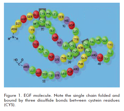
 </font></P >   <FONT size="+1">       ]]></body>
<body><![CDATA[<P   ><font size="3" face="Verdana, Arial, Helvetica, sans-serif"><b>EPIDERMAL GROWTH      FACTOR. THE ACTIVE PHARMACEUTICAL INGREDIENT OF HEBERPROT-P</b></font><font size="2" face="Verdana, Arial, Helvetica, sans-serif">      </font></P >   <FONT size="+1">       <P   ><font size="2" face="Verdana, Arial, Helvetica, sans-serif">EGF is an ancient      polypeptide, conserved throughout evolution, which is present in many animal      species and with a remarkably preserved structure having a high interspecies      homology, and therefore showing cross-species biological activity. This latter      property has been a corner stone in developing a vast series of pharmacological      and toxicological experimental studies using the human natural or recombinant      EGF. A large number of studies and reports in the literature demonstrate that      the human EGF binds to the receptor expressed by the cells of laboratory animals      (1). In fact, the discovery of EGF by Stanley Cohen was supported by the identification      of its epitheliotropic effects in different laboratory rodent species, by      administering a semi-pure preparation extracted from their salivary glands      (2). Undoubtedly, the phylogenetic conservation of EGF in animals and man,      and its ubiquitous distribution in tissues and organic fluids, suggest the      existence of a relevant physiological role in the homeostatic preservation      of epithelial cell populations in the form of mucosae or glands. From the      chemical viewpoint, EGF is a relatively small molecule of only 53 amino acids,      although smaller and larger molecular species or isoforms having physiological      relevance have been described. It is a self-folded single chain with a loop      formed by three disulfide bonds between cystein residues (<a href="#fig1">Figure      1</a>). </font></P >       <P align="center"   ><font size="+1" color="#000000"><font size="+1"><font size="+1"><font size="+1"><font size="+1"><font color="#0000FF"><font size="+1" color="#000000"><font size="+1"><font size="+1"><font size="+1"><font size="+1"><font face="Verdana, Arial, Helvetica, sans-serif"><a name="fig1"></a></font></font></font></font></font></font></font></font></font></font></font></font><font face="Verdana, Arial, Helvetica, sans-serif"><img src="/img/revistas/bta/v27n2/f0102210.gif" width="410" height="367"></font></P >   <FONT size="+1">       
<P   ><font face="Verdana, Arial, Helvetica, sans-serif" size="2">As described for      other growth factors, the biological activities of EGF are induced after binding      to the receptor molecule, which is extensively distributed in many mesenchymal      and epithelial tissues. The receptor, known as HER-1, is a trans-membrane      protein that has a terminal intra-cytoplasmic domain with a tyrosine kinase      activity, significantly involved in signal transduction through several occasionally      convergent and redundant pathways. The systemic administration of EGF at supra-physiological      concentrations is able to induce biological responses that were formerly assumed      as redundant effects. However, in the last 20 years, transgenic mice or those      genetically silenced for specific growth factors have helped unravel its subtle      physiology and biochemistry at the cellular level. </font></P >       <P   ><font face="Verdana, Arial, Helvetica, sans-serif" size="2">Almost since its      discovery and identification, researchers have evaluated the effects of EGF      extracted and purified from salivary glands in experimental systems of rabbit      corneal burn. Preliminary findings at that time demonstrated that the local      administration of EGF stimulated the migration and proliferation of the corneal      epithelium, helping the wounds heal faster than normal. As early as the beginning      of the 1960&rsquo;s, it was known that the healing effect was due to its mitogenic      action on epithelial cells and fibroblasts. Using cell culture models it was      demonstrated that the presence of EGF in cultures of fibroblasts, endothelial      cells and keratinocytes favored their proliferation and, in the latter, their      migration also. In other words, the main cell types responsible for skin wound      healing expressed the EGF receptor and were stimulated by its presence. All      the pieces of knowledge obtained up to that moment from the rabbit corneal      burn <I>in vivo </I>model and from <I>in vitro </I>experimental systems, stimulated      the design of two excellent experiments in mice. They included the surgical      removal of salivary glands from a group of wounded mice, and the exogenous      application of saliva to the wounds. It was conclusively demonstrated that      animals lacking salivary glands, and therefore unable to lick their wounds,      showed significantly deficient wound healing. On the other hand, the exogenous      application of saliva accelerated the healing process. The role of EGF from      saliva in the skin healing process was thus demonstrated. </font></P >       <P   ><font face="Verdana, Arial, Helvetica, sans-serif" size="2">Almost 50 years of      biological research using EGF in different systems and experimental contexts      make it possible to summarize its general functions as a: </font></P >       <P   ><font face="Verdana, Arial, Helvetica, sans-serif" size="2">- Mitogenic agent:      It controls and stimulates cell proliferation, particularly in epithelial      tissue cell lines. </font></P >       <P   ><font face="Verdana, Arial, Helvetica, sans-serif" size="2">- Motogenic agent:      It controls and stimulates cell migration. </font></P >       <P   ><font face="Verdana, Arial, Helvetica, sans-serif" size="2">- Inducing agent      for cell differentiation: It promotes the production of a defined or differentiated      phenotype in undifferentiated or pre-differentiated cells. </font></P >       <P   ><font face="Verdana, Arial, Helvetica, sans-serif" size="2">- Cyto-protective      agent: It stimulates cell survival in episodes or insults that would otherwise      be lethal. </font></P >       ]]></body>
<body><![CDATA[<P   ><font face="Verdana, Arial, Helvetica, sans-serif" size="2">The systemic administration      of EGF at supra-physiological concentrations is able to induce biological      responses as those described above and this can therefore result in pharmacological      actions through the extension or enhancement of the biological response (3).      </font></P >       <P   ><font size="3" face="Verdana, Arial, Helvetica, sans-serif"><b>CUBA PRODUCES      THE HUMAN RECOMBINANT EGF</b></font><font size="2" face="Verdana, Arial, Helvetica, sans-serif">      </font></P >   <FONT size="+1">       <P   ><font size="2" face="Verdana, Arial, Helvetica, sans-serif">The CIGB of Havana      has produced the human recombinant EGF since 1988 as a mixture of isoforms      of 51 and 52 amino acids. At that time, our country was one of the pioneers      in obtaining this protein through genetic engineering techniques. This product      has been subjected to an extensive series of pre-clinical, experimental toxicological      and pharmacological studies. Undoubtedly, it is the most pre-clinically investigated      Active Pharmaceutical Ingredient of the CIGB. Many pharmacological studies      have being carried out by the CIGB and other Cuban institutions with this      product. Other studies have been performed in cooperation with internationally      relevant foreign scientific research groups. The EGF produced in Cuba was      examined and characterized by a group of British researchers, who demonstrated      its high quality based on its physicochemical integrity, purity and biological      potency. Those results allowed and facilitated its use in a successful clinical      trial in the United Kingdom (4, 5). Cuba had produced Hebermin for the last      20 years, a pharmaceutical semi-solid EGF formulation used to stimulate wound      healing in acute cutaneous lesions. </font></P >   <FONT size="+1">       <P   ><font size="3" face="Verdana, Arial, Helvetica, sans-serif"><b>BACKGROUND AND      EXPERIMENTAL STUDIES. PRE-CLINICAL PHARMACOLOGICAL BASIS FOR EGF</b></font><font size="2" face="Verdana, Arial, Helvetica, sans-serif">      </font></P >   <FONT size="+1">       <P   ><font size="2" face="Verdana, Arial, Helvetica, sans-serif">The experimental      models used in EGF pharmacological studies have been exploited extensively      and are fully valid. These biomodels ad integrum have made it possible to      discover new pharmacological effects of this agent, which consequently suggest      new therapeutic indications for the experimentally represented conditions.      The animal models used are confirmed to be solid and useful by their repeatability      and reproducibility. In the 1990&rsquo;s, the development of pre-clinical      pharmacology for the systemic administration of EGF was academically validated      in Cuba and other countries. Here we describe the sequence of studies carried      out at the CIGB. </font></P >   <FONT size="+1">       <P   ><font face="Verdana, Arial, Helvetica, sans-serif" size="2">The cyto-protective      and gastro-protective potentials of EGF, particularly the latter, were studied      in 1992 using an experimental system that is extensively employed in canonical      pharmacology. This system is based on preventing and attenuating gastric damage      in rats exposed to the oral instillation of ethanol. EGF was administered      by the oral route under pre-conditioning prophylactic schedules and a therapeutic      schedule. This simple assay was the first of a series of studies aimed to      evaluate the cyto-protective effects of EGF. Under these experimental conditions,      EGF showed its capacity to prevent and reduce gastric damage in a dose-dependent      way. Interestingly, the preventive effect, associated to the prophylactic      treatment showed clear evidence of the protective potency of EGF in an epithelial      substrate challenged by a necrogenic agent. In other words, EGF seemed to      activate or pre-condition cellular processes opposing the lethal effects of      an external chemical agent. This information was never published but it has      contributed to a more complete documentation within our sanitary records.      </font></P >       <P   ><font face="Verdana, Arial, Helvetica, sans-serif" size="2">
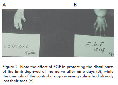
Inspired by the curiosity      and enthusiasm caused by the above results, we decided to evaluate, for the      first time, the effect of the systemic injection of EGF. We hoped to learn      about its protective effect in a clinically and pathologically relevant animal      model, and in tissues unrelated to the upper structures of the digestive system.      At that moment, the literature was full of demonstrations in rats reproducing      Curling&rsquo;s ulcers. Therefore, the examination of the neuroprotective      effect of the perilesional injection of EGF in rats that had been subjected      to total transversal axotomy of the sciatic nerve in the coxofemoral area      was determined. The morpho-functional regenerative response of the axon was      studied, as well as the synthesis of myelin by the nerve and its cells. Two      independent and extemporary studies demonstrated that this treatment produced      the following effects: </font></P >       <P   ><font face="Verdana, Arial, Helvetica, sans-serif" size="2">a) It significantly      favored the recovery of the motor nerve conduction 60 days after provoking      the trauma. This was a delayed effect, since EGF was administered for only      20 days. </font></P >       <P   ><font face="Verdana, Arial, Helvetica, sans-serif" size="2">b) It favored axonal      and myelin recovery. It prevented or reduced the changes suggesting a within      axonal degeneration. EGF also favored, in a dose-dependent way, the survival      of myelin producing cells near the sectioned area, contributing to a more      physiological re-myelination. </font></P >       <P   ><font face="Verdana, Arial, Helvetica, sans-serif" size="2">c) It prevented or      significantly delayed the onset of trophic changes of the soft tissues of      the limbs (skin and related structures). The presence of plantar ulcers and      toe necrosis was delayed (<a href="#fig2">Figure 2</a>). </font></P >       ]]></body>
<body><![CDATA[<P align="center"   ><font size="+1" color="#000000"><font size="+1"><font size="+1"><font size="+1"><font size="+1"><font color="#0000FF"><font size="+1" color="#000000"><font size="+1"><font size="+1"><font size="+1"><font size="+1"><font size="+1"><font size="+1"><font size="+1"><font size="+1"><font size="+1"><font face="Verdana, Arial, Helvetica, sans-serif"><a name="fig2"></a></font></font></font></font></font></font></font></font></font></font></font></font></font></font></font></font></font><font face="Verdana, Arial, Helvetica, sans-serif"><img src="/img/revistas/bta/v27n2/f0202210.gif" width="406" height="291"></font></P >       
<P   ><font face="Verdana, Arial, Helvetica, sans-serif" size="2">The cyto-protective      effects of EGF on the nervous tissues were evident, a conservatively novel      finding at that time. However, the recurring observation that EGF could protect      the soft peripheral tissues in a denervated limb was completely new. The prevention      of hair loss under these conditions was perhaps the most immediate response      to the EGF infiltration treatment. The fact that EGF showed an anti-necrogenic      effect under these experimental conditions, suggested an unusual cyto-protective      capacity with a high potential for therapeutic use, according to the physiopathological      relevance of the model implemented for the lesions. The sectioning of the      sciatic nerve implied a drastic deterioration of the vascular neural tone      and of the vasomotor reserve, which unquestionably will have negative consequences      in the irrigation of oxygenated blood to the skin, as well as in its venous      return. In other words, the possibility of establishing the pharmacological      management of a tissue hypo-perfusion process of neurogenic origin had begun      (6, 7). </font></P >       <P   ><font face="Verdana, Arial, Helvetica, sans-serif" size="2">Other experiments      followed during one decade that were more focused on studying the potential      cytoprotective effect of EGF in experimental ischemic episodes, when administered      by the parenteral route and following pharmacological preconditioning schedules      or therapeutic schedules of multiple injections. Hence, the evaluation of      the hepato-protective effect of EGF was started in an experimental system      involving its anti-inflammatory and anti-oxidant induction effects in order      to rescue the hepatocytes. The EGF was also administered through the intra-peritoneal      route in rats, 30 minutes before being challenged with hepatotoxic carbon      tetra-chloride. The experiment demonstrated the hepato-protective action of      EGF through a substantial reduction of parenchymal necrosis in this organ,      compared to the animals of the control group receiving saline (8). The effects      were dose-dependent, and were a first demonstration of the EGF-induced protection      against a cytotoxic agent having a damaging mechanism involving lipid peroxidation.      An ischemia/reperfusion experiment was carried out to demonstrate in a more      realistic and clinically relevant setting the antioxidant defense recently      described. The most appropriate and available model at that time was the bilateral      renal ischemia, which was reproduced successfully by the late Professor Ernesto      Barber. Depending on the dose, the preconditioning treatment prevented the      morphological and functional deterioration of the kidney tissue. The animals      treated with EGF showed a significant reduction in the levels of all the oxidative      stress markers studied, as well as in the activity of the PLA-A2 as a precursor      of pro-inflammatory mediators and agents related to the spasm of kidney vessels      (9). <a href="/img/revistas/bta/v27n2/f0302210.gif">Figure 3</a> shows      the protective effects of the treatment. </font></P >   <FONT size="+1"><FONT size="+1">       
<P   ><font face="Verdana, Arial, Helvetica, sans-serif" size="2">
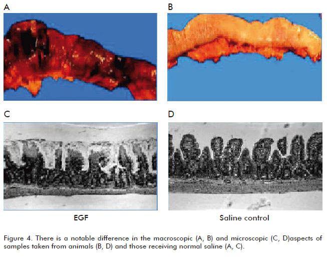
Our group carried      out a second ischemia/reperfusion experiment. It was conceived as the temporary      occlusion of the upper mesenteric artery that irrigates the jejunum and ileum      segments. In addition to demonstrating the cyto-protective effect of EGF against      ischemia, this experiment would be somewhat of a theoretical introduction      to models of multiple organ damage (MOD). Although it was not the main objective,      this experiment in itself produced an excellent reproduction of MOD. It is      known that a systemic inflammatory response syndrome (SIRS) established during      the reperfusion phase of the small intestine, is characterized by high serum      levels of the tumor necrosis factor (TNF-&alpha;) that is morphologically      expressed in several organs, such as in the lungs by an edema of capillary      permeability at the septum (similar to the Adult Respiratory Distress Syndrome,      ARDS) and in the kidneys by acute tubular necrosis. </font></P >       <P   ><font face="Verdana, Arial, Helvetica, sans-serif" size="2">There is a notably      dramatic increase in the levels of malondialdehyde (MDA) as a marker of lipid      peroxidation in different internal organs, as well as of myeloperoxidase (MPO)      as an inflammation marker of activated neutrophils infiltrated within the      parenchyma of tissues. Intestinal damage, such as hemorrhage and partial infarctions,      was significantly prevented in animals receiving a single prophylactic injection      of EGF (<a href="/img/revistas/bta/v27n2/f0402210.gif">Figure 4</a>).      Kidney and lung damage were also significantly reduced. From a biochemical      point of view, it was demonstrated that the treatment with the EGF reduced      the formation of oxygen reactive species, as well as the activity of the enzyme      MPO. An interesting finding was that circulating TNF-&alpha; levels substantially      decreased in the groups treated with EGF (10). To conclude the investigation      we just had to establish an MOD model and assess the EGF in a lethal pathological      and progressive organic deterioration substrate. </font></P >       
<P   ><font face="Verdana, Arial, Helvetica, sans-serif" size="2">
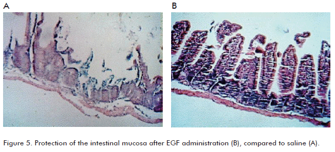
The capacity of EGF      to establish a systemic and simultaneous cyto-protective effect in various      internal epithelial organs was examined, after demonstrating that a single      preconditioning EGF injection generated dose-dependent entero-, hepato- and      nephroprotective effects. For that purpose, a sterile MOD model induced by      severe burns was established in mice and rats receiving hypodermic lesions      in 25 to 30% of their body surface (11). Animals received a single EGF injection,      30 minutes before burning. The results of the experiment were: 1) a significant      reduction in cumulative mortality in three independent experiments, 2) a decrease      in kidney damage, and 3) decreased gastrointestinal damage. <a href="/img/revistas/bta/v27n2/f0502210.gif">Figure      5</a> shows the substantial difference of mucosal integrity at the jejunum      and ileum between animals treated with EGF and those receiving only normal      saline.This was the first study demonstrating the capacity of a single and      prophylactic injection of EGF to induce simultaneous protection in several      organs, an effect that was not disrupted under such a severe stress as burns      affecting the internal organs. In a second demonstration of MOD induced by      a chemical substance (12), a prophylactic or therapeutic intervention with      EGF significantly increased animal survival, preventing kidney, liver and      intestinal damage and also reducing the levels of numerous biochemical markers      indicating functional alterations in different organs. </font></P >       
<P   ><font face="Verdana, Arial, Helvetica, sans-serif" size="2">In a later study,      we were to assess the intestinal response to the continuous exposure to EGF.      Until then it was known that EGF and other growth factors from that same family      exerted a trophic effect on the mucosa of the gastrointestinal system, although      the bases mediating that response had not been yet described. One of the purposes      of this study was to give the scientific support to the systemic administration      of EGF in order to regenerate intestinal mass and tissues. The continuous      infusion of EGF (60 &mu;g/kg per day) was uniformly ensured through the use      of subcutaneous osmotic mini-pumps in periods between days 1 to 14. The study      made it possible to learn that both cell proliferation in the crypt and its      fission were independent but complementary mechanisms for the trophic effect      of EGF in this system (13). It was demonstrated that the colon segment is      particularly sensitive to EGF, where an increase and later regression of the      index of crypt fission was observed, suggesting an endogenous mechanism for      controlling tissue growth. On the other hand, this study demonstrates the      potent effect of EGF on the growth of intestinal villi in different segments      of the small intestine of enterally-fed animals. </font></P >       <P   ><font face="Verdana, Arial, Helvetica, sans-serif" size="2"><a href="/img/revistas/bta/v27n2/f0602210.gif">Figure      6</a> shows the effect of EGF on the growth of villi and crypt fission a few      days after starting the treatment. Compare the difference with animals receiving      normal saline. This study established the potential bases for EGF treatment      in pathological processes requiring the regeneration of intestinal masses      and tissues, and the correction of villi atrophy or flattening, thus being      able to increase its absorption area. </font></P >       
<P   ><font face="Verdana, Arial, Helvetica, sans-serif" size="2">Studies in which      EGF was administered together with the Keratinocyte Growth Factor (KGF) showed      that intestinal atrophy can be reverted by treating with EGF, and that this      response can be enhanced by the concurrent administration of KGF (14). This      experiment confirmed previous results of the trophic effect of EGF on the      gastrointestinal mucosa. This and subsequent studies form part of a series      of experiments on the effect of EGF on the digestive system, thus providing      new theoretical support to correct atrophy and stimulate the healing of the      intestinal mucosa, and to improve the condition of the organism with a malabsorption      syndrome. </font></P >       <P   ><font face="Verdana, Arial, Helvetica, sans-serif" size="2">Finally, the most      recent study of this series (15), shows that a single pre-conditioning administration      of EGF in rats, alone or combined with the growth hormone-releasing peptide      6 (GHRP-6), exerts a potent protective effect on rat liver parenchyma exposed      for 120 min to ischemia and after 3 hours of reperfusion. Damage due to the      activation of the pro-inflammatory cascade became significantly attenuated      in distal organs such as the lungs, small intestine and kidneys. This confirms      the systemic cyto-protective effect of EGF (<a href="/img/revistas/bta/v27n2/f0702210.gif">Figure      7</a>). </font></P >       
]]></body>
<body><![CDATA[<P   ><font face="Verdana, Arial, Helvetica, sans-serif" size="2">After working several      years with EGF in different experimental biological sys-tems, it may be concluded      that there are two basic pharmacological effects that are induced by administering      EGF at supra-physiological concentrations: 1) cyto-protection, which is rapidly      established, even after just a single dose, and 2) trophic- repairing effects,      which appear after repeated and more or less prolonged administrations. Considering      the risk-benefit ratio, the substantial advantages and virtues of using EGF      to treat niches that have been unattended in clinical medicine have been clearly      explained. The rationality of the infiltrating treatment of diabetic foot      ulcers is supported by the findings of our group showing a reduced local bioavailability      of this factor when topically applied on acute and controlled total thickness      wounds. </font></P >       <P   ><font size="3" face="Verdana, Arial, Helvetica, sans-serif"><b>PRE-CLINICAL TOXICOLOGY      STUDIES. CONSIDERATIONS ON THE TRANSFORMING POTENTIAL OF EGF</b></font><font size="2" face="Verdana, Arial, Helvetica, sans-serif">      </font></P >   <FONT size="+1">       <P   ><font size="2" face="Verdana, Arial, Helvetica, sans-serif">As previously mentioned,      EGF has been submitted to a wide series of pre-clinical safety studies with      repeated administration long-term schedules in different laboratory animal      species. Genotoxicity studies have also shown negative results. Until now,      the evidence shows that EGF does not trigger the process of a malignant transformation      of normal cells under the conditions tested and even after its repeated and      exogenous administration (16). However, classical experiments clearly indicate      that EGF promotes chemical and viral carcinogenesis (17) in animals and cells,      respectively. In other words, cells in cultures or within the body of laboratory      animals, which were exposed to viruses or carcinogenic initiating substances,      can become more easily tumorigenic after EGF treatment. It is also well known      that in several types of human epithelial tumors there is an over-expression      of the EGF receptor and its signaling system is over-amplified (18). </font></P >   <FONT size="+1">       <P   ><font face="Verdana, Arial, Helvetica, sans-serif" size="2">The first systemic      intervention with EGF was carried out, as far as we know, in 1975, in patients      with Zollinger-Ellison syndrome. Other severe pathological processes of the      gastrointestinal tract have also been treated with the systemic administration      of EGF (19). Similar studies are being carried out throughout the world (20).      On the other hand, the most recent clinical trials were conducted by our group,      infiltrating EGF locally to diabetic foot ulcers that were refractory to treatment      (21). Throughout these years, no carcinogenic episodes have been observed      in any of the patients treated with these repetitive therapeutic interventions      using EGF at supra-physiological concentrations and with any of the administration      routes used. </font></P >       <P   ><font face="Verdana, Arial, Helvetica, sans-serif" size="2">Is obvious to point      out that EGF therapy, or that of any other growth factor, is strictly contraindicated      in patients having malignant tumors or pre-malignant lesions. The administration      of the EGF or any other growth factor must always be carried out with the      careful selection and inclusion of patients, based on their personal and family      backgrounds, and also considering the risk-benefit ratio. In fact, this rule      is applied in clinical practice for many authorized and internationally prescribed      drugs. </font></P >       <P   ><font face="Verdana, Arial, Helvetica, sans-serif" size="2">Biological and clinical      evidence that support the criterion that EGF does not seem to be an initiator      of cell transformation toward malignancy is summarized below. </font></P >       <P   ><font face="Verdana, Arial, Helvetica, sans-serif" size="2">1. EGF is not genotoxic      or mutagenic according to results of internationally established evaluations.      It does not seem to modify cell stability (22). </font></P >       <P   ><font face="Verdana, Arial, Helvetica, sans-serif" size="2">2. The <I>in vitro      </I>and <I>in vivo </I>pre-clinical systems indicate a tumor-promoting activity      of EGF in cellular niches previously &ldquo;initiated&rdquo; with chemical      or viral carcinogens. The tumor-promoting activity of EGF has not always been      reproduced <I>in vivo </I>(23). </font></P >       <P   ><font face="Verdana, Arial, Helvetica, sans-serif" size="2">3. The exogenous      administration of EGF, of the transforming growth factor alpha and of other      growth factors, has been shown to be insufficient in inducing tumors. It has      been demonstrated that the inadequate exposure to EGF of transgenic mice predisposes      the target tissue to cancer, and EGF could be a necessary, but insufficient      factor for cell transformation (24). </font></P >       <P   ><font face="Verdana, Arial, Helvetica, sans-serif" size="2">4. In other EGF transgenic      models, over-exposure to EGF was associated only to delayed somatic growth      (25) and to the enhanced adaptability of the target cells after small bowel      resection (26). </font></P >       ]]></body>
<body><![CDATA[<P   ><font face="Verdana, Arial, Helvetica, sans-serif" size="2">5. Hyperplastic changes      in animal models mediated by a continuous exo-genous exposure to EGF seem      to be self-limited. The trophic changes mediated by EGF in animal models are      reversible and depend on the dose, frequency, species and sex in susceptible      tissues (27-30). </font></P >       <P   ><font face="Verdana, Arial, Helvetica, sans-serif" size="2">6. It is known that      the healing process comprises a set of complex convergent events, such as      cell migration, differentiation and division, all of which condition the promotion      of tumor growth (31). There are, however, no reports of oncogenic induction      in patients receiving EGF or any other topically-administered growth factor.      On the contrary, the topical administration of EGF has been reported as radioprotective      in patients with a histological diagnosis of skin carcinoma submitted to radiotherapy      (32). </font></P >       <P   ><font face="Verdana, Arial, Helvetica, sans-serif" size="2">7. Pharmacokinetic      and bio-distribution profiles of EGF in animal models and in man have shown      its short half-life with a rapid elimination and no accumulation, thus excluding      the concept of the continuous/ long-term activation of the receptor. </font></P >       <P   ><font size="3" face="Verdana, Arial, Helvetica, sans-serif"><b>CONCLUSIONS</b></font><font size="2" face="Verdana, Arial, Helvetica, sans-serif">      </font></P >   <FONT size="+1">       <P   ><font size="2" face="Verdana, Arial, Helvetica, sans-serif">EGF has proved to      be clinically safe up to now, and its administration may be useful in certain      clinical processes that are refractory to treatment and/or critical conditions.      These are the niches in which its application is adequately adjusted to ethical      and therapeutic standards when following a risk-benefit ratio analysis for      each specific case. </font></P >   <FONT size="+1">       <P   ><font size="3" face="Verdana, Arial, Helvetica, sans-serif"><b>REFERENCES</b></font><font size="2" face="Verdana, Arial, Helvetica, sans-serif">      </font></P >   <FONT size="+1"><FONT size="+1">       <!-- ref --><P   ><font size="2" face="Verdana, Arial, Helvetica, sans-serif">1. Brown KD. The      epidermal growth factor/ transforming growth factor-<font face="Symbol">a</font>      and their receptors. Eur J Gastroenterol Hepatol 1995;7:914-22. </font></P >   <FONT size="+1">       <!-- ref --><P   ><font face="Verdana, Arial, Helvetica, sans-serif" size="2">2. Precocious newborn      mice and Epidermal Growth Factor: the work of Stanley Cohen. Papers in a series      reprinted to celebrate the centenary of the JBC in 2005. J Biol Chem 2006;281:10-1.      </font></P >       <!-- ref --><P   ><font face="Verdana, Arial, Helvetica, sans-serif" size="2">3. Boonstra J, Rijken      P, Humbel B, Cremers F, Verkleij A, van Bergen en Hengowen PB. The epidermal      growth factor. Cell Biol Int 1995;19:413-30. </font></P >       <!-- ref --><P   ><font face="Verdana, Arial, Helvetica, sans-serif" size="2">4. Sinha A, Nightingale      J, West KP, Berlanga-Acosta J, Playford RJ. Epidermal growth factor enemas      with oral mesalamine for mild-to-moderate left-sided ulcerative colitis or      proctitis. N Engl J Med 2003;349:350-7. </font></P >       <!-- ref --><P   ><font face="Verdana, Arial, Helvetica, sans-serif" size="2">5. Calnan DP, Fagbemi      A, Berlanga- Acosta J, Marchbank T, Sizer T, Lakhoo K, <I>et al</I>. Potency      and stability of C truncated human epidermal growth factor. Gut 2000; 47:622-7.      </font></P >       <!-- ref --><P   ><font face="Verdana, Arial, Helvetica, sans-serif" size="2">6. Prats P, Casta&ntilde;eda      L, Berlanga J, Falc&oacute;n V, Rodr&iacute;guez V, Suarez-Muria C. El factor      de crecimiento epid&eacute;rmico en lesiones del sistema nervioso perif&eacute;rico.      Rev Mex Cienc Farm 1998;29:17-23. </font></P >       <!-- ref --><P   ><font face="Verdana, Arial, Helvetica, sans-serif" size="2">7. Prats P, Casta&ntilde;eda      L, Falc&oacute;n V, Ortega R, de la Rosa MC, Men&eacute;ndez I, <I>et al</I>.      Efecto del Factor de Crecimiento Epid&eacute;rmico sobre la regeneraci&oacute;n      del nervio ci&aacute;tico transectado en ratas. Biotecnol Apl 1998; 15:237-41.      </font></P >       <!-- ref --><P   ><font face="Verdana, Arial, Helvetica, sans-serif" size="2">8. Berlanga J, Caballero      ME, Ram&iacute;rez D, Torres A, Valenzuela C, Lodos J, <I>et al</I>. Epidermal      growth factor protects against carbon tetrachloride-induced hepatic injury.      Clinical Science (Lond) 1998;94: 219-23. </font></P >       <!-- ref --><P   ><font face="Verdana, Arial, Helvetica, sans-serif" size="2">9. Caballero ME,      Calunga J, Barber E, Cruz E, L&oacute;pez-Saura P, Boix E, <I>et al</I>. Epidermal      Growth Factor-mediated prevention of renal ischemia/reperfusion injury. Biotecnol      Apl 2000;17:161-5. </font></P >       <!-- ref --><P   ><font face="Verdana, Arial, Helvetica, sans-serif" size="2">10. Berlanga J, Prats      P, Rem&iacute;rez D, Gonz&aacute;lez R, Lopez-Saura P, Aguiar J. Prophylactic      use of epidermal growth factor reduces ischemia/ reperfusion intestinal damage.      Am J Pathol 2002;161:373-9. </font></P >       <!-- ref --><P   ><font face="Verdana, Arial, Helvetica, sans-serif" size="2">11. Berlanga J, Lodos      J, L&oacute;pez-Saura P. Attenuation of internal organs damages by exogenously      administered epidermal growth factor (EGF) in burned rodents. Burns 2002;28:435-42.      </font></P >       <!-- ref --><P   ><font face="Verdana, Arial, Helvetica, sans-serif" size="2">12. Caballero ME,      Berlanga J, Ram&iacute;rez D, Lopez-Saura P, Gonz&aacute;lez R, Floyd DN.      Epidermal growth factor reduces multiorgan failure induced by thioacetamide.      Gut 2001;48:34-40. </font></P >       <!-- ref --><P   ><font face="Verdana, Arial, Helvetica, sans-serif" size="2">13. Berlanga-Acosta      J, Playford RJ, Mandir N, Goodlad RA. Gastrointestinal cell proliferation      and crypt fission are separate but complementary means of increasing tissue      mass following infusion of epidermal growth factor in rats. Gut 2001;48:803-7.      </font></P >       <!-- ref --><P   ><font face="Verdana, Arial, Helvetica, sans-serif" size="2">14. Sasaki M, FitzGerald      AJ, Mandir N, Berlanga-Acosta J, Goodlad RA. Keratinocyte growth factor and      epidermal growth factor can reverse the intestinal atrophy associated with      elemental diets in mice. Exp Physiol 2003;88:261-7. </font></P >       <!-- ref --><P   ><font face="Verdana, Arial, Helvetica, sans-serif" size="2">15. Cibrian D, Ajamieh      H, Berlanga J, Leon OS, Alba JS, Kim MJ, <I>et al</I>. Use of growthhormone-      releasing peptide-6 (GHRP-6) for the prevention of multiple organ failure.      Clin Sci (Lond) 2006;110:563-73. </font></P >       <!-- ref --><P   ><font face="Verdana, Arial, Helvetica, sans-serif" size="2">16. Berlanga J, &Aacute;lvarez      S, de la Fuente J, L&oacute;pez-Saura P. Considerations on the transforming      potential of epidermal growth factor. Biotecnol Apl 1998;(15):65-70. </font></P >       <!-- ref --><P   ><font face="Verdana, Arial, Helvetica, sans-serif" size="2">17. Stoscheck CM,      King LE. Role of Epidermal Growth Factor in carcinogenesis. Cancer Res 1986;46:1030-7.      </font></P >       <P   ><font face="Verdana, Arial, Helvetica, sans-serif" size="2">18. Arteaga CL. Epidermal      Growth Factor receptor dependence in human tumors: more than just expression?      Oncologist 2002;7(Suppl 4):31-9. </font></P >       <!-- ref --><P   ><font face="Verdana, Arial, Helvetica, sans-serif" size="2">19. Guglietta A,      Sullivan PB. Clinical applications of epidermal growth factor. Eur J Gastroenterol      Hepatol 1995;7:945-50. </font></P >       <!-- ref --><P   ><font face="Verdana, Arial, Helvetica, sans-serif" size="2">20. Sullivan PB,      Lewindon PJ, Cheng C, Lenehan PF, Kuo BS, Haskins JR, <I>et al</I>. Intestinal      mucosa remodeling by recombinant human epidermal growth factor (1-48) in neonates      with severe necrotizing enterocolitis. J Pediatr Surg 2007;42(3):462-9. </font></P >       <!-- ref --><P   ><font face="Verdana, Arial, Helvetica, sans-serif" size="2">21. Fern&aacute;ndez-Montequ&iacute;n      JI, Infante-Cristi&aacute; E, Valenzuela-Silva C, Franco-P&eacute;rez N, Savigne-      Gutierrez W, Artaza-Sanz H, <I>et al</I>. Intralesional injections of Citoprot-P      (recombinant human epidermal growth factor) in advanced diabetic foot ulcers      with risk of amputation. Int Wound J 2007;4(4):333-43. </font></P >       <!-- ref --><P   ><font face="Verdana, Arial, Helvetica, sans-serif" size="2">22. Maraschin R,      Bussi R, Conz A, Luciana O, Pirovano R, Nyska A. Toxicological evaluation      of u-hEGF. Toxicol Pathol 1995;23(3):356- 66. </font></P >       <!-- ref --><P   ><font face="Verdana, Arial, Helvetica, sans-serif" size="2">23. Lu Xia, Yao-Zong      Yuan, Chun-Di Xu, Yong-Pin Zhang, Ming-Ming Qiao, Jia-Xu Xu. Effects of epidermal      growth factor on the growth of human gastric cancer cell and the implanted      tumor of nude mice. World J Gastroenterol 2002;8(3):455-8. </font></P >       <!-- ref --><P   ><font face="Verdana, Arial, Helvetica, sans-serif" size="2">24. Borlak J, Meier      T, Halter R, Spanel R, Spanel-Borowski K. Epidermal growth factorinduced hepatocellular      carcinomas: gene expression profiles in precursor lesions, early stage and      solitary tumors. Oncogene 2005;24(11):1809-19. </font></P >       <!-- ref --><P   ><font face="Verdana, Arial, Helvetica, sans-serif" size="2">25. Chan SY, Wong      RWC. Expression of Epidermal Growth Factor in transgenic mice causes growth      retardation. J Biol Chem 2000;275:38693-8. </font></P >       <!-- ref --><P   ><font face="Verdana, Arial, Helvetica, sans-serif" size="2">26. Erwin CR, Helmrath      MA, Shin CE, Falcone RA Jr, Stern LE, Warner BW. Intestinal overexpression      of EGF in transgenic mice enhances adaptation after small bowel resection.      Am J Physiol 1999;277(3 Pt 1):G533-40. </font></P >       <!-- ref --><P   ><font face="Verdana, Arial, Helvetica, sans-serif" size="2">27. Vinter-Jensen      L, Smerup M, Kissmeyer- Nielsen P, Poulsen SS. Chronic systemic treatment      with epidermal growth factor in the rat increases the mucosal surface of the      small intestine. Regul Pept 1995;60(2-3):117-24. </font></P >       <!-- ref --><P   ><font face="Verdana, Arial, Helvetica, sans-serif" size="2">28. Vinter-Jensen      L, Juhl CO, Poulsen SS, Djurhuus JC, Dajani EZ, Nexo E. Chronic administration      of epidermal growth factor to pigs induce growth especially of the urinary      tract with accumulation of epithelial glycoconjugates. Lab Invest 1995;73(6):788-93.      </font></P >       <P   ><font face="Verdana, Arial, Helvetica, sans-serif" size="2">29. Juhl CO, Vinter-Jensen      L, Poulsen SS, Orntoft RF, Dajani EZ. Chronic treatment with epidermal growth      factor causes esophageal epithelial hyperplasia in pigs and rats. Dig Dis      Sci 1995;40(12):2717-2723. </font></P >       <!-- ref --><P   ><font face="Verdana, Arial, Helvetica, sans-serif" size="2">30. Christiansen      JJ, Vinter-Jensen L, Nielsen K. Systemic treatment in the rat with epidermal      growth factor causes polycystic growth of ovaries. APMIS 1996;104:147-52.      </font></P >       <!-- ref --><P   ><font face="Verdana, Arial, Helvetica, sans-serif" size="2">31. Berlanga-Acosta      J, Gavilondo-Cowley J, L&oacute;pez-Saura P; Gonz&aacute;lez-L&oacute;pez      T, Castro-Santana M, <I>et al</I>. Epidermal Growth Factor (EGF) in clinical      practice- A review of its biological actions, clinical indications and safety      implications. Int Wound J 2009;6:331-46. </font></P >       <!-- ref --><P   ><font face="Verdana, Arial, Helvetica, sans-serif" size="2">32. Alert J, Rodr&iacute;guez      J, Lombardero J, P&eacute;rez R. Acci&oacute;n radioprotectora local del factor      de crecimiento epid&eacute;rmico humano recombinante: reporte preliminar.      Interfer&oacute;n Biotecnol 1989;6:62-6. </font></P >       <P   ><font size="2" face="Verdana, Arial, Helvetica, sans-serif">Received in July,      2009. </font><font face="Verdana, Arial, Helvetica, sans-serif">    <br>     <font size="2">Accepted for publication in November, 2009. </font></font></P >   <FONT size="+1">       ]]></body>
<body><![CDATA[<P   ><font face="Verdana, Arial, Helvetica, sans-serif" size="2">Jorge Berlanga, Centro      de Ingenier&iacute;a Gen&eacute;tica y Biotecnolog&iacute;a, Investigaciones      Biom&eacute;dicas, Divisi&oacute;n de Farmac&eacute;uticos, Ave. 31 entre      158 y 190, Cubanac&aacute;n, Playa, CP 10 600, Ciudad de La Habana, Cuba.      E-mail: <a href="mailto:jorge.berlanga@cigb.edu.cu">jorge.berlanga@cigb.edu.cu      </a></font>   <FONT size="+1">       <P   > </P >   </font></font></font></font></font></font></font></font></font></font></font></font></font></font></font></font></font></font></font></font></font></font></font></font></font></font></font></DIV >      ]]></body><back>
<ref-list>
<ref id="B1">
<label>1</label><nlm-citation citation-type="journal">
<person-group person-group-type="author">
<name>
<surname><![CDATA[Brown]]></surname>
<given-names><![CDATA[KD]]></given-names>
</name>
</person-group>
<article-title xml:lang="en"><![CDATA[The epidermal growth factor/ transforming growth factor-&#945; &#61472;and their receptors]]></article-title>
<source><![CDATA[Eur J Gastroenterol Hepatol]]></source>
<year>1995</year>
<volume>7</volume>
<page-range>914-22</page-range></nlm-citation>
</ref>
<ref id="B2">
<label>2</label><nlm-citation citation-type="journal">
<article-title xml:lang="en"><![CDATA[Precocious newborn mice and Epidermal Growth Factor: the work of Stanley Cohen. Papers in a series reprinted to celebrate the centenary of the JBC in 2005]]></article-title>
<source><![CDATA[J Biol Chem]]></source>
<year>2006</year>
<volume>281</volume>
<page-range>10-1</page-range></nlm-citation>
</ref>
<ref id="B3">
<label>3</label><nlm-citation citation-type="journal">
<person-group person-group-type="author">
<name>
<surname><![CDATA[Boonstra]]></surname>
<given-names><![CDATA[J]]></given-names>
</name>
<name>
<surname><![CDATA[Rijken]]></surname>
<given-names><![CDATA[P]]></given-names>
</name>
<name>
<surname><![CDATA[Humbel]]></surname>
<given-names><![CDATA[B]]></given-names>
</name>
<name>
<surname><![CDATA[Cremers]]></surname>
<given-names><![CDATA[F]]></given-names>
</name>
<name>
<surname><![CDATA[Verkleij]]></surname>
<given-names><![CDATA[A]]></given-names>
</name>
<name>
<surname><![CDATA[van Bergen en Hengowen]]></surname>
<given-names><![CDATA[PB]]></given-names>
</name>
</person-group>
<article-title xml:lang="en"><![CDATA[The epidermal growth factor]]></article-title>
<source><![CDATA[Cell Biol Int]]></source>
<year>1995</year>
<volume>19</volume>
<page-range>413-30</page-range></nlm-citation>
</ref>
<ref id="B4">
<label>4</label><nlm-citation citation-type="journal">
<person-group person-group-type="author">
<name>
<surname><![CDATA[Sinha]]></surname>
<given-names><![CDATA[A]]></given-names>
</name>
<name>
<surname><![CDATA[Nightingale]]></surname>
<given-names><![CDATA[J]]></given-names>
</name>
<name>
<surname><![CDATA[West]]></surname>
<given-names><![CDATA[KP]]></given-names>
</name>
<name>
<surname><![CDATA[Berlanga-Acosta]]></surname>
<given-names><![CDATA[J]]></given-names>
</name>
<name>
<surname><![CDATA[Playford]]></surname>
<given-names><![CDATA[RJ]]></given-names>
</name>
</person-group>
<article-title xml:lang="en"><![CDATA[Epidermal growth factor enemas with oral mesalamine for mild-to-moderate left-sided ulcerative colitis or proctitis]]></article-title>
<source><![CDATA[N Engl J Med]]></source>
<year>2003</year>
<volume>349</volume>
<page-range>350-7</page-range></nlm-citation>
</ref>
<ref id="B5">
<label>5</label><nlm-citation citation-type="journal">
<person-group person-group-type="author">
<name>
<surname><![CDATA[Calnan]]></surname>
<given-names><![CDATA[DP]]></given-names>
</name>
<name>
<surname><![CDATA[Fagbemi]]></surname>
<given-names><![CDATA[A]]></given-names>
</name>
<name>
<surname><![CDATA[Berlanga- Acosta]]></surname>
<given-names><![CDATA[J]]></given-names>
</name>
<name>
<surname><![CDATA[Marchbank]]></surname>
<given-names><![CDATA[T]]></given-names>
</name>
<name>
<surname><![CDATA[Sizer]]></surname>
<given-names><![CDATA[T]]></given-names>
</name>
<name>
<surname><![CDATA[Lakhoo]]></surname>
<given-names><![CDATA[K]]></given-names>
</name>
</person-group>
<article-title xml:lang="en"><![CDATA[Potency and stability of C truncated human epidermal growth factor]]></article-title>
<source><![CDATA[Gut]]></source>
<year>2000</year>
<volume>47</volume>
<page-range>622-7</page-range></nlm-citation>
</ref>
<ref id="B6">
<label>6</label><nlm-citation citation-type="journal">
<person-group person-group-type="author">
<name>
<surname><![CDATA[Prats]]></surname>
<given-names><![CDATA[P]]></given-names>
</name>
<name>
<surname><![CDATA[Castañeda]]></surname>
<given-names><![CDATA[L]]></given-names>
</name>
<name>
<surname><![CDATA[Berlanga]]></surname>
<given-names><![CDATA[J]]></given-names>
</name>
<name>
<surname><![CDATA[Falcón]]></surname>
<given-names><![CDATA[V]]></given-names>
</name>
<name>
<surname><![CDATA[Rodríguez]]></surname>
<given-names><![CDATA[V]]></given-names>
</name>
<name>
<surname><![CDATA[Suarez-Muria]]></surname>
<given-names><![CDATA[C]]></given-names>
</name>
</person-group>
<article-title xml:lang="es"><![CDATA[El factor de crecimiento epidérmico en lesiones del sistema nervioso periférico]]></article-title>
<source><![CDATA[Rev Mex Cienc Farm]]></source>
<year>1998</year>
<volume>29</volume>
<page-range>17-23</page-range></nlm-citation>
</ref>
<ref id="B7">
<label>7</label><nlm-citation citation-type="journal">
<person-group person-group-type="author">
<name>
<surname><![CDATA[Prats]]></surname>
<given-names><![CDATA[P]]></given-names>
</name>
<name>
<surname><![CDATA[Castañeda]]></surname>
<given-names><![CDATA[L]]></given-names>
</name>
<name>
<surname><![CDATA[Falcón]]></surname>
<given-names><![CDATA[V]]></given-names>
</name>
<name>
<surname><![CDATA[Ortega]]></surname>
<given-names><![CDATA[R]]></given-names>
</name>
<name>
<surname><![CDATA[de la Rosa]]></surname>
<given-names><![CDATA[MC]]></given-names>
</name>
<name>
<surname><![CDATA[Menéndez]]></surname>
<given-names><![CDATA[I]]></given-names>
</name>
</person-group>
<article-title xml:lang="es"><![CDATA[Efecto del Factor de Crecimiento Epidérmico sobre la regeneración del nervio ciático transectado en ratas]]></article-title>
<source><![CDATA[Biotecnol Apl]]></source>
<year>1998</year>
<volume>15</volume>
<page-range>237-41</page-range></nlm-citation>
</ref>
<ref id="B8">
<label>8</label><nlm-citation citation-type="journal">
<person-group person-group-type="author">
<name>
<surname><![CDATA[Berlanga]]></surname>
<given-names><![CDATA[J]]></given-names>
</name>
<name>
<surname><![CDATA[Caballero]]></surname>
<given-names><![CDATA[ME]]></given-names>
</name>
<name>
<surname><![CDATA[Ramírez]]></surname>
<given-names><![CDATA[D]]></given-names>
</name>
<name>
<surname><![CDATA[Torres]]></surname>
<given-names><![CDATA[A]]></given-names>
</name>
<name>
<surname><![CDATA[Valenzuela]]></surname>
<given-names><![CDATA[C]]></given-names>
</name>
<name>
<surname><![CDATA[Lodos]]></surname>
<given-names><![CDATA[J]]></given-names>
</name>
</person-group>
<article-title xml:lang="en"><![CDATA[Epidermal growth factor protects against carbon tetrachloride-induced hepatic injury]]></article-title>
<source><![CDATA[Clinical Science (Lond)]]></source>
<year>1998</year>
<volume>94</volume>
<page-range>219-23</page-range></nlm-citation>
</ref>
<ref id="B9">
<label>9</label><nlm-citation citation-type="journal">
<person-group person-group-type="author">
<name>
<surname><![CDATA[Caballero]]></surname>
<given-names><![CDATA[ME]]></given-names>
</name>
<name>
<surname><![CDATA[Calunga]]></surname>
<given-names><![CDATA[J]]></given-names>
</name>
<name>
<surname><![CDATA[Barber]]></surname>
<given-names><![CDATA[E]]></given-names>
</name>
<name>
<surname><![CDATA[Cruz]]></surname>
<given-names><![CDATA[E]]></given-names>
</name>
<name>
<surname><![CDATA[López-Saura]]></surname>
<given-names><![CDATA[P]]></given-names>
</name>
<name>
<surname><![CDATA[Boix]]></surname>
<given-names><![CDATA[E]]></given-names>
</name>
</person-group>
<article-title xml:lang="en"><![CDATA[Epidermal Growth Factor-mediated prevention of renal ischemia/reperfusion injury]]></article-title>
<source><![CDATA[Biotecnol Apl]]></source>
<year>2000</year>
<volume>17</volume>
<page-range>161-5</page-range></nlm-citation>
</ref>
<ref id="B10">
<label>10</label><nlm-citation citation-type="journal">
<person-group person-group-type="author">
<name>
<surname><![CDATA[Berlanga]]></surname>
<given-names><![CDATA[J]]></given-names>
</name>
<name>
<surname><![CDATA[Prats]]></surname>
<given-names><![CDATA[P]]></given-names>
</name>
<name>
<surname><![CDATA[Remírez]]></surname>
<given-names><![CDATA[D]]></given-names>
</name>
<name>
<surname><![CDATA[González]]></surname>
<given-names><![CDATA[R]]></given-names>
</name>
<name>
<surname><![CDATA[Lopez-Saura]]></surname>
<given-names><![CDATA[P]]></given-names>
</name>
<name>
<surname><![CDATA[Aguiar]]></surname>
<given-names><![CDATA[J]]></given-names>
</name>
</person-group>
<article-title xml:lang="en"><![CDATA[Prophylactic use of epidermal growth factor reduces ischemia/ reperfusion intestinal damage]]></article-title>
<source><![CDATA[Am J Pathol]]></source>
<year>2002</year>
<volume>161</volume>
<page-range>373-9</page-range></nlm-citation>
</ref>
<ref id="B11">
<label>11</label><nlm-citation citation-type="journal">
<person-group person-group-type="author">
<name>
<surname><![CDATA[Berlanga]]></surname>
<given-names><![CDATA[J]]></given-names>
</name>
<name>
<surname><![CDATA[Lodos]]></surname>
<given-names><![CDATA[J]]></given-names>
</name>
<name>
<surname><![CDATA[López-Saura]]></surname>
<given-names><![CDATA[P]]></given-names>
</name>
</person-group>
<article-title xml:lang="en"><![CDATA[Attenuation of internal organs damages by exogenously administered epidermal growth factor (EGF) in burned rodents]]></article-title>
<source><![CDATA[Burns]]></source>
<year>2002</year>
<volume>28</volume>
<page-range>435-42</page-range></nlm-citation>
</ref>
<ref id="B12">
<label>12</label><nlm-citation citation-type="journal">
<person-group person-group-type="author">
<name>
<surname><![CDATA[Caballero]]></surname>
<given-names><![CDATA[ME]]></given-names>
</name>
<name>
<surname><![CDATA[Berlanga]]></surname>
<given-names><![CDATA[J]]></given-names>
</name>
<name>
<surname><![CDATA[Ramírez]]></surname>
<given-names><![CDATA[D]]></given-names>
</name>
<name>
<surname><![CDATA[Lopez-Saura]]></surname>
<given-names><![CDATA[P]]></given-names>
</name>
<name>
<surname><![CDATA[González]]></surname>
<given-names><![CDATA[R]]></given-names>
</name>
<name>
<surname><![CDATA[Floyd]]></surname>
<given-names><![CDATA[DN]]></given-names>
</name>
</person-group>
<article-title xml:lang="en"><![CDATA[Epidermal growth factor reduces multiorgan failure induced by thioacetamide]]></article-title>
<source><![CDATA[Gut]]></source>
<year>2001</year>
<volume>48</volume>
<page-range>34-40</page-range></nlm-citation>
</ref>
<ref id="B13">
<label>13</label><nlm-citation citation-type="journal">
<person-group person-group-type="author">
<name>
<surname><![CDATA[Berlanga-Acosta]]></surname>
<given-names><![CDATA[J]]></given-names>
</name>
<name>
<surname><![CDATA[Playford]]></surname>
<given-names><![CDATA[RJ]]></given-names>
</name>
<name>
<surname><![CDATA[Mandir]]></surname>
<given-names><![CDATA[N]]></given-names>
</name>
<name>
<surname><![CDATA[Goodlad]]></surname>
<given-names><![CDATA[RA]]></given-names>
</name>
</person-group>
<article-title xml:lang="en"><![CDATA[Gastrointestinal cell proliferation and crypt fission are separate but complementary means of increasing tissue mass following infusion of epidermal growth factor in rats]]></article-title>
<source><![CDATA[Gut]]></source>
<year>2001</year>
<volume>48</volume>
<page-range>803-7</page-range></nlm-citation>
</ref>
<ref id="B14">
<label>14</label><nlm-citation citation-type="journal">
<person-group person-group-type="author">
<name>
<surname><![CDATA[Sasaki]]></surname>
<given-names><![CDATA[M]]></given-names>
</name>
<name>
<surname><![CDATA[FitzGerald]]></surname>
<given-names><![CDATA[AJ]]></given-names>
</name>
<name>
<surname><![CDATA[Mandir]]></surname>
<given-names><![CDATA[N]]></given-names>
</name>
<name>
<surname><![CDATA[Berlanga-Acosta]]></surname>
<given-names><![CDATA[J]]></given-names>
</name>
<name>
<surname><![CDATA[Goodlad]]></surname>
<given-names><![CDATA[RA]]></given-names>
</name>
</person-group>
<article-title xml:lang="en"><![CDATA[Keratinocyte growth factor and epidermal growth factor can reverse the intestinal atrophy associated with elemental diets in mice]]></article-title>
<source><![CDATA[Exp Physiol]]></source>
<year>2003</year>
<volume>88</volume>
<page-range>261-7</page-range></nlm-citation>
</ref>
<ref id="B15">
<label>15</label><nlm-citation citation-type="journal">
<person-group person-group-type="author">
<name>
<surname><![CDATA[Cibrian]]></surname>
<given-names><![CDATA[D]]></given-names>
</name>
<name>
<surname><![CDATA[Ajamieh]]></surname>
<given-names><![CDATA[H]]></given-names>
</name>
<name>
<surname><![CDATA[Berlanga]]></surname>
<given-names><![CDATA[J]]></given-names>
</name>
<name>
<surname><![CDATA[Leon]]></surname>
<given-names><![CDATA[OS]]></given-names>
</name>
<name>
<surname><![CDATA[Alba]]></surname>
<given-names><![CDATA[JS]]></given-names>
</name>
<name>
<surname><![CDATA[Kim]]></surname>
<given-names><![CDATA[MJ]]></given-names>
</name>
</person-group>
<article-title xml:lang="en"><![CDATA[Use of growth hormone- releasing peptide-6 (GHRP-6) for the prevention of multiple organ failure]]></article-title>
<source><![CDATA[Clin Sci (Lond)]]></source>
<year>2006</year>
<volume>110</volume>
<page-range>563-73</page-range></nlm-citation>
</ref>
<ref id="B16">
<label>16</label><nlm-citation citation-type="journal">
<person-group person-group-type="author">
<name>
<surname><![CDATA[Berlanga]]></surname>
<given-names><![CDATA[J]]></given-names>
</name>
<name>
<surname><![CDATA[Álvarez]]></surname>
<given-names><![CDATA[S]]></given-names>
</name>
<name>
<surname><![CDATA[de la Fuente]]></surname>
<given-names><![CDATA[J]]></given-names>
</name>
<name>
<surname><![CDATA[López-Saura]]></surname>
<given-names><![CDATA[P]]></given-names>
</name>
</person-group>
<article-title xml:lang="en"><![CDATA[Considerations on the transforming potential of epidermal growth factor]]></article-title>
<source><![CDATA[Biotecnol Apl]]></source>
<year>1998</year>
<volume>15</volume>
<page-range>65-70</page-range></nlm-citation>
</ref>
<ref id="B17">
<label>17</label><nlm-citation citation-type="journal">
<person-group person-group-type="author">
<name>
<surname><![CDATA[Stoscheck]]></surname>
<given-names><![CDATA[CM]]></given-names>
</name>
<name>
<surname><![CDATA[King]]></surname>
<given-names><![CDATA[LE]]></given-names>
</name>
</person-group>
<article-title xml:lang="en"><![CDATA[Role of Epidermal Growth Factor in carcinogenesis]]></article-title>
<source><![CDATA[Cancer Res]]></source>
<year>1986</year>
<volume>46</volume>
<page-range>1030-7</page-range></nlm-citation>
</ref>
<ref id="B18">
<label>18</label><nlm-citation citation-type="journal">
<person-group person-group-type="author">
<name>
<surname><![CDATA[Arteaga]]></surname>
<given-names><![CDATA[CL]]></given-names>
</name>
</person-group>
<article-title xml:lang="en"><![CDATA[Epidermal Growth Factor receptor dependence in human tumors: more than just expression?]]></article-title>
<source><![CDATA[Oncologist]]></source>
<year>2002</year>
<volume>7</volume>
<page-range>31-9</page-range></nlm-citation>
</ref>
<ref id="B19">
<label>19</label><nlm-citation citation-type="journal">
<person-group person-group-type="author">
<name>
<surname><![CDATA[Guglietta]]></surname>
<given-names><![CDATA[A]]></given-names>
</name>
<name>
<surname><![CDATA[Sullivan]]></surname>
<given-names><![CDATA[PB]]></given-names>
</name>
</person-group>
<article-title xml:lang="en"><![CDATA[Clinical applications of epidermal growth factor]]></article-title>
<source><![CDATA[Eur J Gastroenterol Hepatol]]></source>
<year>1995</year>
<volume>7</volume>
<page-range>945-50</page-range></nlm-citation>
</ref>
<ref id="B20">
<label>20</label><nlm-citation citation-type="journal">
<person-group person-group-type="author">
<name>
<surname><![CDATA[Sullivan]]></surname>
<given-names><![CDATA[PB]]></given-names>
</name>
<name>
<surname><![CDATA[Lewindon]]></surname>
<given-names><![CDATA[PJ]]></given-names>
</name>
<name>
<surname><![CDATA[Cheng]]></surname>
<given-names><![CDATA[C]]></given-names>
</name>
<name>
<surname><![CDATA[Lenehan]]></surname>
<given-names><![CDATA[PF]]></given-names>
</name>
<name>
<surname><![CDATA[Kuo]]></surname>
<given-names><![CDATA[BS]]></given-names>
</name>
<name>
<surname><![CDATA[Haskins]]></surname>
<given-names><![CDATA[JR]]></given-names>
</name>
</person-group>
<article-title xml:lang="en"><![CDATA[Intestinal mucosa remodeling by recombinant human epidermal growth factor (1-48) in neonates with severe necrotizing enterocolitis]]></article-title>
<source><![CDATA[J Pediatr Surg]]></source>
<year>2007</year>
<volume>42</volume>
<numero>3</numero>
<issue>3</issue>
<page-range>462-9</page-range></nlm-citation>
</ref>
<ref id="B21">
<label>21</label><nlm-citation citation-type="journal">
<person-group person-group-type="author">
<name>
<surname><![CDATA[Fernández-Montequín]]></surname>
<given-names><![CDATA[JI]]></given-names>
</name>
<name>
<surname><![CDATA[Infante-Cristiá]]></surname>
<given-names><![CDATA[E]]></given-names>
</name>
<name>
<surname><![CDATA[Valenzuela-Silva]]></surname>
<given-names><![CDATA[C]]></given-names>
</name>
<name>
<surname><![CDATA[Franco-Pérez]]></surname>
<given-names><![CDATA[N]]></given-names>
</name>
<name>
<surname><![CDATA[Savigne-Gutierrez]]></surname>
<given-names><![CDATA[W]]></given-names>
</name>
<name>
<surname><![CDATA[Artaza-Sanz]]></surname>
<given-names><![CDATA[H]]></given-names>
</name>
</person-group>
<article-title xml:lang="en"><![CDATA[Intralesional injections of Citoprot-P (recombinant human epidermal growth factor) in advanced diabetic foot ulcers with risk of amputation]]></article-title>
<source><![CDATA[Int Wound J]]></source>
<year>2007</year>
<volume>4</volume>
<numero>4</numero>
<issue>4</issue>
<page-range>333-43</page-range></nlm-citation>
</ref>
<ref id="B22">
<label>22</label><nlm-citation citation-type="journal">
<person-group person-group-type="author">
<name>
<surname><![CDATA[Maraschin]]></surname>
<given-names><![CDATA[R]]></given-names>
</name>
<name>
<surname><![CDATA[Bussi]]></surname>
<given-names><![CDATA[R]]></given-names>
</name>
<name>
<surname><![CDATA[Conz]]></surname>
<given-names><![CDATA[A]]></given-names>
</name>
<name>
<surname><![CDATA[Luciana]]></surname>
<given-names><![CDATA[O]]></given-names>
</name>
<name>
<surname><![CDATA[Pirovano]]></surname>
<given-names><![CDATA[R]]></given-names>
</name>
<name>
<surname><![CDATA[Nyska]]></surname>
<given-names><![CDATA[A]]></given-names>
</name>
</person-group>
<article-title xml:lang="en"><![CDATA[Toxicological evaluation of u-hEGF]]></article-title>
<source><![CDATA[Toxicol Pathol]]></source>
<year>1995</year>
<volume>23</volume>
<numero>3</numero>
<issue>3</issue>
<page-range>356-66</page-range></nlm-citation>
</ref>
<ref id="B23">
<label>23</label><nlm-citation citation-type="journal">
<person-group person-group-type="author">
<name>
<surname><![CDATA[Lu]]></surname>
<given-names><![CDATA[Xia]]></given-names>
</name>
<name>
<surname><![CDATA[Yao-Zong]]></surname>
<given-names><![CDATA[Yuan]]></given-names>
</name>
<name>
<surname><![CDATA[Chun-Di]]></surname>
<given-names><![CDATA[Xu]]></given-names>
</name>
<name>
<surname><![CDATA[Yong-Pin]]></surname>
<given-names><![CDATA[Zhang]]></given-names>
</name>
<name>
<surname><![CDATA[Ming-Ming]]></surname>
<given-names><![CDATA[Qiao]]></given-names>
</name>
<name>
<surname><![CDATA[Jia-Xu]]></surname>
<given-names><![CDATA[Xu]]></given-names>
</name>
</person-group>
<article-title xml:lang="en"><![CDATA[Effects of epidermal growth factor on the growth of human gastric cancer cell and the implanted tumor of nude mice]]></article-title>
<source><![CDATA[World J Gastroenterol]]></source>
<year>2002</year>
<volume>8</volume>
<numero>3</numero>
<issue>3</issue>
<page-range>455-8</page-range></nlm-citation>
</ref>
<ref id="B24">
<label>24</label><nlm-citation citation-type="journal">
<person-group person-group-type="author">
<name>
<surname><![CDATA[Borlak]]></surname>
<given-names><![CDATA[J]]></given-names>
</name>
<name>
<surname><![CDATA[Meier]]></surname>
<given-names><![CDATA[T]]></given-names>
</name>
<name>
<surname><![CDATA[Halter]]></surname>
<given-names><![CDATA[R]]></given-names>
</name>
<name>
<surname><![CDATA[Spanel]]></surname>
<given-names><![CDATA[R]]></given-names>
</name>
<name>
<surname><![CDATA[Spanel-Borowski]]></surname>
<given-names><![CDATA[K]]></given-names>
</name>
</person-group>
<article-title xml:lang="en"><![CDATA[Epidermal growth factorinduced hepatocellular carcinomas: gene expression profiles in precursor lesions, early stage and solitary tumors]]></article-title>
<source><![CDATA[Oncogene]]></source>
<year>2005</year>
<volume>24</volume>
<numero>11</numero>
<issue>11</issue>
<page-range>1809-19</page-range></nlm-citation>
</ref>
<ref id="B25">
<label>25</label><nlm-citation citation-type="journal">
<person-group person-group-type="author">
<name>
<surname><![CDATA[Chan]]></surname>
<given-names><![CDATA[SY]]></given-names>
</name>
<name>
<surname><![CDATA[Wong]]></surname>
<given-names><![CDATA[RWC]]></given-names>
</name>
</person-group>
<article-title xml:lang="en"><![CDATA[Expression of Epidermal Growth Factor in transgenic mice causes growth retardation]]></article-title>
<source><![CDATA[J Biol Chem]]></source>
<year>2000</year>
<volume>275</volume>
<page-range>38693-8</page-range></nlm-citation>
</ref>
<ref id="B26">
<label>26</label><nlm-citation citation-type="journal">
<person-group person-group-type="author">
<name>
<surname><![CDATA[Erwin]]></surname>
<given-names><![CDATA[CR]]></given-names>
</name>
<name>
<surname><![CDATA[Helmrath]]></surname>
<given-names><![CDATA[MA]]></given-names>
</name>
<name>
<surname><![CDATA[Shin]]></surname>
<given-names><![CDATA[CE]]></given-names>
</name>
<name>
<surname><![CDATA[Falcone]]></surname>
<given-names><![CDATA[RA]]></given-names>
</name>
<name>
<surname><![CDATA[Stern]]></surname>
<given-names><![CDATA[LE]]></given-names>
</name>
<name>
<surname><![CDATA[Warner]]></surname>
<given-names><![CDATA[BW]]></given-names>
</name>
</person-group>
<article-title xml:lang="en"><![CDATA[Intestinal overexpression of EGF in transgenic mice enhances adaptation after small bowel resection]]></article-title>
<source><![CDATA[Am J Physiol]]></source>
<year>1999</year>
<volume>277</volume>
<numero>3</numero>
<issue>3</issue>
<page-range>G533-40</page-range></nlm-citation>
</ref>
<ref id="B27">
<label>27</label><nlm-citation citation-type="journal">
<person-group person-group-type="author">
<name>
<surname><![CDATA[Vinter-Jensen]]></surname>
<given-names><![CDATA[L]]></given-names>
</name>
<name>
<surname><![CDATA[Smerup]]></surname>
<given-names><![CDATA[M]]></given-names>
</name>
<name>
<surname><![CDATA[Kissmeyer- Nielsen]]></surname>
<given-names><![CDATA[P]]></given-names>
</name>
<name>
<surname><![CDATA[Poulsen]]></surname>
<given-names><![CDATA[SS]]></given-names>
</name>
</person-group>
<article-title xml:lang="en"><![CDATA[Chronic systemic treatment with epidermal growth factor in the rat increases the mucosal surface of the small intestine]]></article-title>
<source><![CDATA[Regul Pept]]></source>
<year>1995</year>
<volume>60</volume>
<numero>2-3</numero>
<issue>2-3</issue>
<page-range>117-24</page-range></nlm-citation>
</ref>
<ref id="B28">
<label>28</label><nlm-citation citation-type="journal">
<person-group person-group-type="author">
<name>
<surname><![CDATA[Vinter-Jensen]]></surname>
<given-names><![CDATA[L]]></given-names>
</name>
<name>
<surname><![CDATA[Juhl]]></surname>
<given-names><![CDATA[CO]]></given-names>
</name>
<name>
<surname><![CDATA[Poulsen]]></surname>
<given-names><![CDATA[SS]]></given-names>
</name>
<name>
<surname><![CDATA[Djurhuus]]></surname>
<given-names><![CDATA[JC]]></given-names>
</name>
<name>
<surname><![CDATA[Dajani]]></surname>
<given-names><![CDATA[EZ]]></given-names>
</name>
<name>
<surname><![CDATA[Nexo]]></surname>
<given-names><![CDATA[E]]></given-names>
</name>
</person-group>
<article-title xml:lang="en"><![CDATA[Chronic administration of epidermal growth factor to pigs induce growth especially of the urinary tract with accumulation of epithelial glycoconjugates]]></article-title>
<source><![CDATA[Lab Invest]]></source>
<year>1995</year>
<volume>73</volume>
<numero>6</numero>
<issue>6</issue>
<page-range>788-93</page-range></nlm-citation>
</ref>
<ref id="B29">
<label>29</label><nlm-citation citation-type="journal">
<person-group person-group-type="author">
<name>
<surname><![CDATA[Juhl]]></surname>
<given-names><![CDATA[CO]]></given-names>
</name>
<name>
<surname><![CDATA[Vinter-Jensen]]></surname>
<given-names><![CDATA[L]]></given-names>
</name>
<name>
<surname><![CDATA[Poulsen]]></surname>
<given-names><![CDATA[SS]]></given-names>
</name>
<name>
<surname><![CDATA[Orntoft]]></surname>
<given-names><![CDATA[RF]]></given-names>
</name>
<name>
<surname><![CDATA[Dajani]]></surname>
<given-names><![CDATA[EZ]]></given-names>
</name>
</person-group>
<article-title xml:lang="en"><![CDATA[Chronic treatment with epidermal growth factor causes esophageal epithelial hyperplasia in pigs and rats]]></article-title>
<source><![CDATA[Dig Dis Sci]]></source>
<year>1995</year>
<volume>40</volume>
<numero>12</numero>
<issue>12</issue>
<page-range>2717-23</page-range></nlm-citation>
</ref>
<ref id="B30">
<label>30</label><nlm-citation citation-type="journal">
<person-group person-group-type="author">
<name>
<surname><![CDATA[Christiansen]]></surname>
<given-names><![CDATA[JJ]]></given-names>
</name>
<name>
<surname><![CDATA[Vinter-Jensen]]></surname>
<given-names><![CDATA[L]]></given-names>
</name>
<name>
<surname><![CDATA[Nielsen]]></surname>
<given-names><![CDATA[K]]></given-names>
</name>
</person-group>
<article-title xml:lang="en"><![CDATA[Systemic treatment in the rat with epidermal growth factor causes polycystic growth of ovaries]]></article-title>
<source><![CDATA[APMIS]]></source>
<year>1996</year>
<volume>104</volume>
<page-range>147-52</page-range></nlm-citation>
</ref>
<ref id="B31">
<label>31</label><nlm-citation citation-type="journal">
<person-group person-group-type="author">
<name>
<surname><![CDATA[Berlanga-Acosta]]></surname>
<given-names><![CDATA[J]]></given-names>
</name>
<name>
<surname><![CDATA[Gavilondo-Cowley]]></surname>
<given-names><![CDATA[J]]></given-names>
</name>
<name>
<surname><![CDATA[López-Saura]]></surname>
<given-names><![CDATA[P]]></given-names>
</name>
<name>
<surname><![CDATA[González-López]]></surname>
<given-names><![CDATA[T]]></given-names>
</name>
<name>
<surname><![CDATA[Castro-Santana]]></surname>
<given-names><![CDATA[M]]></given-names>
</name>
</person-group>
<article-title xml:lang="en"><![CDATA[Epidermal Growth Factor (EGF) in clinical practice: A review of its biological actions, clinical indications and safety implications]]></article-title>
<source><![CDATA[Int Wound J]]></source>
<year>2009</year>
<volume>6</volume>
<page-range>331-46</page-range></nlm-citation>
</ref>
<ref id="B32">
<label>32</label><nlm-citation citation-type="journal">
<person-group person-group-type="author">
<name>
<surname><![CDATA[Alert]]></surname>
<given-names><![CDATA[J]]></given-names>
</name>
<name>
<surname><![CDATA[Rodríguez]]></surname>
<given-names><![CDATA[J]]></given-names>
</name>
<name>
<surname><![CDATA[Lombardero]]></surname>
<given-names><![CDATA[J]]></given-names>
</name>
<name>
<surname><![CDATA[Pérez]]></surname>
<given-names><![CDATA[R]]></given-names>
</name>
</person-group>
<article-title xml:lang="es"><![CDATA[Acción radioprotectora local del factor de crecimiento epidérmico humano recombinante: reporte preliminar]]></article-title>
<source><![CDATA[Interferón Biotecnol]]></source>
<year>1989</year>
<volume>6</volume>
<page-range>62-6</page-range></nlm-citation>
</ref>
</ref-list>
</back>
</article>
