<?xml version="1.0" encoding="ISO-8859-1"?><article xmlns:mml="http://www.w3.org/1998/Math/MathML" xmlns:xlink="http://www.w3.org/1999/xlink" xmlns:xsi="http://www.w3.org/2001/XMLSchema-instance">
<front>
<journal-meta>
<journal-id>1027-2852</journal-id>
<journal-title><![CDATA[Biotecnología Aplicada]]></journal-title>
<abbrev-journal-title><![CDATA[Biotecnol Apl]]></abbrev-journal-title>
<issn>1027-2852</issn>
<publisher>
<publisher-name><![CDATA[Editorial Elfos Scientiae]]></publisher-name>
</publisher>
</journal-meta>
<article-meta>
<article-id>S1027-28522011000100002</article-id>
<title-group>
<article-title xml:lang="en"><![CDATA[Glycosylation and Bioinformatics: current status for glycosylation prediction tools]]></article-title>
<article-title xml:lang="es"><![CDATA[Glicosilación y Bioinformática: estado actual de las herramientas para predecir glicosilación]]></article-title>
</title-group>
<contrib-group>
<contrib contrib-type="author">
<name>
<surname><![CDATA[Mazola]]></surname>
<given-names><![CDATA[Yuliet]]></given-names>
</name>
<xref ref-type="aff" rid="A01"/>
</contrib>
<contrib contrib-type="author">
<name>
<surname><![CDATA[Chinea]]></surname>
<given-names><![CDATA[Glay]]></given-names>
</name>
<xref ref-type="aff" rid="A01"/>
</contrib>
<contrib contrib-type="author">
<name>
<surname><![CDATA[Musacchio]]></surname>
<given-names><![CDATA[Alexis]]></given-names>
</name>
<xref ref-type="aff" rid="A01"/>
</contrib>
</contrib-group>
<aff id="A01">
<institution><![CDATA[,Center for Genetic Engineering and Biotechnology, CIGB Department of Bioinformatics ]]></institution>
<addr-line><![CDATA[Havana ]]></addr-line>
<country>Cuba</country>
</aff>
<pub-date pub-type="pub">
<day>00</day>
<month>03</month>
<year>2011</year>
</pub-date>
<pub-date pub-type="epub">
<day>00</day>
<month>03</month>
<year>2011</year>
</pub-date>
<volume>28</volume>
<numero>1</numero>
<fpage>6</fpage>
<lpage>12</lpage>
<copyright-statement/>
<copyright-year/>
<self-uri xlink:href="http://scielo.sld.cu/scielo.php?script=sci_arttext&amp;pid=S1027-28522011000100002&amp;lng=en&amp;nrm=iso"></self-uri><self-uri xlink:href="http://scielo.sld.cu/scielo.php?script=sci_abstract&amp;pid=S1027-28522011000100002&amp;lng=en&amp;nrm=iso"></self-uri><self-uri xlink:href="http://scielo.sld.cu/scielo.php?script=sci_pdf&amp;pid=S1027-28522011000100002&amp;lng=en&amp;nrm=iso"></self-uri><abstract abstract-type="short" xml:lang="en"><p><![CDATA[Glycosylation is an important co-and post-translational modification involved in a variety of critical biological processes. The development of computational algorithms for protein glycosylation prediction has been propelled in the latest years. The localization of potential glycosylated sites facilitates the rational alteration of glycosylationrelated functions in cells. This manuscript gives an overview of current available bioinformatics resources and databases for glycobiology, focusing on glycosylation predictors. As a complement, general features about the different glycosylation types are also exposed.]]></p></abstract>
<abstract abstract-type="short" xml:lang="es"><p><![CDATA[El desarrollo de algoritmos computacionales para la predicción de sitios potenciales de glicosilación en las proteínas ha sido impulsado en los últimos años. La glicosilación constituye una modificación co-y post-traduccional involucrada en una gran variedad de procesos biológicos críticos. La localización de los sitios potenciales de glicosilación facilita la modificación racional de las funciones relacionadas con la glicosilación en las células. Este manuscrito resume el estado actual de las herramientas bioinformáticas y las bases de datos disponibles para la glicobiología, haciendo énfasis en los predictores de glicosilación. Además, como complemento se incluyen las principales características de los diferentes tipos de glicosilación.]]></p></abstract>
<kwd-group>
<kwd lng="en"><![CDATA[glycosylation]]></kwd>
<kwd lng="en"><![CDATA[post-translational modification]]></kwd>
<kwd lng="en"><![CDATA[bioinformatics]]></kwd>
<kwd lng="en"><![CDATA[prediction]]></kwd>
<kwd lng="en"><![CDATA[glycobiology]]></kwd>
<kwd lng="en"><![CDATA[carbohydrate]]></kwd>
<kwd lng="en"><![CDATA[databases]]></kwd>
<kwd lng="es"><![CDATA[glicosilación]]></kwd>
<kwd lng="es"><![CDATA[modificación post-traduccional]]></kwd>
<kwd lng="es"><![CDATA[bioinformática]]></kwd>
<kwd lng="es"><![CDATA[predicción]]></kwd>
<kwd lng="es"><![CDATA[glicobiología]]></kwd>
<kwd lng="es"><![CDATA[carbohidrato]]></kwd>
<kwd lng="es"><![CDATA[bases de datos]]></kwd>
</kwd-group>
</article-meta>
</front><body><![CDATA[ <DIV class="Part"   >    <H4   align="right" ><font size="2" color="#000000" face="Verdana, Arial, Helvetica, sans-serif"><b>REVIEW</b></font></H4 >   <H4   align="right" >&nbsp;</H4 >       <DIV class="Sect"   >      <H4   align="justify" ><font size="4" color="#000000" face="Verdana, Arial, Helvetica, sans-serif"><b>Glycosylation        and Bioinformatics: current status for glycosylation prediction tools</b></font></H4 >     <H4   align="justify" >&nbsp;</H4 >         <DIV class="Sect"   ><FONT size="+1" color="#000000"><B>        <H5   align="justify" ><font size="2" color="#000000" face="Verdana, Arial, Helvetica, sans-serif"><B><font size="3">Glicosilaci&oacute;n          y Bioinform&aacute;tica: estado actual de las herramientas para predecir          glicosilaci&oacute;n</font></b></font></H5 >       <H5   align="justify" >&nbsp;</H5 >       <H5   align="justify" >&nbsp;</H5 >       </b><FONT size="+1">            <P   align="justify" ><font face="Verdana, Arial, Helvetica, sans-serif" size="2"><b><font color="#000000">Yuliet          Mazola, Glay Chinea, Alexis Musacchio</font></b></font></P >           <P   align="justify" ><font face="Verdana, Arial, Helvetica, sans-serif" size="2"><font color="#000000">Department          of Bioinformatics, Center for Genetic Engineering and Biotechnology, CIGB          Ave. 31 / 158 and 190, Playa, PO Box 6162, Havana, Cuba</font></font></P >       </font></font>           <p>&nbsp;</p><hr>       <FONT size="+1" color="#000000"><FONT size="+1"><FONT size="+1"><FONT size="+1"><FONT size="+1">            <DIV class="Sect"   >          <H5   align="justify" ><font size="2" color="#000000" face="Verdana, Arial, Helvetica, sans-serif"><B>ABSTRACT<I>            </I></b></font></H5 >             <P   align="justify" ><font size="2" color="#000000" face="Verdana, Arial, Helvetica, sans-serif">Glycosylation            is an important co-and post-translational modification involved in a            variety of critical biological processes. The development of computational            algorithms for protein glycosylation prediction has been propelled in            the latest years. The localization of potential glycosylated sites facilitates            the rational alteration of glycosylationrelated functions in cells.            This manuscript gives an overview of current available bioinformatics            resources and databases for glycobiology, focusing on glycosylation            predictors. As a complement, general features about the different glycosylation            types are also exposed. </font></P >             <P   align="justify" ><font size="2" color="#000000" face="Verdana, Arial, Helvetica, sans-serif"><b>Keywords</b>:            glycosylation, post-translational modification, bioinformatics, prediction,            glycobiology, carbohydrate, databases. </font></P >         <hr>       </DIV >           <DIV class="Sect"   >          <H5   align="justify" ><font size="2" color="#000000" face="Verdana, Arial, Helvetica, sans-serif"><B>RESUMEN<I>            </I></b></font></H5 >             ]]></body>
<body><![CDATA[<P   align="justify" ><font size="2" color="#000000" face="Verdana, Arial, Helvetica, sans-serif">El            desarrollo de algoritmos computacionales para la predicci&oacute;n de            sitios potenciales de glicosilaci&oacute;n en las prote&iacute;nas ha            sido impulsado en los &uacute;ltimos a&ntilde;os. La glicosilaci&oacute;n            constituye una modificaci&oacute;n co-y post-traduccional involucrada            en una gran variedad de procesos biol&oacute;gicos cr&iacute;ticos.            La localizaci&oacute;n de los sitios potenciales de glicosilaci&oacute;n            facilita la modificaci&oacute;n racional de las funciones relacionadas            con la glicosilaci&oacute;n en las c&eacute;lulas. Este manuscrito resume            el estado actual de las herramientas bioinform&aacute;ticas y las bases            de datos disponibles para la glicobiolog&iacute;a, haciendo &eacute;nfasis            en los predictores de glicosilaci&oacute;n. Adem&aacute;s, como complemento            se incluyen las principales caracter&iacute;sticas de los diferentes            tipos de glicosilaci&oacute;n. </font></P >             <P   align="justify" ><font size="2" color="#000000" face="Verdana, Arial, Helvetica, sans-serif"><b>Palabras            clave</b>: glicosilaci&oacute;n, modificaci&oacute;n post-traduccional,            bioinform&aacute;tica, predicci&oacute;n, glicobiolog&iacute;a, carbohidrato,            bases de datos.</font></P >         <hr>             <P   align="justify" ><font size="2" color="#000000" face="Verdana, Arial, Helvetica, sans-serif">            </font></P >       </DIV >       </font></font></font></font></font></DIV >     <FONT size="+1"><FONT size="+1" color="#000000"><FONT size="+1"><FONT size="+1"><FONT size="+1"><FONT size="+1">         <DIV class="Sect"   >        <H4   align="justify" >&nbsp;</H4 >       <H4   align="justify" ><font size="3" color="#000000" face="Verdana, Arial, Helvetica, sans-serif">INTRODUCTION</font><font size="2" color="#000000" face="Verdana, Arial, Helvetica, sans-serif">          </font></H4 >       <FONT size="+1">           <P   align="justify" ><font size="2" color="#000000" face="Verdana, Arial, Helvetica, sans-serif">Glycosylation          is an essential co-and post-translational modification occurring in cells.          It involves the selective attachment of carbohydrate molecules (also referred          as glycans, sugars or saccharides) to proteins and lipids. Glycans are          composed of monosaccharides covalently linked by glycosidic bonds, adopting          linear and branched structures. There are two major types of glycosylation:          N-glycosylation and O-glycosylation. Besides, another three unusual glycosylation          forms have been described, known as C-glycosylation (or C-mannosylation),          Glypiation (or glycosylphosphatidylinositol (GPI) anchoring) and Glycation          (or non-enzymatic glycosylation). All glycosylation types are enzyme-directed          site-specific processes, with the exception of glycation. Glycation is          a non-enzymatic reaction of glucose and other saccharide derivatives with          proteins, nucleotides and basic phospholipids (1). Since glycation is          not an enzymatic post-translational modification process it will not be          covered in this review. </font></P >       <FONT size="+1">           <P   align="justify" ><font size="2" color="#000000" face="Verdana, Arial, Helvetica, sans-serif">The          biological roles of glycosylation are diverse and influence both at cellular          and protein levels, for example, protein folding and oligomerization (2),          protein degradation (3), protein solubility and stability (4), epitope          recognition (5), cell-cell interactions (6) and protein transport (7).          Hence, as may be expected, the glycoproteins are involved in the development          and progression of several diseases, such as cancer (8), autoimmune diseases          (9) and congenital disorders (10). </font></P >           <P   align="justify" ><font size="2" color="#000000" face="Verdana, Arial, Helvetica, sans-serif">Among          post-translational modifications, glycosylation may be considered the          most complex of all (11). First, carbohydrate structures are indirectly          encoded in the genome. Both sequence and structure of glycan highly depends          on the action of enzymes (<I>e.g.</I>, glycosyl-transferases, carbohydrate-modifying          enzymes and glycosidases) that create, modify or degrade glycosidic bonds          (11). Then, carbohydrate structure is well determined by the enzymes expressed          in a particular cell or tissue. Second, glycoproteins can be modified          with different carbohydrates in the same glycosylated site (leading to          several glycoforms). Besides, not all potential glycosylation sites are          simultaneously occupied (11). </font></P >           <P   align="justify" ><font size="2" color="#000000" face="Verdana, Arial, Helvetica, sans-serif">Glycans          bind to specific motifs within protein sequence depending on the glycosylation          type, as will be described bellow. The recognition of glycan-occupied          sites can be experimentally determined but it is an expensive and laborious          process (12). Thus, the number of verified glycosylated residues is still          limited in relation with the growing number of known protein sequences          (13). The merely knowledge of glycosylated site locations may be a valuable          tool. For example, to improve the 3D protein structure prediction, ensuring          the appearance of glycosylated residues surface-exposed, as well as to          modify the protein pharmacokinetic properties by changing protein-associated          carbohydrate (glycoengineering). In this scenario, the development of          bioinformatics tools to predict glycosylation sites is playing an increasing          important role. Recently, bioinformatics resources for glycomics and glycobiology-related          databases have been nicely reviewed elsewhere (14-16). The application          of bioinformatics tools in any biology field certainly demands a general          understanding of the biological processes involved. Hence, this manuscript          gives an overview of available glycosylation prediction methods, supported          by a description of essential features for the different known glycosylation          types. </font></P >       </font></font></DIV >     <FONT size="+1"><FONT size="+1">         <DIV class="Sect"   >        <H4   align="justify" ><font size="3" color="#000000" face="Verdana, Arial, Helvetica, sans-serif"><b>N-GLYCOSYLATION</b></font><font size="2" color="#000000" face="Verdana, Arial, Helvetica, sans-serif">          </font></H4 >       <FONT size="+1">           <P   align="justify" ><font size="2" color="#000000" face="Verdana, Arial, Helvetica, sans-serif">N-glycosylation          consists in the attachment of a sugar moiety to the amide side chain of          an Asn residue within any of the following consensus sequences: Asn-X-Ser          and Asn-X-Thr (and in some rare cases, Asn-X-Cys), where X could be any          amino acid except Pro (17). These tripeptide sequences are known as sequon.          For many years, it was thought that N-glycosylation was present just in          eukaryotes. But today, Nglycosylated proteins in prokaryotes is a fact          (18). Several differences have been observed in the biosynthesis of eukaryotic          N-glycans compared with bacteria and archaea (19, 20). In eukaryotes,          N-linked protein glycosylation occurs in the endoplasmic reticulum (ER)          (21). It begins by transferring the oligosaccharide portion (Glc<sub>3</sub>Man<sub>9</sub>GlcNAc<sub>2</sub>)          from a lipid-linked precursor (dolichol phosphate) to the protein that          is being translated in the ribosome (<font color="#0000FF"><a href="/img/revistas/bta/v28n1/f0102111.gif">Figure</a></font><a href="/img/revistas/bta/v28n1/">          1</a>) (22). Next, the oligosaccharide immediately undergoes trimming          and processing. First, two terminal glucose residues are removed leading          to Glc<sub>1</sub>Man<sub>9</sub>Glc-NAc<sub>2</sub>-Asn linked protein.          In this form, the newly synthesized glycoprotein enters to the calnexin/calreticulin          cycle (23). Calnexin (membrane-bound) and calreticulin (soluble) are lectin          proteins residing in the ER. They specifically interact with the monoglucosylated          glycoproteins to assist their folding and quality control. Once the glycoprotein          has acquired its native conformation, it exits calnexin/calreticulin cycle          and continues along the secretory pathway. Instead, the glycoprotein is          reglucosylated and re-sent to the calnexin/calreticulin cycle. Upon the          deletion of the remaining glucose residue, one mannose is trimmed leading          to Man<sub>9</sub>GlcNAc<sub>2</sub>-Asn linked protein (<font color="#0000FF"><a href="/img/revistas/bta/v28n1/f0102111.gif">Figure</a></font><a href="/img/revistas/bta/v28n1/f0102111.gif">          1</a>). This emerging N-glycosylated protein is transported to the Golgi          apparatus for other mannose trimmings (22). Finally, the Man<sub>5</sub>GlcNAc<sub>2</sub>-Asn          linked protein is the starting point to generate a huge repertory of N-glycan          types in the Golgi apparatus (24). </font></P >       <FONT size="+1"><FONT size="+1" color="#FF0000"><FONT size="+1" color="#000000"><FONT size="+1" color="#FF0000"><FONT size="+1" color="#000000"><FONT size="+1" color="#FF0000"><FONT size="+1" color="#000000"><FONT size="+1" color="#FF0000"><FONT size="+1" color="#000000"><FONT size="+1" color="#FF0000"><FONT size="+1" color="#000000"><FONT size="+1" color="#FF0000"><FONT size="+1" color="#000000"><FONT size="+1" color="#FF0000"><FONT size="+1" color="#000000"><FONT size="+1" color="#FF0000"><FONT size="+1" color="#000000"><FONT size="+1" color="#FF0000"><FONT size="+1" color="#000000"><FONT size="+1" color="#FF0000"><FONT size="+1" color="#000000">           
]]></body>
<body><![CDATA[<P   align="justify" ><font size="2" color="#000000" face="Verdana, Arial, Helvetica, sans-serif">In          bacteria, the transference of the glycan portion to the nascent protein          is a reaction similar to that occurring in eukaryotes (19). However, both          the enzyme catalyzing such reaction, named as oligosaccharyltransferase          enzyme (OST) and the initial lipid-linked precursor differ in prokaryotes          and eukaryotes (19). The eukaryotic OST is a complex containing several          membrane-associated protein subunits anchored in the lumen of the ER.          The OST complex is involved in other functions besides oligosaccharide          transfer reaction (23). For example: (1) scanning of the polypeptide for          possible N-linked glycosylation sites bearing the tripeptide sequon Asn-X-Ser/Thr,          (2) directing the nascent polypeptide chain to the OST active site in          the proper conformation, (3) positioning the active site of the OST complex          near the retrotranslocon complex, and (4) recognizing and moving the lipid-linker          precursor to the OST active site. Indeed, the availability and location          of the OST complex relative to the nascent glycoprotein affects the N-glycan          site occupancy (23). However, in both bacteria and archaea, the OST enzyme          is a single subunit homologous to the catalytic subunit of the multimeric          OST eukaryotic complex (19). There are other differences in the N-linked          glycosylation process between eukaryotes and prokaryotes. For example,          in bacteria, the N-glycosylation occurs in the periplasm and seems to          be only a post-translational process, although it has not been confirmed          yet (19). Instead, N-glycosylation ineukaryotes is a co-and post-translational          process (21). </font></P >           <DIV class="Sect"   >          <H5   align="justify" ><font size="2" color="#000000" face="Verdana, Arial, Helvetica, sans-serif"><B>N-glycosylation            consensus sequence </b></font></H5 >             <P   align="justify" ><font size="2" color="#000000" face="Verdana, Arial, Helvetica, sans-serif">The            existence of the above described consensus sequon does not guarantee            the occurrence of N-glycosylation (25-33). For example, the Nglycan            occupied sequences usually appear at points of change in secondary structure            and on hydrophobic exposed patches in protein surface (30). Instead,            non-occupied asparagines residues are generally located on non-accessible            surface areas and close to 60 residues from the C-terminal protein end            (30, 34). However, the influence of sequon distance to the protein C-terminal            end in N-glycosylation site occupancy is still controversial. Since            it was demonstrated that the same sequons in similar positions, located            fewer than 60 residues of the C-terminal end, from two different proteins            can be differentially utilized by OST complex in the same cell line            (35). Besides, the nature of amino acids both at position X and surrounding            the sequon strongly modulate the occurrence of N-glycosylation. For            example, occupied sequons from eukaryotes, bacteria or archaea never            contain Pro residues at position X (also referred as position +1) (20,            30-33, 36). In case of bacteria, the existence of Pro residues at position            -1 also inhibits N-glycosylation (32). Besides, the frequency of Pro            residue is very low at position +3 in eukaryal Nglycosylated sequons            (30). An acidic residue (Asp or Glu) at position -2 is required for            N-glycosylation in bacteria (32). Hence, bacterial N-linked sequon was            extensive to Asp/Glu-Y-Asn-X-Ser/Thr, where X and Y are not Pro residues            (32). However, negatively charged amino acids are disfavored at position            -2 in both eukaryal and archaeal N-glycosylated proteins. This position            is preferred for non-polar residues, particularly for aromatic amino            acids in the case of eukaryotes (30). The position X contains a high            incidence of small amino acids (Gly, Ala, Val) in eukaryal modified            sequons (20). By contrast, Ser or Thr residues are found at such position            in archaeal N-linked sequences (20). Large hydrophobic residues (Ile,            Leu, Met, Phe, Trp or Tyr) are located at position +3 in eukaryal occupied            sites (30). However, such position is occupied by small amino acids            (Ala and Gly) in archaeal modified sequons (20). Moreover, in both archaeal            and eukaryal N-glycoproteins, basic residues are poorly represented            at position +3. The frequency of N-glycosylation also varies between            sequons types; Asn-X-Thr sequon is the most glycosylated one (30). The            roles of Ser and Thr residues at position +2 in Asn-X-Ser/Thr sequons            have been already discussed (36, 37). The hydroxyl group of Ser and            Thr residues interacts with the amide side chain of the Asn residue            via hydrogen bond, accepting a hydrogen atom (17, 37). This interaction            is facilitated when sequons are placed on beta-turn or other loops because            the hydroxyl group of Ser or Thr residues may be oriented close to the            Asn amide group (38). It was also noted that, the side chain of the            amino acid at position X is opposite to the hydroxyl and amide groups            from Ser/Thr and Asn residues, respectively. Thus, it was suggested            that Pro residue is not favored at position X because its unusual rigid            structure disrupts the turn structure (38). Additional studies correlating            the frequencies of N-glycosylation with other sequon characteristics            have been done. For example, it was demonstrated that overlapping sequons            in the yeast invertase (<I>e.g.</I>, Asn-Asn-Ser-Ser sequons) can be            both clearly glycosylated (39). Such evidence discharged the idea that            steric hindrance might prevent the N-glycosylation of overlapping sequon,            at least in yeast invertase (39). </font></P >       </DIV >           <DIV class="Sect"   >          <H5   align="justify" ><font size="2" color="#000000" face="Verdana, Arial, Helvetica, sans-serif"><B>N-glycosylation            prediction tools </b></font></H5 >             <P   align="justify" ><font size="2" color="#000000" face="Verdana, Arial, Helvetica, sans-serif">
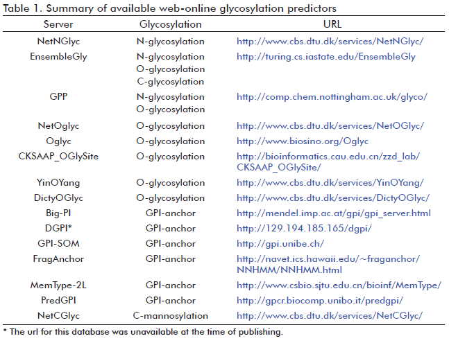
Currently,            three softwares are capable of predicting N-glycosylation; they are            known as NetNGlyc (40), EnsembleGly (41) and GPP (Glycosylation Prediction            Program) (42). EnsembleGly and GPP were recently developed and can be            used not only for N-glycosylation prediction, but also for the prediction            of other glycosylation types (<font color="#FF0000"><a href="/img/revistas/bta/v28n1/t0102111.gif">Table</a></font><a href="/img/revistas/bta/v28n1/t0102111.gif">            1</a>). However, the web-online NetNGlyc server is still the most used            predictor for N-glycosylation (43-45). All prediction methods use machine            learning techniques trained on amino acid sequences. These methods examine            the sequon vicinity to discriminate between possible modified and non-modified            asparagines, since the amino acid composition flanking potential N-glycosylation            sequon is determinant (30). </font></P >       </DIV >       </font></font></font></font></font></font></font></font></font></font></font></font></font></font></font></font></font></font></font></font></font></font></DIV >     <FONT size="+1"><FONT size="+1"><FONT size="+1" color="#FF0000"><FONT size="+1" color="#000000"><FONT size="+1" color="#FF0000"><FONT size="+1" color="#000000"><FONT size="+1" color="#FF0000"><FONT size="+1" color="#000000"><FONT size="+1" color="#FF0000"><FONT size="+1" color="#000000"><FONT size="+1" color="#FF0000"><FONT size="+1" color="#000000"><FONT size="+1" color="#FF0000"><FONT size="+1" color="#000000"><FONT size="+1" color="#FF0000"><FONT size="+1" color="#000000"><FONT size="+1" color="#FF0000"><FONT size="+1" color="#000000"><FONT size="+1" color="#FF0000"><FONT size="+1" color="#000000"><FONT size="+1" color="#FF0000"><FONT size="+1" color="#000000"><FONT color="#800080"><FONT color="#000000">         
<DIV class="Sect"   >        <H4   align="justify" ><font size="3" color="#000000" face="Verdana, Arial, Helvetica, sans-serif"><b>O-GLYCOSYLATION</b></font><font size="2" color="#000000" face="Verdana, Arial, Helvetica, sans-serif">          </font></H4 >       <FONT size="+1">           <P   align="justify" ><font size="2" color="#000000" face="Verdana, Arial, Helvetica, sans-serif">O-linked          glycosylation involves the binding of glycans to hydroxyl side chains          of serine and threonine residues. There is no well defined motif for the          O-glycan acceptor site. The glycans bind to serine and threonine residues          which are usually found in a beta conformation and in close vicinity to          proline residues (46). The O-linked glycosylation occurs in bacteria,          archaea and eukaryotes (47, 48). O-glycosylation is a stepwise process          where one monosaccharide is added at a time, rather than N-glycosylation          where the high-mannose oligosaccharide is transfer en bloc to the target          protein. Examples of O-glycans include: O-N-acetyl-galactosamine (O-GalNAc),          O-N-acetylglucosamine (O-GlcNAc), O-Fucose, O-Glucose, O-Mannose, O-Hexose,          O-Xylose. The most abundant and better characterized O-glycosylation type          is mucin-type glycosylation (49). This reaction is catalyzed by the enzymes          UDP-N-acetyl-D-galactosamine:polypepti-de N-acetylgalactosaminyltransferase          which adds an Nacetyl-galactosamine molecule to serine and threonine residues.          Mucin-type glycans are found on many secreted and membrane-bound mucin          proteins, which are the mucus main components. The function of such proteins          is to protect epithelial surfaces (50, 51). Mucin-type Oglycosilation          occurs in the ER and the Golgi apparatus after N-glycosylation, folding,          and oligomerization (52). </font></P >       <FONT size="+1">           <DIV class="Sect"   >          <H5   align="justify" ><font size="2" color="#000000" face="Verdana, Arial, Helvetica, sans-serif"><B>O-glycosylation            target sequence </b></font></H5 >             <P   align="justify" ><font size="2" color="#000000" face="Verdana, Arial, Helvetica, sans-serif">There            is no clear consensus sequence for O-glycosylation. Some studies have            confirmed a higher frequency of residues like Pro, Ser, Thr and Ala            neighboring mucin-type glycosylated sites (53, 54). Such sites are preferentially            found in coil, turn or linker regions connecting domains. Besides, the            experimentally verified O-glycosylated sites are more surface exposed            than the non-glycosylated ones (55). </font></P >             <P   align="justify" ><font size="2" color="#000000" face="Verdana, Arial, Helvetica, sans-serif"><B>O-glycosylation            prediction methods
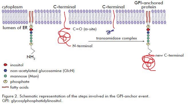
 </b></font></P >             ]]></body>
<body><![CDATA[<P   align="justify" ><font size="2" color="#000000" face="Verdana, Arial, Helvetica, sans-serif">Several            methods for O-glycosylation prediction have been developed (<a href="/img/revistas/bta/v28n1/t0102111.gif"><font color="#0000FF">Table            1</font></a>). Among them, NetOGlyc (55) and Oglyc (56) predictors have            been the most widely used. The former considers primary amino acid sequence,            secondary structure and evolutionary information of both mucintype glycosylated            and non-mucin type glycosylated sites (55). The Oglyc server also predicts            mucin-type O-glycosylation in mammalians, and it is based on a combination            of physical properties of amino acids (56). Recently, three new O-glycosylation            predictors have been available: EnsembleGly (41), CKSAAP_OGlySite (13)            and GPP (42). CKSAAP_OGlySite server only predicts mucin-type O-glycosylation,            like NetOGlyc and Oglyc servers (13). It considers the local conformation            and the short-range interactions of amino acids close to the mucin-type            glycosylated sites. The latest published software, GPP, is based on            pairwise sequence patterns combined with prediction of protein secondary            structure, surface accessibility and hydrophobicity (42). Actually,            GPP is the most accurate program for both N-and O-glycosylation prediction            available (42). </font></P >       </DIV >       </font></font></DIV >     <FONT size="+1"><FONT size="+1">         
<DIV class="Sect"   >        <H4   align="justify" ><font size="3" color="#000000" face="Verdana, Arial, Helvetica, sans-serif"><b>GLYPIATION</b></font><font size="2" color="#000000" face="Verdana, Arial, Helvetica, sans-serif">          </font></H4 >       <FONT size="+1">           <P   align="justify" ><font size="2" color="#000000" face="Verdana, Arial, Helvetica, sans-serif">Glypiation          is ubiquitous in eukaryotes and also possible in a reduced subset of <I>archaea          </I>species (57), but probably absent in other Archaea and Eubacteria          (58). It involves the addition of a GPI molecule to the C-terminal end          of the target protein. The GPI molecule is composed by a phosphatidylinositol          group and a sugar moiety. The sugar moiety comprises a non-acetylated          glucosamine attached to three mannosyl residues and to the phosphatidylinositol          group. A phosphoethanolamine residue connected to the terminal mannose          mediates the binding of the GPI to the C-terminal end of the mature protein          by an amide linkage. Two long-chain fatty acids included in the phosphatidylinositol          group anchor the protein to the cell membrane. Although all GPI molecules          share a common core, some variability have been observed depending on          the organism and the cell type in which they are synthesized (59). Such          differences correspond to substitutions in the oligosaccharidic and lipidic          portions of the GPI residue. Proteins susceptible to glypiation contain          a C-terminal signal sequence which is sufficient for GPI attachment (60).          Glypiation begins with the recognition of the &omega;-site in the C-terminal          protein end embedded in the ER membrane. Then, about 20-30 residues downstream          from the &omega;-site (propeptide) are removed and replaced by the GPI          molecule via an amide bond between the phospho-ethanolamine group and          the new C-terminal residue (&omega;-site). The transamidase complex is          responsible for the above mentioned events that occur during glypiation          (61). After glypiation process, proteins pass through the secretory pathway          in vesicles to Golgi apparatus and finally, most of them are translocated          to the cell membrane (62). Then, glypiated proteins reside attached to          the cell membrane facing the extracellular environment, where they perform          different important functions (61). <font color="#0000FF"><a href="/img/revistas/bta/v28n1/f0202111.gif">Figure</a></font><a href="/img/revistas/bta/v28n1/f0202111.gif">          2</a> schematizes the steps involved in the glypiation process. Glypiated          proteins may operate as enzymes (63), membrane receptors (64), surface          antigens (65) and adhesion molecules (66). However, some GPI-anchored          proteins are released by enzymatic cleavage of their anchor to achieve          other functions (67). Glypiation process is relevant for cell functions          and development. Indeed, mice lacking the GPI synthesis were unable to          grow at embryonic stages (68). </font></P >       <FONT size="+1">           
<DIV class="Sect"   >          <H5   align="justify" ><font size="2" color="#000000" face="Verdana, Arial, Helvetica, sans-serif"><B>GPI-anchor            signal sequence </b></font></H5 >             <P   align="justify" ><font size="2" color="#000000" face="Verdana, Arial, Helvetica, sans-serif">None            consensus sequence characterizes the amino acid at the &omega;-site            (69). Some residues experimentally verified in &omega;-site include:            cysteine, aspartic acid, glycine, asparagine, and serine (69). However,            some generally features can be found in the non-cleaved C-terminal end            of glypiated proteins like: an unfolded linker region comprising about            11 residues (upstream from position &omega;-1), a region comprised by            small amino acids surrounding the cleavage site (from positions &omega;-1            to &omega;+2) which is followed by a moderately polar spacer region            (from positions &omega;+3 to &omega;+9) and finally, a hydrophobic tail            extended from the position &omega;+10 up to the C-terminal end (70).            </font></P >       </DIV >           <DIV class="Sect"   >          <H5   align="justify" ><font size="2" color="#000000" face="Verdana, Arial, Helvetica, sans-serif"><B>GPI-anchor            prediction </b></font></H5 >             <P   align="justify" ><font size="2" color="#000000" face="Verdana, Arial, Helvetica, sans-serif">Various            computational methods have been developed for the prediction of glypiated            proteins (<a href="/img/revistas/bta/v28n1/t0102111.gif"><font color="#0000FF">Table            1</font></a>). Such predictors identify the C-terminal GPI-anchor signals            and most of them also distinguish the &omega;-site. Methods like Big-PI            (70) and DGPI (71) are based on the rules concerning the amino acid            composition around the &omega;-site observed in glypiated proteins.            The Big-PI predictions are divided in relation to life-kingdoms, for            example, for animals, fungi and plants. The performance of these softwares            is better for the already verified glypiated proteins compared to the            non-confirmed ones (72). More recently, the GPI-SOM program was developed            (72). This one achieved better results than Big-PI and DGPI (71). The            improvement of GPI-SOM is related with its power to discriminate false            positives GPI-anchor proteins. The main source of false positive predictions            is the existence of integral membrane proteins having a transmembrane            domain at the C-terminal end. GPI-SOM overcomes this difficulty by the            simulation of mutagenesis experiments at the C-terminus leading to the            identification of key residues that discriminates GPI-anchored proteins            from nonGPI-anchored proteins. This procedure follows an experimental            demonstration that a single point mutation may be enough to convert            a GPI-anchor signal into a transmembrane domain (73). The latest prediction            tools are FragAnchor (74) and PredGPI (75). Both are able to recognize            a higher number of GPI-anchored proteins with a lower rate of false            positive errors, in comparison with other earlier described algorithms.            But, PredGPI outperforms all available prediction methods (75). MemType-2L            is another resource capable to predict not only GPI-anchored protein,            but also other seven types of membrane proteins (76). However, both            FragAnchor and MemType-2L are not able to detect the &omega;-site (74,            76). </font></P >       </DIV >       </font></font></DIV >     <FONT size="+1"><FONT size="+1">         
<DIV class="Sect"   >        <H4   align="justify" ><font size="3" color="#000000" face="Verdana, Arial, Helvetica, sans-serif"><b>C-MANNOSYLATION</b></font><font size="2" color="#000000" face="Verdana, Arial, Helvetica, sans-serif">          </font></H4 >       <FONT size="+1">           <P   align="justify" ><font size="2" color="#000000" face="Verdana, Arial, Helvetica, sans-serif">C-mannosylation          was originally found in human ribonuclease protein (RNase 2) from urine          (77). This post-translational modification involves the attachment of          a mannopyranosyl residue to the C2 atom indole moiety of a tryptophan          residue via a C-C bond (77, 78). The transfer of the mannose residue to          the target protein is catalyzed by the enzyme C-mannosyltransferase (77).          It has been suggested that this reaction probably occurs in the ER since          already folded proteins are poor substrates <I>in vitro </I>(79). Although          C-glycosylation appears to be common in mammalian proteins, it has not          been observed in yeast and bacteria (80). At present, the knowledge related          with the C-mannosylation functions and disorders is still very limited.          However, at least three functions have been recently described. For example,          C-mannosylation is required for an adequate folding of Cys subdomains          contained in two mucin proteins (MUC5AC and MUC5B)(79). Also, C-glycosylation          appears to control the secretion of the puntion-1 protein (81) and it          may be involved in the development of diabetic complications under hyperglycemic          conditions (82). </font></P >       <FONT size="+1">           <DIV class="Sect"   >          <H5   align="justify" ><font size="2" color="#000000" face="Verdana, Arial, Helvetica, sans-serif"><B>C-mannosylation            consensus sequence </b></font></H5 >             ]]></body>
<body><![CDATA[<P   align="justify" ><font size="2" color="#000000" face="Verdana, Arial, Helvetica, sans-serif">C-mannosylation            generally occurs at the first trypto-phan residue (position 0) contained            in the Trp-X-X-Trp sequence motif, where X could be any amino acid (77).However,            other studies revealed that mannosylated tryptophan has also been detected            in other sequences motifs, for example in Trp-X-X-Phe, Trp-X-X-Tyr and            Trp-X-X-Cys (83-85). Since some sequence motifs having another aromatic            residue instead of Trp at the position +3 can also be mannosylated,            the Trp-X-X-Trp pattern seems to be sufficient but not strictly required            for C-mannosylation. Indeed, it was demonstrated that only two-thirds            of known mannosylated sites are found in Trp-X-X-Trp motifs (83). Besides,            small and/or polar residues (Ala, Gly, Ser and Thr) are preferred for            occupying X positions within the sequence motif while Phe and Leu residues            are not well tolerated at the mentioned position (83). Other general            features related with the structure of C-mannosylation motif, have also            been described. For example, modified tryptophan residues instead of            non-modified ones, are partly solvent exposed (83). Besides, a particular            interaction takes place at the Trp-X-X-Trp motif, whe-re both tryptophan            residues interact via an aromatic stacking. It was suggested that this            type of interaction may account for the recognition of the C-mannosyltransferase            enzyme (83). </font></P >       </DIV >           <DIV class="Sect"   >          <H5   align="justify" ><font size="2" color="#000000" face="Verdana, Arial, Helvetica, sans-serif"><B>C-mannosylation            prediction </b></font></H5 >             <P   align="justify" ><font size="2" color="#000000" face="Verdana, Arial, Helvetica, sans-serif">There            is only one computational method reserved for the solely prediction            of C-mannosylation and it is called NetCGlyc (83) (<font color="#FF0000"><a href="/img/revistas/bta/v28n1/t0102111.gif">Table</a></font><a href="/img/revistas/bta/v28n1/t0102111.gif">            1</a>). It predicts not only the typical mannosylated site (Trp-X-X-Trp),            but others like those previously mentioned (Trp-X-X-Phe, Trp-X-X-Tyr            and TrpX-X-Cys). However, better predictions are achieved for the first            case (83). Recently, EnsembleGly could be also used for C-mannosylation            prediction (41) (<font color="#FF0000"><a href="/img/revistas/bta/v28n1/t0102111.gif">Table</a></font><a href="/img/revistas/bta/v28n1/t0102111.gif">            1</a>). </font></P >       </DIV >       </font></font></DIV >     <FONT size="+1"><FONT size="+1"><FONT color="#800080"><FONT color="#000000"><FONT color="#800080"><FONT color="#000000">         
<DIV class="Sect"   >        <H4   align="justify" ><font size="3" color="#000000" face="Verdana, Arial, Helvetica, sans-serif"><b>DATABASES</b></font><font size="2" color="#000000" face="Verdana, Arial, Helvetica, sans-serif">          </font></H4 >       <FONT size="+1">           <P   align="justify" ><font size="2" color="#000000" face="Verdana, Arial, Helvetica, sans-serif">
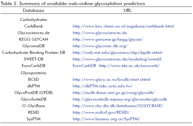
Undoubtedly,          the development of accuracy glycosylation prediction methods requires          the existence of databases with experimentally verified glycosylated sites.          Usually, the data is extracted from O-GlycBase database (86), but in some          cases, the Swiss-Prot database has been also used (86, 87). O-GlycBase          contains proteins having at least one experimentally verified O-or C-glycosylation          site. Glycosylation data may also be found in databases including different          types of post-translational modifications, like dbPTM (88), SysPTM (89),          RESID (90) and others (<font color="#FF0000"><a href="/img/revistas/bta/v28n1/t0202111.gif">Table</a></font><a href="/img/revistas/bta/v28n1/">          2</a>). Besides glycoprotein databases, carbohydrate molecules databases          also exists. For example, the major available databases of complex carbohydrates          are: Complex Carbohydrate Structure Database (CCSD or CarbBank) (91),          Glycosciences.de (92), KEGG GLYCAN (93), GlycomeDB (94), Carbohydrate          DB from the Consortium Functional Genomic (95) and others (<font color="#FF0000"><a href="/img/revistas/bta/v28n1/t0202111.gif">Table</a></font><a href="/img/revistas/bta/v28n1/t0202111.gif">          2</a>). Other available databases include the carbohydrate tertiary structures          (<I>e.g.</I>, 3D Disaccharides (96), GDB:Structures (97) and GlycoMapsDB          (98)). Others like GlyTorsionDB, GlySeqDB and GlyVicinityDB are integrated          in the Carbohydrate Structure Suite (99). Each database comprises the          carbohydrate torsion angles, the glycoprotein sequences and the carbohydrate-protein          interactions, respectively (99). </font></P >       </font></DIV >     <FONT size="+1"><FONT size="+1"><FONT color="#800080"><FONT color="#000000">         
<DIV class="Sect"   >        <H4   align="justify" ><font size="3" color="#000000" face="Verdana, Arial, Helvetica, sans-serif"><b>CONCLUDING          REMARKS</b></font><font size="2" color="#000000" face="Verdana, Arial, Helvetica, sans-serif">          </font></H4 >       <FONT size="+1">           <P   align="justify" ><font size="2" color="#000000" face="Verdana, Arial, Helvetica, sans-serif">Currently,          about 15 web-online softwares for the prediction of all glycosylation          types are available. The possibility to perform prediction analysis using          at least two or three different methods in parallel now exists. However,          in our opinion, with upcoming prediction methods, a critical comparative          study would be required to define the most accurate one. The combination          of such prediction methods with other non-carbohydrate tools facilities          the characterization and rational modification of the native protein glycosylation          pattern. </font></P >       </font></DIV >     <FONT size="+1"><FONT size="+1">         <DIV class="Sect"   >        <H4   align="justify" ><font size="3" color="#000000" face="Verdana, Arial, Helvetica, sans-serif"><b>ACKNOWLEDGMENTS</b></font><font size="2" color="#000000" face="Verdana, Arial, Helvetica, sans-serif">          </font></H4 >       <FONT size="+1">           <P   align="justify" ><font size="2" color="#000000" face="Verdana, Arial, Helvetica, sans-serif">We          thank Dr. Jose Alberto Cremata, from the CIGB Carbohydrate Department,          for his useful comments and suggestions on the manuscript. </font></P >       </font></DIV >     <FONT size="+1"><FONT size="+1">         <DIV class="Sect"   >        <H4   align="justify" ><font size="3" color="#000000" face="Verdana, Arial, Helvetica, sans-serif"><b>REFERENCES</b></font><font size="2" color="#000000" face="Verdana, Arial, Helvetica, sans-serif">          </font></H4 >           ]]></body>
<body><![CDATA[<!-- ref --><p><font size="2" face="Verdana, Arial, Helvetica, sans-serif">1. Ramamurthy          B, Hook P, Larsson L. An overview of carbohydrate-protein interactions          with specific reference to myosin and ageing. Acta Physiol Scand. 1999;          167:327-9.    <br>         </font></p>           <!-- ref --><p><font size="2" face="Verdana, Arial, Helvetica, sans-serif">2. Mitra          N, Sinha S, Ramya TN, Surolia A. N-linked oligosaccharides as outfitters          for glycoprotein folding, form and function. Trends Biochem Sci. 2006;31:156-63.    <br>         </font></p>           <!-- ref --><p><font size="2" face="Verdana, Arial, Helvetica, sans-serif">3. Mbonye          UR, Yuan C, Harris CE, Sidhu RS, Song I, Arakawa T, <i>et al</i>. Two          distinct pathways for cyclooxygenase-2 protein degradation. J Biol Chem.          2008;283:8611-23.    <br>         </font></p>           <!-- ref --><p><font size="2" face="Verdana, Arial, Helvetica, sans-serif">4. Sola RJ,          Griebenow K. Effects of glycosylation on the stability of protein pharmaceuticals.          J Pharm Sci. 2009;98:1223-45.    <br>         </font></p>           <!-- ref --><p><font size="2" face="Verdana, Arial, Helvetica, sans-serif">5. Specks          U, Fass DN, Finkielman JD, Hummel AM, Viss MA, Litwiller RD, <i>et al</i>.          Functional significance of Asn-linked glycosylation of proteinase 3 for          enzymatic activity, processing, targeting, and recognition by anti-neutrophil          cytoplasmic antibodies. J Biochem. 2007;141:101-12.    <br>         </font></p>           ]]></body>
<body><![CDATA[<!-- ref --><p><font size="2" face="Verdana, Arial, Helvetica, sans-serif">6. Corthay          A, Backlund J, Broddefalk J, Michaelsson E, Goldschmidt TJ, Kihlberg J,          <i>et al</i>. Epitope glycosylation plays a critical role for T cell recognition          of type II collagen in collagen-induced arthritis. Eur J Immunol. 1998;28:2580-90.    <br>         </font></p>           <!-- ref --><p><font size="2" face="Verdana, Arial, Helvetica, sans-serif">7. Vagin          O, Kraut JA, Sachs G. Role of N-glycosylation in trafficking of apical          membrane proteins in epithelia. Am J Physiol Renal Physiol. 2009;296:F459-F469.    <br>         </font></p>           <!-- ref --><p><font size="2" face="Verdana, Arial, Helvetica, sans-serif">8. Couldrey          C, Green JE. Metastases: the glycan connection. Breast Cancer Res. 2000;2:321-3.    <br>         </font></p>           <!-- ref --><p><font size="2" face="Verdana, Arial, Helvetica, sans-serif">9. Corthay          A, Backlund J, Holmdahl R. Role of glycopeptide-specific T cells in collagen-induced          arthritis: an example how post-translational modification of proteins          may be involved in autoimmune disease. Ann Med. 2001;33:456-65.    <br>         </font></p>           <!-- ref --><p><font size="2" face="Verdana, Arial, Helvetica, sans-serif">10. Freeze          HH. Update and perspectives on congenital disorders of glycosylation.          Glycobiology. 2001;11:129R-43R.    <br>         </font></p>           ]]></body>
<body><![CDATA[<p><font size="2" face="Verdana, Arial, Helvetica, sans-serif">11. Varki          A, Cummings RD, Esko JD, <i>et al</i>., editors. Essentials of glycobiology.          2nd ed. New York: Cold Spring Harbor Laboratory press; 2008.    <br>         </font></p>           <!-- ref --><p><font size="2" face="Verdana, Arial, Helvetica, sans-serif">12. Zaia          J. Mass spectrometry and the emerging field of glycomics. Chem Biol. 2008;15:881-92.    <br>         </font></p>           <!-- ref --><p><font size="2" face="Verdana, Arial, Helvetica, sans-serif">13. Chen          YZ, Tang YR, Sheng ZY, Zhang Z. Prediction of mucin-type O-glycosylation          sites in mammalian proteins using the composition of k-spaced amino acid          pairs. BMC Bioinformatics. 2008;9:101.    <br>         </font></p>           <!-- ref --><p><font size="2" face="Verdana, Arial, Helvetica, sans-serif">14. Aoki-Kinoshita          KF. An introduction to bioinformatics for glycomics research. PLoS Comput          Biol. 2008;4:e1000075.    <br>         </font></p>           <!-- ref --><p><font size="2" face="Verdana, Arial, Helvetica, sans-serif">15. der Lieth          CW, Bohne-Lang A, Lohmann KK, Frank M. Bioinformatics for glycomics: status,          methods, requirements and perspectives. Brief Bioinform. 2004;5:164-78.    <br>         </font></p>           ]]></body>
<body><![CDATA[<!-- ref --><p><font size="2" face="Verdana, Arial, Helvetica, sans-serif">16. Frank          M, Schloissnig S. Bioinformatics and molecular modeling in glycobiology.          Cell Mol Life Sci. 2010;67:2749-72.    <br>         </font></p>           <p><font size="2" face="Verdana, Arial, Helvetica, sans-serif">17. Bause          E, Legler G. The role of the hydroxy amino acid in the triplet sequence          Asn-Xaa-Thr(Ser) for the N-glycosylation step during glycoprotein biosynthesis.          Biochem J. 1981;195:639-44.    <br>         </font></p>           <!-- ref --><p><font size="2" face="Verdana, Arial, Helvetica, sans-serif">18. Schaffer          C, Graninger M, Messner P. Prokaryotic glycosylation. Proteomics. 2001;1:248-61.    <br>         </font></p>           <!-- ref --><p><font size="2" face="Verdana, Arial, Helvetica, sans-serif">19. Weerapana          E, Imperiali B. Asparagine-linked protein glycosylation: from eukaryotic          to prokaryotic systems. Glycobiology. 2006;16:91R-101R.    <br>         </font></p>           <!-- ref --><p><font size="2" face="Verdana, Arial, Helvetica, sans-serif">20. Yurist-Doutsch          S, Chaban B, VanDyke DJ, Jarrell KF, Eichler J. Sweet to the extreme:          protein glycosylation in Archaea. Mol Microbiol. 2008;68:1079-84.    <br>         </font></p>           ]]></body>
<body><![CDATA[<!-- ref --><p><font size="2" face="Verdana, Arial, Helvetica, sans-serif">21. Helenius          A, Aebi M. Intracellular functions of N-linked glycans. Science. 2001;291:2364-9.    <br>         </font></p>           <!-- ref --><p><font size="2" face="Verdana, Arial, Helvetica, sans-serif">22. Helenius          A, Aebi M. Roles of N-linked glycans in the endoplasmic reticulum. Annu          Rev Biochem. 2004;73:1019-49.    <br>         </font></p>           <!-- ref --><p><font size="2" face="Verdana, Arial, Helvetica, sans-serif">23. Jones          J, Krag SS, Betenbaugh MJ. Controlling N-linked glycan site occupancy.          Biochim Biophys Acta. 2005;1726:121-37.    <br>         </font></p>           <!-- ref --><p><font size="2" face="Verdana, Arial, Helvetica, sans-serif">24. Munro          S. What can yeast tell us about N-linked glycosylation in the Golgi apparatus?          FEBS Lett. 2001;498:223-7.    <br>         </font></p>           <!-- ref --><p><font size="2" face="Verdana, Arial, Helvetica, sans-serif">25. Roitsch          T, Lehle L. Structural requirements for protein N-glycosylation. Influence          of acceptor peptides on cotranslational glycosylation of yeast invertase          and site-directed mutagenesis around a sequon sequence. Eur J Biochem.          1989;181:525-9.    <br>         </font></p>           ]]></body>
<body><![CDATA[<!-- ref --><p><font size="2" face="Verdana, Arial, Helvetica, sans-serif">26. Gavel          Y, von Heijne G. Sequence differences between glycosylated and non-glycosylated          Asn-X-Thr/Ser acceptor sites: implications for protein engineering. Protein          Eng. 1990;3:433-42.    <br>         </font></p>           <!-- ref --><p><font size="2" face="Verdana, Arial, Helvetica, sans-serif">27. Shakin-Eshleman          SH, Spitalnik SL, Kasturi L. The amino acid at the X position of an Asn-X-Ser          sequon is an important determinant of N-linked core-glycosylation efficiency.          J Biol Chem. 1996;271:6363-6.    <br>         </font></p>           <!-- ref --><p><font size="2" face="Verdana, Arial, Helvetica, sans-serif">28. Kasturi          L, Chen H, Shakin-Eshleman SH. Regulation of N-linked core glycosylation:          use of a site-directed mutagenesis approach to identify Asn-Xaa-Ser/Thr          sequons that are poor oligosaccharide acceptors. Biochem J. 1997;323 (          Pt 2):415-9.    <br>         </font></p>           <!-- ref --><p><font size="2" face="Verdana, Arial, Helvetica, sans-serif">29. Christlet          TH, Biswas M, Veluraja K. A database analysis of potential glycosylating          Asn-X-Ser/Thr consensus sequences. Acta Crystallogr D Biol Crystallogr.          1999;55:1414-20.    <br>         </font></p>           <!-- ref --><p><font size="2" face="Verdana, Arial, Helvetica, sans-serif">30. Petrescu          AJ, Milac AL, Petrescu SM, Dwek RA, Wormald MR. Statistical analysis of          the protein environment of N-glycosylation sites: implications for occupancy,          structure, and folding. Glycobiology. 2004; 14:103-14.    <br>         </font></p>           ]]></body>
<body><![CDATA[<!-- ref --><p><font size="2" face="Verdana, Arial, Helvetica, sans-serif">31. Ben Dor          S, Esterman N, Rubin E, Sharon N. Biases and complex patterns in the residues          flanking protein N-glycosylation sites. Glycobiology. 2004;14:95-101.    <br>         </font></p>           <!-- ref --><p><font size="2" face="Verdana, Arial, Helvetica, sans-serif">32. Kowarik          M, Young NM, Numao S, Schulz BL, Hug I, Callewaert N, <i>et al</i>. Definition          of the bacterial N-glycosylation site consensus sequence. EMBO J. 2006;          25:1957-66.    <br>         </font></p>           <!-- ref --><p><font size="2" face="Verdana, Arial, Helvetica, sans-serif">33. Abu-Qarn          M, Eichler J. An analysis of amino acid sequences surrounding archaeal          glycoprotein sequons. Archaea. 2007;2:73-81.    <br>         </font></p>           <!-- ref --><p><font size="2" face="Verdana, Arial, Helvetica, sans-serif">34. Nilsson          I, von Heijne G. Glycosylation efficiency of Asn-Xaa-Thr sequons depends          both on the distance from the C terminus and on the presence of a downstream          transmembrane segment. J Biol Chem. 2000;275:17338-43.    <br>         </font></p>           <!-- ref --><p><font size="2" face="Verdana, Arial, Helvetica, sans-serif">35. Walmsley          AR, Hooper NM. Glycosylation efficiency of Asn-Xaa-Thr sequons is independent          of distance from the C-terminus in membrane dipeptidase. Glycobiology.          2003;13:641-6.    <br>         </font></p>           ]]></body>
<body><![CDATA[<!-- ref --><p><font size="2" face="Verdana, Arial, Helvetica, sans-serif">36. Bause          E. Structural requirements of N-glycosylation of proteins. Studies with          proline peptides as conformational probes. Biochem J. 1983;209:331-6.    <br>         </font></p>           <!-- ref --><p><font size="2" face="Verdana, Arial, Helvetica, sans-serif">37. Bause          E, Hettkamp H, Legler G. Conformational aspects of N-glycosylation of          proteins. Studies with linear and cyclic peptides as probes. Biochem J.          1982;203:761-8.    <br>         </font></p>           <!-- ref --><p><font size="2" face="Verdana, Arial, Helvetica, sans-serif">38. Yan A,          Lennarz WJ. Unraveling the mechanism of protein N-glycosylation. J Biol          Chem. 2005;280:3121-4.    <br>         </font></p>           <!-- ref --><p><font size="2" face="Verdana, Arial, Helvetica, sans-serif">39. Reddy          A, Gibbs BS, Liu YL, Coward JK, Changchien LM, Maley F. Glycosylation          of the overlapping sequons in yeast external invertase: effect of amino          acid variation on site selectivity <i>in vivo</i> and <i>in vitro</i>.          Glycobiology. 1999;9:547-55.    <br>         </font></p>           <!-- ref --><p><font size="2" face="Verdana, Arial, Helvetica, sans-serif">40. NetNGlyc          1.0 Server (Internet; updated 2007 Mar 09; cited 2011 Jan 17). Available          from: <a href="http://www.cbs.dtu.dk/services/NetNGlyc/">http://www.cbs.dtu.dk/services/NetNGlyc/</a>.    <br>         </font></p>           ]]></body>
<body><![CDATA[<!-- ref --><p><font size="2" face="Verdana, Arial, Helvetica, sans-serif">41. Caragea          C, Sinapov J, Silvescu A, Dobbs D, Honavar V. Glycosylation site prediction          using ensembles of Support Vector Machine classifiers. BMC Bioinformatics.          2007;8:438.    <br>         </font></p>           <!-- ref --><p><font size="2" face="Verdana, Arial, Helvetica, sans-serif">42. Hamby          SE, Hirst JD. Prediction of glycosylation sites using random forests.          BMC Bioinformatics. 2008;9:500.    <br>         </font></p>           <!-- ref --><p><font size="2" face="Verdana, Arial, Helvetica, sans-serif">43. Liu Y,          Nguyen A, Wolfert RL, Zhuo S. Enhancing the secretion of recombinant proteins          by engineering N-glycosylation sites. Biotechnol Prog. 2009;25:1468-75.    <br>         </font></p>           <!-- ref --><p><font size="2" face="Verdana, Arial, Helvetica, sans-serif">44. Radoslavov          G, Jordanova R, Teofanova D, Georgieva K, Hristov P, Salomone-Stagni M,          <i>et al</i>. A novel secretory poly-cysteine and histidine-tailed metalloprotein          (Ts-PCHTP) from Trichinella spiralis (Nematoda). PLoS One. 2010;5:e13343.    <br>         </font></p>           <!-- ref --><p><font size="2" face="Verdana, Arial, Helvetica, sans-serif">45. Lu C,          Walker WH, Sun J, Weisz OA, Gibbs RB, Witchel SF, <i>et al</i>. Insulin-like          peptide 6: characterization of secretory status and posttranslational          modifications. Endocrinology. 2006;147:5611-23.    <br>         </font></p>           ]]></body>
<body><![CDATA[<!-- ref --><p><font size="2" face="Verdana, Arial, Helvetica, sans-serif">46. Gupta          R, Brunak S. Prediction of glycosylation across the human proteome and          the correlation to protein function. Pac Symp Biocomput. 2002;7:310-22.    <br>         </font></p>           <!-- ref --><p><font size="2" face="Verdana, Arial, Helvetica, sans-serif">47. Nothaft          H, Szymanski CM. Protein glycosylation in bacteria: sweeter than ever.          Nat Rev Microbiol. 2010;8:765-78.    <br>         </font></p>           <!-- ref --><p><font size="2" face="Verdana, Arial, Helvetica, sans-serif">48. Gentzsch          M, Tanner W. Protein-O-glycosylation in yeast: protein-specific mannosyltransferases.          Glycobiology. 1997;7:481-6.    <br>         </font></p>           <!-- ref --><p><font size="2" face="Verdana, Arial, Helvetica, sans-serif">49. Hanisch          FG. O-glycosylation of the mucin type. Biol Chem. 2001;382:143-9.    <br>         </font></p>           <!-- ref --><p><font size="2" face="Verdana, Arial, Helvetica, sans-serif">50. Carraway          KL, Hull SR. Cell surface mucin-type glycoproteins and mucin-like domains.          Glycobiology. 1991;1:131-8.    <br>         </font></p>           ]]></body>
<body><![CDATA[<!-- ref --><p><font size="2" face="Verdana, Arial, Helvetica, sans-serif">51. Strous          GJ, Dekker J. Mucin-type gly-coproteins. Crit Rev Biochem Mol Biol. 1992;27:57-92.    <br>         </font></p>           <!-- ref --><p><font size="2" face="Verdana, Arial, Helvetica, sans-serif">52. Asker          N, Baeckstrom D, Axelsson MA, Carlstedt I, Hansson GC. The human MUC2          mucin apoprotein appears to dimerize before O-glycosylation and shares          epitopes with the 'insoluble' mucin of rat small intestine. Biochem J.          1995;308(Pt 3):873-80.    <br>         </font></p>           <!-- ref --><p><font size="2" face="Verdana, Arial, Helvetica, sans-serif">53. Thanka          Christlet TH, Veluraja K. Database analysis of O-glycosylation sites in          proteins. Biophys J. 2001;80:952-60.    <br>         </font></p>           <!-- ref --><p><font size="2" face="Verdana, Arial, Helvetica, sans-serif">54. Wilson          IB, Gavel Y, von Heijne G. Amino acid distributions around O-linked glycosylation          sites. Biochem J. 1991;275(Pt 2):529-34.    <br>         </font></p>           <!-- ref --><p><font size="2" face="Verdana, Arial, Helvetica, sans-serif">55. Julenius          K, Molgaard A, Gupta R, Brunak S. Prediction, conservation analysis, and          structural characterization of mammalian mucin-type O-glycosylation sites.          Glycobiology. 2005;15:153-64.    <br>         </font></p>           ]]></body>
<body><![CDATA[<!-- ref --><p><font size="2" face="Verdana, Arial, Helvetica, sans-serif">56. Li S,          Liu B, Zeng R, Cai Y, Li Y. Predicting O-glycosylation sites in mammalian          proteins by using SVMs. Comput Biol Chem. 2006;30:203-8.    <br>         </font></p>           <!-- ref --><p><font size="2" face="Verdana, Arial, Helvetica, sans-serif">57. Kobayashi          T, Nishizaki R, Ikezawa H. The presence of GPI-linked protein(s) in an          archaeobacterium, Sulfolobus acidocaldarius, closely related to eukaryotes.          Biochim Biophys Acta. 1997;1334:1-4.    <br>         </font></p>           <!-- ref --><p><font size="2" face="Verdana, Arial, Helvetica, sans-serif">58. Eisenhaber          B, Bork P, Eisenhaber F. Post-translational GPI lipid anchor modification          of proteins in kingdoms of life: analysis of protein sequence data from          complete genomes. Protein Eng. 2001;14:17-25.    <br>         </font></p>           <!-- ref --><p><font size="2" face="Verdana, Arial, Helvetica, sans-serif">59. Menon          AK. Structural analysis of glycosylphosphatidylinositol anchors. Methods          Enzymol. 1994;230:418-42.    <br>         </font></p>           <!-- ref --><p><font size="2" face="Verdana, Arial, Helvetica, sans-serif">60. Caras          IW, Weddell GN, Davitz MA, Nussenzweig V, Martin DW Jr. Signal for attachment          of a phospholipid membrane anchor in decay accelerating factor. Science.          1987;238:1280-3.    <br>         </font></p>           ]]></body>
<body><![CDATA[<!-- ref --><p><font size="2" face="Verdana, Arial, Helvetica, sans-serif">61. Udenfriend          S, Kodukula K. How glycosylphosphatidylinositol-anchored membrane proteins          are made. Annu Rev Biochem. 1995;64:563-91.    <br>         </font></p>           <!-- ref --><p><font size="2" face="Verdana, Arial, Helvetica, sans-serif">62. Chatterjee          S, Mayor S. The GPI-anchor and protein sorting. Cell Mol Life Sci. 2001;58:1969-87.    <br>         </font></p>           <!-- ref --><p><font size="2" face="Verdana, Arial, Helvetica, sans-serif">63. Koelsch          R, Gottwald S, Lasch J. Release of GPI-anchored membrane aminopeptidase          P by enzymes and detergents has some peculiarities. Biochim Biophys Acta.          1994;1190:170-2.    <br>         </font></p>           <!-- ref --><p><font size="2" face="Verdana, Arial, Helvetica, sans-serif">64. Bergelson          JM, Chan M, Solomon KR, St John NF, Lin H, Finberg RW. Decay-accelerating          factor (CD55), a glycosylphosphatidylinositol-anchored complement regulatory          protein, is a receptor for several echoviruses. Proc Natl Acad Sci USA.          1994;91:6245-8.    <br>         </font></p>           <!-- ref --><p><font size="2" face="Verdana, Arial, Helvetica, sans-serif">65. Chan          CH, Stanners CP. Recent advances in the tumour biology of the GPI-anchored          carcinoembryonic antigen family members CEACAM5 and CEACAM6. Curr Oncol.          2007;14:70-3.    <br>         </font></p>           ]]></body>
<body><![CDATA[<!-- ref --><p><font size="2" face="Verdana, Arial, Helvetica, sans-serif">66. Cervello          M, Matranga V, Durbec P, Rougon G, Gomez S. The GPI-anchored adhesion          molecule F3 induces tyrosine phosphorylation: involvement of the FNIII          repeats. J Cell Sci. 1996;109(Pt 3):699-704.    <br>         </font></p>           <!-- ref --><p><font size="2" face="Verdana, Arial, Helvetica, sans-serif">67. Frieman          MB, Cormack BP. Multiple sequence signals determine the distribution of          glycosylphosphatidylinositol proteins between the plasma membrane and          cell wall in Saccharomyces cerevisiae. Microbiology. 2004;150:3105-14.    <br>         </font></p>           <!-- ref --><p><font size="2" face="Verdana, Arial, Helvetica, sans-serif">68. Nozaki          M, Ohishi K, Yamada N, Kinoshita T, Nagy A, Takeda J. Developmental abnormalities          of glycosylphosphatidylinositol-anchor-deficient embryos revealed by Cre/loxP          system. Lab Invest. 1999;79:293-9.    <br>         </font></p>           <!-- ref --><p><font size="2" face="Verdana, Arial, Helvetica, sans-serif">69. Orlean          P, Menon AK. Thematic review series: lipid posttranslational modifications.          GPI anchoring of protein in yeast and mammalian cells, or: how we learned          to stop worrying and love glycophospholipids. J Lipid Res. 2007;48:993-1011.    <br>         </font></p>           <!-- ref --><p><font size="2" face="Verdana, Arial, Helvetica, sans-serif">70. Eisenhaber          B, Bork P, Eisenhaber F. Prediction of potential GPI-modification sites          in proprotein sequences. J Mol Biol. 1999;292:741-58.    <br>         </font></p>           ]]></body>
<body><![CDATA[<!-- ref --><p><font size="2" face="Verdana, Arial, Helvetica, sans-serif">71. Kronegg          J, Buloz D. Detection/prediction of GPI cleavage site (GPI-anchor) in          a protein (DGPI). 1999. Available from: <a href="http://129.194.185.165/dgpi/index_en.html">http://129.194.185.165/dgpi/index_en.html</a>    <br>       </font></p>           <!-- ref --><p><font size="2" face="Verdana, Arial, Helvetica, sans-serif">72. Fankhauser          N, Maser P. Identification of GPI anchor attachment signals by a Kohonen          self-organizing map. Bioinformatics. 2005;21:1846-52.    <br>         </font></p>           <!-- ref --><p><font size="2" face="Verdana, Arial, Helvetica, sans-serif">73. Dalley          JA, Bulleid NJ. The endoplasmic reticulum (ER) translocon can differentiate          between hydrophobic sequences allowing signals for glycosylphosphatidylinositol          anchor addition to be fully translocated into the ER lumen. J Biol Chem.          2003;278:51749-57.    <br>         </font></p>           <!-- ref --><p><font size="2" face="Verdana, Arial, Helvetica, sans-serif">74. Poisson          G, Chauve C, Chen X, Bergeron A. FragAnchor: a large-scale predictor of          glycosylphosphatidylinositol anchors in eukaryote protein sequences by          qualitative scoring. Genomics Proteomics Bioinformatics. 2007;5:121-30.    <br>         </font></p>           <!-- ref --><p><font size="2" face="Verdana, Arial, Helvetica, sans-serif">75. Pierleoni          A, Martelli PL, Casadio R. PredGPI: a GPI-anchor predictor. BMC Bioinformatics.          2008;9:392.    <br>         </font></p>           ]]></body>
<body><![CDATA[<!-- ref --><p><font size="2" face="Verdana, Arial, Helvetica, sans-serif">76. Chou          KC, Shen HB. MemType-2L: a web server for predicting membrane proteins          and their types by incorporating evolution information through Pse-PSSM.          Biochem Biophys Res Commun. 2007;360:339-45.    <br>         </font></p>           <!-- ref --><p><font size="2" face="Verdana, Arial, Helvetica, sans-serif">77. Hofsteenge          J, Muller DR, de Beer T, Loffler A, Richter WJ, Vliegenthart JF. New type          of linkage between a carbohydrate and a protein: C-glycosylation of a          specific tryptophan residue in human RNase Us. Biochemistry. 1994;33:13524-30.    <br>         </font></p>           <!-- ref --><p><font size="2" face="Verdana, Arial, Helvetica, sans-serif">78. de Beer          T, Vliegenthart JF, Loffler A, Hofsteenge J. The hexopyranosyl residue          that is C-glycosidically linked to the side chain of tryptophan-7 in human          RNase Us is alpha-mannopyranose. Biochemistry. 1995;34:11785-9.    <br>         </font></p>           <!-- ref --><p><font size="2" face="Verdana, Arial, Helvetica, sans-serif">79. Perez-Vilar          J, Randell SH, Boucher RC. C-Mannosylation of MUC5AC and MUC5B Cys subdomains.          Glycobiology. 2004;14:325-37.    <br>         </font></p>           <!-- ref --><p><font size="2" face="Verdana, Arial, Helvetica, sans-serif">80. Zanetta          JP, Pons A, Richet C, Huet G, Timmerman P, Leroy Y, <i>et al</i>. Quantitative          gas chromatography/mass spectrometry determination of C-mannosylation          of tryptophan residues in glycoproteins. Anal Biochem. 2004;329:199-206.    <br>         </font></p>           ]]></body>
<body><![CDATA[<!-- ref --><p><font size="2" face="Verdana, Arial, Helvetica, sans-serif">81. Wang          LW, Leonhard-Melief C, Haltiwanger RS, Apte SS. Post-translational modification          of thrombospondin type-1 repeats in ADAMTS-like 1/punctin-1 by C-mannosylation          of tryp-tophan. J Biol Chem. 2009;284:30004-15.    <br>         </font></p>           <!-- ref --><p><font size="2" face="Verdana, Arial, Helvetica, sans-serif">82. Ihara          Y, Manabe S, Kanda M, Kawano H, Nakayama T, Sekine I, <i>et al</i>. Increased          expression of protein C-mannosylation in the aortic vessels of diabetic          Zucker rats. Glycobiology. 2005;15:383-92.    <br>         </font></p>           <!-- ref --><p><font size="2" face="Verdana, Arial, Helvetica, sans-serif">83. Julenius          K. NetCGlyc 1.0: prediction of mammalian C-mannosylation sites. Glycobiology.          2007;17:868-76.    <br>         </font></p>           <!-- ref --><p><font size="2" face="Verdana, Arial, Helvetica, sans-serif">84. Krieg          J, Hartmann S, Vicentini A, Glasner W, Hess D, Hofsteenge J. Recognition          signal for C-mannosylation of Trp-7 in RNase 2 consists of sequence Trp-x-x-Trp.          Mol Biol Cell. 1998;9:301-9.    <br>         </font></p>           <!-- ref --><p><font size="2" face="Verdana, Arial, Helvetica, sans-serif">85. Hofsteenge          J, Blommers M, Hess D, Furmanek A, Miroshnichenko O. The four terminal          components of the complement system are C-mannosylated on multiple tryptophan          residues. J Biol Chem. 1999;274:32786-94.    <br>         </font></p>           ]]></body>
<body><![CDATA[<!-- ref --><p><font size="2" face="Verdana, Arial, Helvetica, sans-serif">86. Gupta          R, Birch H, Rapacki K, Brunak S, Hansen JE. O-GLYCBASE version 4.0: a          revised database of O-glycosylated proteins. Nucleic Acids Res. 1999;27:370-2.    <br>         </font></p>           <!-- ref --><p><font size="2" face="Verdana, Arial, Helvetica, sans-serif">87. Boeckmann          B, Bairoch A, Apweiler R, Blatter MC, Estreicher A, Gasteiger E, <i>et          al</i>. The SWISS-PROT protein knowledgebase and its supplement TrEMBL          in 2003. Nucleic Acids Res. 2003;31:365-70.    <br>         </font></p>           <!-- ref --><p><font size="2" face="Verdana, Arial, Helvetica, sans-serif">88. Lee TY,          Hsu JB, Chang WC, Wang TY, Hsu PC, Huang HD. A comprehensive resource          for integrating and displaying protein post-translational modifications.          BMC Res Notes. 2009;2:111.    <br>         </font></p>           <!-- ref --><p><font size="2" face="Verdana, Arial, Helvetica, sans-serif">89. Li H,          Xing X, Ding G, Li Q, Wang C, Xie L, <i>et al</i>. SysPTM: a systematic          resource for proteomic research on post-translational modifications. Mol          Cell Proteomics. 2009;8:1839-49.    <br>         </font></p>           <!-- ref --><p><font size="2" face="Verdana, Arial, Helvetica, sans-serif">90. Garavelli          JS. The RESID Database of Protein Modifications as a resource and annotation          tool. Proteomics. 2004;4:1527-33.    <br>         </font></p>           ]]></body>
<body><![CDATA[<!-- ref --><p><font size="2" face="Verdana, Arial, Helvetica, sans-serif">91. Doubet          S, Bock K, Smith D, Darvill A, Albersheim P. The complex carbohydrate          structure database. Trends Biochem Sci. 1989;14:475-7.    <br>         </font></p>           <!-- ref --><p><font size="2" face="Verdana, Arial, Helvetica, sans-serif">92. Lutteke          T, Bohne-Lang A, Loss A, Goetz T, Frank M, der Lieth CW. GLYCOSCIENCES.de:          an Internet portal to support glycomics and glycobiology research. Glycobiology.          2006;16:71R-81R.    <br>         </font></p>           <!-- ref --><p><font size="2" face="Verdana, Arial, Helvetica, sans-serif">93. Hashimoto          K, Goto S, Kawano S, Aoki-Kinoshita KF, Ueda N, Hamajima M, <i>et al</i>.          KEGG as a glycome informatics resource. Glycobiology. 2006;16:63R-70R.    <br>         </font></p>           <!-- ref --><p><font size="2" face="Verdana, Arial, Helvetica, sans-serif">94. Ranzinger          R, Herget S, Wetter T, der Lieth CW. GlycomeDB - integration of open-access          carbohydrate structure databases. BMC Bioinformatics. 2008;9:384.    <br>         </font></p>           <!-- ref --><p><font size="2" face="Verdana, Arial, Helvetica, sans-serif">95. Consortium          for Functional Glycomics [Internet]. Consortium for Functional Glycomics          funded by NIGMS [updated 2007 Apr 05; cited 2011 Jan 17]. Available from:          <a href="http://www.functionalglycomics.org/static/consortium/consortium.shtml">http://www.functionalglycomics.org/static/consortium/consortium.shtml</a>    <br>         </font></p>           ]]></body>
<body><![CDATA[<!-- ref --><p><font size="2" face="Verdana, Arial, Helvetica, sans-serif">96. Imberty          A, Delage MM, Bourne Y, Cambillau C, Perez S. Data bank of three-dimensional          structures of disaccharides: Part II, N-acetyllactosaminic type N-glycans.          Comparison with the crystal structure of a biantennary octasaccharide.          Glycoconj J. 1991;8:456-83.    <br>         </font></p>           <!-- ref --><p><font size="2" face="Verdana, Arial, Helvetica, sans-serif">97. Lutteke          T, der Lieth CW. pdb-care (PDB carbohydrate residue check): a program          to support annotation of complex carbohydrate structures in PDB files.          BMC Bioinformatics. 2004;5:69.    <br>         </font></p>           <!-- ref --><p><font size="2" face="Verdana, Arial, Helvetica, sans-serif">98. Frank          M, Lutteke T,der Lieth CW. GlycoMapsDB: a database of the accessible conformational          space of glycosidic linkages. Nucleic Acids Res. 2007;35:287-90.    <br>         </font></p>           <!-- ref --><p><font size="2" face="Verdana, Arial, Helvetica, sans-serif">99. Lutteke          T, Frank M, der Lieth CW. Carbohydrate Structure Suite (CSS): analysis          of carbohydrate 3D structures derived from the PDB. Nucleic Acids Res.          2005;33:D242-D246.</font><p>&nbsp;</p>           <p><font face="Verdana, Arial, Helvetica, sans-serif" size="2">Received          in February, 2011.    <br>         Accepted for publication in March, 2011.</font></p>           ]]></body>
<body><![CDATA[<p><font face="Verdana, Arial, Helvetica, sans-serif" size="2">Yuliet Mazola,          Department of Bioinformatics, Center for Genetic Engineering and Biotechnology,          CIGB Ave. 31 / 158 and 190, Playa, PO Box 6162, Havana, Cuba, E-mail:          <a href="mailto:yuliet.mazola@cigb.edu.cu">yuliet.mazola@cigb.edu.cu</a></font>        </p>     </DIV >     </font></font></font></font></font></font></font></font></font></font></font></font></font></font></font></font></font></font></font></font></font></font></font></font></font></font></font></font></font></font></font></font></font></font></font></font></font></font></font></font></font></font></font></font></font></font></font></font></font></font></DIV > </DIV >      ]]></body><back>
<ref-list>
<ref id="B1">
<label>1</label><nlm-citation citation-type="journal">
<person-group person-group-type="author">
<name>
<surname><![CDATA[Ramamurthy]]></surname>
<given-names><![CDATA[B]]></given-names>
</name>
<name>
<surname><![CDATA[Hook]]></surname>
<given-names><![CDATA[P]]></given-names>
</name>
<name>
<surname><![CDATA[Larsson]]></surname>
<given-names><![CDATA[L]]></given-names>
</name>
</person-group>
<article-title xml:lang="en"><![CDATA[An overview of carbohydrate-protein interactions with specific reference to myosin and ageing]]></article-title>
<source><![CDATA[Acta Physiol Scand]]></source>
<year>1999</year>
<volume>167</volume>
<page-range>327-9</page-range></nlm-citation>
</ref>
<ref id="B2">
<label>2</label><nlm-citation citation-type="journal">
<person-group person-group-type="author">
<name>
<surname><![CDATA[Mitra]]></surname>
<given-names><![CDATA[N]]></given-names>
</name>
<name>
<surname><![CDATA[Sinha]]></surname>
<given-names><![CDATA[S]]></given-names>
</name>
<name>
<surname><![CDATA[Ramya]]></surname>
<given-names><![CDATA[TN]]></given-names>
</name>
<name>
<surname><![CDATA[Surolia]]></surname>
<given-names><![CDATA[A]]></given-names>
</name>
</person-group>
<article-title xml:lang="en"><![CDATA[N-linked oligosaccharides as outfitters for glycoprotein folding, form and function]]></article-title>
<source><![CDATA[Trends Biochem Sci]]></source>
<year>2006</year>
<volume>31</volume>
<page-range>156-63</page-range></nlm-citation>
</ref>
<ref id="B3">
<label>3</label><nlm-citation citation-type="journal">
<person-group person-group-type="author">
<name>
<surname><![CDATA[Mbonye]]></surname>
<given-names><![CDATA[UR]]></given-names>
</name>
<name>
<surname><![CDATA[Yuan]]></surname>
<given-names><![CDATA[C]]></given-names>
</name>
<name>
<surname><![CDATA[Harris]]></surname>
<given-names><![CDATA[CE]]></given-names>
</name>
<name>
<surname><![CDATA[Sidhu]]></surname>
<given-names><![CDATA[RS]]></given-names>
</name>
<name>
<surname><![CDATA[Song]]></surname>
<given-names><![CDATA[I]]></given-names>
</name>
<name>
<surname><![CDATA[Arakawa]]></surname>
<given-names><![CDATA[T]]></given-names>
</name>
</person-group>
<article-title xml:lang="en"><![CDATA[Two distinct pathways for cyclooxygenase-2 protein degradation]]></article-title>
<source><![CDATA[J Biol Chem]]></source>
<year>2008</year>
<volume>283</volume>
<page-range>8611-23</page-range></nlm-citation>
</ref>
<ref id="B4">
<label>4</label><nlm-citation citation-type="journal">
<person-group person-group-type="author">
<name>
<surname><![CDATA[Sola]]></surname>
<given-names><![CDATA[RJ]]></given-names>
</name>
<name>
<surname><![CDATA[Griebenow]]></surname>
<given-names><![CDATA[K]]></given-names>
</name>
</person-group>
<article-title xml:lang="en"><![CDATA[Effects of glycosylation on the stability of protein pharmaceuticals]]></article-title>
<source><![CDATA[J Pharm Sci]]></source>
<year>2009</year>
<volume>98</volume>
<page-range>1223-45</page-range></nlm-citation>
</ref>
<ref id="B5">
<label>5</label><nlm-citation citation-type="journal">
<person-group person-group-type="author">
<name>
<surname><![CDATA[Specks]]></surname>
<given-names><![CDATA[U]]></given-names>
</name>
<name>
<surname><![CDATA[Fass]]></surname>
<given-names><![CDATA[DN]]></given-names>
</name>
<name>
<surname><![CDATA[Finkielman]]></surname>
<given-names><![CDATA[JD]]></given-names>
</name>
<name>
<surname><![CDATA[Hummel]]></surname>
<given-names><![CDATA[AM]]></given-names>
</name>
<name>
<surname><![CDATA[Viss]]></surname>
<given-names><![CDATA[MA]]></given-names>
</name>
<name>
<surname><![CDATA[Litwiller]]></surname>
<given-names><![CDATA[RD]]></given-names>
</name>
</person-group>
<article-title xml:lang="en"><![CDATA[Functional significance of Asn-linked glycosylation of proteinase 3 for enzymatic activity, processing, targeting, and recognition by anti-neutrophil cytoplasmic antibodies]]></article-title>
<source><![CDATA[J Biochem]]></source>
<year>2007</year>
<volume>141</volume>
<page-range>101-12</page-range></nlm-citation>
</ref>
<ref id="B6">
<label>6</label><nlm-citation citation-type="journal">
<person-group person-group-type="author">
<name>
<surname><![CDATA[Corthay]]></surname>
<given-names><![CDATA[A]]></given-names>
</name>
<name>
<surname><![CDATA[Backlund]]></surname>
<given-names><![CDATA[J]]></given-names>
</name>
<name>
<surname><![CDATA[Broddefalk]]></surname>
<given-names><![CDATA[J]]></given-names>
</name>
<name>
<surname><![CDATA[Michaelsson]]></surname>
<given-names><![CDATA[E]]></given-names>
</name>
<name>
<surname><![CDATA[Goldschmidt]]></surname>
<given-names><![CDATA[TJ]]></given-names>
</name>
<name>
<surname><![CDATA[Kihlberg]]></surname>
<given-names><![CDATA[J]]></given-names>
</name>
</person-group>
<article-title xml:lang="en"><![CDATA[Epitope glycosylation plays a critical role for T cell recognition of type II collagen in collagen-induced arthritis]]></article-title>
<source><![CDATA[Eur J Immunol]]></source>
<year>1998</year>
<volume>28</volume>
<page-range>2580-90</page-range></nlm-citation>
</ref>
<ref id="B7">
<label>7</label><nlm-citation citation-type="journal">
<person-group person-group-type="author">
<name>
<surname><![CDATA[Vagin]]></surname>
<given-names><![CDATA[O]]></given-names>
</name>
<name>
<surname><![CDATA[Kraut]]></surname>
<given-names><![CDATA[JA]]></given-names>
</name>
<name>
<surname><![CDATA[Sachs]]></surname>
<given-names><![CDATA[G]]></given-names>
</name>
</person-group>
<article-title xml:lang="en"><![CDATA[Role of N-glycosylation in trafficking of apical membrane proteins in epithelia]]></article-title>
<source><![CDATA[Am J Physiol Renal Physiol]]></source>
<year>2009</year>
<volume>296</volume>
<page-range>F459-F469</page-range></nlm-citation>
</ref>
<ref id="B8">
<label>8</label><nlm-citation citation-type="journal">
<person-group person-group-type="author">
<name>
<surname><![CDATA[Couldrey]]></surname>
<given-names><![CDATA[C]]></given-names>
</name>
<name>
<surname><![CDATA[Green]]></surname>
<given-names><![CDATA[JE]]></given-names>
</name>
</person-group>
<article-title xml:lang="en"><![CDATA[Metastases: the glycan connection]]></article-title>
<source><![CDATA[Breast Cancer Res]]></source>
<year>2000</year>
<volume>2</volume>
<page-range>321-3</page-range></nlm-citation>
</ref>
<ref id="B9">
<label>9</label><nlm-citation citation-type="journal">
<person-group person-group-type="author">
<name>
<surname><![CDATA[Corthay]]></surname>
<given-names><![CDATA[A]]></given-names>
</name>
<name>
<surname><![CDATA[Backlund]]></surname>
<given-names><![CDATA[J]]></given-names>
</name>
<name>
<surname><![CDATA[Holmdahl]]></surname>
<given-names><![CDATA[R]]></given-names>
</name>
</person-group>
<article-title xml:lang="en"><![CDATA[Role of glycopeptide-specific T cells in collagen-induced arthritis: an example how post-translational modification of proteins may be involved in autoimmune disease]]></article-title>
<source><![CDATA[Ann Med]]></source>
<year>2001</year>
<volume>33</volume>
<page-range>456-65</page-range></nlm-citation>
</ref>
<ref id="B10">
<label>10</label><nlm-citation citation-type="journal">
<person-group person-group-type="author">
<name>
<surname><![CDATA[Freeze]]></surname>
<given-names><![CDATA[HH]]></given-names>
</name>
</person-group>
<article-title xml:lang="en"><![CDATA[Update and perspectives on congenital disorders of glycosylation]]></article-title>
<source><![CDATA[Glycobiology]]></source>
<year>2001</year>
<volume>11</volume>
<page-range>129R-43R</page-range></nlm-citation>
</ref>
<ref id="B11">
<label>12</label><nlm-citation citation-type="journal">
<person-group person-group-type="author">
<name>
<surname><![CDATA[Zaia]]></surname>
<given-names><![CDATA[J]]></given-names>
</name>
</person-group>
<article-title xml:lang="en"><![CDATA[Mass spectrometry and the emerging field of glycomics]]></article-title>
<source><![CDATA[Chem Biol]]></source>
<year>2008</year>
<volume>15</volume>
<page-range>881-92</page-range></nlm-citation>
</ref>
<ref id="B12">
<label>13</label><nlm-citation citation-type="journal">
<person-group person-group-type="author">
<name>
<surname><![CDATA[Chen]]></surname>
<given-names><![CDATA[YZ]]></given-names>
</name>
<name>
<surname><![CDATA[Tang]]></surname>
<given-names><![CDATA[YR]]></given-names>
</name>
<name>
<surname><![CDATA[Sheng]]></surname>
<given-names><![CDATA[ZY]]></given-names>
</name>
<name>
<surname><![CDATA[Zhang]]></surname>
<given-names><![CDATA[Z]]></given-names>
</name>
</person-group>
<article-title xml:lang="en"><![CDATA[Prediction of mucin-type O-glycosylation sites in mammalian proteins using the composition of k-spaced amino acid pairs]]></article-title>
<source><![CDATA[BMC Bioinformatics]]></source>
<year>2008</year>
<volume>9</volume>
<page-range>101</page-range></nlm-citation>
</ref>
<ref id="B13">
<label>14</label><nlm-citation citation-type="journal">
<person-group person-group-type="author">
<name>
<surname><![CDATA[Aoki-Kinoshita]]></surname>
<given-names><![CDATA[KF]]></given-names>
</name>
</person-group>
<article-title xml:lang="en"><![CDATA[An introduction to bioinformatics for glycomics research]]></article-title>
<source><![CDATA[PLoS Comput Biol]]></source>
<year>2008</year>
<volume>4</volume>
<page-range>e1000075</page-range></nlm-citation>
</ref>
<ref id="B14">
<label>15</label><nlm-citation citation-type="journal">
<person-group person-group-type="author">
<name>
<surname><![CDATA[der Lieth]]></surname>
<given-names><![CDATA[CW]]></given-names>
</name>
<name>
<surname><![CDATA[Bohne-Lang]]></surname>
<given-names><![CDATA[A]]></given-names>
</name>
<name>
<surname><![CDATA[Lohmann]]></surname>
<given-names><![CDATA[KK]]></given-names>
</name>
<name>
<surname><![CDATA[Frank]]></surname>
<given-names><![CDATA[M]]></given-names>
</name>
</person-group>
<article-title xml:lang="en"><![CDATA[Bioinformatics for glycomics: status, methods, requirements and perspectives]]></article-title>
<source><![CDATA[Brief Bioinform]]></source>
<year>2004</year>
<volume>5</volume>
<page-range>164-78</page-range></nlm-citation>
</ref>
<ref id="B15">
<label>16</label><nlm-citation citation-type="journal">
<person-group person-group-type="author">
<name>
<surname><![CDATA[Frank]]></surname>
<given-names><![CDATA[M]]></given-names>
</name>
<name>
<surname><![CDATA[Schloissnig]]></surname>
<given-names><![CDATA[S]]></given-names>
</name>
</person-group>
<article-title xml:lang="en"><![CDATA[Bioinformatics and molecular modeling in glycobiology]]></article-title>
<source><![CDATA[Cell Mol Life Sci]]></source>
<year>2010</year>
<volume>67</volume>
<page-range>2749-72</page-range></nlm-citation>
</ref>
<ref id="B16">
<label>18</label><nlm-citation citation-type="journal">
<person-group person-group-type="author">
<name>
<surname><![CDATA[Schaffer]]></surname>
<given-names><![CDATA[C]]></given-names>
</name>
<name>
<surname><![CDATA[Graninger]]></surname>
<given-names><![CDATA[M]]></given-names>
</name>
<name>
<surname><![CDATA[Messner]]></surname>
<given-names><![CDATA[P]]></given-names>
</name>
</person-group>
<article-title xml:lang="en"><![CDATA[Prokaryotic glycosylation]]></article-title>
<source><![CDATA[Proteomics]]></source>
<year>2001</year>
<volume>1</volume>
<page-range>248-61</page-range></nlm-citation>
</ref>
<ref id="B17">
<label>19</label><nlm-citation citation-type="journal">
<person-group person-group-type="author">
<name>
<surname><![CDATA[Weerapana]]></surname>
<given-names><![CDATA[E]]></given-names>
</name>
<name>
<surname><![CDATA[Imperiali]]></surname>
<given-names><![CDATA[B]]></given-names>
</name>
</person-group>
<article-title xml:lang="en"><![CDATA[Asparagine-linked protein glycosylation: from eukaryotic to prokaryotic systems]]></article-title>
<source><![CDATA[Glycobiology]]></source>
<year>2006</year>
<volume>16</volume>
<page-range>91R-101R</page-range></nlm-citation>
</ref>
<ref id="B18">
<label>20</label><nlm-citation citation-type="journal">
<person-group person-group-type="author">
<name>
<surname><![CDATA[Yurist-Doutsch]]></surname>
<given-names><![CDATA[S]]></given-names>
</name>
<name>
<surname><![CDATA[Chaban]]></surname>
<given-names><![CDATA[B]]></given-names>
</name>
<name>
<surname><![CDATA[VanDyke]]></surname>
<given-names><![CDATA[DJ]]></given-names>
</name>
<name>
<surname><![CDATA[Jarrell]]></surname>
<given-names><![CDATA[KF]]></given-names>
</name>
<name>
<surname><![CDATA[Eichler]]></surname>
<given-names><![CDATA[J]]></given-names>
</name>
</person-group>
<article-title xml:lang="en"><![CDATA[Sweet to the extreme: protein glycosylation in Archaea]]></article-title>
<source><![CDATA[Mol Microbiol]]></source>
<year>2008</year>
<volume>68</volume>
<page-range>1079-84</page-range></nlm-citation>
</ref>
<ref id="B19">
<label>21</label><nlm-citation citation-type="journal">
<person-group person-group-type="author">
<name>
<surname><![CDATA[Helenius]]></surname>
<given-names><![CDATA[A]]></given-names>
</name>
<name>
<surname><![CDATA[Aebi]]></surname>
<given-names><![CDATA[M]]></given-names>
</name>
</person-group>
<article-title xml:lang="en"><![CDATA[Intracellular functions of N-linked glycans]]></article-title>
<source><![CDATA[Science]]></source>
<year>2001</year>
<volume>291</volume>
<page-range>2364-9</page-range></nlm-citation>
</ref>
<ref id="B20">
<label>22</label><nlm-citation citation-type="journal">
<person-group person-group-type="author">
<name>
<surname><![CDATA[Helenius]]></surname>
<given-names><![CDATA[A]]></given-names>
</name>
<name>
<surname><![CDATA[Aebi]]></surname>
<given-names><![CDATA[M]]></given-names>
</name>
</person-group>
<article-title xml:lang="en"><![CDATA[Roles of N-linked glycans in the endoplasmic reticulum]]></article-title>
<source><![CDATA[Annu Rev Biochem]]></source>
<year>2004</year>
<volume>73</volume>
<page-range>1019-49</page-range></nlm-citation>
</ref>
<ref id="B21">
<label>23</label><nlm-citation citation-type="journal">
<person-group person-group-type="author">
<name>
<surname><![CDATA[Jones]]></surname>
<given-names><![CDATA[J]]></given-names>
</name>
<name>
<surname><![CDATA[Krag]]></surname>
<given-names><![CDATA[SS]]></given-names>
</name>
<name>
<surname><![CDATA[Betenbaugh]]></surname>
<given-names><![CDATA[MJ]]></given-names>
</name>
</person-group>
<article-title xml:lang="en"><![CDATA[Controlling N-linked glycan site occupancy]]></article-title>
<source><![CDATA[Biochim Biophys Acta]]></source>
<year>2005</year>
<volume>1726</volume>
<page-range>121-37</page-range></nlm-citation>
</ref>
<ref id="B22">
<label>24</label><nlm-citation citation-type="journal">
<person-group person-group-type="author">
<name>
<surname><![CDATA[Munro]]></surname>
<given-names><![CDATA[S]]></given-names>
</name>
</person-group>
<article-title xml:lang="en"><![CDATA[What can yeast tell us about N-linked glycosylation in the Golgi apparatus?]]></article-title>
<source><![CDATA[FEBS Lett]]></source>
<year>2001</year>
<volume>498</volume>
<page-range>223-7</page-range></nlm-citation>
</ref>
<ref id="B23">
<label>25</label><nlm-citation citation-type="journal">
<person-group person-group-type="author">
<name>
<surname><![CDATA[Roitsch]]></surname>
<given-names><![CDATA[T]]></given-names>
</name>
<name>
<surname><![CDATA[Lehle]]></surname>
<given-names><![CDATA[L]]></given-names>
</name>
</person-group>
<article-title xml:lang="en"><![CDATA[Structural requirements for protein N-glycosylation: Influence of acceptor peptides on cotranslational glycosylation of yeast invertase and site-directed mutagenesis around a sequon sequence]]></article-title>
<source><![CDATA[Eur J Biochem]]></source>
<year>1989</year>
<volume>181</volume>
<page-range>525-9</page-range></nlm-citation>
</ref>
<ref id="B24">
<label>26</label><nlm-citation citation-type="journal">
<person-group person-group-type="author">
<name>
<surname><![CDATA[Gavel]]></surname>
<given-names><![CDATA[Y]]></given-names>
</name>
<name>
<surname><![CDATA[von Heijne]]></surname>
<given-names><![CDATA[G]]></given-names>
</name>
</person-group>
<article-title xml:lang="en"><![CDATA[Sequence differences between glycosylated and non-glycosylated Asn-X-Thr/Ser acceptor sites: implications for protein engineering]]></article-title>
<source><![CDATA[Protein Eng]]></source>
<year>1990</year>
<volume>3</volume>
<page-range>433-42</page-range></nlm-citation>
</ref>
<ref id="B25">
<label>27</label><nlm-citation citation-type="journal">
<person-group person-group-type="author">
<name>
<surname><![CDATA[Shakin-Eshleman]]></surname>
<given-names><![CDATA[SH]]></given-names>
</name>
<name>
<surname><![CDATA[Spitalnik]]></surname>
<given-names><![CDATA[SL]]></given-names>
</name>
<name>
<surname><![CDATA[Kasturi]]></surname>
<given-names><![CDATA[L]]></given-names>
</name>
</person-group>
<article-title xml:lang="en"><![CDATA[The amino acid at the X position of an Asn-X-Ser sequon is an important determinant of N-linked core-glycosylation efficiency]]></article-title>
<source><![CDATA[J Biol Chem]]></source>
<year>1996</year>
<volume>271</volume>
<page-range>6363-6</page-range></nlm-citation>
</ref>
<ref id="B26">
<label>28</label><nlm-citation citation-type="journal">
<person-group person-group-type="author">
<name>
<surname><![CDATA[Kasturi]]></surname>
<given-names><![CDATA[L]]></given-names>
</name>
<name>
<surname><![CDATA[Chen]]></surname>
<given-names><![CDATA[H]]></given-names>
</name>
<name>
<surname><![CDATA[Shakin-Eshleman]]></surname>
<given-names><![CDATA[SH]]></given-names>
</name>
</person-group>
<article-title xml:lang="en"><![CDATA[Regulation of N-linked core glycosylation: use of a site-directed mutagenesis approach to identify Asn-Xaa-Ser/Thr sequons that are poor oligosaccharide acceptors]]></article-title>
<source><![CDATA[Biochem J]]></source>
<year>1997</year>
<volume>323</volume>
<numero>2</numero>
<issue>2</issue>
<page-range>415-9</page-range></nlm-citation>
</ref>
<ref id="B27">
<label>29</label><nlm-citation citation-type="journal">
<person-group person-group-type="author">
<name>
<surname><![CDATA[Christlet]]></surname>
<given-names><![CDATA[TH]]></given-names>
</name>
<name>
<surname><![CDATA[Biswas]]></surname>
<given-names><![CDATA[M]]></given-names>
</name>
<name>
<surname><![CDATA[Veluraja]]></surname>
<given-names><![CDATA[K]]></given-names>
</name>
</person-group>
<article-title xml:lang="en"><![CDATA[A database analysis of potential glycosylating Asn-X-Ser/Thr consensus sequences]]></article-title>
<source><![CDATA[Acta Crystallogr D Biol Crystallogr]]></source>
<year>1999</year>
<volume>55</volume>
<page-range>1414-20</page-range></nlm-citation>
</ref>
<ref id="B28">
<label>30</label><nlm-citation citation-type="journal">
<person-group person-group-type="author">
<name>
<surname><![CDATA[Petrescu]]></surname>
<given-names><![CDATA[AJ]]></given-names>
</name>
<name>
<surname><![CDATA[Milac]]></surname>
<given-names><![CDATA[AL]]></given-names>
</name>
<name>
<surname><![CDATA[Petrescu]]></surname>
<given-names><![CDATA[SM]]></given-names>
</name>
<name>
<surname><![CDATA[Dwek]]></surname>
<given-names><![CDATA[RA]]></given-names>
</name>
<name>
<surname><![CDATA[Wormald]]></surname>
<given-names><![CDATA[MR]]></given-names>
</name>
</person-group>
<article-title xml:lang="en"><![CDATA[Statistical analysis of the protein environment of N-glycosylation sites: implications for occupancy, structure, and folding]]></article-title>
<source><![CDATA[Glycobiology]]></source>
<year>2004</year>
<volume>14</volume>
<page-range>103-14</page-range></nlm-citation>
</ref>
<ref id="B29">
<label>31</label><nlm-citation citation-type="journal">
<person-group person-group-type="author">
<name>
<surname><![CDATA[Ben Dor]]></surname>
<given-names><![CDATA[S]]></given-names>
</name>
<name>
<surname><![CDATA[Esterman]]></surname>
<given-names><![CDATA[N]]></given-names>
</name>
<name>
<surname><![CDATA[Rubin]]></surname>
<given-names><![CDATA[E]]></given-names>
</name>
<name>
<surname><![CDATA[Sharon]]></surname>
<given-names><![CDATA[N]]></given-names>
</name>
</person-group>
<article-title xml:lang="en"><![CDATA[Biases and complex patterns in the residues flanking protein N-glycosylation sites]]></article-title>
<source><![CDATA[Glycobiology]]></source>
<year>2004</year>
<volume>14</volume>
<page-range>95-101</page-range></nlm-citation>
</ref>
<ref id="B30">
<label>32</label><nlm-citation citation-type="journal">
<person-group person-group-type="author">
<name>
<surname><![CDATA[Kowarik]]></surname>
<given-names><![CDATA[M]]></given-names>
</name>
<name>
<surname><![CDATA[Young]]></surname>
<given-names><![CDATA[NM]]></given-names>
</name>
<name>
<surname><![CDATA[Numao]]></surname>
<given-names><![CDATA[S]]></given-names>
</name>
<name>
<surname><![CDATA[Schulz]]></surname>
<given-names><![CDATA[BL]]></given-names>
</name>
<name>
<surname><![CDATA[Hug]]></surname>
<given-names><![CDATA[I]]></given-names>
</name>
<name>
<surname><![CDATA[Callewaert]]></surname>
<given-names><![CDATA[N]]></given-names>
</name>
</person-group>
<article-title xml:lang="en"><![CDATA[Definition of the bacterial N-glycosylation site consensus sequence]]></article-title>
<source><![CDATA[EMBO J]]></source>
<year>2006</year>
<volume>25</volume>
<page-range>1957-66</page-range></nlm-citation>
</ref>
<ref id="B31">
<label>33</label><nlm-citation citation-type="journal">
<person-group person-group-type="author">
<name>
<surname><![CDATA[Abu-Qarn]]></surname>
<given-names><![CDATA[M]]></given-names>
</name>
<name>
<surname><![CDATA[Eichler]]></surname>
<given-names><![CDATA[J]]></given-names>
</name>
</person-group>
<article-title xml:lang="en"><![CDATA[An analysis of amino acid sequences surrounding archaeal glycoprotein sequons]]></article-title>
<source><![CDATA[Archaea]]></source>
<year>2007</year>
<volume>2</volume>
<page-range>73-81</page-range></nlm-citation>
</ref>
<ref id="B32">
<label>34</label><nlm-citation citation-type="journal">
<person-group person-group-type="author">
<name>
<surname><![CDATA[Nilsson]]></surname>
<given-names><![CDATA[I]]></given-names>
</name>
<name>
<surname><![CDATA[von Heijne]]></surname>
<given-names><![CDATA[G]]></given-names>
</name>
</person-group>
<article-title xml:lang="en"><![CDATA[Glycosylation efficiency of Asn-Xaa-Thr sequons depends both on the distance from the C terminus and on the presence of a downstream transmembrane segment]]></article-title>
<source><![CDATA[J Biol Chem]]></source>
<year>2000</year>
<volume>275</volume>
<page-range>17338-43</page-range></nlm-citation>
</ref>
<ref id="B33">
<label>35</label><nlm-citation citation-type="journal">
<person-group person-group-type="author">
<name>
<surname><![CDATA[Walmsley]]></surname>
<given-names><![CDATA[AR]]></given-names>
</name>
<name>
<surname><![CDATA[Hooper]]></surname>
<given-names><![CDATA[NM]]></given-names>
</name>
</person-group>
<article-title xml:lang="en"><![CDATA[Glycosylation efficiency of Asn-Xaa-Thr sequons is independent of distance from the C-terminus in membrane dipeptidase]]></article-title>
<source><![CDATA[Glycobiology]]></source>
<year>2003</year>
<volume>13</volume>
<page-range>641-6</page-range></nlm-citation>
</ref>
<ref id="B34">
<label>36</label><nlm-citation citation-type="journal">
<person-group person-group-type="author">
<name>
<surname><![CDATA[Bause]]></surname>
<given-names><![CDATA[E]]></given-names>
</name>
</person-group>
<article-title xml:lang="en"><![CDATA[Structural requirements of N-glycosylation of proteins: Studies with proline peptides as conformational probes]]></article-title>
<source><![CDATA[Biochem J]]></source>
<year>1983</year>
<volume>209</volume>
<page-range>331-6</page-range></nlm-citation>
</ref>
<ref id="B35">
<label>37</label><nlm-citation citation-type="journal">
<person-group person-group-type="author">
<name>
<surname><![CDATA[Bause]]></surname>
<given-names><![CDATA[E]]></given-names>
</name>
<name>
<surname><![CDATA[Hettkamp]]></surname>
<given-names><![CDATA[H]]></given-names>
</name>
<name>
<surname><![CDATA[Legler]]></surname>
<given-names><![CDATA[G]]></given-names>
</name>
</person-group>
<article-title xml:lang="en"><![CDATA[Conformational aspects of N-glycosylation of proteins: Studies with linear and cyclic peptides as probes]]></article-title>
<source><![CDATA[Biochem J]]></source>
<year>1982</year>
<volume>203</volume>
<page-range>761-8</page-range></nlm-citation>
</ref>
<ref id="B36">
<label>38</label><nlm-citation citation-type="journal">
<person-group person-group-type="author">
<name>
<surname><![CDATA[Yan]]></surname>
<given-names><![CDATA[A]]></given-names>
</name>
<name>
<surname><![CDATA[Lennarz]]></surname>
<given-names><![CDATA[WJ]]></given-names>
</name>
</person-group>
<article-title xml:lang="en"><![CDATA[Unraveling the mechanism of protein N-glycosylation]]></article-title>
<source><![CDATA[J Biol Chem]]></source>
<year>2005</year>
<volume>280</volume>
<page-range>3121-4</page-range></nlm-citation>
</ref>
<ref id="B37">
<label>39</label><nlm-citation citation-type="journal">
<person-group person-group-type="author">
<name>
<surname><![CDATA[Reddy]]></surname>
<given-names><![CDATA[A]]></given-names>
</name>
<name>
<surname><![CDATA[Gibbs]]></surname>
<given-names><![CDATA[BS]]></given-names>
</name>
<name>
<surname><![CDATA[Liu]]></surname>
<given-names><![CDATA[YL]]></given-names>
</name>
<name>
<surname><![CDATA[Coward]]></surname>
<given-names><![CDATA[JK]]></given-names>
</name>
<name>
<surname><![CDATA[Changchien]]></surname>
<given-names><![CDATA[LM]]></given-names>
</name>
<name>
<surname><![CDATA[Maley]]></surname>
<given-names><![CDATA[F]]></given-names>
</name>
</person-group>
<article-title xml:lang="en"><![CDATA[Glycosylation of the overlapping sequons in yeast external invertase: effect of amino acid variation on site selectivity in vivo and in vitro]]></article-title>
<source><![CDATA[Glycobiology]]></source>
<year>1999</year>
<volume>9</volume>
<page-range>547-55</page-range></nlm-citation>
</ref>
<ref id="B38">
<label>40</label><nlm-citation citation-type="">
<source><![CDATA[NetNGlyc 1.0 Server]]></source>
<year>2007</year>
<month> M</month>
<day>ar</day>
</nlm-citation>
</ref>
<ref id="B39">
<label>41</label><nlm-citation citation-type="journal">
<person-group person-group-type="author">
<name>
<surname><![CDATA[Caragea]]></surname>
<given-names><![CDATA[C]]></given-names>
</name>
<name>
<surname><![CDATA[Sinapov]]></surname>
<given-names><![CDATA[J]]></given-names>
</name>
<name>
<surname><![CDATA[Silvescu]]></surname>
<given-names><![CDATA[A]]></given-names>
</name>
<name>
<surname><![CDATA[Dobbs]]></surname>
<given-names><![CDATA[D]]></given-names>
</name>
<name>
<surname><![CDATA[Honavar]]></surname>
<given-names><![CDATA[V]]></given-names>
</name>
</person-group>
<article-title xml:lang="en"><![CDATA[Glycosylation site prediction using ensembles of Support Vector Machine classifiers]]></article-title>
<source><![CDATA[BMC Bioinformatics]]></source>
<year>2007</year>
<volume>8</volume>
<page-range>438</page-range></nlm-citation>
</ref>
<ref id="B40">
<label>42</label><nlm-citation citation-type="journal">
<person-group person-group-type="author">
<name>
<surname><![CDATA[Hamby]]></surname>
<given-names><![CDATA[SE]]></given-names>
</name>
<name>
<surname><![CDATA[Hirst]]></surname>
<given-names><![CDATA[JD]]></given-names>
</name>
</person-group>
<article-title xml:lang="en"><![CDATA[Prediction of glycosylation sites using random forests]]></article-title>
<source><![CDATA[BMC Bioinformatics]]></source>
<year>2008</year>
<volume>9</volume>
<page-range>500</page-range></nlm-citation>
</ref>
<ref id="B41">
<label>43</label><nlm-citation citation-type="journal">
<person-group person-group-type="author">
<name>
<surname><![CDATA[Liu]]></surname>
<given-names><![CDATA[Y]]></given-names>
</name>
<name>
<surname><![CDATA[Nguyen]]></surname>
<given-names><![CDATA[A]]></given-names>
</name>
<name>
<surname><![CDATA[Wolfert]]></surname>
<given-names><![CDATA[RL]]></given-names>
</name>
<name>
<surname><![CDATA[Zhuo]]></surname>
<given-names><![CDATA[S]]></given-names>
</name>
</person-group>
<article-title xml:lang="en"><![CDATA[Enhancing the secretion of recombinant proteins by engineering N-glycosylation sites]]></article-title>
<source><![CDATA[Biotechnol Prog]]></source>
<year>2009</year>
<volume>25</volume>
<page-range>1468-75</page-range></nlm-citation>
</ref>
<ref id="B42">
<label>44</label><nlm-citation citation-type="journal">
<person-group person-group-type="author">
<name>
<surname><![CDATA[Radoslavov]]></surname>
<given-names><![CDATA[G]]></given-names>
</name>
<name>
<surname><![CDATA[Jordanova]]></surname>
<given-names><![CDATA[R]]></given-names>
</name>
<name>
<surname><![CDATA[Teofanova]]></surname>
<given-names><![CDATA[D]]></given-names>
</name>
<name>
<surname><![CDATA[Georgieva]]></surname>
<given-names><![CDATA[K]]></given-names>
</name>
<name>
<surname><![CDATA[Hristov]]></surname>
<given-names><![CDATA[P]]></given-names>
</name>
<name>
<surname><![CDATA[Salomone-Stagni]]></surname>
<given-names><![CDATA[M]]></given-names>
</name>
</person-group>
<article-title xml:lang="en"><![CDATA[A novel secretory poly-cysteine and histidine-tailed metalloprotein (Ts-PCHTP) from Trichinella spiralis (Nematoda)]]></article-title>
<source><![CDATA[PLoS One]]></source>
<year>2010</year>
<volume>5</volume>
<page-range>e13343</page-range></nlm-citation>
</ref>
<ref id="B43">
<label>45</label><nlm-citation citation-type="journal">
<person-group person-group-type="author">
<name>
<surname><![CDATA[Lu]]></surname>
<given-names><![CDATA[C]]></given-names>
</name>
<name>
<surname><![CDATA[Walker]]></surname>
<given-names><![CDATA[WH]]></given-names>
</name>
<name>
<surname><![CDATA[Sun]]></surname>
<given-names><![CDATA[J]]></given-names>
</name>
<name>
<surname><![CDATA[Weisz]]></surname>
<given-names><![CDATA[OA]]></given-names>
</name>
<name>
<surname><![CDATA[Gibbs]]></surname>
<given-names><![CDATA[RB]]></given-names>
</name>
<name>
<surname><![CDATA[Witchel]]></surname>
<given-names><![CDATA[SF]]></given-names>
</name>
</person-group>
<article-title xml:lang="en"><![CDATA[Insulin-like peptide 6: characterization of secretory status and posttranslational modifications]]></article-title>
<source><![CDATA[Endocrinology]]></source>
<year>2006</year>
<volume>147</volume>
<page-range>5611-23</page-range></nlm-citation>
</ref>
<ref id="B44">
<label>46</label><nlm-citation citation-type="journal">
<person-group person-group-type="author">
<name>
<surname><![CDATA[Gupta]]></surname>
<given-names><![CDATA[R]]></given-names>
</name>
<name>
<surname><![CDATA[Brunak]]></surname>
<given-names><![CDATA[S]]></given-names>
</name>
</person-group>
<article-title xml:lang="en"><![CDATA[Prediction of glycosylation across the human proteome and the correlation to protein function]]></article-title>
<source><![CDATA[Pac Symp Biocomput]]></source>
<year>2002</year>
<volume>7</volume>
<page-range>310-22</page-range></nlm-citation>
</ref>
<ref id="B45">
<label>47</label><nlm-citation citation-type="journal">
<person-group person-group-type="author">
<name>
<surname><![CDATA[Nothaft]]></surname>
<given-names><![CDATA[H]]></given-names>
</name>
<name>
<surname><![CDATA[Szymanski]]></surname>
<given-names><![CDATA[CM]]></given-names>
</name>
</person-group>
<article-title xml:lang="en"><![CDATA[Protein glycosylation in bacteria: sweeter than ever]]></article-title>
<source><![CDATA[Nat Rev Microbiol]]></source>
<year>2010</year>
<volume>8</volume>
<page-range>765-78</page-range></nlm-citation>
</ref>
<ref id="B46">
<label>48</label><nlm-citation citation-type="journal">
<person-group person-group-type="author">
<name>
<surname><![CDATA[Gentzsch]]></surname>
<given-names><![CDATA[M]]></given-names>
</name>
<name>
<surname><![CDATA[Tanner]]></surname>
<given-names><![CDATA[W]]></given-names>
</name>
</person-group>
<article-title xml:lang="en"><![CDATA[Protein-O-glycosylation in yeast: protein-specific mannosyltransferases]]></article-title>
<source><![CDATA[Glycobiology]]></source>
<year>1997</year>
<volume>7</volume>
<page-range>481-6</page-range></nlm-citation>
</ref>
<ref id="B47">
<label>49</label><nlm-citation citation-type="journal">
<person-group person-group-type="author">
<name>
<surname><![CDATA[Hanisch]]></surname>
<given-names><![CDATA[FG]]></given-names>
</name>
</person-group>
<article-title xml:lang="en"><![CDATA[O-glycosylation of the mucin type]]></article-title>
<source><![CDATA[Biol Chem]]></source>
<year>2001</year>
<volume>382</volume>
<page-range>143-9</page-range></nlm-citation>
</ref>
<ref id="B48">
<label>50</label><nlm-citation citation-type="journal">
<person-group person-group-type="author">
<name>
<surname><![CDATA[Carraway]]></surname>
<given-names><![CDATA[KL]]></given-names>
</name>
<name>
<surname><![CDATA[Hull]]></surname>
<given-names><![CDATA[SR]]></given-names>
</name>
</person-group>
<article-title xml:lang="en"><![CDATA[Cell surface mucin-type glycoproteins and mucin-like domains]]></article-title>
<source><![CDATA[Glycobiology]]></source>
<year>1991</year>
<volume>1</volume>
<page-range>131-8</page-range></nlm-citation>
</ref>
<ref id="B49">
<label>51</label><nlm-citation citation-type="journal">
<person-group person-group-type="author">
<name>
<surname><![CDATA[Strous]]></surname>
<given-names><![CDATA[GJ]]></given-names>
</name>
<name>
<surname><![CDATA[Dekker]]></surname>
<given-names><![CDATA[J]]></given-names>
</name>
</person-group>
<article-title xml:lang="en"><![CDATA[Mucin-type glycoproteins]]></article-title>
<source><![CDATA[Crit Rev Biochem Mol Biol]]></source>
<year>1992</year>
<volume>27</volume>
<page-range>57-92</page-range></nlm-citation>
</ref>
<ref id="B50">
<label>52</label><nlm-citation citation-type="journal">
<person-group person-group-type="author">
<name>
<surname><![CDATA[Asker]]></surname>
<given-names><![CDATA[N]]></given-names>
</name>
<name>
<surname><![CDATA[Baeckstrom]]></surname>
<given-names><![CDATA[D]]></given-names>
</name>
<name>
<surname><![CDATA[Axelsson]]></surname>
<given-names><![CDATA[MA]]></given-names>
</name>
<name>
<surname><![CDATA[Carlstedt]]></surname>
<given-names><![CDATA[I]]></given-names>
</name>
<name>
<surname><![CDATA[Hansson]]></surname>
<given-names><![CDATA[GC]]></given-names>
</name>
</person-group>
<article-title xml:lang="en"><![CDATA[The human MUC2 mucin apoprotein appears to dimerize before O-glycosylation and shares epitopes with the 'insoluble' mucin of rat small intestine]]></article-title>
<source><![CDATA[Biochem J]]></source>
<year>1995</year>
<volume>308</volume>
<numero>3</numero>
<issue>3</issue>
<page-range>873-80</page-range></nlm-citation>
</ref>
<ref id="B51">
<label>53</label><nlm-citation citation-type="journal">
<person-group person-group-type="author">
<name>
<surname><![CDATA[Thanka Christlet]]></surname>
<given-names><![CDATA[TH]]></given-names>
</name>
<name>
<surname><![CDATA[Veluraja]]></surname>
<given-names><![CDATA[K]]></given-names>
</name>
</person-group>
<article-title xml:lang="en"><![CDATA[Database analysis of O-glycosylation sites in proteins]]></article-title>
<source><![CDATA[Biophys J]]></source>
<year>2001</year>
<volume>80</volume>
<page-range>952-60</page-range></nlm-citation>
</ref>
<ref id="B52">
<label>54</label><nlm-citation citation-type="journal">
<person-group person-group-type="author">
<name>
<surname><![CDATA[Wilson]]></surname>
<given-names><![CDATA[IB]]></given-names>
</name>
<name>
<surname><![CDATA[Gavel]]></surname>
<given-names><![CDATA[Y]]></given-names>
</name>
<name>
<surname><![CDATA[von Heijne]]></surname>
<given-names><![CDATA[G]]></given-names>
</name>
</person-group>
<article-title xml:lang="en"><![CDATA[Amino acid distributions around O-linked glycosylation sites.]]></article-title>
<source><![CDATA[Biochem J.]]></source>
<year>1991</year>
<volume>275</volume>
<page-range>529-34</page-range></nlm-citation>
</ref>
<ref id="B53">
<label>55</label><nlm-citation citation-type="journal">
<person-group person-group-type="author">
<name>
<surname><![CDATA[Julenius]]></surname>
<given-names><![CDATA[K]]></given-names>
</name>
<name>
<surname><![CDATA[Molgaard]]></surname>
<given-names><![CDATA[A]]></given-names>
</name>
<name>
<surname><![CDATA[Gupta]]></surname>
<given-names><![CDATA[R]]></given-names>
</name>
<name>
<surname><![CDATA[Brunak]]></surname>
<given-names><![CDATA[S]]></given-names>
</name>
</person-group>
<article-title xml:lang="en"><![CDATA[Prediction, conservation analysis, and structural characterization of mammalian mucin-type O-glycosylation sites]]></article-title>
<source><![CDATA[Glycobiology]]></source>
<year>2005</year>
<volume>15</volume>
<page-range>153-64</page-range></nlm-citation>
</ref>
<ref id="B54">
<label>56</label><nlm-citation citation-type="journal">
<person-group person-group-type="author">
<name>
<surname><![CDATA[Li]]></surname>
<given-names><![CDATA[S]]></given-names>
</name>
<name>
<surname><![CDATA[Liu]]></surname>
<given-names><![CDATA[B]]></given-names>
</name>
<name>
<surname><![CDATA[Zeng]]></surname>
<given-names><![CDATA[R]]></given-names>
</name>
<name>
<surname><![CDATA[Cai]]></surname>
<given-names><![CDATA[Y]]></given-names>
</name>
<name>
<surname><![CDATA[Li]]></surname>
<given-names><![CDATA[Y]]></given-names>
</name>
</person-group>
<article-title xml:lang="en"><![CDATA[Predicting O-glycosylation sites in mammalian proteins by using SVMs]]></article-title>
<source><![CDATA[Comput Biol Chem]]></source>
<year>2006</year>
<volume>30</volume>
<page-range>203-8</page-range></nlm-citation>
</ref>
<ref id="B55">
<label>57</label><nlm-citation citation-type="journal">
<person-group person-group-type="author">
<name>
<surname><![CDATA[Kobayashi]]></surname>
<given-names><![CDATA[T]]></given-names>
</name>
<name>
<surname><![CDATA[Nishizaki]]></surname>
<given-names><![CDATA[R]]></given-names>
</name>
<name>
<surname><![CDATA[Ikezawa]]></surname>
<given-names><![CDATA[H]]></given-names>
</name>
</person-group>
<article-title xml:lang="en"><![CDATA[The presence of GPI-linked protein(s) in an archaeobacterium, Sulfolobus acidocaldarius, closely related to eukaryotes]]></article-title>
<source><![CDATA[Biochim Biophys Acta]]></source>
<year>1997</year>
<volume>1334</volume>
<page-range>1-4</page-range></nlm-citation>
</ref>
<ref id="B56">
<label>58</label><nlm-citation citation-type="journal">
<person-group person-group-type="author">
<name>
<surname><![CDATA[Eisenhaber]]></surname>
<given-names><![CDATA[B]]></given-names>
</name>
<name>
<surname><![CDATA[Bork]]></surname>
<given-names><![CDATA[P]]></given-names>
</name>
<name>
<surname><![CDATA[Eisenhaber]]></surname>
<given-names><![CDATA[F]]></given-names>
</name>
</person-group>
<article-title xml:lang="en"><![CDATA[Post-translational GPI lipid anchor modification of proteins in kingdoms of life: analysis of protein sequence data from complete genomes]]></article-title>
<source><![CDATA[Protein Eng]]></source>
<year>2001</year>
<volume>14</volume>
<page-range>17-25</page-range></nlm-citation>
</ref>
<ref id="B57">
<label>59</label><nlm-citation citation-type="journal">
<person-group person-group-type="author">
<name>
<surname><![CDATA[Menon]]></surname>
<given-names><![CDATA[AK]]></given-names>
</name>
</person-group>
<article-title xml:lang="en"><![CDATA[Structural analysis of glycosylphosphatidylinositol anchors]]></article-title>
<source><![CDATA[Methods Enzymol]]></source>
<year>1994</year>
<volume>230</volume>
<page-range>418-42</page-range></nlm-citation>
</ref>
<ref id="B58">
<label>60</label><nlm-citation citation-type="journal">
<person-group person-group-type="author">
<name>
<surname><![CDATA[Caras]]></surname>
<given-names><![CDATA[IW]]></given-names>
</name>
<name>
<surname><![CDATA[Weddell]]></surname>
<given-names><![CDATA[GN]]></given-names>
</name>
<name>
<surname><![CDATA[Davitz]]></surname>
<given-names><![CDATA[MA]]></given-names>
</name>
<name>
<surname><![CDATA[Nussenzweig]]></surname>
<given-names><![CDATA[V]]></given-names>
</name>
<name>
<surname><![CDATA[Martin]]></surname>
<given-names><![CDATA[DW]]></given-names>
</name>
</person-group>
<article-title xml:lang="en"><![CDATA[Signal for attachment of a phospholipid membrane anchor in decay accelerating factor]]></article-title>
<source><![CDATA[Science]]></source>
<year>1987</year>
<volume>238</volume>
<page-range>1280-3</page-range></nlm-citation>
</ref>
<ref id="B59">
<label>61</label><nlm-citation citation-type="journal">
<person-group person-group-type="author">
<name>
<surname><![CDATA[Udenfriend]]></surname>
<given-names><![CDATA[S]]></given-names>
</name>
<name>
<surname><![CDATA[Kodukula]]></surname>
<given-names><![CDATA[K]]></given-names>
</name>
</person-group>
<article-title xml:lang="en"><![CDATA[How glycosylphosphatidylinositol-anchored membrane proteins are made]]></article-title>
<source><![CDATA[Annu Rev Biochem]]></source>
<year>1995</year>
<volume>64</volume>
<page-range>563-91</page-range></nlm-citation>
</ref>
<ref id="B60">
<label>62</label><nlm-citation citation-type="journal">
<person-group person-group-type="author">
<name>
<surname><![CDATA[Chatterjee]]></surname>
<given-names><![CDATA[S]]></given-names>
</name>
<name>
<surname><![CDATA[Mayor]]></surname>
<given-names><![CDATA[S]]></given-names>
</name>
</person-group>
<article-title xml:lang="en"><![CDATA[The GPI-anchor and protein sorting]]></article-title>
<source><![CDATA[Cell Mol Life Sci]]></source>
<year>2001</year>
<volume>58</volume>
<page-range>1969-87</page-range></nlm-citation>
</ref>
<ref id="B61">
<label>63</label><nlm-citation citation-type="journal">
<person-group person-group-type="author">
<name>
<surname><![CDATA[Koelsch]]></surname>
<given-names><![CDATA[R]]></given-names>
</name>
<name>
<surname><![CDATA[Gottwald]]></surname>
<given-names><![CDATA[S]]></given-names>
</name>
<name>
<surname><![CDATA[Lasch]]></surname>
<given-names><![CDATA[J]]></given-names>
</name>
</person-group>
<article-title xml:lang="en"><![CDATA[Release of GPI-anchored membrane aminopeptidase P by enzymes and detergents has some peculiarities]]></article-title>
<source><![CDATA[Biochim Biophys Acta]]></source>
<year>1994</year>
<volume>1190</volume>
<page-range>170-2</page-range></nlm-citation>
</ref>
<ref id="B62">
<label>64</label><nlm-citation citation-type="journal">
<person-group person-group-type="author">
<name>
<surname><![CDATA[Bergelson]]></surname>
<given-names><![CDATA[JM]]></given-names>
</name>
<name>
<surname><![CDATA[Chan]]></surname>
<given-names><![CDATA[M]]></given-names>
</name>
<name>
<surname><![CDATA[Solomon]]></surname>
<given-names><![CDATA[KR]]></given-names>
</name>
<name>
<surname><![CDATA[St John]]></surname>
<given-names><![CDATA[NF]]></given-names>
</name>
<name>
<surname><![CDATA[Lin]]></surname>
<given-names><![CDATA[H]]></given-names>
</name>
<name>
<surname><![CDATA[Finberg]]></surname>
<given-names><![CDATA[RW]]></given-names>
</name>
</person-group>
<article-title xml:lang="en"><![CDATA[Decay-accelerating factor (CD55), a glycosylphosphatidylinositol-anchored complement regulatory protein, is a receptor for several echoviruses]]></article-title>
<source><![CDATA[Proc Natl Acad Sci USA]]></source>
<year>1994</year>
<volume>91</volume>
<page-range>6245-8</page-range></nlm-citation>
</ref>
<ref id="B63">
<label>65</label><nlm-citation citation-type="journal">
<person-group person-group-type="author">
<name>
<surname><![CDATA[Chan]]></surname>
<given-names><![CDATA[CH]]></given-names>
</name>
<name>
<surname><![CDATA[Stanners]]></surname>
<given-names><![CDATA[CP]]></given-names>
</name>
</person-group>
<article-title xml:lang="en"><![CDATA[Recent advances in the tumour biology of the GPI-anchored carcinoembryonic antigen family members CEACAM5 and CEACAM6]]></article-title>
<source><![CDATA[Curr Oncol]]></source>
<year>2007</year>
<volume>14</volume>
<page-range>70-3</page-range></nlm-citation>
</ref>
<ref id="B64">
<label>66</label><nlm-citation citation-type="journal">
<person-group person-group-type="author">
<name>
<surname><![CDATA[Cervello]]></surname>
<given-names><![CDATA[M]]></given-names>
</name>
<name>
<surname><![CDATA[Matranga]]></surname>
<given-names><![CDATA[V]]></given-names>
</name>
<name>
<surname><![CDATA[Durbec]]></surname>
<given-names><![CDATA[P]]></given-names>
</name>
<name>
<surname><![CDATA[Rougon]]></surname>
<given-names><![CDATA[G]]></given-names>
</name>
<name>
<surname><![CDATA[Gomez]]></surname>
<given-names><![CDATA[S]]></given-names>
</name>
</person-group>
<article-title xml:lang="en"><![CDATA[The GPI-anchored adhesion molecule F3 induces tyrosine phosphorylation: involvement of the FNIII repeats]]></article-title>
<source><![CDATA[J Cell Sci]]></source>
<year>1996</year>
<volume>109</volume>
<numero>3</numero>
<issue>3</issue>
<page-range>699-704</page-range></nlm-citation>
</ref>
<ref id="B65">
<label>67</label><nlm-citation citation-type="journal">
<person-group person-group-type="author">
<name>
<surname><![CDATA[Frieman]]></surname>
<given-names><![CDATA[MB]]></given-names>
</name>
<name>
<surname><![CDATA[Cormack]]></surname>
<given-names><![CDATA[BP]]></given-names>
</name>
</person-group>
<article-title xml:lang="en"><![CDATA[Multiple sequence signals determine the distribution of glycosylphosphatidylinositol proteins between the plasma membrane and cell wall in Saccharomyces cerevisiae]]></article-title>
<source><![CDATA[Microbiology]]></source>
<year>2004</year>
<volume>150</volume>
<page-range>3105-14</page-range></nlm-citation>
</ref>
<ref id="B66">
<label>68</label><nlm-citation citation-type="journal">
<person-group person-group-type="author">
<name>
<surname><![CDATA[Nozaki]]></surname>
<given-names><![CDATA[M]]></given-names>
</name>
<name>
<surname><![CDATA[Ohishi]]></surname>
<given-names><![CDATA[K]]></given-names>
</name>
<name>
<surname><![CDATA[Yamada]]></surname>
<given-names><![CDATA[N]]></given-names>
</name>
<name>
<surname><![CDATA[Kinoshita]]></surname>
<given-names><![CDATA[T]]></given-names>
</name>
<name>
<surname><![CDATA[Nagy]]></surname>
<given-names><![CDATA[A]]></given-names>
</name>
<name>
<surname><![CDATA[Takeda]]></surname>
<given-names><![CDATA[J]]></given-names>
</name>
</person-group>
<article-title xml:lang="en"><![CDATA[Developmental abnormalities of glycosylphosphatidylinositol-anchor-deficient embryos revealed by Cre/loxP system]]></article-title>
<source><![CDATA[Lab Invest]]></source>
<year>1999</year>
<volume>79</volume>
<page-range>293-9</page-range></nlm-citation>
</ref>
<ref id="B67">
<label>69</label><nlm-citation citation-type="journal">
<person-group person-group-type="author">
<name>
<surname><![CDATA[Orlean]]></surname>
<given-names><![CDATA[P]]></given-names>
</name>
<name>
<surname><![CDATA[Menon]]></surname>
<given-names><![CDATA[AK]]></given-names>
</name>
</person-group>
<article-title xml:lang="en"><![CDATA[Thematic review series: lipid posttranslational modifications. GPI anchoring of protein in yeast and mammalian cells, or: how we learned to stop worrying and love glycophospholipids]]></article-title>
<source><![CDATA[J Lipid Res]]></source>
<year>2007</year>
<volume>48</volume>
<page-range>993-1011</page-range></nlm-citation>
</ref>
<ref id="B68">
<label>70</label><nlm-citation citation-type="journal">
<person-group person-group-type="author">
<name>
<surname><![CDATA[Eisenhaber]]></surname>
<given-names><![CDATA[B]]></given-names>
</name>
<name>
<surname><![CDATA[Bork]]></surname>
<given-names><![CDATA[P]]></given-names>
</name>
<name>
<surname><![CDATA[Eisenhaber]]></surname>
<given-names><![CDATA[F]]></given-names>
</name>
</person-group>
<article-title xml:lang="en"><![CDATA[Prediction of potential GPI-modification sites in proprotein sequences]]></article-title>
<source><![CDATA[J Mol Biol]]></source>
<year>1999</year>
<volume>292</volume>
<page-range>741-58</page-range></nlm-citation>
</ref>
<ref id="B69">
<label>71</label><nlm-citation citation-type="">
<person-group person-group-type="author">
<name>
<surname><![CDATA[Kronegg]]></surname>
<given-names><![CDATA[J]]></given-names>
</name>
<name>
<surname><![CDATA[Buloz]]></surname>
<given-names><![CDATA[D]]></given-names>
</name>
</person-group>
<source><![CDATA[Detection/prediction of GPI cleavage site (GPI-anchor) in a protein (DGPI)]]></source>
<year>1999</year>
</nlm-citation>
</ref>
<ref id="B70">
<label>72</label><nlm-citation citation-type="journal">
<person-group person-group-type="author">
<name>
<surname><![CDATA[Fankhauser]]></surname>
<given-names><![CDATA[N]]></given-names>
</name>
<name>
<surname><![CDATA[Maser]]></surname>
<given-names><![CDATA[P]]></given-names>
</name>
</person-group>
<article-title xml:lang="en"><![CDATA[Identification of GPI anchor attachment signals by a Kohonen self-organizing map]]></article-title>
<source><![CDATA[Bioinformatics]]></source>
<year>2005</year>
<volume>21</volume>
<page-range>1846-52</page-range></nlm-citation>
</ref>
<ref id="B71">
<label>73</label><nlm-citation citation-type="journal">
<person-group person-group-type="author">
<name>
<surname><![CDATA[Dalley]]></surname>
<given-names><![CDATA[JA]]></given-names>
</name>
<name>
<surname><![CDATA[Bulleid]]></surname>
<given-names><![CDATA[NJ]]></given-names>
</name>
</person-group>
<article-title xml:lang="en"><![CDATA[The endoplasmic reticulum (ER) translocon can differentiate between hydrophobic sequences allowing signals for glycosylphosphatidylinositol anchor addition to be fully translocated into the ER lumen]]></article-title>
<source><![CDATA[J Biol Chem]]></source>
<year>2003</year>
<volume>278</volume>
<page-range>51749-57</page-range></nlm-citation>
</ref>
<ref id="B72">
<label>74</label><nlm-citation citation-type="journal">
<person-group person-group-type="author">
<name>
<surname><![CDATA[Poisson]]></surname>
<given-names><![CDATA[G]]></given-names>
</name>
<name>
<surname><![CDATA[Chauve]]></surname>
<given-names><![CDATA[C]]></given-names>
</name>
<name>
<surname><![CDATA[Chen]]></surname>
<given-names><![CDATA[X]]></given-names>
</name>
<name>
<surname><![CDATA[Bergeron]]></surname>
<given-names><![CDATA[A]]></given-names>
</name>
</person-group>
<article-title xml:lang="en"><![CDATA[FragAnchor: a large-scale predictor of glycosylphosphatidylinositol anchors in eukaryote protein sequences by qualitative scoring]]></article-title>
<source><![CDATA[Genomics Proteomics Bioinformatics]]></source>
<year>2007</year>
<volume>5</volume>
<page-range>121-30</page-range></nlm-citation>
</ref>
<ref id="B73">
<label>75</label><nlm-citation citation-type="journal">
<person-group person-group-type="author">
<name>
<surname><![CDATA[Pierleoni]]></surname>
<given-names><![CDATA[A]]></given-names>
</name>
<name>
<surname><![CDATA[Martelli]]></surname>
<given-names><![CDATA[PL]]></given-names>
</name>
<name>
<surname><![CDATA[Casadio]]></surname>
<given-names><![CDATA[R]]></given-names>
</name>
</person-group>
<article-title xml:lang="en"><![CDATA[PredGPI: a GPI-anchor predictor]]></article-title>
<source><![CDATA[BMC Bioinformatics]]></source>
<year>2008</year>
<volume>9</volume>
<page-range>392</page-range></nlm-citation>
</ref>
<ref id="B74">
<label>76</label><nlm-citation citation-type="journal">
<person-group person-group-type="author">
<name>
<surname><![CDATA[Chou]]></surname>
<given-names><![CDATA[KC]]></given-names>
</name>
<name>
<surname><![CDATA[Shen]]></surname>
<given-names><![CDATA[HB]]></given-names>
</name>
</person-group>
<article-title xml:lang="en"><![CDATA[MemType-2L: a web server for predicting membrane proteins and their types by incorporating evolution information through Pse-PSSM]]></article-title>
<source><![CDATA[Biochem Biophys Res Commun]]></source>
<year>2007</year>
<volume>360</volume>
<page-range>339-45</page-range></nlm-citation>
</ref>
<ref id="B75">
<label>77</label><nlm-citation citation-type="journal">
<person-group person-group-type="author">
<name>
<surname><![CDATA[Hofsteenge]]></surname>
<given-names><![CDATA[J]]></given-names>
</name>
<name>
<surname><![CDATA[Muller]]></surname>
<given-names><![CDATA[DR]]></given-names>
</name>
<name>
<surname><![CDATA[de Beer]]></surname>
<given-names><![CDATA[T]]></given-names>
</name>
<name>
<surname><![CDATA[Loffler]]></surname>
<given-names><![CDATA[A]]></given-names>
</name>
<name>
<surname><![CDATA[Richter]]></surname>
<given-names><![CDATA[WJ]]></given-names>
</name>
<name>
<surname><![CDATA[Vliegenthart]]></surname>
<given-names><![CDATA[JF]]></given-names>
</name>
</person-group>
<article-title xml:lang="en"><![CDATA[New type of linkage between a carbohydrate and a protein: C-glycosylation of a specific tryptophan residue in human RNase Us]]></article-title>
<source><![CDATA[Biochemistry]]></source>
<year>1994</year>
<volume>33</volume>
<page-range>13524-30</page-range></nlm-citation>
</ref>
<ref id="B76">
<label>78</label><nlm-citation citation-type="journal">
<person-group person-group-type="author">
<name>
<surname><![CDATA[de Beer]]></surname>
<given-names><![CDATA[T]]></given-names>
</name>
<name>
<surname><![CDATA[Vliegenthart]]></surname>
<given-names><![CDATA[JF]]></given-names>
</name>
<name>
<surname><![CDATA[Loffler]]></surname>
<given-names><![CDATA[A]]></given-names>
</name>
<name>
<surname><![CDATA[Hofsteenge]]></surname>
<given-names><![CDATA[J]]></given-names>
</name>
</person-group>
<article-title xml:lang="en"><![CDATA[The hexopyranosyl residue that is C-glycosidically linked to the side chain of tryptophan-7 in human RNase Us is alpha-mannopyranose]]></article-title>
<source><![CDATA[Biochemistry]]></source>
<year>1995</year>
<volume>34</volume>
<page-range>11785-9</page-range></nlm-citation>
</ref>
<ref id="B77">
<label>79</label><nlm-citation citation-type="journal">
<person-group person-group-type="author">
<name>
<surname><![CDATA[Perez-Vilar]]></surname>
<given-names><![CDATA[J]]></given-names>
</name>
<name>
<surname><![CDATA[Randell]]></surname>
<given-names><![CDATA[SH]]></given-names>
</name>
<name>
<surname><![CDATA[Boucher]]></surname>
<given-names><![CDATA[RC]]></given-names>
</name>
</person-group>
<article-title xml:lang="en"><![CDATA[C-Mannosylation of MUC5AC and MUC5B Cys subdomains]]></article-title>
<source><![CDATA[Glycobiology]]></source>
<year>2004</year>
<volume>14</volume>
<page-range>325-37</page-range></nlm-citation>
</ref>
<ref id="B78">
<label>80</label><nlm-citation citation-type="journal">
<person-group person-group-type="author">
<name>
<surname><![CDATA[Zanetta]]></surname>
<given-names><![CDATA[JP]]></given-names>
</name>
<name>
<surname><![CDATA[Pons]]></surname>
<given-names><![CDATA[A]]></given-names>
</name>
<name>
<surname><![CDATA[Richet]]></surname>
<given-names><![CDATA[C]]></given-names>
</name>
<name>
<surname><![CDATA[Huet]]></surname>
<given-names><![CDATA[G]]></given-names>
</name>
<name>
<surname><![CDATA[Timmerman]]></surname>
<given-names><![CDATA[P]]></given-names>
</name>
<name>
<surname><![CDATA[Leroy]]></surname>
<given-names><![CDATA[Y]]></given-names>
</name>
</person-group>
<article-title xml:lang="en"><![CDATA[Quantitative gas chromatography/mass spectrometry determination of C-mannosylation of tryptophan residues in glycoproteins]]></article-title>
<source><![CDATA[Anal Biochem]]></source>
<year>2004</year>
<volume>329</volume>
<page-range>199-206</page-range></nlm-citation>
</ref>
<ref id="B79">
<label>81</label><nlm-citation citation-type="journal">
<person-group person-group-type="author">
<name>
<surname><![CDATA[Wang]]></surname>
<given-names><![CDATA[LW]]></given-names>
</name>
<name>
<surname><![CDATA[Leonhard-Melief]]></surname>
<given-names><![CDATA[C]]></given-names>
</name>
<name>
<surname><![CDATA[Haltiwanger]]></surname>
<given-names><![CDATA[RS]]></given-names>
</name>
<name>
<surname><![CDATA[Apte]]></surname>
<given-names><![CDATA[SS]]></given-names>
</name>
</person-group>
<article-title xml:lang="en"><![CDATA[Post-translational modification of thrombospondin type-1 repeats in ADAMTS-like 1/punctin-1 by C-mannosylation of tryptophan]]></article-title>
<source><![CDATA[J Biol Chem]]></source>
<year>2009</year>
<volume>284</volume>
<page-range>30004-15</page-range></nlm-citation>
</ref>
<ref id="B80">
<label>82</label><nlm-citation citation-type="journal">
<person-group person-group-type="author">
<name>
<surname><![CDATA[Ihara]]></surname>
<given-names><![CDATA[Y]]></given-names>
</name>
<name>
<surname><![CDATA[Manabe]]></surname>
<given-names><![CDATA[S]]></given-names>
</name>
<name>
<surname><![CDATA[Kanda]]></surname>
<given-names><![CDATA[M]]></given-names>
</name>
<name>
<surname><![CDATA[Kawano]]></surname>
<given-names><![CDATA[H]]></given-names>
</name>
<name>
<surname><![CDATA[Nakayama]]></surname>
<given-names><![CDATA[T]]></given-names>
</name>
<name>
<surname><![CDATA[Sekine]]></surname>
<given-names><![CDATA[I]]></given-names>
</name>
</person-group>
<article-title xml:lang="en"><![CDATA[Increased expression of protein C-mannosylation in the aortic vessels of diabetic Zucker rats]]></article-title>
<source><![CDATA[Glycobiology]]></source>
<year>2005</year>
<volume>15</volume>
<page-range>383-92</page-range></nlm-citation>
</ref>
<ref id="B81">
<label>83</label><nlm-citation citation-type="journal">
<person-group person-group-type="author">
<name>
<surname><![CDATA[Julenius]]></surname>
<given-names><![CDATA[K]]></given-names>
</name>
</person-group>
<article-title xml:lang="en"><![CDATA[NetCGlyc 1.0: prediction of mammalian C-mannosylation sites]]></article-title>
<source><![CDATA[Glycobiology]]></source>
<year>2007</year>
<volume>17</volume>
<page-range>868-76</page-range></nlm-citation>
</ref>
<ref id="B82">
<label>84</label><nlm-citation citation-type="journal">
<person-group person-group-type="author">
<name>
<surname><![CDATA[Krieg]]></surname>
<given-names><![CDATA[J]]></given-names>
</name>
<name>
<surname><![CDATA[Hartmann]]></surname>
<given-names><![CDATA[S]]></given-names>
</name>
<name>
<surname><![CDATA[Vicentini]]></surname>
<given-names><![CDATA[A]]></given-names>
</name>
<name>
<surname><![CDATA[Glasner]]></surname>
<given-names><![CDATA[W]]></given-names>
</name>
<name>
<surname><![CDATA[Hess]]></surname>
<given-names><![CDATA[D]]></given-names>
</name>
<name>
<surname><![CDATA[Hofsteenge]]></surname>
<given-names><![CDATA[J]]></given-names>
</name>
</person-group>
<article-title xml:lang="en"><![CDATA[Recognition signal for C-mannosylation of Trp-7 in RNase 2 consists of sequence Trp-x-x-Trp]]></article-title>
<source><![CDATA[Mol Biol Cell]]></source>
<year>1998</year>
<volume>9</volume>
<page-range>301-9</page-range></nlm-citation>
</ref>
<ref id="B83">
<label>85</label><nlm-citation citation-type="journal">
<person-group person-group-type="author">
<name>
<surname><![CDATA[Hofsteenge]]></surname>
<given-names><![CDATA[J]]></given-names>
</name>
<name>
<surname><![CDATA[Blommers]]></surname>
<given-names><![CDATA[M]]></given-names>
</name>
<name>
<surname><![CDATA[Hess]]></surname>
<given-names><![CDATA[D]]></given-names>
</name>
<name>
<surname><![CDATA[Furmanek]]></surname>
<given-names><![CDATA[A]]></given-names>
</name>
<name>
<surname><![CDATA[Miroshnichenko]]></surname>
<given-names><![CDATA[O]]></given-names>
</name>
</person-group>
<article-title xml:lang="en"><![CDATA[The four terminal components of the complement system are C-mannosylated on multiple tryptophan residues]]></article-title>
<source><![CDATA[J Biol Chem]]></source>
<year>1999</year>
<volume>274</volume>
<page-range>32786-94</page-range></nlm-citation>
</ref>
<ref id="B84">
<label>86</label><nlm-citation citation-type="journal">
<person-group person-group-type="author">
<name>
<surname><![CDATA[Gupta]]></surname>
<given-names><![CDATA[R]]></given-names>
</name>
<name>
<surname><![CDATA[Birch]]></surname>
<given-names><![CDATA[H]]></given-names>
</name>
<name>
<surname><![CDATA[Rapacki]]></surname>
<given-names><![CDATA[K]]></given-names>
</name>
<name>
<surname><![CDATA[Brunak]]></surname>
<given-names><![CDATA[S]]></given-names>
</name>
<name>
<surname><![CDATA[Hansen]]></surname>
<given-names><![CDATA[JE]]></given-names>
</name>
</person-group>
<article-title xml:lang="en"><![CDATA[O-GLYCBASE version 4.0: a revised database of O-glycosylated proteins]]></article-title>
<source><![CDATA[Nucleic Acids Res]]></source>
<year>1999</year>
<volume>27</volume>
<page-range>370-2</page-range></nlm-citation>
</ref>
<ref id="B85">
<label>87</label><nlm-citation citation-type="journal">
<person-group person-group-type="author">
<name>
<surname><![CDATA[Boeckmann]]></surname>
<given-names><![CDATA[B]]></given-names>
</name>
<name>
<surname><![CDATA[Bairoch]]></surname>
<given-names><![CDATA[A]]></given-names>
</name>
<name>
<surname><![CDATA[Apweiler]]></surname>
<given-names><![CDATA[R]]></given-names>
</name>
<name>
<surname><![CDATA[Blatter]]></surname>
<given-names><![CDATA[MC]]></given-names>
</name>
<name>
<surname><![CDATA[Estreicher]]></surname>
<given-names><![CDATA[A]]></given-names>
</name>
<name>
<surname><![CDATA[Gasteiger]]></surname>
<given-names><![CDATA[E]]></given-names>
</name>
</person-group>
<article-title xml:lang="en"><![CDATA[The SWISS-PROT protein knowledgebase and its supplement TrEMBL in 2003]]></article-title>
<source><![CDATA[Nucleic Acids Res]]></source>
<year>2003</year>
<volume>31</volume>
<page-range>365-70</page-range></nlm-citation>
</ref>
<ref id="B86">
<label>88</label><nlm-citation citation-type="journal">
<person-group person-group-type="author">
<name>
<surname><![CDATA[Lee]]></surname>
<given-names><![CDATA[TY]]></given-names>
</name>
<name>
<surname><![CDATA[Hsu]]></surname>
<given-names><![CDATA[JB]]></given-names>
</name>
<name>
<surname><![CDATA[Chang]]></surname>
<given-names><![CDATA[WC]]></given-names>
</name>
<name>
<surname><![CDATA[Wang]]></surname>
<given-names><![CDATA[TY]]></given-names>
</name>
<name>
<surname><![CDATA[Hsu]]></surname>
<given-names><![CDATA[PC]]></given-names>
</name>
<name>
<surname><![CDATA[Huang]]></surname>
<given-names><![CDATA[HD]]></given-names>
</name>
</person-group>
<article-title xml:lang="en"><![CDATA[A comprehensive resource for integrating and displaying protein post-translational modifications]]></article-title>
<source><![CDATA[BMC Res Notes]]></source>
<year>2009</year>
<volume>2</volume>
<page-range>111</page-range></nlm-citation>
</ref>
<ref id="B87">
<label>89</label><nlm-citation citation-type="journal">
<person-group person-group-type="author">
<name>
<surname><![CDATA[Li]]></surname>
<given-names><![CDATA[H]]></given-names>
</name>
<name>
<surname><![CDATA[Xing]]></surname>
<given-names><![CDATA[X]]></given-names>
</name>
<name>
<surname><![CDATA[Ding]]></surname>
<given-names><![CDATA[G]]></given-names>
</name>
<name>
<surname><![CDATA[Li]]></surname>
<given-names><![CDATA[Q]]></given-names>
</name>
<name>
<surname><![CDATA[Wang]]></surname>
<given-names><![CDATA[C]]></given-names>
</name>
<name>
<surname><![CDATA[Xie]]></surname>
<given-names><![CDATA[L]]></given-names>
</name>
</person-group>
<article-title xml:lang="en"><![CDATA[SysPTM: a systematic resource for proteomic research on post-translational modifications]]></article-title>
<source><![CDATA[Mol Cell Proteomics]]></source>
<year>2009</year>
<volume>8</volume>
<page-range>1839-49</page-range></nlm-citation>
</ref>
<ref id="B88">
<label>90</label><nlm-citation citation-type="journal">
<person-group person-group-type="author">
<name>
<surname><![CDATA[Garavelli]]></surname>
<given-names><![CDATA[JS]]></given-names>
</name>
</person-group>
<article-title xml:lang="en"><![CDATA[The RESID Database of Protein Modifications as a resource and annotation tool]]></article-title>
<source><![CDATA[Proteomics]]></source>
<year>2004</year>
<volume>4</volume>
<page-range>1527-33</page-range></nlm-citation>
</ref>
<ref id="B89">
<label>91</label><nlm-citation citation-type="journal">
<person-group person-group-type="author">
<name>
<surname><![CDATA[Doubet]]></surname>
<given-names><![CDATA[S]]></given-names>
</name>
<name>
<surname><![CDATA[Bock]]></surname>
<given-names><![CDATA[K]]></given-names>
</name>
<name>
<surname><![CDATA[Smith]]></surname>
<given-names><![CDATA[D]]></given-names>
</name>
<name>
<surname><![CDATA[Darvill]]></surname>
<given-names><![CDATA[A]]></given-names>
</name>
<name>
<surname><![CDATA[Albersheim]]></surname>
<given-names><![CDATA[P]]></given-names>
</name>
</person-group>
<article-title xml:lang="en"><![CDATA[The complex carbohydrate structure database]]></article-title>
<source><![CDATA[Trends Biochem Sci]]></source>
<year>1989</year>
<volume>14</volume>
<page-range>475-7</page-range></nlm-citation>
</ref>
<ref id="B90">
<label>92</label><nlm-citation citation-type="journal">
<person-group person-group-type="author">
<name>
<surname><![CDATA[Lutteke]]></surname>
<given-names><![CDATA[T]]></given-names>
</name>
<name>
<surname><![CDATA[Bohne-Lang]]></surname>
<given-names><![CDATA[A]]></given-names>
</name>
<name>
<surname><![CDATA[Loss]]></surname>
<given-names><![CDATA[A]]></given-names>
</name>
<name>
<surname><![CDATA[Goetz]]></surname>
<given-names><![CDATA[T]]></given-names>
</name>
<name>
<surname><![CDATA[Frank]]></surname>
<given-names><![CDATA[M]]></given-names>
</name>
<name>
<surname><![CDATA[der Lieth]]></surname>
<given-names><![CDATA[CW]]></given-names>
</name>
</person-group>
<article-title xml:lang="en"><![CDATA[GLYCOSCIENCES.de: an Internet portal to support glycomics and glycobiology research]]></article-title>
<source><![CDATA[Glycobiology]]></source>
<year>2006</year>
<volume>16</volume>
<page-range>71R-81R</page-range></nlm-citation>
</ref>
<ref id="B91">
<label>93</label><nlm-citation citation-type="journal">
<person-group person-group-type="author">
<name>
<surname><![CDATA[Hashimoto]]></surname>
<given-names><![CDATA[K]]></given-names>
</name>
<name>
<surname><![CDATA[Goto]]></surname>
<given-names><![CDATA[S]]></given-names>
</name>
<name>
<surname><![CDATA[Kawano]]></surname>
<given-names><![CDATA[S]]></given-names>
</name>
<name>
<surname><![CDATA[Aoki-Kinoshita]]></surname>
<given-names><![CDATA[KF]]></given-names>
</name>
<name>
<surname><![CDATA[Ueda]]></surname>
<given-names><![CDATA[N]]></given-names>
</name>
<name>
<surname><![CDATA[Hamajima]]></surname>
<given-names><![CDATA[M]]></given-names>
</name>
</person-group>
<article-title xml:lang="en"><![CDATA[KEGG as a glycome informatics resource]]></article-title>
<source><![CDATA[Glycobiology]]></source>
<year>2006</year>
<volume>16</volume>
<page-range>63R-70R</page-range></nlm-citation>
</ref>
<ref id="B92">
<label>94</label><nlm-citation citation-type="journal">
<person-group person-group-type="author">
<name>
<surname><![CDATA[Ranzinger]]></surname>
<given-names><![CDATA[R]]></given-names>
</name>
<name>
<surname><![CDATA[Herget]]></surname>
<given-names><![CDATA[S]]></given-names>
</name>
<name>
<surname><![CDATA[Wetter]]></surname>
<given-names><![CDATA[T]]></given-names>
</name>
<name>
<surname><![CDATA[der Lieth]]></surname>
<given-names><![CDATA[CW]]></given-names>
</name>
</person-group>
<article-title xml:lang="en"><![CDATA[GlycomeDB: integration of open-access carbohydrate structure databases]]></article-title>
<source><![CDATA[BMC Bioinformatics]]></source>
<year>2008</year>
<volume>9</volume>
<page-range>384</page-range></nlm-citation>
</ref>
<ref id="B93">
<label>95</label><nlm-citation citation-type="book">
<source><![CDATA[Consortium for Functional Glycomics]]></source>
<year>2007</year>
<month> A</month>
<day>pr</day>
<publisher-name><![CDATA[NIGMS]]></publisher-name>
</nlm-citation>
</ref>
<ref id="B94">
<label>96</label><nlm-citation citation-type="journal">
<person-group person-group-type="author">
<name>
<surname><![CDATA[Imberty]]></surname>
<given-names><![CDATA[A]]></given-names>
</name>
<name>
<surname><![CDATA[Delage]]></surname>
<given-names><![CDATA[MM]]></given-names>
</name>
<name>
<surname><![CDATA[Bourne]]></surname>
<given-names><![CDATA[Y]]></given-names>
</name>
<name>
<surname><![CDATA[Cambillau]]></surname>
<given-names><![CDATA[C]]></given-names>
</name>
<name>
<surname><![CDATA[Perez]]></surname>
<given-names><![CDATA[S]]></given-names>
</name>
</person-group>
<article-title xml:lang="en"><![CDATA[Data bank of three-dimensional structures of disaccharides: Part II, N-acetyllactosaminic type N-glycans. Comparison with the crystal structure of a biantennary octasaccharide]]></article-title>
<source><![CDATA[Glycoconj J]]></source>
<year>1991</year>
<volume>8</volume>
<page-range>456-83</page-range></nlm-citation>
</ref>
<ref id="B95">
<label>97</label><nlm-citation citation-type="journal">
<person-group person-group-type="author">
<name>
<surname><![CDATA[Lutteke]]></surname>
<given-names><![CDATA[T]]></given-names>
</name>
<name>
<surname><![CDATA[der Lieth]]></surname>
<given-names><![CDATA[CW]]></given-names>
</name>
</person-group>
<article-title xml:lang="en"><![CDATA[pdb-care (PDB carbohydrate residue check): a program to support annotation of complex carbohydrate structures in PDB files]]></article-title>
<source><![CDATA[BMC Bioinformatics]]></source>
<year>2004</year>
<volume>5</volume>
<page-range>69</page-range></nlm-citation>
</ref>
<ref id="B96">
<label>98</label><nlm-citation citation-type="journal">
<person-group person-group-type="author">
<name>
<surname><![CDATA[Frank]]></surname>
<given-names><![CDATA[M]]></given-names>
</name>
<name>
<surname><![CDATA[Lutteke]]></surname>
<given-names><![CDATA[T]]></given-names>
</name>
<name>
<surname><![CDATA[der Lieth]]></surname>
<given-names><![CDATA[CW]]></given-names>
</name>
</person-group>
<article-title xml:lang="en"><![CDATA[GlycoMapsDB: a database of the accessible conformational space of glycosidic linkages]]></article-title>
<source><![CDATA[Nucleic Acids Res]]></source>
<year>2007</year>
<volume>35</volume>
<page-range>287-90</page-range></nlm-citation>
</ref>
<ref id="B97">
<label>99</label><nlm-citation citation-type="journal">
<person-group person-group-type="author">
<name>
<surname><![CDATA[Lutteke]]></surname>
<given-names><![CDATA[T]]></given-names>
</name>
<name>
<surname><![CDATA[Frank]]></surname>
<given-names><![CDATA[M]]></given-names>
</name>
<name>
<surname><![CDATA[der Lieth]]></surname>
<given-names><![CDATA[CW]]></given-names>
</name>
</person-group>
<article-title xml:lang="en"><![CDATA[Carbohydrate Structure Suite (CSS): analysis of carbohydrate 3D structures derived from the PDB]]></article-title>
<source><![CDATA[Nucleic Acids Res]]></source>
<year>2005</year>
<volume>33</volume>
<page-range>D242-D246</page-range></nlm-citation>
</ref>
</ref-list>
</back>
</article>
