<?xml version="1.0" encoding="ISO-8859-1"?><article xmlns:mml="http://www.w3.org/1998/Math/MathML" xmlns:xlink="http://www.w3.org/1999/xlink" xmlns:xsi="http://www.w3.org/2001/XMLSchema-instance">
<front>
<journal-meta>
<journal-id>1027-2852</journal-id>
<journal-title><![CDATA[Biotecnología Aplicada]]></journal-title>
<abbrev-journal-title><![CDATA[Biotecnol Apl]]></abbrev-journal-title>
<issn>1027-2852</issn>
<publisher>
<publisher-name><![CDATA[Editorial Elfos Scientiae]]></publisher-name>
</publisher>
</journal-meta>
<article-meta>
<article-id>S1027-28522012000200002</article-id>
<title-group>
<article-title xml:lang="en"><![CDATA[Growth Hormone Releasing Peptide 6 (GHRP6) reduces liver fibrosis in CCl4 chronically intoxicated rats]]></article-title>
<article-title xml:lang="es"><![CDATA[El péptido liberador de la Hormona de Crecimiento-6 (GHRP6) reduce la fibrosis del hígado en ratas intoxicadas crónicamente con CCl4]]></article-title>
</title-group>
<contrib-group>
<contrib contrib-type="author">
<name>
<surname><![CDATA[Berlanga-Acosta]]></surname>
<given-names><![CDATA[Jorge]]></given-names>
</name>
<xref ref-type="aff" rid="A01"/>
</contrib>
<contrib contrib-type="author">
<name>
<surname><![CDATA[Vázquez-Blomquist]]></surname>
<given-names><![CDATA[Dania]]></given-names>
</name>
<xref ref-type="aff" rid="A02"/>
</contrib>
<contrib contrib-type="author">
<name>
<surname><![CDATA[Cibrián]]></surname>
<given-names><![CDATA[Danay]]></given-names>
</name>
<xref ref-type="aff" rid="A01"/>
</contrib>
<contrib contrib-type="author">
<name>
<surname><![CDATA[Mendoza]]></surname>
<given-names><![CDATA[Yssel]]></given-names>
</name>
<xref ref-type="aff" rid="A01"/>
</contrib>
<contrib contrib-type="author">
<name>
<surname><![CDATA[Ochagavía]]></surname>
<given-names><![CDATA[María E]]></given-names>
</name>
<xref ref-type="aff" rid="A03"/>
</contrib>
<contrib contrib-type="author">
<name>
<surname><![CDATA[Miranda]]></surname>
<given-names><![CDATA[Jamilet]]></given-names>
</name>
<xref ref-type="aff" rid="A03"/>
</contrib>
<contrib contrib-type="author">
<name>
<surname><![CDATA[Suárez]]></surname>
<given-names><![CDATA[José]]></given-names>
</name>
<xref ref-type="aff" rid="A04"/>
</contrib>
<contrib contrib-type="author">
<name>
<surname><![CDATA[González-Ferrer]]></surname>
<given-names><![CDATA[Yolanda]]></given-names>
</name>
<xref ref-type="aff" rid="A07"/>
</contrib>
<contrib contrib-type="author">
<name>
<surname><![CDATA[Vila]]></surname>
<given-names><![CDATA[José M]]></given-names>
</name>
<xref ref-type="aff" rid="A07"/>
</contrib>
<contrib contrib-type="author">
<name>
<surname><![CDATA[Abreu]]></surname>
<given-names><![CDATA[Angel]]></given-names>
</name>
<xref ref-type="aff" rid="A07"/>
</contrib>
<contrib contrib-type="author">
<name>
<surname><![CDATA[Ugarte-Moreno]]></surname>
<given-names><![CDATA[Dayana]]></given-names>
</name>
<xref ref-type="aff" rid="A07"/>
</contrib>
<contrib contrib-type="author">
<name>
<surname><![CDATA[Cruz]]></surname>
<given-names><![CDATA[Yolanda]]></given-names>
</name>
<xref ref-type="aff" rid="A07"/>
</contrib>
<contrib contrib-type="author">
<name>
<surname><![CDATA[Howland]]></surname>
<given-names><![CDATA[Ivon]]></given-names>
</name>
<xref ref-type="aff" rid="A07"/>
</contrib>
<contrib contrib-type="author">
<name>
<surname><![CDATA[Coro-Antich]]></surname>
<given-names><![CDATA[Rosa]]></given-names>
</name>
<xref ref-type="aff" rid="A08"/>
</contrib>
<contrib contrib-type="author">
<name>
<surname><![CDATA[León]]></surname>
<given-names><![CDATA[Olga S]]></given-names>
</name>
<xref ref-type="aff" rid="A09"/>
</contrib>
<contrib contrib-type="author">
<name>
<surname><![CDATA[Bringas]]></surname>
<given-names><![CDATA[Ricardo]]></given-names>
</name>
<xref ref-type="aff" rid="A03"/>
</contrib>
<contrib contrib-type="author">
<name>
<surname><![CDATA[García del Barco]]></surname>
<given-names><![CDATA[Diana]]></given-names>
</name>
<xref ref-type="aff" rid="A01"/>
</contrib>
<contrib contrib-type="author">
<name>
<surname><![CDATA[Cosme-Díaz]]></surname>
<given-names><![CDATA[Karelia]]></given-names>
</name>
<xref ref-type="aff" rid="A04"/>
</contrib>
<contrib contrib-type="author">
<name>
<surname><![CDATA[Palenzuela]]></surname>
<given-names><![CDATA[Daniel]]></given-names>
</name>
<xref ref-type="aff" rid="A02"/>
</contrib>
<contrib contrib-type="author">
<name>
<surname><![CDATA[Fernández]]></surname>
<given-names><![CDATA[Julio R]]></given-names>
</name>
<xref ref-type="aff" rid="A02"/>
</contrib>
<contrib contrib-type="author">
<name>
<surname><![CDATA[Nazabal]]></surname>
<given-names><![CDATA[Marcelo]]></given-names>
</name>
<xref ref-type="aff" rid="A02"/>
</contrib>
<contrib contrib-type="author">
<name>
<surname><![CDATA[Guillén]]></surname>
<given-names><![CDATA[Isabel]]></given-names>
</name>
<xref ref-type="aff" rid="A02"/>
</contrib>
<contrib contrib-type="author">
<name>
<surname><![CDATA[Cintado]]></surname>
<given-names><![CDATA[Alberto]]></given-names>
</name>
<xref ref-type="aff" rid="A02"/>
</contrib>
<contrib contrib-type="author">
<name>
<surname><![CDATA[Inés]]></surname>
<given-names><![CDATA[Lidia]]></given-names>
</name>
<xref ref-type="aff" rid="A02"/>
</contrib>
<contrib contrib-type="author">
<name>
<surname><![CDATA[López-Mola]]></surname>
<given-names><![CDATA[Ernesto]]></given-names>
</name>
<xref ref-type="aff" rid="A05"/>
</contrib>
<contrib contrib-type="author">
<name>
<surname><![CDATA[Guillén-Nieto]]></surname>
<given-names><![CDATA[Gerardo E]]></given-names>
</name>
<xref ref-type="aff" rid="A06"/>
</contrib>
</contrib-group>
<aff id="A02">
<institution><![CDATA[,Center for Genetic Engineering and Biotechnology, CIGB Genomics Department ]]></institution>
<addr-line><![CDATA[Havana ]]></addr-line>
<country>Cuba</country>
</aff>
<aff id="A03">
<institution><![CDATA[,Center for Genetic Engineering and Biotechnology, CIGB Bioinformatics Department ]]></institution>
<addr-line><![CDATA[Havana ]]></addr-line>
<country>Cuba</country>
</aff>
<aff id="A04">
<institution><![CDATA[,Center for Genetic Engineering and Biotechnology, CIGB Animal Care Department ]]></institution>
<addr-line><![CDATA[Havana ]]></addr-line>
<country>Cuba</country>
</aff>
<aff id="A05">
<institution><![CDATA[,Center for Genetic Engineering and Biotechnology, CIGB Business Development Group ]]></institution>
<addr-line><![CDATA[Havana ]]></addr-line>
<country>Cuba</country>
</aff>
<aff id="A06">
<institution><![CDATA[,Center for Genetic Engineering and Biotechnology, CIGB Direction of Biomedical Research ]]></institution>
<addr-line><![CDATA[Havana ]]></addr-line>
<country>Cuba</country>
</aff>
<aff id="A07">
<institution><![CDATA[,Center for Medical and Surgical Research  ]]></institution>
<addr-line><![CDATA[Havana ]]></addr-line>
<country>Cuba</country>
</aff>
<aff id="A08">
<institution><![CDATA[,Institute of Neurology and Neurosurgery, INN  ]]></institution>
<addr-line><![CDATA[Havana ]]></addr-line>
<country>Cuba</country>
</aff>
<aff id="A09">
<institution><![CDATA[,University of Havana Institute of Pharmacy and Food Center for Biological Studies]]></institution>
<addr-line><![CDATA[Havana ]]></addr-line>
<country>Cuba</country>
</aff>
<aff id="A01">
<institution><![CDATA[,Center for Genetic Engineering and Biotechnology, CIGB Direction of Biomedical Research Department of Tissue Repair and Cytoprotection]]></institution>
<addr-line><![CDATA[Havana ]]></addr-line>
<country>Cuba</country>
</aff>
<pub-date pub-type="pub">
<day>00</day>
<month>06</month>
<year>2012</year>
</pub-date>
<pub-date pub-type="epub">
<day>00</day>
<month>06</month>
<year>2012</year>
</pub-date>
<volume>29</volume>
<numero>2</numero>
<fpage>60</fpage>
<lpage>72</lpage>
<copyright-statement/>
<copyright-year/>
<self-uri xlink:href="http://scielo.sld.cu/scielo.php?script=sci_arttext&amp;pid=S1027-28522012000200002&amp;lng=en&amp;nrm=iso"></self-uri><self-uri xlink:href="http://scielo.sld.cu/scielo.php?script=sci_abstract&amp;pid=S1027-28522012000200002&amp;lng=en&amp;nrm=iso"></self-uri><self-uri xlink:href="http://scielo.sld.cu/scielo.php?script=sci_pdf&amp;pid=S1027-28522012000200002&amp;lng=en&amp;nrm=iso"></self-uri><abstract abstract-type="short" xml:lang="en"><p><![CDATA[Tissue fibrosis is a leading cause of morbidity and mortality. Current treatments for conditions such as hepatic fibrosis have been unsuccessful. The growth hormone relasing peptide 6 (GHRP6) is endowed with cardioprotective actions but its antifibrotic effect had not been anticipated. We examined the GHRP6 ability to prevent and revert liver cirrhosis after induction in Wistar rats by a subcutaneous administration of CCl4. GHRP6 effects were examined after concomitant and delayed administration to toxic respectively. The percentages of hepatic fat, fibrosis, nodularity and septae thickness were histologically and morphometrically determined. Ascitis and portal dilation were judged by ultrasound and serum biochemical profile and oxidative stress parameters determined. Mechanistic involvement of selective gene/proteins was assessed by RT-PCR and immunohistochemistry. Microarrays showed gene expression profiles of GHRP6-treated liver samples on CapitalBio Rat Genome Oligo Array. GHRP6 concomitant intervention prevented in more than 85% parenchymal fibrotic induration (p < 0.0001) and therapeutic administration for only 15 days allowed for 37% fibrotic clearance (p = 0.0004) with more than 30% reduction of septae thickness (p = 0.0011). The 60 days GHRP6 administration scheme produced a 75% reduction of the fibrotic area with more than 60% reduction of nodularity. GHRP6 reduced oxidative damage enhancing the activity of antioxidant enzymes. Vimentin and alpha smooth muscle actin immunodetection profile indicated GHRP6 reduced the number of activated stellate cells. GHRP6 administration reduced fibrogenic factors as TGF-ß and CTGF on Kupffer cells. Differentially expressed genes in the microarray experiment indicated GHRP6 modulate the redox balance and parenchymal cells response to injury. These evidences suggest GHRP6 may control the liver's fibroplastic response.]]></p></abstract>
<abstract abstract-type="short" xml:lang="es"><p><![CDATA[La fibrosis es causa fundamental de morbilidad y mortalidad. Los tratamientos actuales han fracasado. El péptido liberador de la hormona de crecimiento-6 (GHRP6) ejerce efectos cardioprotectores, pero no se ha descrito su acción antifibrótica. Se examinó la propiedad del GHRP6 para prevenir y revertir la cirrosis hepática, luego de su inducción en ratas mediante la administración de CCl4. Se evaluó el porcentaje de grasa hepática, fibrosis, nodularidad y grosor septal mediante estudios histomorfométricos y la ascitis o dilatación portal por ultrasonido. Se determinó el perfil bioquímico y los parámetros de estrés oxidativo en suero, así como la participación de genes y proteínas, mediante reacción en cadena de la polimerasa con transcripción inversa e inmunohistoquímica. Se utilizó un microarreglo de oligonucleótidos del genoma de rata para estudiar el perfil de expresión de los genes inducidos por el GHRP6. La intervención concomitante con GHRP6 previno la induración fibrótica en más del 85% (p < 0.0001). La administración terapéutica durante 15 días permitió la remoción fibrótica del 37% (p = 0.0004), la reducción del grosor septal superó el 30% (p = 0.0011). La administración durante 60 días redujo las áreas fibróticas en 75%, y la reducción de la nodularidad fue de más del 60%. El GHRP6 redujo el daño oxidativo porque aumentó la actividad de las enzimas antioxidantes y las células estrelladas activadas positivas a vimentina y actina alfa de músculo liso. También redujo la expresión de factores fibrogénicos sobre las células Kupffer. El perfil de expresión de genes indicó que el GHRP6 modula el balance redox y la respuesta al daño tisular en las células parenquimales. Estas evidencias sugieren que el GHRP6 pudiera controlar la respuesta fibroplásica del hígado.]]></p></abstract>
<kwd-group>
<kwd lng="en"><![CDATA[Secretagogue]]></kwd>
<kwd lng="en"><![CDATA[GHRP6]]></kwd>
<kwd lng="en"><![CDATA[liver]]></kwd>
<kwd lng="en"><![CDATA[fibrosis]]></kwd>
<kwd lng="en"><![CDATA[cirrhosis]]></kwd>
<kwd lng="en"><![CDATA[carbon tetrachloride]]></kwd>
<kwd lng="en"><![CDATA[cytoprotection]]></kwd>
<kwd lng="es"><![CDATA[Secretagogo]]></kwd>
<kwd lng="es"><![CDATA[GHRP6]]></kwd>
<kwd lng="es"><![CDATA[hígado]]></kwd>
<kwd lng="es"><![CDATA[fibrosis]]></kwd>
<kwd lng="es"><![CDATA[cirrosis]]></kwd>
<kwd lng="es"><![CDATA[tetracloruro de carbono]]></kwd>
<kwd lng="es"><![CDATA[citoprotección]]></kwd>
</kwd-group>
</article-meta>
</front><body><![CDATA[ <DIV class="Sect"   >        <P align="right"   ><font size="2" color="#000000" face="Verdana, Arial, Helvetica, sans-serif"><b>RESEARCH</b></font></P >       <P align="right"   >&nbsp;</P >   <FONT size="+1" color="#000000">        <P   > </P >       <P   ><font size="4"><b><font face="Verdana, Arial, Helvetica, sans-serif">Growth Hormone      Releasing Peptide 6 (GHRP6) reduces liver fibrosis in CCl<Sub>4</Sub> chronically      intoxicated rats</font></b></font></P >       <P   >&nbsp;</P >   <FONT size="+1"><FONT size="+1">        <P   ><font face="Verdana, Arial, Helvetica, sans-serif" size="2"><b><font size="3">El      p&eacute;ptido liberador de la Hormona de Crecimiento-6 (GHRP6) reduce la      fibrosis del h&iacute;gado en ratas intoxicadas cr&oacute;nicamente con CCl<Sub>4</Sub></font></b></font></P >       <P   >&nbsp;</P >       <P   >&nbsp;</P >   <FONT size="+1"><FONT size="+1">        <P   > </P >       ]]></body>
<body><![CDATA[<P   ><b><font face="Verdana, Arial, Helvetica, sans-serif" size="2">Jorge Berlanga-Acosta<Sup>1</Sup>,      Dania V&aacute;zquez-Blomquist<Sup>2</Sup>, Danay Cibri&aacute;n<Sup>1</Sup>,      Yssel Mendoza<Sup>1</Sup>, Mar&iacute;a E Ochagav&iacute;a<Sup>3</Sup>, Jamilet      Miranda<Sup>3</Sup>, Jos&eacute; Su&aacute;rez<Sup>4</Sup>, Yolanda Gonz&aacute;lez-Ferrer<Sup>7</Sup>,      Jos&eacute; M Vila<Sup>7</Sup>, Angel Abreu<Sup>7</Sup>, Dayana Ugarte-Moreno<Sup>7</Sup>,      Yolanda Cruz<Sup>7</Sup>, Ivon Howland<Sup>7</Sup>, Rosa Coro-Antich<Sup>8</Sup>,      Olga S Le&oacute;n<Sup>9</Sup>, Ricardo Bringas<Sup>3</Sup>, Diana Garc&iacute;a-del      Barco<Sup>1</Sup>, Karelia Cosme-D&iacute;az<Sup>4</Sup>, Daniel Palenzuela<Sup>2</Sup>,      Julio R Fern&aacute;ndez<Sup>2</Sup>, Marcelo Nazabal<Sup>2</Sup>, Isabel      Guill&eacute;n<Sup>2</Sup>, Alberto Cintado<Sup>2</Sup>, Lidia In&eacute;s<Sup>2</Sup>,      Ernesto L&oacute;pez-Mola<Sup>5</Sup>, Gerardo E Guill&eacute;n-Nieto<Sup>6      </Sup></font></b></P >   <FONT size="+1"><FONT size="+1"><FONT size="+1"><FONT size="+1"><FONT size="+1"><FONT size="+1"><FONT size="+1"><FONT size="+1"><FONT size="+1"><FONT size="+1"><FONT size="+1"><FONT size="+1"><FONT size="+1"><FONT size="+1"><FONT size="+1"><FONT size="+1"><FONT size="+1"><FONT size="+1"><FONT size="+1"><FONT size="+1"><FONT size="+1"><FONT size="+1"><FONT size="+1"><FONT size="+1"><FONT size="+1"><FONT size="+1"><FONT size="+1"><FONT size="+1"><FONT size="+1"><FONT size="+1"><FONT size="+1"><FONT size="+1"><FONT size="+1"><FONT size="+1"><FONT size="+1"><FONT size="+1"><FONT size="+1"><FONT size="+1"><FONT size="+1"><FONT size="+1"><FONT size="+1"><FONT size="+1"><FONT size="+1"><FONT size="+1"><FONT size="+1"><FONT size="+1"><FONT size="+1"><FONT size="+1"><FONT size="+1"><FONT size="+1"><FONT size="+1"><FONT size="+1">        <P   > </P >       <P   ><font face="Verdana, Arial, Helvetica, sans-serif" size="2"><Sup>1</Sup> Departments      of Tissue Repair and Cytoprotection, <Sup>2</Sup> Genomics, <Sup>3</Sup> Bioinformatics      and <Sup>4</Sup> Animal Care; 5 Business Development Group and 6 Direction      of Biomedical Research. Center for Genetic Engineering and Biotechnology,      CIGB. Ave. 31 / 158 and 190, Playa, PO Box 6162, CP 10 600, Havana, Cuba.          <br>     <Sup>7</Sup> Center for Medical and Surgical Research.     <br>     <Sup>8</Sup> Institute of Neurology and Neurosurgery, INN.     <br>     <Sup>9</Sup> Center for Biological Studies, Institute of Pharmacy and Food,      University of Havana, UH.</font></P >       <P   >&nbsp;</P >       <P   >&nbsp;</P >   </font></font></font></font></font></font></font></font></font></font></font></font></font></font></font></font></font></font></font></font></font></font></font></font></font></font></font></font></font></font></font></font></font></font></font></font></font></font></font></font></font></font></font></font></font></font></font></font></font></font></font></font></font></font></font></font></font>   <hr>   <FONT size="+1" color="#000000"><FONT size="+1"><FONT size="+1"><FONT size="+1"><FONT size="+1"><FONT size="+1"><FONT size="+1"><FONT size="+1"><FONT size="+1"><FONT size="+1"><FONT size="+1"><FONT size="+1"><FONT size="+1"><FONT size="+1"><FONT size="+1"><FONT size="+1"><FONT size="+1"><FONT size="+1"><FONT size="+1"><FONT size="+1"><FONT size="+1"><FONT size="+1"><FONT size="+1"><FONT size="+1"><FONT size="+1"><FONT size="+1"><FONT size="+1"><FONT size="+1"><FONT size="+1"><FONT size="+1"><FONT size="+1"><FONT size="+1"><FONT size="+1"><FONT size="+1"><FONT size="+1"><FONT size="+1"><FONT size="+1"><FONT size="+1"><FONT size="+1"><FONT size="+1"><FONT size="+1"><FONT size="+1"><FONT size="+1"><FONT size="+1"><FONT size="+1"><FONT size="+1"><FONT size="+1"><FONT size="+1"><FONT size="+1"><FONT size="+1"><FONT size="+1"><FONT size="+1"><FONT size="+1"><FONT size="+1"><FONT size="+1"><FONT size="+1"><FONT size="+1">        <P   ><b><font face="Verdana, Arial, Helvetica, sans-serif" size="2">ABSTRACT </font></b></P >   <FONT size="+1"><FONT size="+1"><FONT size="+1"><FONT size="+1"><FONT size="+1"><FONT size="+1"><FONT size="+1"><FONT size="+1"><FONT size="+1"><FONT size="+1"><FONT size="+1"><FONT size="+1"><FONT size="+1"><FONT size="+1">        <P   ><font face="Verdana, Arial, Helvetica, sans-serif" size="2">Tissue fibrosis is      a leading cause of morbidity and mortality. Current treatments for conditions      such as hepatic fibrosis have been unsuccessful. The growth hormone relasing      peptide 6 (GHRP6) is endowed with cardioprotective actions but its antifibrotic      effect had not been anticipated. We examined the GHRP6 ability to prevent      and revert liver cirrhosis after induction in Wistar rats by a subcutaneous      administration of CCl<Sub>4</Sub>. GHRP6 effects were examined after concomitant      and delayed administration to toxic respectively. The percentages of hepatic      fat, fibrosis, nodularity and septae thickness were histologically and morphometrically      determined. Ascitis and portal dilation were judged by ultrasound and serum      biochemical profile and oxidative stress parameters determined. Mechanistic      involvement of selective gene/proteins was assessed by RT-PCR and immunohistochemistry.      Microarrays showed gene expression profiles of GHRP6-treated liver samples      on CapitalBio Rat Genome Oligo Array. GHRP6 concomitant intervention prevented      in more than 85% parenchymal fibrotic induration (p &lt; 0.0001) and therapeutic      administration for only 15 days allowed for 37% fibrotic clearance (p = 0.0004)      with more than 30% reduction of septae thickness (p = 0.0011). The 60 days      GHRP6 administration scheme produced a 75% reduction of the fibrotic area      with more than 60% reduction of nodularity. GHRP6 reduced oxidative damage      enhancing the activity of antioxidant enzymes. Vimentin and alpha smooth muscle      actin immunodetection profile indicated GHRP6 reduced the number of activated      stellate cells. GHRP6 administration reduced fibrogenic factors as TGF-&beta;      and CTGF on Kupffer cells. Differentially expressed genes in the microarray      experiment indicated GHRP6 modulate the redox balance and parenchymal cells      response to injury. These evidences suggest GHRP6 may control the liver&rsquo;s      fibroplastic response. </font></P >   <FONT size="+1"><FONT size="+1">        ]]></body>
<body><![CDATA[<P   ><font face="Verdana, Arial, Helvetica, sans-serif" size="2"><b>Keywords: </b>Secretagogue,      GHRP6, liver, fibrosis, cirrhosis, carbon tetrachloride, cytoprotection.</font></P >   </font></font></font></font></font></font></font></font></font></font></font></font></font></font></font></font></font></font></font></font></font></font></font></font></font></font></font></font></font></font></font></font></font></font></font></font></font></font></font></font></font></font></font></font></font></font></font></font></font></font></font></font></font></font></font></font></font></font></font></font></font></font></font></font></font></font></font></font></font></font></font></font></font>   <hr>   <FONT size="+1" color="#000000"><FONT size="+1"><FONT size="+1"><FONT size="+1"><FONT size="+1"><FONT size="+1"><FONT size="+1"><FONT size="+1"><FONT size="+1"><FONT size="+1"><FONT size="+1"><FONT size="+1"><FONT size="+1"><FONT size="+1"><FONT size="+1"><FONT size="+1"><FONT size="+1"><FONT size="+1"><FONT size="+1"><FONT size="+1"><FONT size="+1"><FONT size="+1"><FONT size="+1"><FONT size="+1"><FONT size="+1"><FONT size="+1"><FONT size="+1"><FONT size="+1"><FONT size="+1"><FONT size="+1"><FONT size="+1"><FONT size="+1"><FONT size="+1"><FONT size="+1"><FONT size="+1"><FONT size="+1"><FONT size="+1"><FONT size="+1"><FONT size="+1"><FONT size="+1"><FONT size="+1"><FONT size="+1"><FONT size="+1"><FONT size="+1"><FONT size="+1"><FONT size="+1"><FONT size="+1"><FONT size="+1"><FONT size="+1"><FONT size="+1"><FONT size="+1"><FONT size="+1"><FONT size="+1"><FONT size="+1"><FONT size="+1"><FONT size="+1"><FONT size="+1"><FONT size="+1"><FONT size="+1"><FONT size="+1"><FONT size="+1"><FONT size="+1"><FONT size="+1"><FONT size="+1"><FONT size="+1"><FONT size="+1"><FONT size="+1"><FONT size="+1"><FONT size="+1"><FONT size="+1"><FONT size="+1"><FONT size="+1"><FONT size="+1">        <P   ><font face="Verdana, Arial, Helvetica, sans-serif" size="2"><b>RESUMEN </b></font></P >       <P   ><font face="Verdana, Arial, Helvetica, sans-serif" size="2">La fibrosis es causa      fundamental de morbilidad y mortalidad. Los tratamientos actuales han fracasado.      El p&eacute;ptido liberador de la hormona de crecimiento-6 (GHRP6) ejerce      efectos cardioprotectores, pero no se ha descrito su acci&oacute;n antifibr&oacute;tica.      Se examin&oacute; la propiedad del GHRP6 para prevenir y revertir la cirrosis      hep&aacute;tica, luego de su inducci&oacute;n en ratas mediante la administraci&oacute;n      de CCl<Sub>4</Sub>. Se evalu&oacute; el porcentaje de grasa hep&aacute;tica,      fibrosis, nodularidad y grosor septal mediante estudios histomorfom&eacute;tricos      y la ascitis o dilataci&oacute;n portal por ultrasonido. Se determin&oacute;      el perfil bioqu&iacute;mico y los par&aacute;metros de estr&eacute;s oxidativo      en suero, as&iacute; como la participaci&oacute;n de genes y prote&iacute;nas,      mediante reacci&oacute;n en cadena de la polimerasa con transcripci&oacute;n      inversa e inmunohistoqu&iacute;mica. Se utiliz&oacute; un microarreglo de      oligonucle&oacute;tidos del genoma de rata para estudiar el perfil de expresi&oacute;n      de los genes inducidos por el GHRP6. La intervenci&oacute;n concomitante con      GHRP6 previno la induraci&oacute;n fibr&oacute;tica en m&aacute;s del 85%      (p &lt; 0.0001). La administraci&oacute;n terap&eacute;utica durante 15 d&iacute;as      permiti&oacute; la remoci&oacute;n fibr&oacute;tica del 37% (p = 0.0004),      la reducci&oacute;n del grosor septal super&oacute; el 30% (p = 0.0011). La      administraci&oacute;n durante 60 d&iacute;as redujo las &aacute;reas fibr&oacute;ticas      en 75%, y la reducci&oacute;n de la nodularidad fue de m&aacute;s del 60%.      El GHRP6 redujo el da&ntilde;o oxidativo porque aument&oacute; la actividad      de las enzimas antioxidantes y las c&eacute;lulas estrelladas activadas positivas      a vimentina y actina alfa de m&uacute;sculo liso. Tambi&eacute;n redujo la      expresi&oacute;n de factores fibrog&eacute;nicos sobre las c&eacute;lulas      Kupffer. El perfil de expresi&oacute;n de genes indic&oacute; que el GHRP6      modula el balance redox y la respuesta al da&ntilde;o tisular en las c&eacute;lulas      parenquimales. Estas evidencias sugieren que el GHRP6 pudiera controlar la      respuesta fibropl&aacute;sica del h&iacute;gado. </font></P >   <FONT size="+1"><FONT size="+1">        <P   ><font face="Verdana, Arial, Helvetica, sans-serif" size="2"><b>Palabras clave:</b>      Secretagogo, GHRP6, h&iacute;gado, fibrosis, cirrosis, tetracloruro de carbono,      citoprotecci&oacute;n.</font></P >   </font></font></font></font></font></font></font></font></font></font></font></font></font></font></font></font></font></font></font></font></font></font></font></font></font></font></font></font></font></font></font></font></font></font></font></font></font></font></font></font></font></font></font></font></font></font></font></font></font></font></font></font></font></font></font></font></font></font></font></font></font></font></font></font></font></font></font></font></font></font></font></font></font></font></font>   <hr>   <FONT size="+1" color="#000000"><FONT size="+1"><FONT size="+1"><FONT size="+1"><FONT size="+1"><FONT size="+1"><FONT size="+1"><FONT size="+1"><FONT size="+1"><FONT size="+1"><FONT size="+1"><FONT size="+1"><FONT size="+1"><FONT size="+1"><FONT size="+1"><FONT size="+1"><FONT size="+1"><FONT size="+1"><FONT size="+1"><FONT size="+1"><FONT size="+1"><FONT size="+1"><FONT size="+1"><FONT size="+1"><FONT size="+1"><FONT size="+1"><FONT size="+1"><FONT size="+1"><FONT size="+1"><FONT size="+1"><FONT size="+1"><FONT size="+1"><FONT size="+1"><FONT size="+1"><FONT size="+1"><FONT size="+1"><FONT size="+1"><FONT size="+1"><FONT size="+1"><FONT size="+1"><FONT size="+1"><FONT size="+1"><FONT size="+1"><FONT size="+1"><FONT size="+1"><FONT size="+1"><FONT size="+1"><FONT size="+1"><FONT size="+1"><FONT size="+1"><FONT size="+1"><FONT size="+1"><FONT size="+1"><FONT size="+1"><FONT size="+1"><FONT size="+1"><FONT size="+1"><FONT size="+1"><FONT size="+1"><FONT size="+1"><FONT size="+1"><FONT size="+1"><FONT size="+1"><FONT size="+1"><FONT size="+1"><FONT size="+1"><FONT size="+1"><FONT size="+1"><FONT size="+1"><FONT size="+1"><FONT size="+1"><FONT size="+1"><FONT size="+1"><FONT size="+1"><FONT size="+1">        <P   >&nbsp;</P >       <P   >&nbsp;</P >       <P   > </P >       <P   ><b><font face="Verdana, Arial, Helvetica, sans-serif" size="3">INTRODUCTION </font></b></P >       <P   ><font size="2" face="Verdana, Arial, Helvetica, sans-serif">Liver fibrosis is      the final common pathway of many human hepatic diseases and represents a major      source of morbidity and mortality worldwide [1]. In contrast to the traditional      view that liver fibrosis is an irreversible disease, recent evidences obtained      from animal models and patients indicate that advanced cirrhosis may be ameliorated      [2-5]. </font></P >   <FONT size="+1">        <P   align="justify" ><font face="Verdana, Arial, Helvetica, sans-serif" size="2">Hepatic fibrosis      is the wound-healing response of the liver to chronic injury [6]. After an      acute liver injury, parenchymal cells regenerate and replace the necrotic      and/or apoptotic hepatocytes. This process is associated with a controlled      inflammatory response and a limited deposition of extracellular matrix (ECM)      proteins. If the injury persists, the liver regeneration process fails and      the parenchymal hepatocytes are replaced by abundant ECM proteins that disrupt      the hepatic architecture by forming cirrhotic nodules. This induces hepatocellular      dysfunction and increases intrahepatic resistance to blood flow, which results      in liver insufficiency and portal hypertension, respectively [7]. </font></P >       ]]></body>
<body><![CDATA[<P   align="justify" ><font face="Verdana, Arial, Helvetica, sans-serif" size="2">The growth hormone      releasing peptide 6 (GHRP6) is a six-amino acids synthetic peptide that belongs      to the growth hormone secretagogues (GHS) family. Besides its first described      GH-releasing activity [8], mounting evidences substantiate that GHRP6 and      their analogs exhibit ever expanding pharmacological effects including cytoprotection      [9-13]. We had previously demonstrated that a single GHRP6-prophylactic administration      prevented hepatocytes demise in a stringent setting of hepatic ischemia [11].      Whether this GHRP6-induced hepatoprotective effect had impact in liver fibrosis      remained unexplored. Nevertheless, serendipitous observations inspired this      study, when we observed that rats affected by doxorubicin-induced dilated      myocardiopathy and treated with GHRP6 exhibited far less fibrosis in their      major parenchymal organs than their saline-treated counterparts. </font></P >       <P   align="justify" ><font face="Verdana, Arial, Helvetica, sans-serif" size="2">This work demonstrates      for the first time that GHRP6 intervention substantially attenuates the onset      of a fibrotic process as well as triggers the regression of cirrhosis in CCl<Sub>4</Sub>      chronically intoxicated rats. The intervention appeared to amplify hepatic      cells detoxification mechanisms which may ultimately attenuate hepatic stellate      cells (HSC) activation and the onset of a fibrogenic program. </font></P >       <P   align="justify" >&nbsp;</P >   <FONT size="+1"><FONT size="+1">        <P   ><font size="3" face="Verdana, Arial, Helvetica, sans-serif"><b>MATERIALS AND      METHODS </b> </font></P >   <FONT size="+1">        <P   ><font size="2" face="Verdana, Arial, Helvetica, sans-serif"><b>Reagents </b></font></P >   <FONT size="+1">        <P   align="justify" ><font face="Verdana, Arial, Helvetica, sans-serif" size="2">The GHRP6 (His-D-Trp-Ala-Trp-D-Phe-Lys-NH<Sub>2</Sub>)      was purchased from BCN-Peptides (Barcelona, Spain). The product was certified      as a sterile, pyrogen-free white powder with 95% purity. For animal administration,      fresh solutions were always prepared by diluting the peptide in sterile normal      saline solution. CCl<Sub>4</Sub> and mineral oil were purchased from Merck      (Darmstadt, Germany). The antibodies anti-Transforming growth factor beta      (TGF-&beta;), anti-p53, anti-Cyclin D1 and anti-FasL were purchased from Santa      Cruz Biotechnology Inc. (USA). The anti- alpha smooth muscle actin (&alpha;-SMA)      and anti-Vimentin monoclonal antibodies were purchased from DakoCytomation      (Denmark). </font></P >   <FONT size="+1"><FONT size="+1"><FONT size="+1"><FONT size="+1">        <P   ><font face="Verdana, Arial, Helvetica, sans-serif" size="2"><b>Animals </b></font></P >       <P   align="justify" ><font face="Verdana, Arial, Helvetica, sans-serif" size="2">A total of 75 male      Wistar rats (250-270 g, 9-10 weeks) were purchased from the National Center      for the Production of Laboratory Animals (Havana, Cuba). The rats were maintained      in a certified room at the Animal Facility of the Center for Genetic Engineering      and Biotechnology (Havana, Cuba), under controlled environmental conditions      and with unrestricted animals&rsquo; access to food and water. The study protocol      was approved by the institutional board on laboratory animals&rsquo; welfare.      </font></P >       <P   ><font face="Verdana, Arial, Helvetica, sans-serif" size="2"><b>Fibrosis induction      protocol </b></font></P >       <P   align="justify" ><font face="Verdana, Arial, Helvetica, sans-serif" size="2">Liver fibrosis was      induced by the subcutaneous injection of CCl<Sub>4</Sub> twice a week (Monday      and Friday), for five or seven months. The CCl<Sub>4 </Sub>selected dose was      1 mL/kg, diluted as 1:1 proportion with mineral oil before injection [14].      </font></P >   <FONT size="+1"><FONT size="+1"><FONT size="+1"><FONT size="+1">        ]]></body>
<body><![CDATA[<P   ><font face="Verdana, Arial, Helvetica, sans-serif" size="2"><b>Phases and experimental      groups </b></font></P >       <P   align="justify" ><font face="Verdana, Arial, Helvetica, sans-serif" size="2">Seven intact animals      received mineral oil for seven months and were terminated once the study was      completed (Intact control group). The 68 remaining rats were subjected to      the fibrosis induction protocol as mentioned above. </font></P >       <P   align="justify" ><font face="Verdana, Arial, Helvetica, sans-serif" size="2">As this study aimed      to examine the potential effect of the GHRP6 intervention toward both fibrosis      prevention and regression, two experimental blocks were established. The first      one, developed during the five initial months, included the concomitant administration      of GHRP6 with CCl<Sub>4</Sub> to assess hepatic fibrosis prevention. The second      was conducted for the sixth and seventh months while GHRP6 was therapeutically      administered to assess its potential in promoting cirrhosis regression. </font></P >   <FONT size="+1"><FONT size="+1">        <P   ><font face="Verdana, Arial, Helvetica, sans-serif" size="2"><b>Experimental groups      for the prevention trial </b></font></P >       <P   align="justify" ><font face="Verdana, Arial, Helvetica, sans-serif" size="2">The groups named      CCl<Sub>4</Sub> + GHRP6 and CCl<Sub>4</Sub> + Saline received CCl<Sub>4</Sub>      as previously described while receiving concomitantly two daily intraperitoneal      (i.p.) doses of GHRP6 (400 &micro;g/kg) or normal saline injections, respectively,      for five months. Both groups were of 12 rats each and all the animals were      autopsied upon concluding the fifth month of the study. </font></P >   <FONT size="+1"><FONT size="+1"><FONT size="+1"><FONT size="+1"><FONT size="+1"><FONT size="+1">        <P   ><font face="Verdana, Arial, Helvetica, sans-serif" size="2"><b>Experimental groups      for the regression trial </b></font></P >       <P   align="justify" ><font face="Verdana, Arial, Helvetica, sans-serif" size="2">The 44 remaining      rats also received CCl<Sub>4</Sub> as previously described during the five      initial months of the study. Afterwards, all these animals were individually      subjected to a diagnostic laparotomy (see below). This allowed for allocating      the animals into four balanced groups according to their hepatic disease severity.      </font></P >   <FONT size="+1"><FONT size="+1">        <P   align="justify" ><font face="Verdana, Arial, Helvetica, sans-serif" size="2">Two groups of 10      rats each (denominated GHRP6-15d and Saline-15d) received two daily i.p. injections      of GHRP6 (dose 400 &micro;g/kg) or normal saline, respectively, for 15 days,      starting at the beginning of the sixth month. The CCl<Sub>4</Sub> administration      was interrupted during the application of treatments. All these animals were      autopsied when the short time intervention was completed. </font></P >   <FONT size="+1"><FONT size="+1">        <P   align="justify" ><font face="Verdana, Arial, Helvetica, sans-serif" size="2">The last two groups      comprised 12 rats each, denominated GHRP6-60d and Saline-60d, respectively.      These animals were treated either with GHRP6 or normal saline as described      for groups GHRP6-15d and Saline-15d, but for 60 days (sixth and seventh experimental      months).The CCl<Sub>4</Sub> administration continued in both groups until      the end of the seventh month, when all these rats were finally autopsied.      </font></P >   <FONT size="+1"><FONT size="+1">        <P   ><font face="Verdana, Arial, Helvetica, sans-serif" size="2"><b>Hepatic ultrasound      </b> </font></P >       ]]></body>
<body><![CDATA[<P   align="justify" ><font face="Verdana, Arial, Helvetica, sans-serif" size="2">All the animals of      the CCl<Sub>4</Sub> + GHRP6 and CCl<Sub>4</Sub> + Saline groups were subjected      to a comparative ultrasonic study after five months of concomitant CCl<Sub>4</Sub>      and treatments administrations. Animals from GHRP6-60d and Saline-60d groups      were comparatively evaluated by ultrasound at the end of the seventh month.      The rats from the Intact control group were concurrently evaluated in each      case. Ultrasounds were performed with anAloka apparatus (Japan) connected      to an 11 MHz transducer in previously anesthetized (Ketamine, 50 mg/kg) animals.      The studied parameters included: portal diameter (mm), ascites, parenchymal      nodularity, echogenicity increase and liver texture evaluation. Ascites was      scored according to the following criteria: 0- no ascites, 1- small quantity      of ascites that is only detectable by ultrasound, 2- clinically evident ascites.      For parenchymal nodularity a scale from 0 to 3 was used to score the result:      0- no nodules, 1- one or two nodules, 2- a faint multinodularity and 3- multinodular      images with large size nodules included. Echogenicity increase was qualitatively      graded according to the following scale: 0- no increase, 1- moderate increase      and 2- significant increase of parenchymal echogenicity. Liver texture was      graded as follows: 0- homogeneous texture; 1- heterogeneous, faintly granulated;      and 2- heterogeneous, grossly granulated. The ultrasonic fibrosis index (UFI)      was defined as the total sum of the values obtained in these four gradation      scales. To avoid a biased judgment all the images from hepatic ultrasound      were blindly scored. </font></P >   <FONT size="+1"><FONT size="+1"><FONT size="+1"><FONT size="+1"><FONT size="+1"><FONT size="+1">        <P   ><font face="Verdana, Arial, Helvetica, sans-serif" size="2"><b>Diagnostic study      by laparotomy </b></font></P >       <P   align="justify" ><font face="Verdana, Arial, Helvetica, sans-serif" size="2">The 44 rats included      in the fibrosis regression phase were subjected to a diagnostic study by laparotomy      once concluding the five initial months of CCl<Sub>4</Sub> administration.      Briefly, a small abdominal incision was performed to previously anesthetized      animals (Ketamine, 50 mg/kg), and by gentle manipulation the whole liver mass      was fully exposed for macroscopic inspection, classification and scoring according      to the World Health Organization score: macronodular, micronodular, mixed      and fat organ gross appearance [15]. Finally, the liver was appropriately      returned to its cavity and the small wound was sutured. Balanced groups were      made up as judged by the hepatic macroscopic aspect. </font></P >   <FONT size="+1"><FONT size="+1">        <P   ><font face="Verdana, Arial, Helvetica, sans-serif" size="2"><b>Liver histology</b>      </font></P >       <P   align="justify" ><font face="Verdana, Arial, Helvetica, sans-serif" size="2">For histopathology,      three liver fragments from all the animals were harvested during the autopsy,      each one from a different hepatic lobe, and were 10% buffered formalin fixed      and paraffin embedded. Slides with semi-thin sections (2-3 &micro;m) were      prepared and stained with hematoxylin/eosin and Mallory trichrome. All the      histological evaluations and morphometric protocols were conducted in a blinded      manner. Mallory stained slides were used to assess the cirrhotic nodularity      for each animal. For this purpose, the total number of cirrhotic nodules was      counted in three equally randomly selected microscopic fields (5 &times;)      per liver fragment (a total examined area of 10 mm<Sup>2</Sup> by hepatic      lobe). The final result was presented as the average of the nodules/mm<Sup>2      </Sup>among the three hepatic fragments. </font></P >   <FONT size="+1"><FONT size="+1"><FONT size="+1"><FONT size="+1">        <P   align="justify" ><font face="Verdana, Arial, Helvetica, sans-serif" size="2">Histomorphometric      analysis was conducted using the ImageJ program (NIH, USA). Digital images      were captured from the Mallory stained slides in RGB format, with 24 bit true      colors and at 3072 &times; 2304 pixels resolution, through a Carl Zeiss Axiotron      microscope (Germany) coupled to a Canon PC1089 camera (Canon, Japan). Fat      and fibrosis percentages were assessed for all the animals using images from      three equally randomly selected microscopic fields (10 &times;) per liver      fragment. The final values from the nine captured images were averaged to      obtain the representative percentages of fat and fibrosis hepatic covered      area for each animal. </font></P >       <P   align="justify" ><font face="Verdana, Arial, Helvetica, sans-serif" size="2">In order to determinate      the averaged fibrotic septum thickness of each animal, a total of 100 randomly      selected microscopic fields (40 &times;) was captured by animal, and processed      with the calibrated ImageJ software. The thickness was always measured amidst      the whole septum length and was reported in microns. </font></P >       <P   ><font face="Verdana, Arial, Helvetica, sans-serif" size="2"><b>Serum biochemical      determinations </b></font></P >       <P   align="justify" ><font face="Verdana, Arial, Helvetica, sans-serif" size="2">Blood samples were      collected from the retro-orbital plexus of previously anesthetized rats from      the intact control group, on the first experimental day and from all the animals      of the CCl<Sub>4</Sub> + GHRP6 and CCl<Sub>4</Sub> + Saline groups after two      months of experiment initiation. The final blood samples were harvested by      myocardial punction during the autopsies. Serum samples were aliquoted and      kept at &ndash;20 &deg;C until processing. alanine aminotransferase (ALAT)      and aspartate aminotransferase (ASAT) serum activities; as the serum concentration      of total proteins (TP), albumin, very low density lipoprotein, cholesterol      and triglycerides were assessed in an automatic analyzer Hitachi 747 (Boehringer      Mannheim, Germany). Commercial kits and analytical procedures were conducted      according to the manufacturer&rsquo;s instructions. </font></P >   <FONT size="+1"><FONT size="+1"><FONT size="+1"><FONT size="+1">        <P   ><font face="Verdana, Arial, Helvetica, sans-serif" size="2"><b>Hepatic oxidative      stress assessment </b></font></P >       ]]></body>
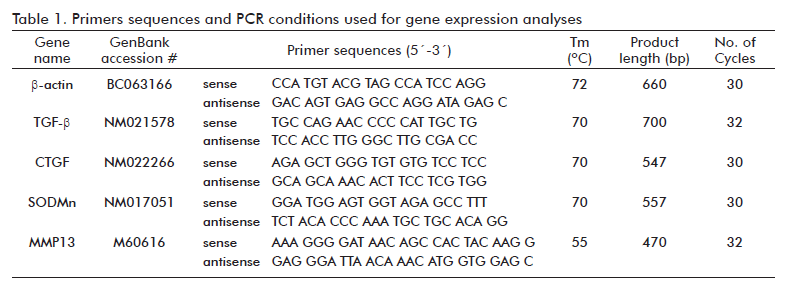
<body><![CDATA[<P   align="justify" ><font face="Verdana, Arial, Helvetica, sans-serif" size="2">Liver central lobe      fragments collected during the autopsies from all the rats were used to assess      the hepatic oxidative stress. The protocols for tissue homogenates and superoxide      dismutase (SOD) and catalase enzymes activities were followed as previously      described [11]. The lipid peroxidation potential (LPP) and malondialdehyde      (MDA) were measured using the Bioxytech LPO-586 commercial kit; while the      total hydroperoxide content was assayed by the Bioxytech H<Sub>2</Sub>O<Sub>2</Sub>-560      commercial kit, both according to manufacturer&rsquo;s instructions (Bio-Rad      Laboratories, Germany). The advanced oxidation protein products (AOPP) content      was assessed according to the Witko-Sarsat described technique [16]. All the      hepatic biochemical data were adjusted to the total protein concentration      determined in the tissue homogenates using a commercial kit (Bio-Rad Laboratories,      Germany). </font></P >   <FONT size="+1"><FONT size="+1"><FONT size="+1"><FONT size="+1">        <P   ><font face="Verdana, Arial, Helvetica, sans-serif" size="2"><b>Immunohistochemistry      </b> </font></P >       <P   align="justify" ><font face="Verdana, Arial, Helvetica, sans-serif" size="2">For immunohistochemistry      studies, liver sections (2-3 &micro;m) were mounted on sialinized slides (DAKO,      Denmark), heat-treated for antigen exposure, and processed according to the      manufacturer&rsquo;s instructions from DakoCytomation LSAB<Sup>TM</Sup>+ System-HRP      commercial kit. Tissue samples were incubated for 30 minwith: anti-&alpha;SMA      (1:100), anti-TGF-&beta; (1:250), anti-p53 (1:200), anti-Cyclin D1 (1:100),      anti-FasL (1:200) and anti-Vimentin (1:100). Antibodies were diluted in Dako      background reducing solution. Immunohistochemistry was accomplished on material      retrieved from three representative animals per group purposely selected according      to the histopathology judgment. Healthy animals were also included. Slides      were counterstained with hematoxilin or light green and were blindly analyzed      by two different investigators. The number of Cyclin D1 positively labeled      hepatocytes nuclei and FasL positively labeled Kupffer cells within the hepatic      parenchyma were quantified in 15 microscopic fields (20 &times;), evenly distributed      in the three liver fragments collected from GHRP6-15d and Saline-15d groups      only. Internal controls included liver sections from healthy intact rats and      omission or replacement of the commercial primary antibody by pre-immune isospecies      serum. Data are presented as the averaged value of the 45 microscopic fields      studied by group (three rats in each one). </font></P >   <FONT size="+1"><FONT size="+1">        <P   ><font face="Verdana, Arial, Helvetica, sans-serif" size="2"><b>Gene expression      analyses by semiquantitative RT-PCR </b></font></P >       <P   align="justify" ><font face="Verdana, Arial, Helvetica, sans-serif" size="2">Liver central lobe      fragments collected from five randomly selected rats of the Saline-15d, GHRP6-15d      and Intact control group were processed to isolate total RNA using TRI Reagent      (Sigma, St. Louis, USA). Total RNA was digested with RNase-free DNase I (Epicentre      Technologies, USA) according to the manufacturer&rsquo;s instructions for      DNA contaminant removal. Afterward, one microgram of total RNA was reverse      transcribed using a commercial available kit (GeneAmp&reg; RNA PCR Core Kit.      Applied Biosystems, USA) with an oligo-dT primer. PCR were performed using      specific primers and annealing temperatures referred in <a href="/img/revistas/bta/v29n2/t0102212.gif">table      1</a>. Final PCR products were detected in a 1% (w/v) agarose gel and were      quantified using the Kodak ID 3.6 software package (Kodak Inc, USA). &beta;-actin      was used as housekeeping gene for normalization. </font></P >       
<P   ><font face="Verdana, Arial, Helvetica, sans-serif" size="2"><b>RNA extraction      for microarray experiment </b></font></P >       <P   align="justify" ><font face="Verdana, Arial, Helvetica, sans-serif" size="2">Rats in groups GHRP6-60d      and Saline-60d were checked for their percentage of fibrosis at the end of      the seventh month and fibrosis reduction was then calculated. Total RNA isolation      was carried out as for semiquantitative RT-PCR experiments and further purified      using a NucleoSpin RNA clean-up kit (Macherey-Nagel, Germany). The quality      of the total RNA (<I>i.e.</I>, the purity and integrity of the intact RNA)      was assessed by Nanodrop 1000 (ThermoScientific, USA) and Bioanalizer Agilent      2100 (Agilent, USA), reporting the concentration, absorbance 260/280 nm ratio      of 1.8 or higher, and RNA integrity number equal to or higher than 7, respectively.      Five paired samples from GHRP6-60d and Saline-60d that met RNA quality requirements      and exhibited a fibrosis reduction superior to 69% were used for the experiment.      </font></P >       <P   ><font face="Verdana, Arial, Helvetica, sans-serif" size="2"><b>Microarray experiment      </b> </font></P >       <P   align="justify" ><font face="Verdana, Arial, Helvetica, sans-serif" size="2">We chose a reference      design with five samples per groups GHRP6-60d and Saline-60d compared to a      reference, representing the seven pooled samples from the Intact control group.      </font></P >       <P   align="justify" ><font face="Verdana, Arial, Helvetica, sans-serif" size="2">The amplification      and labeling of mRNA were performed using the CapitalBio cRNA Amplification      and Labeling kit according to manufacturing instructions (CapitalBio, Beijing,      China). The rat 27 K oligonucleotide microarray comprises 26 962 oligo probes      of 70-mer (Capitol-Bio Corporation, Beijing, China) from the Operon Company      (Rat Genome Oligo Set, Version 3.0.5). Dual channel microarray hybridization      was performed with 70-80 total pmol of Cy3-labeled control sample and Cy5-labeled      test samples (from GPHR6-60d and Saline-60d) onto 25 &times; 75 mm chips.      Hybridization and washing of slides were carried out according to the manufacturer      instructions (Capitol-Bio Corporation, Beijing, China).The slides were scanned      with a confocal LuxScan scanner (CapitalBio Corp, China) and the raw data      were extracted using LuxScan&trade; 3.0 software (CapitalBio Corp). For dual-channels      microarray data, the scanning setting for Cy3 and Cy5 channels were balanced.      The signals detected from housekeeping and Hex genes, and those detected from      the exogenous controls were used as positive controls. Negative controls were      at background levels. </font></P >       ]]></body>
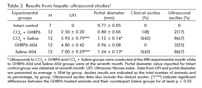
<body><![CDATA[<P   ><font face="Verdana, Arial, Helvetica, sans-serif" size="2"><b>Statistical analysis      </b> </font></P >       <P   align="justify" ><font face="Verdana, Arial, Helvetica, sans-serif" size="2">All the experimental      data were initially evaluated for a normal distribution using the Kolmogorov-Smirnov      test (p &lt; 0.05). When a normal distribution was established, an unpaired      Student&rsquo;s test was used for comparisons between the groups of prevention      phase; while paired Student&rsquo;s t test was used for the groups of the      regression phase. In case of multiple comparisons, the one way ANOVA followed      by the Student-Newman- Keuls test was used. The percentage values were compared      using the Fisher&rsquo;s exact test. A value of p &lt; 0.05 was used to indicate      a significant difference. </font></P >       <P   align="justify" ><font face="Verdana, Arial, Helvetica, sans-serif" size="2">The R Limma package      (<a href="http://bioinf.wehi.edu.au/limma" target="_blank">http://bioinf.wehi.edu.au/limma</a>)      was used for preprocessing and differential expression analysis of microarray      data [17]. The median average intensity of foreground and background were      extracted from the lsr files. A quality criterion [18] was applied to identify      low intensity or high background spots assigning weights 0 or 1, which were      later used as inputs for limma approach. The normexp+offset method was selected      for adaptively adjusting the foreground for the background intensities. The      within-arrays normalization was performed with the print-tip loess method      using between channels non-differentially expressed controls and for between-array      normalization the scale method using all control probes. The moderated paired      t-test for each gene was calculated. Genes with p &lt; 0.001 [18] and fold      change greater than 1.5 were considered for bioinformatics analysis and biological      interpretation. </font></P >       <P   ><font face="Verdana, Arial, Helvetica, sans-serif" size="2"><b>Bioinformatics      analysis of differentially expressed gene </b></font></P >       <P   align="justify" ><font face="Verdana, Arial, Helvetica, sans-serif" size="2">Bioinformatics analysis      was performed using, as input, the list of differentially expressed genes      in rat (GHRP6-60d <I>vs</I>. Saline-60d) and also the list of their human      orthologous in order to take benefit of the functional annotation available      on human genes aiming to predict significant biological process involved in      the putative GHRP6-mediated antifibrotic mechanisms. Human orthologous for      rat genes were identified by searching the Homologene database. Whenever orthologous      genes were not found in Homologene, sequence similarity searches of rat&rsquo;s      transcripts against the human RefSeq transcripts were performed with Blastn.      The most similar gene, for every rat gene, was chosen as the best Blastn hit      (gene with lowest E-value, E-value &lt; 1 &times; 10<Sup>-7</Sup>). To identify      putative protein-protein interactions, in which the protein products of the      input genes are involved, gene networks were constructed using the Cytoscape&rsquo;s      [19] plugin BisoGenet [20]. All molecular interaction data sources available      at BisoGenet were used to generate the gene networks. These networks were      enriched with transcription regulation data extracted from the literature.      The Cytoscape&rsquo;s plugin BiNGO [21] was used to identify the Gene Ontology      (GO) [22] biological proc[19] plugin BisoGenet [20]. All molecular interaction      data sources available at BisoGenet were used to generate the gene networks.      These networks were enriched with transcription regulation data extracted      from the literature. The Cytoscape&rsquo;s plugin BiNGO [21] was used to identify      the Gene Ontology (GO) [22] biological processes enriched in the lists of      input genes. The statistically significant GO processes were determined by      using the hypergeometric test and the Benjamini-Hochberg False Discovery Rate      (FDR) correction for multiple testing [23] with a threshold of 0.05.</font></P >       <P   align="justify" >&nbsp;</P >   <FONT size="+1"><FONT size="+1">        <P   ><font size="2" face="Verdana, Arial, Helvetica, sans-serif"><b><font size="3">RESULTS      </font></b></font></P >   <FONT size="+1">        <P   ><font size="2" face="Verdana, Arial, Helvetica, sans-serif"><b>Hepatic ultrasound      exploration </b></font></P >   <FONT size="+1">        <P   align="justify" ><font face="Verdana, Arial, Helvetica, sans-serif" size="2">At the fifth month,      the comparative ultrasonic study between CCl<Sub>4</Sub> + GHRP6 and CCl<Sub>4</Sub>      + Saline groups demonstrated a significant difference (p = 0.0009) between      the calculated UFI values, thus suggesting that GHRP6 prevented fibrosis (<a href="/img/revistas/bta/v29n2/t0202212.gif">Table      2</a>). The animals of the GHRP6-60d and Saline-60d groups were also subjected      to a comparative study by ultrasound at the seventh month. At this time point,      the Saline-60d achieved the largest UFI value registered (<a href="/img/revistas/bta/v29n2/t0202212.gif">Table      2</a>), which was significantly different (p = 0.0005) to that of GHRP6-60d      group; thus indicating that a process of fibrosis regression had been set      forth. </font></P >   <FONT size="+1"><FONT size="+1"><FONT size="+1"><FONT size="+1">        
<P   align="justify" ><font face="Verdana, Arial, Helvetica, sans-serif" size="2">The portal vein appeared      dilated over 60% in the animals from the CCl<Sub>4</Sub> + Saline group at      the fifth month, as compared to the Intact control group (p = 0.0052; <a href="/img/revistas/bta/v29n2/t0202212.gif">table      2</a>). In contrast, the averaged portal diameter for the CCl<Sub>4</Sub>      + GHRP6 group did not differ from the normal values (p = 0.2938). At the seventh      month, once the regression phase concluded, the GHRP6-60d group showed a significant      reduction of portal dilation as compared to the Saline-60d group (p = 0.0201).      </font></P >   <FONT size="+1"><FONT size="+1"><FONT size="+1"><FONT size="+1">        
]]></body>
<body><![CDATA[<P   align="justify" ><font face="Verdana, Arial, Helvetica, sans-serif" size="2">As shown in <a href="/img/revistas/bta/v29n2/t0202212.gif">table      2</a>, a significant reduction of the animals bearing ascites was detected      in the concomitant (p = 0.0361) and the therapeutic GHRP6 for 60 days (p =      0.0373). No ascites was detected for the GHRP6-15d and Saline-15d groups during      the short time of therapeutic intervention. </font></P >       
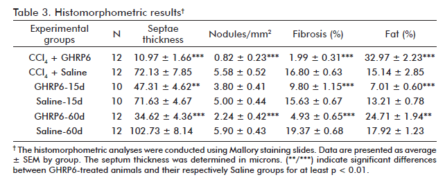
<P   ><font face="Verdana, Arial, Helvetica, sans-serif" size="2"><b>Liver histopathology</b>      </font></P >       <P   align="justify" ><font face="Verdana, Arial, Helvetica, sans-serif" size="2">After five months      of continuous CCl<Sub>4</Sub> administration, the animals from the CCl<Sub>4      </Sub>+Saline group showed dense collagen bundles surrounding cirrhotic nodules      (<a href="/img/revistas/bta/v29n2/f0102212.gif">Figure 1A</a>) which      represented about 17% of hepatic area covered by fibrosis (<a href="/img/revistas/bta/v29n2/t0302212.gif">Table      3</a>). However, the concomitant intervention with GHRP6 prevented in more      than 85% the fibrotic induration (p &lt; 0.0001; <a href="/img/revistas/bta/v29n2/f0102212.gif">figure      1B</a>). In line with this, the CCl<Sub>4</Sub> + GHRP6 group showed far less      cirrhotic nodules and averaged lesser septum thickness as compared to the      CCl<Sub>4</Sub>+Saline group (both p &lt; 0.0001). These findings sustain      the ultrasound evidences of GHRP6-mediated anti-fibrotic response. Moreover,      the percent of fibrosis, the number of cirrhotic nodules, and the averaged      septum thickness of the saline-treated animals within the reversion trial      for 15 days were very close to those of the CCl<Sub>4</Sub> + Saline group      (all p &gt; 0.05); which indicated that no relevant spontaneous fibrosis resolution      took place during the 15 days in which the CCl<Sub>4</Sub> was not injected      (<a href="/img/revistas/bta/v29n2/f0102212.gif">Figures 1C and D</a>;      <a href="/img/revistas/bta/v29n2/t0302212.gif">table 3</a>). </font></P >   <FONT size="+1"><FONT size="+1"><FONT size="+1"><FONT size="+1"><FONT size="+1"><FONT size="+1"><FONT size="+1"><FONT size="+1"><FONT size="+1"><FONT size="+1"><FONT size="+1"><FONT size="+1">        
<P   align="justify" ><font face="Verdana, Arial, Helvetica, sans-serif" size="2">The therapeutic administration      of GHRP6 for 15 days in the first reversion protocol, allowed for 37% of fibrosis      clearance (p = 0.0004). It was mainly due to reduction of more than 30% of      septae thickness (p = 0.0011). No differences were found in the number of      cirrhotic nodules (p = 0.0602) between the groups (<a href="/img/revistas/bta/v29n2/t0302212.gif">Table      3</a>). </font></P >       
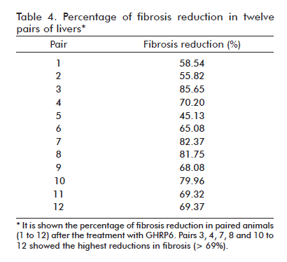
<P   align="justify" ><font face="Verdana, Arial, Helvetica, sans-serif" size="2">After seven months      of CCl<Sub>4</Sub> continuous administration, the CCl<Sub>4</Sub> + Saline-60d      group reached the largest percent of fibrosis which was significantly different      from the CCl<Sub>4</Sub> + Saline group (p = 0.0139) at the fifth experimental      month, indicating a progression in the disease severity due to a substantial      increase in septal thickness. As shown in <a href="/img/revistas/bta/v29n2/t0302212.gif">table      3</a>, the therapeutic administration of GHRP6 by 60 days achieved a 75% reduction      of the fibrotic area when compared to the Saline-60d group (p &lt; 0.0001),      even though the CCl<Sub>4</Sub> injections were not interrupted (<a href="/img/revistas/bta/v29n2/f0102212.gif">Figure      1F</a>). <a href="#tab4">Table 4</a> shows the reduction in fibrosis from      each of the twelve pairs of liver from GHRP6-60d and Saline-60d groups. A      reduction higher than 69% is observed in pairs 3, 4, 7, 8, 10, 11, 12 and      higher than 80% in pairs 3, 7 and 8. Accordingly, the GHRP6-60d group showed      significant reductions in the cirrhotic nodules/mm<Sup>2</Sup> and the septae      thickness as compared to the Saline-60d group (both p &lt; 0.0001).</font></P >       
<P   align="center" ><img src="/img/revistas/bta/v29n2/t0402212.gif" width="410" height="374"><a name="tab4"></a></P >   <FONT size="+1"><FONT size="+1"><FONT size="+1"><FONT size="+1"><FONT size="+1"><FONT size="+1"><FONT size="+1"><FONT size="+1"><FONT size="+1"><FONT size="+1">        
<P   align="justify" ><font face="Verdana, Arial, Helvetica, sans-serif" size="2">The CCl<Sub>4</Sub>      + GHRP6 group exhibited a two-fold increase of hepatic fat percentage with      respect to the CCl<Sub>4</Sub> + Saline group (p = 0.0002; <a href="/img/revistas/bta/v29n2/t0302212.gif">table      3</a>). At the seventh month, the GHRP6-60d group showed a 38% increase of      fat deposition compared to the Saline-60d group (p = 0.0067). In contrast,      in the rats from the GHRP6-15d group, where the CCl<Sub>4</Sub> injections      were interrupted, a notorious reduction of fat was detected as compared to      the Saline-15d group (p &lt; 0.0001). </font></P >   <FONT size="+1"><FONT size="+1"><FONT size="+1"><FONT size="+1"><FONT size="+1"><FONT size="+1">        
<P   ><font face="Verdana, Arial, Helvetica, sans-serif" size="2"><b>Serum biochemistry      </b></font></P >       <P   align="justify" ><font face="Verdana, Arial, Helvetica, sans-serif" size="2">In the fibrosis prevention      trial the serum biochemical analysis was done after the second and the fifth      month of continuous CCl<Sub>4</Sub> and GHRP6/Saline administrations. At the      second month, the CCl<Sub>4 </Sub>+ Saline group showed the highest ASAT and      ALAT levels detected as compared to the Intact control group (both p &lt;      0.001; <a href="/img/revistas/bta/v29n2/f0202212.gif">figures 2A and      B</a>). These values dropped by the fifth experimental month although remained      significantly superior to the healthy animals (both p &lt; 0.001). A similar      biphasic behavior for both transaminases was observed for the CCl<Sub>4</Sub>      + GHRP6 group; which resulted significantly lower than those detected for      the CCl<Sub>4</Sub> + Saline group at the second month (both p &lt; 0.001).      At the fifth month, the ASAT level of the CCl<Sub>4</Sub> + GHRP6 group was      also significantly lower than the CCl<Sub>4</Sub> + Saline group (p &lt; 0.001)      while similar values for ALAT were observed for both groups. The CCl<Sub>4</Sub>-induced      liver damage was associated to a reduction of the organ&rsquo;s biosynthetic      function in both groups (<a href="/img/revistas/bta/v29n2/f0202212.gif">Figures      2C to F</a>). However, at the two evaluation time points, the CCl<Sub>4</Sub>      + GHRP6 group exhibited a significantly better liver function as compared      to the CCl<Sub>4</Sub> + Saline group for all the evaluated parameters (at      least p &lt; 0.05). </font></P >   <FONT size="+1"><FONT size="+1"><FONT size="+1"><FONT size="+1"><FONT size="+1"><FONT size="+1"><FONT size="+1"><FONT size="+1"><FONT size="+1"><FONT size="+1"><FONT size="+1"><FONT size="+1"><FONT size="+1"><FONT size="+1"><FONT size="+1"><FONT size="+1"><FONT size="+1"><FONT size="+1">        
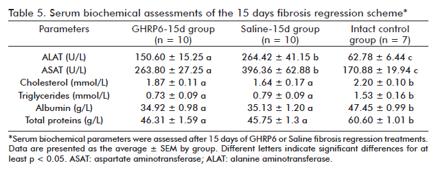
<P   align="justify" ><font face="Verdana, Arial, Helvetica, sans-serif" size="2">In the fibrosis regression      trial, serum chemical parameters in the GHRP6-15d and Saline-15d groups were      evaluated once the short therapeutic scheme was completed. As showed in <a href="/img/revistas/bta/v29n2/t0502212.gif">table      5</a>, significant differences for ALAT (p &lt; 0.01) and ASAT (p &lt; 0.05)      values were detected between these groups reflecting the GHRP6-induced hepatoprotective      effect. Despite this, no significant differences were detected between the      GHRP6-15d and the Saline-15d groups in any of the evaluated hepatic synthesis      indicators, which remained lower than those of the Intact control group (at      least p &lt; 0.05). The serum biochemical parameters from the GHRP6-60d and      the Saline-60d groups were also assessed following autopsy (<a href="/img/revistas/bta/v29n2/t0602212.gif">Table      6</a>). No significant differences were detected between these groups for      the serum transaminases as for any of the organ&rsquo;s functional parameters      (at least p &lt; 0.05). </font></P >       
]]></body>
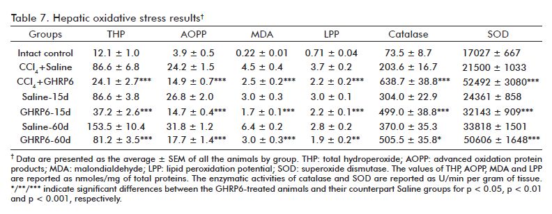
<body><![CDATA[<P   ><font face="Verdana, Arial, Helvetica, sans-serif" size="2"><b>Hepatic oxidative      stress </b></font></P >       <P   align="justify" ><font face="Verdana, Arial, Helvetica, sans-serif" size="2">An etiopathogenic      ingredient of the CCl<Sub>4</Sub> hepatotoxic mechanism is the oxidative damage      to liver cells [24]. It was confirmed by the detected increase of the evaluated      oxidative stress markers (total hydroperoxide content, MDA, LPP and AOPP)      in all the experimental groups, as compared to the Intact control group (at      least p &lt; 0.001; <a href="/img/revistas/bta/v29n2/t0702212.gif">table      7</a>). The GHRP6 intervention significantly attenuated all these oxidative      markers as compared with each respective saline group (at least p &lt; 0.01),      in the three interventional approaches assessed. Concurrently, the GHRP6-treated      rats exhibited a remarkable increase of Catalase and SOD activities as compared      to their counterpart saline groups (at least p &lt; 0.05). </font></P >   <FONT size="+1"><FONT size="+1">        
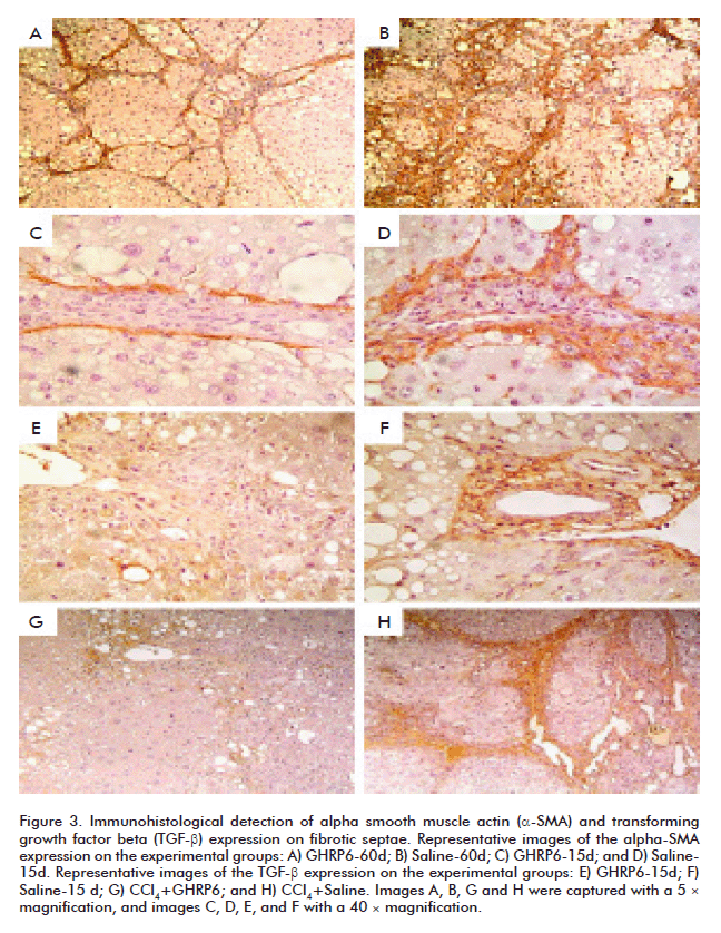
<P   ><font face="Verdana, Arial, Helvetica, sans-serif" size="2"><b>Immunohistochemical      results </b></font></P >       <P   align="justify" ><font face="Verdana, Arial, Helvetica, sans-serif" size="2">The &alpha;-SMA protein      is a molecular marker broadly used to detect activated HSC [25, 26]. Although      based on qualitative judgment, animals intervened with GHRP6 either under      concomitant (data not shown) or therapeutic approaches exhibited far less      &alpha;-SMA labeling than their saline counterparts (<a href="/img/revistas/bta/v29n2/f0302212.gif">Figures      3A to D</a>). The immuno-detection of the &alpha;-SMA antibody appears particularly      restricted to the external sides of the fibrotic septae where it has been      reported that activated HSC are confined [27]. It is worthy to highlight that      an appreciable reduction in the number of labeled cells was a hallmark in      the GHRP6-derived samples, including to those fields in which the fibrotic      septae appeared similar in terms of thickening and cellular density (<a href="/img/revistas/bta/v29n2/f0302212.gif">Figures      3C and D</a>). Similarly, TGF-&beta; appeared far less expressed in those      samples derived from GHRP6-treated rats from both preventive and regression      phases (<a href="/img/revistas/bta/v29n2/f0302212.gif">Figures 3E to      H</a>). As discussed below, our immunostaining for TGF-&beta; also appeared      restricted to the fibrotic septae where it is anchored to the ECM proteins      [28]. </font></P >       
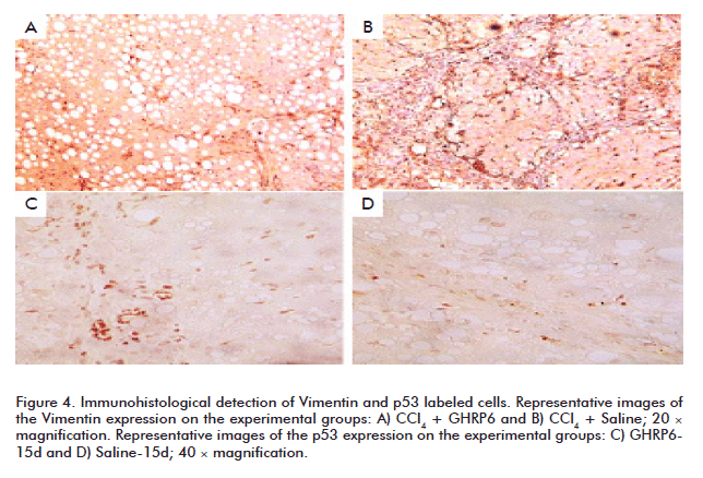
<P   align="justify" ><font face="Verdana, Arial, Helvetica, sans-serif" size="2">Vimentin is also      used as a molecular marker for activated HSC and Kupffer cells [29, 30]. GHRP6-treated      animals within the prevention trial showed far less Vimentin positive cells      (in the fibrotic septae as within the hepatic parenchyma) than the saline      group (<a href="/img/revistas/bta/v29n2/f0402212.gif">Figures 4A and      B</a>). This result was also similar for the samples of the fibrosis regression      trial (data not shown). As judged by cell morphology and topographic location      (core of the fibrotic septae and hepatic parenchyma), lineages immunolabeled      with the anti-p53 correspond to recruited round mononuclear cells, Kupffer      and HSC. Remarkably, the most intense signal and amount of positive anti-p53      cells were detected in the GHRP6 treated animals; in both prevention (data      not shown) and regression trials (<a href="/img/revistas/bta/v29n2/f0402212.gif">Figures      4C and D</a>). </font></P >       
<P   align="justify" ><font face="Verdana, Arial, Helvetica, sans-serif" size="2">Although double immunohistochemistry      was not conducted, FasL expression specifically matched to Kupffer cells,      as suggested by its morphology and location. A larger increase of FasL positively      labeled Kupffer cells was detected in the Saline-15d group (14.22 &plusmn;      6.17 <I>vs</I>. 2.09 &plusmn; 2.12; p &lt; 0.0001). Conversely, the animals      of the GHRP6-15d group showed a remarkable increase of Cyclin D1 positively      labeled hepatocytes nuclei, as compared to Saline-15d group (8.65 &plusmn;      2.36 <I>vs</I>. 2.22 &plusmn; 1.59; p &lt; 0.0001), which is likely an indicative      of GHRP6-induced parenchymal regeneration [31]. A similar result of FasL and      Cyclin D1 expression patterns was obtained in the remaining experimental groups      (data not shown). </font></P >       <P   ><font face="Verdana, Arial, Helvetica, sans-serif" size="2"><b>RT-PCR analysis      </b></font></P >       <P   align="justify" ><font face="Verdana, Arial, Helvetica, sans-serif" size="2">The transcriptional      profile of fibrosis-committed target genes was studied in the GHRP6-15d and      Saline-15d groups (<a href="/img/revistas/bta/v29n2/f0502212.gif">Figure      5</a>). In comparison to intact rats, the saline treated animals showed a      significant enhancement on the transcriptional expression of TGF-&beta; and      connective tissue growth factor (CTGF; both p &lt; 0.001), two growth factors      with well-characterized role in the fibrogenic process [28]. A significantly      sharp expression reduction for both fibrogenic cytokines was readily observed      in the GHRP6-15d group as compared to the saline counterparts (at least p      &lt; 0.05).The CCl<Sub>4</Sub> also induced a significant decrease of the      superoxide dismutase manganese enzyme (SODMn) transcriptional expression in      the Saline-15d group as compared to the intact control group (p &lt; 0.05;      <a href="/img/revistas/bta/v29n2/f0502212.gif">figure 5</a>). However,      the SODMn transcriptional expression of the GHRP6-treated rats did not differ      from the constitutive levels found in the Intact control group and was significantly      superior to the Saline-15d group (p &lt; 0.01). Matrix metalloprotease-13      (MMP13) is the major interstitial collagenase that reduces liver fibrosis      by degrading the ECM proteins [32]. A dramatic expression enhancement of MMP13      transcriptional levels appeared with the GHRP6 intervention as compared to      the saline group (p &lt; 0.01). </font></P >   <FONT size="+1"><FONT size="+1">        
<P   ><font face="Verdana, Arial, Helvetica, sans-serif" size="2"><b>Microarray and      bioinformatic analyses </b></font></P >       <P   align="justify" ><font face="Verdana, Arial, Helvetica, sans-serif" size="2">Microarray experiment      was carried out comparing paired samples 4, 7, 8, 11, 12 in Groups GHRP6-60d      and Saline-60d with the reference sample from the Intact control group. The      1.5 -fold differentially-expressed rat genes and their homologous human genes      (ORT-Human), reported in Homologene or identified by Blastn are shown in <a href="/img/revistas/bta/v29n2/t0802212.gif">table      8</a>. The molecular interaction network that was generated with BisoGenet,      using differentially-expressed rat genes as input data, contained only 41      genes and 25 molecular interactions. </font></P >       
]]></body>
<body><![CDATA[<P   align="justify" ><font face="Verdana, Arial, Helvetica, sans-serif" size="2">However, a similar      network generated from ORT-Human genes included 386 genes and 1883 interactions.      After a gene enrichment analysis performed with BiNGO, the most biologically      significant processes were underscored as consequence of the GHRP6 intervention.      These were: &#145;oxidation-reduction&#146; and &#145;response to wounding&#146;      (<a href="/img/revistas/bta/v29n2/t0902212.gif">Table 9</a>). For the      former, members of the cytochrome P450 family as CYP2A13, CYP2C18, CYP2C19      and CYP2C9; aldo-ketoreductase (AKR1C1) and UDP glucuronosyltransferase, also      committed in drugs and xenobiotic metabolism pathways, were included. Cysteine      dioxygenase type I (CDO1), participating in redox process, and the NADP+-dependent      isocitrate dehydrogenase 1 (IDH1)/Pipecolic acid oxidase (PIPOX), which participate      in peroxisome pathway, were also incorporated within the &#145;oxidation-reduction&#146;      process. Besides, Cdo1, alpha-1-inhibitor 3 (A1i3), coagulation factor X (F10),      histidine-rich glycoprotein (Hrg), serine (or cysteine) peptidase inhibitor,      clade A, members 3N (Serpina3n) and 3M (Serpina3m), transferrin (TF) and hepcidin      antimicrobial peptide (HAMP) genes were related to &#145;response to wounding&#146;.</font></P >       
<P   align="justify" >&nbsp;</P >       <P   ><font size="2" face="Verdana, Arial, Helvetica, sans-serif"><b><font size="3">DISCUSSION      </font></b></font></P >   <FONT size="+1">        <P   align="justify" ><font size="2" face="Verdana, Arial, Helvetica, sans-serif">CCl<Sub>4</Sub> chronic      administration induced an overt cirrhotic disease to otherwise normal rats,      which engendered systemic disturbances for the animal homeostasis. Herein,      we provide the first evidences suggesting that a classic member of the Bower&rsquo;s      synthetic secretagogue peptides, GHRP6, is not solely endowed with cardioprotective      actions but also with anti-fibrotic effect. The evidences derived from these      experiments provide the fundamentals to consider that the GHRP6 intervention      prevented the progression of a liver fibrogenic process and also triggered      its regression. This result substantiates previous findings of our group [11]      and others [13] in terms of GHRP6-mediated hepatic tissue protection. </font></P >   <FONT size="+1"><FONT size="+1"><FONT size="+1">        <P   align="justify" ><font face="Verdana, Arial, Helvetica, sans-serif" size="2">In this study three      experimental settings were established. The prophylactic approach attempted      to recreate a clinical condition in which a patient is threatened to evolve      to fibrosis following a triggering event. Further, the therapeutic intervention      trial was split in two clinical schemes, one in which the hepatic challenge      was interrupted and an alternative one related to a chronic liver insult.      This work seems to be the first preclinical study in which a fibrosis regression      effect is examined in homogeneously allocated groups according to a scale      of liver gross pathology [15]. </font></P >       <P   align="justify" ><font face="Verdana, Arial, Helvetica, sans-serif" size="2">The histomorphometric      assessment as the ultrasound study concurrently indicated less fibrosis in      all the GHRP6 intervened animals. The most remarkable result in terms of fibrosis      material reduction was associated to the GHRP6 preventive scheme, where the      liver parenchyma appeared spared of nodular organization. Accordingly, the      lowest UFI value was calculated for this group. This fact highlights the hepatoprotective      effect induced by GHRP6 in order to prevent parenchymal cells downfall and      a subsequent fibrogenic response. The short term therapeutic intervention      (15 days) accounted for a significant regression of liver fibrosis, mainly      expressed in fibrotic septae involution. Importantly, this effect was attained      in a scenario where no spontaneous fibrotic resolution took place in the saline      group. Parenchymal nodules clearance along with septae thickness reduction      was also confirmed in the 60 days GHRP6-therapeutic scheme. It is noteworthy      that this effect appeared in a scenario of continuous liver aggression as      the CCl<Sub>4</Sub> administration was intentionally maintained. </font></P >   <FONT size="+1"><FONT size="+1">        <P   align="justify" ><font face="Verdana, Arial, Helvetica, sans-serif" size="2">One of the methodological      limitations of this study is the lack of portal venous pressure/flow measurements.      Alternatively, the most reliable approach undertaken was to assess portal      dilation by ultrasound as described elsewhere [33, 34]. Generally speaking,      the incidence of ascites and portal dilation was scarcely registered within      the GHRP6 groups, thus suggesting a lesser amount of fibrotic accumulation      and a more physiologic haemodynamic performance. We do not rule out however,      the hypothetical involvement of the GHRP6 induction of endothelial nitric      oxide release as a key factor controlling portal haemodynamic balance [35].      </font></P >       <P   align="justify" ><font face="Verdana, Arial, Helvetica, sans-serif" size="2">Fat quantification      results suggest that a magnification of the fatty liver phenotype is associated      to the co-administration of CCl<Sub>4</Sub> and GHRP6 and not to the GHRP6      intervention alone. Long term clinical interventions with the cognate GHRP2      have proved to be safe in dwarf children [36]. In line with this, our long-term      systemic administration toxicity studies in healthy rats proved that GHRP6      does not harm the hepatic tissue (Cosme-D&iacute;az K; unpublished data).      The Cyclin D1 expression profile incites to speculate that GHRP6 stimulates      hepatocytes mitosis. Whether this mitogenic response is directly triggered      by GHRP6 itself and/or through the GH/Insulin-like growth factor 1 axis remains      to be examined [37]; moreover, regeneration of the hepatic mass could hypothetically      explain the increase of fat storage in concomitantly CCl<Sub>4</Sub> + GHRP6-treated      rats. It could also explain the noticeable fat storage reduction in those      animals solely exposed to GHRP6 without a correlate increase in fat serum      markers. </font></P >   <FONT size="+1"><FONT size="+1"><FONT size="+1"><FONT size="+1">        <P   align="justify" ><font face="Verdana, Arial, Helvetica, sans-serif" size="2">In general terms,      ALAT and ASAT serum levels exhibited a biphasic behavior. The acute damage      phase, histologically expressed as hepatic steatosis and non-parenchymal cells      reactivity (data not shown), correlated with the highest transaminases levels.      From this point onward, however, fibrosis severity increase appeared associated      to a drastic transaminases drop-down, irrespective to the medication, which      has been previously reported [38, 39]. The fact that ALAT levels from the      GHRP6-preventively treated group remained similar to the saline counterpart      at the fifth month could speculatively be associated to the increase of fat      storage and/or to hepatocytes regeneration. These two factors could be also      related to the fact that in the regression phase, transaminases were only      reduced within the GHRP6-short treatment scheme where CCl<Sub>4</Sub> injections      were interrupted. The liver synthesis function remained preserved with the      concomitant GHRP6 scheme, as indicative of its hepatoprotective potential      and/or to a hepatocytes&rsquo; regeneration. However, the liver biosynthetic      function did not improve with any of the GHRP6 therapeutic schemes. The CCl<Sub>4</Sub>-induced      irreversible inhibition of macromolecules exocytosis mechanism in the hepatocytes      may explain this drawback [40, 41]. It is reasonable to deem that a more prolonged      treatment period, associated to a liver-free insult, would be translated into      a superior GHRP6 therapeutic impact in terms of hepatic synthesis function.      </font></P >   <FONT size="+1"><FONT size="+1"><FONT size="+1"><FONT size="+1">        <P   align="justify" ><font face="Verdana, Arial, Helvetica, sans-serif" size="2">Aside from the limitations      of this work to fully elucidate the GHRP6 anti-fibrogenic mechanism, a major      contribution in this context is the finding that GHRP6 induces an enhancement      of SOD activity, which appears mediated via the transcriptional activation      of at least the manganese-dependent isoform. In line with this observation,      the bioinformatics analysis of the microarray experiment results indicated      that GHRP6 enhances the action of a series of genes encompassed in the redox      homeostasis as Cdo1. In addition, two other enzymes whose expression appeared      upregulated by GHRP6 (PIPOX and IDH1) play instrumental roles in peroxisomal      processes as free radicals detoxification. Furthermore, increases of CYP2A13,      CYP2C18, CYP2C19, CYP2C9, AKR1C1 and UDP glucuronosyltransferase support the      idea of an active detoxification process, contributing to revert fibrogenesis      [42]. This has extended previous findings in which GHRP6 proved to ameliorate      radicalary cytotoxicity and to amplify antioxidant defenses [11]. As reactive      oxygen species are key mediators of the fibrogenic process by directly activating      HSC transdifferentiation [28], its neutralization and/or pool reduction has      been considered an appropriate strategy to mitigate the fibrogenic responses      [43-46]. </font></P >       ]]></body>
<body><![CDATA[<P   align="justify" ><font face="Verdana, Arial, Helvetica, sans-serif" size="2">The inflammatory      response associated to the hepatocytes&rsquo; injury amplifies the generation      of reactive oxygen species, the recruitment of inflammatory cells, as the      local release of profibrogenic cytokines [47]. The fact that in the GHRP6      short term therapeutic intervention, Kupffer cells expressed p53 in concomitance      to an obvious suppression of FasL expression, may translate as an ongoing      process of Kupffer cells arrest or apoptotic induction. It would represent      a possible disruption of hepatocytes&rsquo; demise and the ensued activation      cascade [48]. It is known that Kupffer cells promote hepatocytes apoptosis      via FasL pathway [49, 50]. Overall, these observations indicate that GHRP6      contributed to quench the liver inflammatory response as has been previously      described [11, 13]. <I>In vitro</I> models are in progress to delineate the      actual potential of GHRP6 to prevent HSC activation. </font></P >       <P   align="justify" ><font face="Verdana, Arial, Helvetica, sans-serif" size="2">The notion that GHRP6      intervention reduced local inflammation and the consequent HSC activation-perpetuation      may be supported by the consistent reduction of &alpha;-SMA and Vimentin expression,      as markers of an activation phenotype. In line with this, GHRP6-treated animals      exhibited far less expression of the two major fibrogenic growth factors TGF-&beta;      and CTGF which may anticipate a decrease in ECM protein synthesis and accumulation      [28]. Furthermore, GHRP6 increased MMP13 transcriptional expression which      may be obviously linked to fibrosis reduction due to collagen degradation      [32]. It obviously would be complemented by a functional enzymatic assay.      </font></P >       <P   align="justify" ><font face="Verdana, Arial, Helvetica, sans-serif" size="2">Growth hormone secretagogues      regulate the transcriptional activation of the peroxisome proliferator-activated      receptor gamma (PPAR&gamma;) through the concerted interaction with the two      types of receptors identified: CD36 and the ghrelin GHS-R1a. PPAR&gamma; can      also induce CD36 expression thus establishing a mutually positive regulatory      loop [51]. PPAR&gamma; agonistic stimulation markedly inhibited HSC proliferation,      induced cells apoptosis and significantly suppressed TGF-&beta;1-induced CTGF      expression [52]; which is in correspondence to our immunohistology and microarray      data, suggesting an abortion of the HSC activation program. Likewise, Milam      and co-workers recently confirmed the PPAR&gamma; agonist ligands inhibit      the ability of TGF-&beta; to promote myofibroblast trans-differentiation as      the ensuing collagen secretion and fibrotic induration in a murine model of      lung fibrosis [53]. The above demonstrations render hypothetical mechanistic      involvement for the GHRP6-mediated PPAR&gamma; activation via CD36 occupation.      This may represent a not redundant pathway for novel anti-fibrotic agents.      </font></P >       <P   align="justify" ><font face="Verdana, Arial, Helvetica, sans-serif" size="2">In summary, the main      contribution of this work is the unprecedented demonstration that GHRP6 reduces      the fibrotic induration of a parenchymal organ in a renowned experimental      model. The mechanism whereby GHRP6 reduces fibroplasias seems to be multifactorial      involving a broad number of gene products concerned to detoxification pathways,      redox homeostasis, and response to injury. Distally, it appears to nurture      hepatocytes and deactivate stroma-associated cells. These preliminary findings      justify further studies on the role of the GHS for the control of fibrotic      diseases.</font></P >       <P   align="justify" >&nbsp;</P >       <P   ><font size="2" face="Verdana, Arial, Helvetica, sans-serif"><B><font size="3">ACKNOWLEDGEMENTS</font></B></font></P >   <FONT size="+1">        <P   align="justify" ><font size="2" face="Verdana, Arial, Helvetica, sans-serif">Authors are indebted      to Dr. Bienvenido Gra, Head of the Pathology Department from the Institute      of Gastroenterology, Havana, Cuba, for his valuable assistance while examining      the slides.</font></P >       <P   align="justify" >&nbsp;</P >   <FONT size="+1">        <P   align="justify" > </P >       <P   align="justify" ><b><font face="Verdana, Arial, Helvetica, sans-serif" size="3">REFERENCES</font></b><font face="Verdana, Arial, Helvetica, sans-serif" size="2">      </font></P >       ]]></body>
<body><![CDATA[<!-- ref --><P   align="justify" ><font size="2" face="Verdana, Arial, Helvetica, sans-serif">1. Lefton HB, Rosa      A, Cohen M. Diagnosis and epidemiology of cirrhosis. Med Clin North Am. 2009;93(4):787-99.          </font></P >   <FONT size="+1">        <P   align="justify" ><font face="Verdana, Arial, Helvetica, sans-serif" size="2">2. Hammel P, Couvelard      A, O&rsquo;Toole D, Ratouis A, Sauvanet A, Flejou JF, et al. Regression of      liver fibrosis after biliary drainage in patients with chronic pancreatitis      and stenosis of the common bile duct. N Engl J Med. 2001;344(6):418-23. </font></P >       <!-- ref --><P   align="justify" ><font face="Verdana, Arial, Helvetica, sans-serif" size="2">3. Arima M, Terao      H, Kashima K, Arita T, Nasu M, Nishizono A. Regression of liver fibrosis in      cases of chronic liver disease type C: quantitative evaluation by using computed      image analysis. Intern Med. 2004;43(10):902-10.     </font></P >       <!-- ref --><P   align="justify" ><font face="Verdana, Arial, Helvetica, sans-serif" size="2">4. Issa R, Zhou X,      Constandinou CM, Fallowfield J, Millward-Sadler H, Gaca MD, et al. Spontaneous      recovery from micronodular cirrhosis: evidence for incomplete resolution associated      with matrix cross-linking. Gastroenterology. 2004; 126(7):1795-808.     </font></P >       <!-- ref --><P   align="justify" ><font face="Verdana, Arial, Helvetica, sans-serif" size="2">5. Bourliere M, Kahloun      A, Gascou-Tessonnier G. Analogs and fibrosis regression in hepatitis B. Gastroenterol      Clin Biol. 2009;33(10-11):923-9.     </font></P >       <!-- ref --><P   align="justify" ><font face="Verdana, Arial, Helvetica, sans-serif" size="2">6. Friedman SL. Liver      fibrosis -- from bench to bedside. J Hepatol. 2003;38 Suppl 1:S38-53.     </font></P >       <!-- ref --><P   align="justify" ><font face="Verdana, Arial, Helvetica, sans-serif" size="2">7. Gines P, Cardenas      A, Arroyo V, Rodes J. Management of cirrhosis and ascites. N Engl J Med. 2004;350(16):1646-54.          </font></P >       <!-- ref --><P   align="justify" ><font face="Verdana, Arial, Helvetica, sans-serif" size="2">8. Bowers CY, Momany      FA, Reynolds GA, Hong A. On the in vitro and in vivo activity of a new synthetic      hexapeptide that acts on the pituitary to specifically release growth hormone.      Endocrinology. 1984; 114(5):1537-45.     </font></P >       <!-- ref --><P   align="justify" ><font face="Verdana, Arial, Helvetica, sans-serif" size="2">9. Shen YT, Lynch      JJ, Hargreaves RJ, Gould RJ. A growth hormone secretagogue prevents ischemic-induced      mortality independently of the growth hormone pathway in dogs with chronic      dilated cardiomyopathy. J Pharmacol Exp Ther. 2003; 306(2):815-20.     </font></P >       <!-- ref --><P   align="justify" ><font face="Verdana, Arial, Helvetica, sans-serif" size="2">10. Paneda C, Arroba      AI, Frago LM, Holm AM, Romer J, Argente J, et al. Growth hormone-releasing      peptide-6 inhibits cerebellar cell death in aged rats. Neuroreport. 2003;14(12):1633-5.          </font></P >       <!-- ref --><P   align="justify" ><font face="Verdana, Arial, Helvetica, sans-serif" size="2">11. Cibrian D, Ajamieh      H, Berlanga J, Leon OS, Alba JS, Kim MJ, et al. Use of growth-hormone-releasing      peptide-6 (GHRP-6) for the prevention of multiple organ failure. Clin Sci      (Lond). 2006;110(5):563-73.     </font></P >       <!-- ref --><P   align="justify" ><font face="Verdana, Arial, Helvetica, sans-serif" size="2">12. Berlanga J, Cibrian      D, Guevara L, Dominguez H, Alba JS, Seralena A, et al. Growth-hormone-releasing      peptide 6 (GHRP6) prevents oxidant cytotoxicity and reduces myocardial necrosis      in a model of acute myocardial infarction. Clin Sci (Lond). 2007;112(4):241-50.          </font></P >       <!-- ref --><P   align="justify" ><font face="Verdana, Arial, Helvetica, sans-serif" size="2">13. Granado M, Martin      AI, Lopez-Menduina M, Lopez-Calderon A, Villanua MA. GH-releasing peptide-2      administration prevents liver inflammatory response in endotoxemia. Am J Physiol      Endocrinol Metab. 2008;294(1):E131-41.     </font></P >       <!-- ref --><P   align="justify" ><font face="Verdana, Arial, Helvetica, sans-serif" size="2">14. Abe W, Ikejima      K, Lang T, Okumura K, Enomoto N, Kitamura T, et al. Low molecular weight heparin      prevents hepatic fibrogenesis caused by carbon tetrachloride in the rat. J      Hepatol. 2007;46(2):286-94.     </font></P >       <!-- ref --><P   align="justify" ><font face="Verdana, Arial, Helvetica, sans-serif" size="2">15. Anthony PP, Ishak      KG, Nayak NC, Poulsen HE, Scheuer PJ, Sobin LH. The morphology of cirrhosis:      definition, nomenclature, and classification. Bull World Health Organ. 1977;55(4):521-40.          </font></P >       <!-- ref --><P   align="justify" ><font face="Verdana, Arial, Helvetica, sans-serif" size="2">16. Witko-Sarsat      V, Gausson V, Nguyen AT, Touam M, Drueke T, Santangelo F, et al. AOPP-induced      activation of human neutrophil and monocyte oxidative metabolism: a potential      target for N-acetylcysteine treatment in dialysis patients. Kidney Int. 2003;64(1):82-91.          </font></P >       <!-- ref --><P   align="justify" ><font face="Verdana, Arial, Helvetica, sans-serif" size="2">17. Smyth GK. Linear      models and empirical bayes methods for assessing differential expression in      microarray experiments. Stat Appl Genet Mol Biol. 2004;3:Article 3.     </font></P >       <!-- ref --><P   align="justify" ><font face="Verdana, Arial, Helvetica, sans-serif" size="2">18. Simon R, Korn      E, McShane L, Radmacher M, Wright G, Zhao Y. Design and Analysis of DNA Microarray      Investigations. New York: Springer-Verlag; 2004.     </font></P >       <!-- ref --><P   align="justify" ><font face="Verdana, Arial, Helvetica, sans-serif" size="2">19. Shannon P, Markiel      A, Ozier O, Baliga NS, Wang JT, Ramage D, et al. Cytoscape: a software environment      for integrated models of biomolecular interaction networks. Genome Res. 2003;13(11):2498-504.          </font></P >       <!-- ref --><P   align="justify" ><font face="Verdana, Arial, Helvetica, sans-serif" size="2">20. Martin A, Ochagavia      ME, Rabasa LC, Miranda J, Fernandez-de-Cossio J, Bringas R. BisoGenet: a new      tool for gene network building, visualization and analysis. BMC Bioinformatics.      2010;11:91.     </font></P >       <!-- ref --><P   align="justify" ><font face="Verdana, Arial, Helvetica, sans-serif" size="2">21. Maere S, Heymans      K, Kuiper M. BiNGO: a Cytoscape plugin to assess overrepresentation of gene      ontology categories in biological networks. Bioinformatics. 2005;21(16):3448-9.          </font></P >       <!-- ref --><P   align="justify" ><font face="Verdana, Arial, Helvetica, sans-serif" size="2">22. Ashburner M,      Ball CA, Blake JA, Botstein D, Butler H, Cherry JM, et al. Gene ontology:      tool for the unification of biology. The Gene Ontology Consortium. Nat Genet.      2000;25(1):25-9.     </font></P >       <!-- ref --><P   align="justify" ><font face="Verdana, Arial, Helvetica, sans-serif" size="2">23. Benjamini Y,      Hochberg Y. Controlling the false discovery rate: a practical and powerful      approach to multiple testing. J R Stat Soc Ser B. 1995;57(1):289-300.     </font></P >       <!-- ref --><P   align="justify" ><font face="Verdana, Arial, Helvetica, sans-serif" size="2">24. Biasi F, Albano      E, Chiarpotto E, Corongiu FP, Pronzato MA, Marinari UM, et al. In vivo and      in vitro evidence concerning the role of lipid peroxidation in the mechanism      of hepatocyte death due to carbon tetrachloride. Cell Biochem Funct. 1991;9(2):111-8.          </font></P >       <!-- ref --><P   align="justify" ><font face="Verdana, Arial, Helvetica, sans-serif" size="2">25. Carpino G, Morini      S, Ginanni Corradini S, Franchitto A, Merli M, Siciliano M, et al. Alpha-SMA      expression in hepatic stellate cells and quantitative analysis of hepatic      fibrosis in cirrhosis and in recurrent chronic hepatitis after liver transplantation.      Dig Liver Dis. 2005;37(5):349-56.     </font></P >       <!-- ref --><P   align="justify" ><font face="Verdana, Arial, Helvetica, sans-serif" size="2">26. Tomanovic N,      Boricic I, Brasanac D. Immunohistochemical analysis of alpha-SMA and GFAP      expression in liver stellate cells. Vojnosanit Pregl. 2006;63(6):553-7.     </font></P >       <!-- ref --><P   align="justify" ><font face="Verdana, Arial, Helvetica, sans-serif" size="2">27. Ramm GA, Nair      VG, Bridle KR, Shepherd RW, Crawford DH. Contribution of hepatic parenchymal      and nonparenchymal cells to hepatic fibrogenesis in biliary atresia. Am J      Pathol. 1998;153(2):527-35.     </font></P >       <!-- ref --><P   align="justify" ><font face="Verdana, Arial, Helvetica, sans-serif" size="2">28. Gressner OA,      Weiskirchen R, Gressner AM. Evolving concepts of liver fibrogenesis provide      new diagnostic and therapeutic options. Comp Hepatol. 2007;6:7.     </font></P >       <!-- ref --><P   align="justify" ><font face="Verdana, Arial, Helvetica, sans-serif" size="2">29. Wu HH, Tao LC,      Cramer HM. Vimentin-positive spider-shaped Kupffer cells. A new clue to cytologic      diagnosis of primary and metastatic hepatocellular carcinoma by fine-needle      aspiration biopsy. Am J Clin Pathol. 1996;106(4):517-21.     </font></P >       <!-- ref --><P   align="justify" ><font face="Verdana, Arial, Helvetica, sans-serif" size="2">30. Geerts A, Eliasson      C, Niki T, Wielant A, Vaeyens F, Pekny M. Formation of normal desmin intermediate      filaments in mouse hepatic stellate cells requires vimentin. Hepatology. 2001;33(1):177-88.          </font></P >       <!-- ref --><P   align="justify" ><font face="Verdana, Arial, Helvetica, sans-serif" size="2">31. Stacey DW. Cyclin      D1 serves as a cell cycle regulatory switch in actively proliferating cells.      Curr Opin Cell Biol. 2003;15(2):158-63.     </font></P >       <!-- ref --><P   align="justify" ><font face="Verdana, Arial, Helvetica, sans-serif" size="2">32. Watanabe T, Niioka      M, Hozawa S, Kameyama K, Hayashi T, Arai M, et al. Gene expression of interstitial      collagenase in both progressive and recovery phase of rat liver fibrosis induced      by carbon tetrachloride. J Hepatol. 2000;33(2):224-35.     </font></P >       <!-- ref --><P   align="justify" ><font face="Verdana, Arial, Helvetica, sans-serif" size="2">33. Liu Y, Li L,      Yu Z, Liu Q, Li Z, Wang Y, et al. Correlative study between portal vein pressure      and portal hemodynamics in patients with portal hypertension. Zhonghua Gan      Zang Bing Za Zhi. 2002;10(2):135-7.     </font></P >       <!-- ref --><P   align="justify" ><font face="Verdana, Arial, Helvetica, sans-serif" size="2">34. Quintanilha LF,      Mannheimer EG, Carvalho AB, Paredes BD, Dias JV, Almeida AS, et al. Bone marrow      cell transplant does not prevent or reverse murine liver cirrhosis. Cell Transplant.      2008;17(8):943-53.     </font></P >       <!-- ref --><P   align="justify" ><font face="Verdana, Arial, Helvetica, sans-serif" size="2">35. Fiorucci S, Antonelli      E, Morelli A. Nitric oxide and portal hypertension: a nitric oxide-releasing      derivative of ursodeoxycholic acid that selectively releases nitric oxide      in the liver. Dig Liver Dis. 2003;35 Suppl 2:S61-9.     </font></P >       <!-- ref --><P   align="justify" ><font face="Verdana, Arial, Helvetica, sans-serif" size="2">36. Mericq V, Cassorla      F, Salazar T, Avila A, Iniguez G, Bowers CY, et al. Effects of eight months      treatment with graded doses of a growth hormone (GH)-releasing peptide in      GH-deficient children. J Clin Endocrinol Metab. 1998;83(7):2355-60.     </font></P >       <!-- ref --><P   align="justify" ><font face="Verdana, Arial, Helvetica, sans-serif" size="2">37. Desbois-Mouthon      C, Wendum D, Cadoret A, Rey C, Leneuve P, Blaise A, et al. Hepatocyte proliferation      during liver regeneration is impaired in mice with liver-specific IGF-1R knockout.      FASEB J. 2006;20(6):773-5.     </font></P >       <!-- ref --><P   align="justify" ><font face="Verdana, Arial, Helvetica, sans-serif" size="2">38. Poynard T, Munteanu      M, Ngo Y, Moussalli J, Lebray P, Thabut D, et al. FibroTest is effective in      patients with normal transaminases, when accuracy is standardized on fibrosis      stage prevalence. J Viral Hepat. 2008;15(6):472-3.     </font></P >       <!-- ref --><P   align="justify" ><font face="Verdana, Arial, Helvetica, sans-serif" size="2">39. Sebastiani G,      Vario A, Guido M, Alberti A. Performance of noninvasive markers for liver      fibrosis is reduced in chronic hepatitis C with normal transaminases. J Viral      Hepat. 2008;15(3):212-8.     </font></P >       <!-- ref --><P   align="justify" ><font face="Verdana, Arial, Helvetica, sans-serif" size="2">40. Becker E, Messner      B, Berndt J. Two mechanisms of CCl4-induced fatty liver: lipid peroxidation      or covalent binding studied in cultured rat hepatocytes. Free Radic Res Commun.      1987;3(1-5):299-308.     </font></P >       <!-- ref --><P   align="justify" ><font face="Verdana, Arial, Helvetica, sans-serif" size="2">41. Poli G, Cottalasso      D, Pronzato MA, Chiarpotto E, Biasi F, Corongiu FP, et al. Lipid peroxidation      and covalent binding in the early functional impairment of liver Golgi apparatus      by carbon tetrachloride. Cell Biochem Funct. 1990;8(1):1-10.     </font></P >       <!-- ref --><P   align="justify" ><font face="Verdana, Arial, Helvetica, sans-serif" size="2">42. Takahara Y, Takahashi      M, Wagatsuma H, Yokoya F, Zhang QW, Yamaguchi M, et al. Gene expression profiles      of hepatic cell-type specific marker genes in progression of liver fibrosis.      World J Gastroenterol. 2006;12(40):6473-99.     </font></P >       <!-- ref --><P   align="justify" ><font face="Verdana, Arial, Helvetica, sans-serif" size="2">43. Shen XH, Cheng      WF, Li XH, Xie LM, Sun J, Li F, et al. Effect of antioxidant on hepatic stellate      cell proliferation and apoptosis during recovery from liver fibrosis. Wei      Sheng Yan Jiu. 2005;34(2):194-6.     </font></P >       <!-- ref --><P   align="justify" ><font face="Verdana, Arial, Helvetica, sans-serif" size="2">44. Di Sario A, Candelaresi      C, Omenetti A, Benedetti A. Vitamin E in chronic liver diseases and liver      fibrosis. Vitam Horm. 2007;76:551-73.     </font></P >       <!-- ref --><P   align="justify" ><font face="Verdana, Arial, Helvetica, sans-serif" size="2">45. El-Demerdash      E, Salam OM, El-Batran SA, Abdallah HM, Shaffie NM. Inhibition of the renin-angiotensin      system attenuates the development of liver fibrosis and oxidative stress in      rats. Clin Exp Pharmacol Physiol. 2008;35(2):159-67.     </font></P >       <!-- ref --><P   align="justify" ><font face="Verdana, Arial, Helvetica, sans-serif" size="2">46. Ogeturk M, Kus      I, Pekmez H, Yekeler H, Sahin S, Sarsilmaz M. Inhibition of carbon tetrachloride-mediated      apoptosis and oxidative stress by melatonin in experimental liver fibrosis.      Toxicol Ind Health. 2008;24(4):201-8.     </font></P >       <!-- ref --><P   align="justify" ><font face="Verdana, Arial, Helvetica, sans-serif" size="2">47. Bataller R, Brenner      DA. Liver fibrosis. J Clin Invest. 2005;115(2):209-18.     </font></P >       <!-- ref --><P   align="justify" ><font face="Verdana, Arial, Helvetica, sans-serif" size="2">48. Laptenko O, Prives      C. Transcriptional regulation by p53: one protein, many possibilities. Cell      Death Differ. 2006;13(6):951-61.     </font></P >       <!-- ref --><P   align="justify" ><font face="Verdana, Arial, Helvetica, sans-serif" size="2">49. Pianko S, Patella      S, Ostapowicz G, Desmond P, Sievert W. Fas-mediated hepatocyte apoptosis is      increased by hepatitis C virus infection and alcohol consumption, and may      be associated with hepatic fibrosis: mechanisms of liver cell injury in chronic      hepatitis C virus infection. J Viral Hepat. 2001;8(6):406-13.     </font></P >       <!-- ref --><P   align="justify" ><font face="Verdana, Arial, Helvetica, sans-serif" size="2">50. Canbay A, Higuchi      H, Bronk SF, Taniai M, Sebo TJ, Gores GJ. Fas enhances fibrogenesis in the      bile duct ligated mouse: a link between apoptosis and fibrosis. Gastroenterology.      2002;123(4):1323-30.     </font></P >       <!-- ref --><P   align="justify" ><font face="Verdana, Arial, Helvetica, sans-serif" size="2">51. Demers A, Rodrigue-Way      A, Tremblay A. Hexarelin Signaling to PPARgamma in Metabolic Diseases. PPAR      Res. 2008;2008:364784.     </font></P >       <!-- ref --><P   align="justify" ><font face="Verdana, Arial, Helvetica, sans-serif" size="2">52. Sun K, Wang Q,      Huang XH. PPAR gamma inhibits growth of rat hepatic stellate cells and TGF      beta-induced connective tissue growth factor expression. Acta Pharmacol Sin.      2006;27(6):715-23.     </font></P >       <!-- ref --><P   align="justify" ><font face="Verdana, Arial, Helvetica, sans-serif" size="2">53. Milam JE, Keshamouni      VG, Phan SH, Hu B, Gangireddy SR, Hogaboam CM, et al. PPAR-gamma agonists      inhibit profibrotic phenotypes in human lung fibroblasts and bleomycin-induced      pulmonary fibrosis. Am J Physiol Lung Cell Mol Physiol. 2008;294(5):L891-901.    </font></P >       <P   align="justify" >&nbsp;</P >       <P   align="justify" > </P >       <P   align="justify" ><font face="Verdana, Arial, Helvetica, sans-serif" size="2">Received in October,      2011.    <br>     Accepted for publication in February, 2012.</font></P >       <P   align="justify" >&nbsp;</P >       ]]></body>
<body><![CDATA[<P   align="justify" > </P >       <P   ><font size="2" face="Verdana, Arial, Helvetica, sans-serif"><i>Jorge Berlanga-Acosta</i>.      Department of Tissue Repair and Cytoprotection. Direction of Biomedical Research.      Center for Genetic Engineering and Biotechnology, CIGB. </font><font size="+1" color="#000000"><font size="+1"><font size="+1"><font size="+1"><font size="+1"><font size="+1"><font size="+1"><font size="+1"><font size="+1"><font size="+1"><font size="+1"><font size="+1"><font size="+1"><font size="+1"><font size="+1"><font size="+1"><font size="+1"><font size="+1"><font size="+1"><font size="+1"><font size="+1"><font size="+1"><font size="+1"><font size="+1"><font size="+1"><font size="+1"><font size="+1"><font size="+1"><font size="+1"><font size="+1"><font size="+1"><font size="+1"><font size="+1"><font size="+1"><font size="+1"><font size="+1"><font size="+1"><font size="+1"><font size="+1"><font size="+1"><font size="+1"><font size="+1"><font size="+1"><font size="+1"><font size="+1"><font size="+1"><font size="+1"><font size="+1"><font size="+1"><font size="+1"><font size="+1"><font size="+1"><font size="+1"><font size="+1"><font size="+1"><font size="+1"><font size="+1"><font face="Verdana, Arial, Helvetica, sans-serif" size="2">Ave.      31 e/ 158 and 190, Playa, PO Box 6162, CP 10 600, Havana, Cuba.</font></font></font></font></font></font></font></font></font></font></font></font></font></font></font></font></font></font></font></font></font></font></font></font></font></font></font></font></font></font></font></font></font></font></font></font></font></font></font></font></font></font></font></font></font></font></font></font></font></font></font></font></font></font></font></font></font></font><font size="2" face="Verdana, Arial, Helvetica, sans-serif">E-mail:      <A href="mailto:jorge.berlanga@cigb.edu.cu"> <U><U><FONT color="#0000FF">jorge.berlanga@cigb.edu.cu</font></U></U></A></font></P >         ]]></body><back>
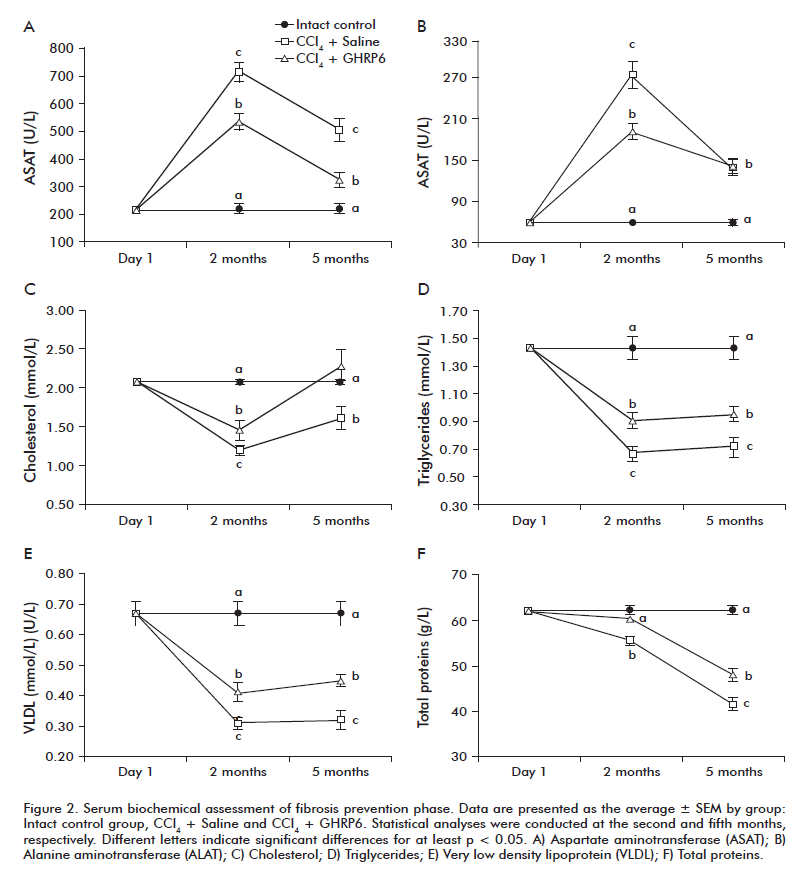
<ref-list>
<ref id="B1">
<label>1</label><nlm-citation citation-type="journal">
<person-group person-group-type="author">
<name>
<surname><![CDATA[Lefton]]></surname>
<given-names><![CDATA[HB]]></given-names>
</name>
<name>
<surname><![CDATA[Rosa]]></surname>
<given-names><![CDATA[A]]></given-names>
</name>
<name>
<surname><![CDATA[Cohen]]></surname>
<given-names><![CDATA[M]]></given-names>
</name>
</person-group>
<article-title xml:lang="en"><![CDATA[Diagnosis and epidemiology of cirrhosis]]></article-title>
<source><![CDATA[Med Clin North Am]]></source>
<year>2009</year>
<volume>93</volume>
<numero>4</numero>
<issue>4</issue>
<page-range>787-99</page-range></nlm-citation>
</ref>
<ref id="B2">
<label>2</label><nlm-citation citation-type="journal">
<person-group person-group-type="author">
<name>
<surname><![CDATA[Hammel]]></surname>
<given-names><![CDATA[P]]></given-names>
</name>
<name>
<surname><![CDATA[Couvelard]]></surname>
<given-names><![CDATA[A]]></given-names>
</name>
<name>
<surname><![CDATA[O'Toole]]></surname>
<given-names><![CDATA[D]]></given-names>
</name>
<name>
<surname><![CDATA[Ratouis]]></surname>
<given-names><![CDATA[A]]></given-names>
</name>
<name>
<surname><![CDATA[Sauvanet]]></surname>
<given-names><![CDATA[A]]></given-names>
</name>
<name>
<surname><![CDATA[Flejou]]></surname>
<given-names><![CDATA[JF]]></given-names>
</name>
</person-group>
<article-title xml:lang="en"><![CDATA[Regression of liver fibrosis after biliary drainage in patients with chronic pancreatitis and stenosis of the common bile duct]]></article-title>
<source><![CDATA[N Engl J Med]]></source>
<year>2001</year>
<volume>344</volume>
<numero>6</numero>
<issue>6</issue>
<page-range>418-23</page-range></nlm-citation>
</ref>
<ref id="B3">
<label>3</label><nlm-citation citation-type="journal">
<person-group person-group-type="author">
<name>
<surname><![CDATA[Arima]]></surname>
<given-names><![CDATA[M]]></given-names>
</name>
<name>
<surname><![CDATA[Terao]]></surname>
<given-names><![CDATA[H]]></given-names>
</name>
<name>
<surname><![CDATA[Kashima]]></surname>
<given-names><![CDATA[K]]></given-names>
</name>
<name>
<surname><![CDATA[Arita]]></surname>
<given-names><![CDATA[T]]></given-names>
</name>
<name>
<surname><![CDATA[Nasu]]></surname>
<given-names><![CDATA[M]]></given-names>
</name>
<name>
<surname><![CDATA[Nishizono]]></surname>
<given-names><![CDATA[A]]></given-names>
</name>
</person-group>
<article-title xml:lang="en"><![CDATA[Regression of liver fibrosis in cases of chronic liver disease type C: quantitative evaluation by using computed image analysis]]></article-title>
<source><![CDATA[Intern Med]]></source>
<year>2004</year>
<volume>43</volume>
<numero>10</numero>
<issue>10</issue>
<page-range>902-10</page-range></nlm-citation>
</ref>
<ref id="B4">
<label>4</label><nlm-citation citation-type="journal">
<person-group person-group-type="author">
<name>
<surname><![CDATA[Issa]]></surname>
<given-names><![CDATA[R]]></given-names>
</name>
<name>
<surname><![CDATA[Zhou]]></surname>
<given-names><![CDATA[X]]></given-names>
</name>
<name>
<surname><![CDATA[Constandinou]]></surname>
<given-names><![CDATA[CM]]></given-names>
</name>
<name>
<surname><![CDATA[Fallowfield]]></surname>
<given-names><![CDATA[J]]></given-names>
</name>
<name>
<surname><![CDATA[Millward-Sadler]]></surname>
<given-names><![CDATA[H]]></given-names>
</name>
<name>
<surname><![CDATA[Gaca]]></surname>
<given-names><![CDATA[MD]]></given-names>
</name>
</person-group>
<article-title xml:lang="en"><![CDATA[Spontaneous recovery from micronodular cirrhosis: evidence for incomplete resolution associated with matrix cross-linking]]></article-title>
<source><![CDATA[Gastroenterology]]></source>
<year>2004</year>
<volume>126</volume>
<numero>7</numero>
<issue>7</issue>
<page-range>1795-808</page-range></nlm-citation>
</ref>
<ref id="B5">
<label>5</label><nlm-citation citation-type="journal">
<person-group person-group-type="author">
<name>
<surname><![CDATA[Bourliere]]></surname>
<given-names><![CDATA[M]]></given-names>
</name>
<name>
<surname><![CDATA[Kahloun]]></surname>
<given-names><![CDATA[A]]></given-names>
</name>
<name>
<surname><![CDATA[Gascou-Tessonnier]]></surname>
<given-names><![CDATA[G]]></given-names>
</name>
</person-group>
<article-title xml:lang="en"><![CDATA[Analogs and fibrosis regression in hepatitis B]]></article-title>
<source><![CDATA[Gastroenterol Clin Biol]]></source>
<year>2009</year>
<volume>33</volume>
<numero>10-11</numero>
<issue>10-11</issue>
<page-range>923-9</page-range></nlm-citation>
</ref>
<ref id="B6">
<label>6</label><nlm-citation citation-type="journal">
<person-group person-group-type="author">
<name>
<surname><![CDATA[Friedman]]></surname>
<given-names><![CDATA[SL]]></given-names>
</name>
</person-group>
<article-title xml:lang="en"><![CDATA[Liver fibrosis: from bench to bedside]]></article-title>
<source><![CDATA[J Hepatol]]></source>
<year>2003</year>
<volume>38</volume>
<numero>^s1</numero>
<issue>^s1</issue>
<supplement>1</supplement>
<page-range>S38-53</page-range></nlm-citation>
</ref>
<ref id="B7">
<label>7</label><nlm-citation citation-type="journal">
<person-group person-group-type="author">
<name>
<surname><![CDATA[Gines]]></surname>
<given-names><![CDATA[P]]></given-names>
</name>
<name>
<surname><![CDATA[Cardenas]]></surname>
<given-names><![CDATA[A]]></given-names>
</name>
<name>
<surname><![CDATA[Arroyo]]></surname>
<given-names><![CDATA[V]]></given-names>
</name>
<name>
<surname><![CDATA[Rodes]]></surname>
<given-names><![CDATA[J]]></given-names>
</name>
</person-group>
<article-title xml:lang="en"><![CDATA[Management of cirrhosis and ascites]]></article-title>
<source><![CDATA[N Engl J Med]]></source>
<year>2004</year>
<volume>350</volume>
<numero>16</numero>
<issue>16</issue>
<page-range>1646-54</page-range></nlm-citation>
</ref>
<ref id="B8">
<label>8</label><nlm-citation citation-type="journal">
<person-group person-group-type="author">
<name>
<surname><![CDATA[Bowers]]></surname>
<given-names><![CDATA[CY]]></given-names>
</name>
<name>
<surname><![CDATA[Momany]]></surname>
<given-names><![CDATA[FA]]></given-names>
</name>
<name>
<surname><![CDATA[Reynolds]]></surname>
<given-names><![CDATA[GA]]></given-names>
</name>
<name>
<surname><![CDATA[Hong]]></surname>
<given-names><![CDATA[A]]></given-names>
</name>
</person-group>
<article-title xml:lang="en"><![CDATA[On the in vitro and in vivo activity of a new synthetic hexapeptide that acts on the pituitary to specifically release growth hormone]]></article-title>
<source><![CDATA[Endocrinology]]></source>
<year>1984</year>
<volume>114</volume>
<numero>5</numero>
<issue>5</issue>
<page-range>1537-45</page-range></nlm-citation>
</ref>
<ref id="B9">
<label>9</label><nlm-citation citation-type="journal">
<person-group person-group-type="author">
<name>
<surname><![CDATA[Shen]]></surname>
<given-names><![CDATA[YT]]></given-names>
</name>
<name>
<surname><![CDATA[Lynch]]></surname>
<given-names><![CDATA[JJ]]></given-names>
</name>
<name>
<surname><![CDATA[Hargreaves]]></surname>
<given-names><![CDATA[RJ]]></given-names>
</name>
<name>
<surname><![CDATA[Gould]]></surname>
<given-names><![CDATA[RJ]]></given-names>
</name>
</person-group>
<article-title xml:lang="en"><![CDATA[A growth hormone secretagogue prevents ischemic-induced mortality independently of the growth hormone pathway in dogs with chronic dilated cardiomyopathy]]></article-title>
<source><![CDATA[J Pharmacol Exp Ther]]></source>
<year>2003</year>
<volume>306</volume>
<numero>2</numero>
<issue>2</issue>
<page-range>815-20</page-range></nlm-citation>
</ref>
<ref id="B10">
<label>10</label><nlm-citation citation-type="journal">
<person-group person-group-type="author">
<name>
<surname><![CDATA[Paneda]]></surname>
<given-names><![CDATA[C]]></given-names>
</name>
<name>
<surname><![CDATA[Arroba]]></surname>
<given-names><![CDATA[AI]]></given-names>
</name>
<name>
<surname><![CDATA[Frago]]></surname>
<given-names><![CDATA[LM]]></given-names>
</name>
<name>
<surname><![CDATA[Holm]]></surname>
<given-names><![CDATA[AM]]></given-names>
</name>
<name>
<surname><![CDATA[Romer]]></surname>
<given-names><![CDATA[J]]></given-names>
</name>
<name>
<surname><![CDATA[Argente]]></surname>
<given-names><![CDATA[J]]></given-names>
</name>
</person-group>
<article-title xml:lang="en"><![CDATA[Growth hormone-releasing peptide-6 inhibits cerebellar cell death in aged rats]]></article-title>
<source><![CDATA[Neuroreport]]></source>
<year>2003</year>
<volume>14</volume>
<numero>12</numero>
<issue>12</issue>
<page-range>1633-5</page-range></nlm-citation>
</ref>
<ref id="B11">
<label>11</label><nlm-citation citation-type="journal">
<person-group person-group-type="author">
<name>
<surname><![CDATA[Cibrian]]></surname>
<given-names><![CDATA[D]]></given-names>
</name>
<name>
<surname><![CDATA[Ajamieh]]></surname>
<given-names><![CDATA[H]]></given-names>
</name>
<name>
<surname><![CDATA[Berlanga]]></surname>
<given-names><![CDATA[J]]></given-names>
</name>
<name>
<surname><![CDATA[Leon]]></surname>
<given-names><![CDATA[OS]]></given-names>
</name>
<name>
<surname><![CDATA[Alba]]></surname>
<given-names><![CDATA[JS]]></given-names>
</name>
<name>
<surname><![CDATA[Kim]]></surname>
<given-names><![CDATA[MJ]]></given-names>
</name>
</person-group>
<article-title xml:lang="en"><![CDATA[Use of growth-hormone-releasing peptide-6 (GHRP-6) for the prevention of multiple organ failure]]></article-title>
<source><![CDATA[Clin Sci (Lond)]]></source>
<year>2006</year>
<volume>110</volume>
<numero>5</numero>
<issue>5</issue>
<page-range>563-73</page-range></nlm-citation>
</ref>
<ref id="B12">
<label>12</label><nlm-citation citation-type="journal">
<person-group person-group-type="author">
<name>
<surname><![CDATA[Berlanga]]></surname>
<given-names><![CDATA[J]]></given-names>
</name>
<name>
<surname><![CDATA[Cibrian]]></surname>
<given-names><![CDATA[D]]></given-names>
</name>
<name>
<surname><![CDATA[Guevara]]></surname>
<given-names><![CDATA[L]]></given-names>
</name>
<name>
<surname><![CDATA[Dominguez]]></surname>
<given-names><![CDATA[H]]></given-names>
</name>
<name>
<surname><![CDATA[Alba]]></surname>
<given-names><![CDATA[JS]]></given-names>
</name>
<name>
<surname><![CDATA[Seralena]]></surname>
<given-names><![CDATA[A]]></given-names>
</name>
</person-group>
<article-title xml:lang="en"><![CDATA[Growth-hormone-releasing peptide 6 (GHRP6) prevents oxidant cytotoxicity and reduces myocardial necrosis in a model of acute myocardial infarction]]></article-title>
<source><![CDATA[Clin Sci (Lond)]]></source>
<year>2007</year>
<volume>112</volume>
<numero>4</numero>
<issue>4</issue>
<page-range>241-50</page-range></nlm-citation>
</ref>
<ref id="B13">
<label>13</label><nlm-citation citation-type="journal">
<person-group person-group-type="author">
<name>
<surname><![CDATA[Granado]]></surname>
<given-names><![CDATA[M]]></given-names>
</name>
<name>
<surname><![CDATA[Martin]]></surname>
<given-names><![CDATA[AI]]></given-names>
</name>
<name>
<surname><![CDATA[Lopez-Menduina]]></surname>
<given-names><![CDATA[M]]></given-names>
</name>
<name>
<surname><![CDATA[Lopez-Calderon]]></surname>
<given-names><![CDATA[A]]></given-names>
</name>
<name>
<surname><![CDATA[Villanua]]></surname>
<given-names><![CDATA[MA]]></given-names>
</name>
</person-group>
<article-title xml:lang="en"><![CDATA[GH-releasing peptide-2 administration prevents liver inflammatory response in endotoxemia]]></article-title>
<source><![CDATA[Am J Physiol Endocrinol Metab]]></source>
<year>2008</year>
<volume>294</volume>
<numero>1</numero>
<issue>1</issue>
<page-range>E131-41</page-range></nlm-citation>
</ref>
<ref id="B14">
<label>14</label><nlm-citation citation-type="journal">
<person-group person-group-type="author">
<name>
<surname><![CDATA[Abe]]></surname>
<given-names><![CDATA[W]]></given-names>
</name>
<name>
<surname><![CDATA[Ikejima]]></surname>
<given-names><![CDATA[K]]></given-names>
</name>
<name>
<surname><![CDATA[Lang]]></surname>
<given-names><![CDATA[T]]></given-names>
</name>
<name>
<surname><![CDATA[Okumura]]></surname>
<given-names><![CDATA[K]]></given-names>
</name>
<name>
<surname><![CDATA[Enomoto]]></surname>
<given-names><![CDATA[N]]></given-names>
</name>
<name>
<surname><![CDATA[Kitamura]]></surname>
<given-names><![CDATA[T]]></given-names>
</name>
</person-group>
<article-title xml:lang="en"><![CDATA[Low molecular weight heparin prevents hepatic fibrogenesis caused by carbon tetrachloride in the rat]]></article-title>
<source><![CDATA[J Hepatol]]></source>
<year>2007</year>
<volume>46</volume>
<numero>2</numero>
<issue>2</issue>
<page-range>286-94</page-range></nlm-citation>
</ref>
<ref id="B15">
<label>15</label><nlm-citation citation-type="journal">
<person-group person-group-type="author">
<name>
<surname><![CDATA[Anthony]]></surname>
<given-names><![CDATA[PP]]></given-names>
</name>
<name>
<surname><![CDATA[Ishak]]></surname>
<given-names><![CDATA[KG]]></given-names>
</name>
<name>
<surname><![CDATA[Nayak]]></surname>
<given-names><![CDATA[NC]]></given-names>
</name>
<name>
<surname><![CDATA[Poulsen]]></surname>
<given-names><![CDATA[HE]]></given-names>
</name>
<name>
<surname><![CDATA[Scheuer]]></surname>
<given-names><![CDATA[PJ]]></given-names>
</name>
<name>
<surname><![CDATA[Sobin]]></surname>
<given-names><![CDATA[LH]]></given-names>
</name>
</person-group>
<article-title xml:lang="en"><![CDATA[The morphology of cirrhosis: definition, nomenclature, and classification]]></article-title>
<source><![CDATA[Bull World Health Organ]]></source>
<year>1977</year>
<volume>55</volume>
<numero>4</numero>
<issue>4</issue>
<page-range>521-40</page-range></nlm-citation>
</ref>
<ref id="B16">
<label>16</label><nlm-citation citation-type="journal">
<person-group person-group-type="author">
<name>
<surname><![CDATA[Witko-Sarsat]]></surname>
<given-names><![CDATA[V]]></given-names>
</name>
<name>
<surname><![CDATA[Gausson]]></surname>
<given-names><![CDATA[V]]></given-names>
</name>
<name>
<surname><![CDATA[Nguyen]]></surname>
<given-names><![CDATA[AT]]></given-names>
</name>
<name>
<surname><![CDATA[Touam]]></surname>
<given-names><![CDATA[M]]></given-names>
</name>
<name>
<surname><![CDATA[Drueke]]></surname>
<given-names><![CDATA[T]]></given-names>
</name>
<name>
<surname><![CDATA[Santangelo]]></surname>
<given-names><![CDATA[F]]></given-names>
</name>
</person-group>
<article-title xml:lang="en"><![CDATA[AOPP-induced activation of human neutrophil and monocyte oxidative metabolism: a potential target for N-acetylcysteine treatment in dialysis patients]]></article-title>
<source><![CDATA[Kidney Int]]></source>
<year>2003</year>
<volume>64</volume>
<numero>1</numero>
<issue>1</issue>
<page-range>82-91</page-range></nlm-citation>
</ref>
<ref id="B17">
<label>17</label><nlm-citation citation-type="journal">
<person-group person-group-type="author">
<name>
<surname><![CDATA[Smyth]]></surname>
<given-names><![CDATA[GK]]></given-names>
</name>
</person-group>
<article-title xml:lang="en"><![CDATA[Linear models and empirical bayes methods for assessing differential expression in microarray experiments]]></article-title>
<source><![CDATA[Stat Appl Genet Mol Biol]]></source>
<year>2004</year>
<volume>3</volume>
<page-range>Article3</page-range></nlm-citation>
</ref>
<ref id="B18">
<label>18</label><nlm-citation citation-type="book">
<person-group person-group-type="author">
<name>
<surname><![CDATA[Simon]]></surname>
<given-names><![CDATA[R]]></given-names>
</name>
<name>
<surname><![CDATA[Korn]]></surname>
<given-names><![CDATA[E]]></given-names>
</name>
<name>
<surname><![CDATA[McShane]]></surname>
<given-names><![CDATA[L]]></given-names>
</name>
<name>
<surname><![CDATA[Radmacher]]></surname>
<given-names><![CDATA[M]]></given-names>
</name>
<name>
<surname><![CDATA[Wright]]></surname>
<given-names><![CDATA[G]]></given-names>
</name>
<name>
<surname><![CDATA[Zhao]]></surname>
<given-names><![CDATA[Y]]></given-names>
</name>
</person-group>
<source><![CDATA[Design and Analysis of DNA Microarray Investigations]]></source>
<year>2004</year>
<publisher-loc><![CDATA[New York ]]></publisher-loc>
<publisher-name><![CDATA[Springer-Verlag]]></publisher-name>
</nlm-citation>
</ref>
<ref id="B19">
<label>19</label><nlm-citation citation-type="journal">
<person-group person-group-type="author">
<name>
<surname><![CDATA[Shannon]]></surname>
<given-names><![CDATA[P]]></given-names>
</name>
<name>
<surname><![CDATA[Markiel]]></surname>
<given-names><![CDATA[A]]></given-names>
</name>
<name>
<surname><![CDATA[Ozier]]></surname>
<given-names><![CDATA[O]]></given-names>
</name>
<name>
<surname><![CDATA[Baliga]]></surname>
<given-names><![CDATA[NS]]></given-names>
</name>
<name>
<surname><![CDATA[Wang]]></surname>
<given-names><![CDATA[JT]]></given-names>
</name>
<name>
<surname><![CDATA[Ramage]]></surname>
<given-names><![CDATA[D]]></given-names>
</name>
</person-group>
<article-title xml:lang="en"><![CDATA[Cytoscape: a software environment for integrated models of biomolecular interaction networks]]></article-title>
<source><![CDATA[Genome Res]]></source>
<year>2003</year>
<volume>13</volume>
<numero>11</numero>
<issue>11</issue>
<page-range>2498-504</page-range></nlm-citation>
</ref>
<ref id="B20">
<label>20</label><nlm-citation citation-type="journal">
<person-group person-group-type="author">
<name>
<surname><![CDATA[Martin]]></surname>
<given-names><![CDATA[A]]></given-names>
</name>
<name>
<surname><![CDATA[Ochagavia]]></surname>
<given-names><![CDATA[ME]]></given-names>
</name>
<name>
<surname><![CDATA[Rabasa]]></surname>
<given-names><![CDATA[LC]]></given-names>
</name>
<name>
<surname><![CDATA[Miranda]]></surname>
<given-names><![CDATA[J]]></given-names>
</name>
<name>
<surname><![CDATA[Fernandez-de-Cossio]]></surname>
<given-names><![CDATA[J]]></given-names>
</name>
<name>
<surname><![CDATA[Bringas]]></surname>
<given-names><![CDATA[R]]></given-names>
</name>
</person-group>
<article-title xml:lang="en"><![CDATA[BisoGenet: a new tool for gene network building, visualization and analysis]]></article-title>
<source><![CDATA[BMC Bioinformatics]]></source>
<year>2010</year>
<volume>11</volume>
<page-range>91</page-range></nlm-citation>
</ref>
<ref id="B21">
<label>21</label><nlm-citation citation-type="journal">
<person-group person-group-type="author">
<name>
<surname><![CDATA[Maere]]></surname>
<given-names><![CDATA[S]]></given-names>
</name>
<name>
<surname><![CDATA[Heymans]]></surname>
<given-names><![CDATA[K]]></given-names>
</name>
<name>
<surname><![CDATA[Kuiper]]></surname>
<given-names><![CDATA[M]]></given-names>
</name>
</person-group>
<article-title xml:lang="en"><![CDATA[BiNGO: a Cytoscape plugin to assess overrepresentation of gene ontology categories in biological networks]]></article-title>
<source><![CDATA[Bioinformatics]]></source>
<year>2005</year>
<volume>21</volume>
<numero>16</numero>
<issue>16</issue>
<page-range>3448-9</page-range></nlm-citation>
</ref>
<ref id="B22">
<label>22</label><nlm-citation citation-type="journal">
<person-group person-group-type="author">
<name>
<surname><![CDATA[Ashburner]]></surname>
<given-names><![CDATA[M]]></given-names>
</name>
<name>
<surname><![CDATA[Ball]]></surname>
<given-names><![CDATA[CA]]></given-names>
</name>
<name>
<surname><![CDATA[Blake]]></surname>
<given-names><![CDATA[JA]]></given-names>
</name>
<name>
<surname><![CDATA[Botstein]]></surname>
<given-names><![CDATA[D]]></given-names>
</name>
<name>
<surname><![CDATA[Butler]]></surname>
<given-names><![CDATA[H]]></given-names>
</name>
<name>
<surname><![CDATA[Cherry]]></surname>
<given-names><![CDATA[JM]]></given-names>
</name>
</person-group>
<article-title xml:lang="en"><![CDATA[Gene ontology: tool for the unification of biology. The Gene Ontology Consortium]]></article-title>
<source><![CDATA[Nat Genet]]></source>
<year>2000</year>
<volume>25</volume>
<numero>1</numero>
<issue>1</issue>
<page-range>25-9</page-range></nlm-citation>
</ref>
<ref id="B23">
<label>23</label><nlm-citation citation-type="journal">
<person-group person-group-type="author">
<name>
<surname><![CDATA[Benjamini]]></surname>
<given-names><![CDATA[Y]]></given-names>
</name>
<name>
<surname><![CDATA[Hochberg]]></surname>
<given-names><![CDATA[Y]]></given-names>
</name>
</person-group>
<article-title xml:lang="en"><![CDATA[Controlling the false discovery rate: a practical and powerful approach to multiple testing]]></article-title>
<source><![CDATA[J R Stat Soc Ser B]]></source>
<year>1995</year>
<volume>57</volume>
<numero>1</numero>
<issue>1</issue>
<page-range>289-300</page-range></nlm-citation>
</ref>
<ref id="B24">
<label>24</label><nlm-citation citation-type="journal">
<person-group person-group-type="author">
<name>
<surname><![CDATA[Biasi]]></surname>
<given-names><![CDATA[F]]></given-names>
</name>
<name>
<surname><![CDATA[Albano]]></surname>
<given-names><![CDATA[E]]></given-names>
</name>
<name>
<surname><![CDATA[Chiarpotto]]></surname>
<given-names><![CDATA[E]]></given-names>
</name>
<name>
<surname><![CDATA[Corongiu]]></surname>
<given-names><![CDATA[FP]]></given-names>
</name>
<name>
<surname><![CDATA[Pronzato]]></surname>
<given-names><![CDATA[MA]]></given-names>
</name>
<name>
<surname><![CDATA[Marinari]]></surname>
<given-names><![CDATA[UM]]></given-names>
</name>
</person-group>
<article-title xml:lang="en"><![CDATA[In vivo and in vitro evidence concerning the role of lipid peroxidation in the mechanism of hepatocyte death due to carbon tetrachloride]]></article-title>
<source><![CDATA[Cell Biochem Funct]]></source>
<year>1991</year>
<volume>9</volume>
<numero>2</numero>
<issue>2</issue>
<page-range>111-8</page-range></nlm-citation>
</ref>
<ref id="B25">
<label>25</label><nlm-citation citation-type="journal">
<person-group person-group-type="author">
<name>
<surname><![CDATA[Carpino]]></surname>
<given-names><![CDATA[G]]></given-names>
</name>
<name>
<surname><![CDATA[Morini]]></surname>
<given-names><![CDATA[S]]></given-names>
</name>
<name>
<surname><![CDATA[Ginanni Corradini]]></surname>
<given-names><![CDATA[S]]></given-names>
</name>
<name>
<surname><![CDATA[Franchitto]]></surname>
<given-names><![CDATA[A]]></given-names>
</name>
<name>
<surname><![CDATA[Merli]]></surname>
<given-names><![CDATA[M]]></given-names>
</name>
<name>
<surname><![CDATA[Siciliano]]></surname>
<given-names><![CDATA[M]]></given-names>
</name>
</person-group>
<article-title xml:lang="en"><![CDATA[Alpha-SMA expression in hepatic stellate cells and quantitative analysis of hepatic fibrosis in cirrhosis and in recurrent chronic hepatitis after liver transplantation]]></article-title>
<source><![CDATA[Dig Liver Dis]]></source>
<year>2005</year>
<volume>37</volume>
<numero>5</numero>
<issue>5</issue>
<page-range>349-56</page-range></nlm-citation>
</ref>
<ref id="B26">
<label>26</label><nlm-citation citation-type="journal">
<person-group person-group-type="author">
<name>
<surname><![CDATA[Tomanovic]]></surname>
<given-names><![CDATA[N]]></given-names>
</name>
<name>
<surname><![CDATA[Boricic]]></surname>
<given-names><![CDATA[I]]></given-names>
</name>
<name>
<surname><![CDATA[Brasanac]]></surname>
<given-names><![CDATA[D]]></given-names>
</name>
</person-group>
<article-title xml:lang="en"><![CDATA[Immunohistochemical analysis of alpha-SMA and GFAP expression in liver stellate cells]]></article-title>
<source><![CDATA[Vojnosanit Pregl]]></source>
<year>2006</year>
<volume>63</volume>
<numero>6</numero>
<issue>6</issue>
<page-range>553-7</page-range></nlm-citation>
</ref>
<ref id="B27">
<label>27</label><nlm-citation citation-type="journal">
<person-group person-group-type="author">
<name>
<surname><![CDATA[Ramm]]></surname>
<given-names><![CDATA[GA]]></given-names>
</name>
<name>
<surname><![CDATA[Nair]]></surname>
<given-names><![CDATA[VG]]></given-names>
</name>
<name>
<surname><![CDATA[Bridle]]></surname>
<given-names><![CDATA[KR]]></given-names>
</name>
<name>
<surname><![CDATA[Shepherd]]></surname>
<given-names><![CDATA[RW]]></given-names>
</name>
<name>
<surname><![CDATA[Crawford]]></surname>
<given-names><![CDATA[DH]]></given-names>
</name>
</person-group>
<article-title xml:lang="en"><![CDATA[Contribution of hepatic parenchymal and nonparenchymal cells to hepatic fibrogenesis in biliary atresia]]></article-title>
<source><![CDATA[Am J Pathol]]></source>
<year>1998</year>
<volume>153</volume>
<numero>2</numero>
<issue>2</issue>
<page-range>527-35</page-range></nlm-citation>
</ref>
<ref id="B28">
<label>28</label><nlm-citation citation-type="journal">
<person-group person-group-type="author">
<name>
<surname><![CDATA[Gressner]]></surname>
<given-names><![CDATA[OA]]></given-names>
</name>
<name>
<surname><![CDATA[Weiskirchen]]></surname>
<given-names><![CDATA[R]]></given-names>
</name>
<name>
<surname><![CDATA[Gressner]]></surname>
<given-names><![CDATA[AM]]></given-names>
</name>
</person-group>
<article-title xml:lang="en"><![CDATA[Evolving concepts of liver fibrogenesis provide new diagnostic and therapeutic options]]></article-title>
<source><![CDATA[Comp Hepatol]]></source>
<year>2007</year>
<volume>6</volume>
<page-range>7</page-range></nlm-citation>
</ref>
<ref id="B29">
<label>29</label><nlm-citation citation-type="journal">
<person-group person-group-type="author">
<name>
<surname><![CDATA[Wu]]></surname>
<given-names><![CDATA[HH]]></given-names>
</name>
<name>
<surname><![CDATA[Tao]]></surname>
<given-names><![CDATA[LC]]></given-names>
</name>
<name>
<surname><![CDATA[Cramer]]></surname>
<given-names><![CDATA[HM]]></given-names>
</name>
</person-group>
<article-title xml:lang="en"><![CDATA[Vimentin-positive spider-shaped Kupffer cells: A new clue to cytologic diagnosis of primary and metastatic hepatocellular carcinoma by fine-needle aspiration biopsy]]></article-title>
<source><![CDATA[Am J Clin Pathol]]></source>
<year>1996</year>
<volume>106</volume>
<numero>4</numero>
<issue>4</issue>
<page-range>517-21</page-range></nlm-citation>
</ref>
<ref id="B30">
<label>30</label><nlm-citation citation-type="journal">
<person-group person-group-type="author">
<name>
<surname><![CDATA[Geerts]]></surname>
<given-names><![CDATA[A]]></given-names>
</name>
<name>
<surname><![CDATA[Eliasson]]></surname>
<given-names><![CDATA[C]]></given-names>
</name>
<name>
<surname><![CDATA[Niki]]></surname>
<given-names><![CDATA[T]]></given-names>
</name>
<name>
<surname><![CDATA[Wielant]]></surname>
<given-names><![CDATA[A]]></given-names>
</name>
<name>
<surname><![CDATA[Vaeyens]]></surname>
<given-names><![CDATA[F]]></given-names>
</name>
<name>
<surname><![CDATA[Pekny]]></surname>
<given-names><![CDATA[M]]></given-names>
</name>
</person-group>
<article-title xml:lang="en"><![CDATA[Formation of normal desmin intermediate filaments in mouse hepatic stellate cells requires vimentin]]></article-title>
<source><![CDATA[Hepatology]]></source>
<year>2001</year>
<volume>33</volume>
<numero>1</numero>
<issue>1</issue>
<page-range>177-88</page-range></nlm-citation>
</ref>
<ref id="B31">
<label>31</label><nlm-citation citation-type="journal">
<person-group person-group-type="author">
<name>
<surname><![CDATA[Stacey]]></surname>
<given-names><![CDATA[DW]]></given-names>
</name>
</person-group>
<article-title xml:lang="en"><![CDATA[Cyclin D1 serves as a cell cycle regulatory switch in actively proliferating cells]]></article-title>
<source><![CDATA[Curr Opin Cell Biol]]></source>
<year>2003</year>
<volume>15</volume>
<numero>2</numero>
<issue>2</issue>
<page-range>158-63</page-range></nlm-citation>
</ref>
<ref id="B32">
<label>32</label><nlm-citation citation-type="journal">
<person-group person-group-type="author">
<name>
<surname><![CDATA[Watanabe]]></surname>
<given-names><![CDATA[T]]></given-names>
</name>
<name>
<surname><![CDATA[Niioka]]></surname>
<given-names><![CDATA[M]]></given-names>
</name>
<name>
<surname><![CDATA[Hozawa]]></surname>
<given-names><![CDATA[S]]></given-names>
</name>
<name>
<surname><![CDATA[Kameyama]]></surname>
<given-names><![CDATA[K]]></given-names>
</name>
<name>
<surname><![CDATA[Hayashi]]></surname>
<given-names><![CDATA[T]]></given-names>
</name>
<name>
<surname><![CDATA[Arai]]></surname>
<given-names><![CDATA[M]]></given-names>
</name>
</person-group>
<article-title xml:lang="en"><![CDATA[Gene expression of interstitial collagenase in both progressive and recovery phase of rat liver fibrosis induced by carbon tetrachloride]]></article-title>
<source><![CDATA[J Hepatol]]></source>
<year>2000</year>
<volume>33</volume>
<numero>2</numero>
<issue>2</issue>
<page-range>224-35</page-range></nlm-citation>
</ref>
<ref id="B33">
<label>33</label><nlm-citation citation-type="journal">
<person-group person-group-type="author">
<name>
<surname><![CDATA[Liu]]></surname>
<given-names><![CDATA[Y]]></given-names>
</name>
<name>
<surname><![CDATA[Li]]></surname>
<given-names><![CDATA[L]]></given-names>
</name>
<name>
<surname><![CDATA[Yu]]></surname>
<given-names><![CDATA[Z]]></given-names>
</name>
<name>
<surname><![CDATA[Liu]]></surname>
<given-names><![CDATA[Q]]></given-names>
</name>
<name>
<surname><![CDATA[Li]]></surname>
<given-names><![CDATA[Z]]></given-names>
</name>
<name>
<surname><![CDATA[Wang]]></surname>
<given-names><![CDATA[Y]]></given-names>
</name>
</person-group>
<article-title xml:lang="en"><![CDATA[Correlative study between portal vein pressure and portal hemodynamics in patients with portal hypertension]]></article-title>
<source><![CDATA[Zhonghua Gan Zang Bing Za Zhi]]></source>
<year>2002</year>
<volume>10</volume>
<numero>2</numero>
<issue>2</issue>
<page-range>135-7</page-range></nlm-citation>
</ref>
<ref id="B34">
<label>34</label><nlm-citation citation-type="journal">
<person-group person-group-type="author">
<name>
<surname><![CDATA[Quintanilha]]></surname>
<given-names><![CDATA[LF]]></given-names>
</name>
<name>
<surname><![CDATA[Mannheimer]]></surname>
<given-names><![CDATA[EG]]></given-names>
</name>
<name>
<surname><![CDATA[Carvalho]]></surname>
<given-names><![CDATA[AB]]></given-names>
</name>
<name>
<surname><![CDATA[Paredes]]></surname>
<given-names><![CDATA[BD]]></given-names>
</name>
<name>
<surname><![CDATA[Dias]]></surname>
<given-names><![CDATA[JV]]></given-names>
</name>
<name>
<surname><![CDATA[Almeida]]></surname>
<given-names><![CDATA[AS]]></given-names>
</name>
</person-group>
<article-title xml:lang="en"><![CDATA[Bone marrow cell transplant does not prevent or reverse murine liver cirrhosis]]></article-title>
<source><![CDATA[Cell Transplant]]></source>
<year>2008</year>
<volume>17</volume>
<numero>8</numero>
<issue>8</issue>
<page-range>943-53</page-range></nlm-citation>
</ref>
<ref id="B35">
<label>35</label><nlm-citation citation-type="journal">
<person-group person-group-type="author">
<name>
<surname><![CDATA[Fiorucci]]></surname>
<given-names><![CDATA[S]]></given-names>
</name>
<name>
<surname><![CDATA[Antonelli]]></surname>
<given-names><![CDATA[E]]></given-names>
</name>
<name>
<surname><![CDATA[Morelli]]></surname>
<given-names><![CDATA[A]]></given-names>
</name>
</person-group>
<article-title xml:lang="en"><![CDATA[Nitric oxide and portal hypertension: a nitric oxide-releasing derivative of ursodeoxycholic acid that selectively releases nitric oxide in the liver]]></article-title>
<source><![CDATA[Dig Liver Dis]]></source>
<year>2003</year>
<volume>35</volume>
<numero>^s2</numero>
<issue>^s2</issue>
<supplement>2</supplement>
<page-range>S61-9</page-range></nlm-citation>
</ref>
<ref id="B36">
<label>36</label><nlm-citation citation-type="journal">
<person-group person-group-type="author">
<name>
<surname><![CDATA[Mericq]]></surname>
<given-names><![CDATA[V]]></given-names>
</name>
<name>
<surname><![CDATA[Cassorla]]></surname>
<given-names><![CDATA[F]]></given-names>
</name>
<name>
<surname><![CDATA[Salazar]]></surname>
<given-names><![CDATA[T]]></given-names>
</name>
<name>
<surname><![CDATA[Avila]]></surname>
<given-names><![CDATA[A]]></given-names>
</name>
<name>
<surname><![CDATA[Iniguez]]></surname>
<given-names><![CDATA[G]]></given-names>
</name>
<name>
<surname><![CDATA[Bowers]]></surname>
<given-names><![CDATA[CY]]></given-names>
</name>
</person-group>
<article-title xml:lang="en"><![CDATA[Effects of eight months treatment with graded doses of a growth hormone (GH)-releasing peptide in GH-deficient children]]></article-title>
<source><![CDATA[J Clin Endocrinol Metab]]></source>
<year>1998</year>
<volume>83</volume>
<numero>7</numero>
<issue>7</issue>
<page-range>2355-60</page-range></nlm-citation>
</ref>
<ref id="B37">
<label>37</label><nlm-citation citation-type="journal">
<person-group person-group-type="author">
<name>
<surname><![CDATA[Desbois-Mouthon]]></surname>
<given-names><![CDATA[C]]></given-names>
</name>
<name>
<surname><![CDATA[Wendum]]></surname>
<given-names><![CDATA[D]]></given-names>
</name>
<name>
<surname><![CDATA[Cadoret]]></surname>
<given-names><![CDATA[A]]></given-names>
</name>
<name>
<surname><![CDATA[Rey]]></surname>
<given-names><![CDATA[C]]></given-names>
</name>
<name>
<surname><![CDATA[Leneuve]]></surname>
<given-names><![CDATA[P]]></given-names>
</name>
<name>
<surname><![CDATA[Blaise]]></surname>
<given-names><![CDATA[A]]></given-names>
</name>
</person-group>
<article-title xml:lang="en"><![CDATA[Hepatocyte proliferation during liver regeneration is impaired in mice with liver-specific IGF-1R knockout]]></article-title>
<source><![CDATA[FASEB J]]></source>
<year>2006</year>
<volume>20</volume>
<numero>6</numero>
<issue>6</issue>
<page-range>773-5</page-range></nlm-citation>
</ref>
<ref id="B38">
<label>38</label><nlm-citation citation-type="journal">
<person-group person-group-type="author">
<name>
<surname><![CDATA[Poynard]]></surname>
<given-names><![CDATA[T]]></given-names>
</name>
<name>
<surname><![CDATA[Munteanu]]></surname>
<given-names><![CDATA[M]]></given-names>
</name>
<name>
<surname><![CDATA[Ngo]]></surname>
<given-names><![CDATA[Y]]></given-names>
</name>
<name>
<surname><![CDATA[Moussalli]]></surname>
<given-names><![CDATA[J]]></given-names>
</name>
<name>
<surname><![CDATA[Lebray]]></surname>
<given-names><![CDATA[P]]></given-names>
</name>
<name>
<surname><![CDATA[Thabut]]></surname>
<given-names><![CDATA[D]]></given-names>
</name>
</person-group>
<article-title xml:lang="en"><![CDATA[FibroTest is effective in patients with normal transaminases, when accuracy is standardized on fibrosis stage prevalence]]></article-title>
<source><![CDATA[J Viral Hepat]]></source>
<year>2008</year>
<volume>15</volume>
<numero>6</numero>
<issue>6</issue>
<page-range>472-3</page-range></nlm-citation>
</ref>
<ref id="B39">
<label>39</label><nlm-citation citation-type="journal">
<person-group person-group-type="author">
<name>
<surname><![CDATA[Sebastiani]]></surname>
<given-names><![CDATA[G]]></given-names>
</name>
<name>
<surname><![CDATA[Vario]]></surname>
<given-names><![CDATA[A]]></given-names>
</name>
<name>
<surname><![CDATA[Guido]]></surname>
<given-names><![CDATA[M]]></given-names>
</name>
<name>
<surname><![CDATA[Alberti]]></surname>
<given-names><![CDATA[A]]></given-names>
</name>
</person-group>
<article-title xml:lang="en"><![CDATA[Performance of noninvasive markers for liver fibrosis is reduced in chronic hepatitis C with normal transaminases]]></article-title>
<source><![CDATA[J Viral Hepat]]></source>
<year>2008</year>
<volume>15</volume>
<numero>3</numero>
<issue>3</issue>
<page-range>212-8</page-range></nlm-citation>
</ref>
<ref id="B40">
<label>40</label><nlm-citation citation-type="journal">
<person-group person-group-type="author">
<name>
<surname><![CDATA[Becker]]></surname>
<given-names><![CDATA[E]]></given-names>
</name>
<name>
<surname><![CDATA[Messner]]></surname>
<given-names><![CDATA[B]]></given-names>
</name>
<name>
<surname><![CDATA[Berndt]]></surname>
<given-names><![CDATA[J]]></given-names>
</name>
</person-group>
<article-title xml:lang="en"><![CDATA[Two mechanisms of CCl4-induced fatty liver: lipid peroxidation or covalent binding studied in cultured rat hepatocytes]]></article-title>
<source><![CDATA[Free Radic Res Commun]]></source>
<year>1987</year>
<volume>3</volume>
<numero>1-5</numero>
<issue>1-5</issue>
<page-range>299-308</page-range></nlm-citation>
</ref>
<ref id="B41">
<label>41</label><nlm-citation citation-type="journal">
<person-group person-group-type="author">
<name>
<surname><![CDATA[Poli]]></surname>
<given-names><![CDATA[G]]></given-names>
</name>
<name>
<surname><![CDATA[Cottalasso]]></surname>
<given-names><![CDATA[D]]></given-names>
</name>
<name>
<surname><![CDATA[Pronzato]]></surname>
<given-names><![CDATA[MA]]></given-names>
</name>
<name>
<surname><![CDATA[Chiarpotto]]></surname>
<given-names><![CDATA[E]]></given-names>
</name>
<name>
<surname><![CDATA[Biasi]]></surname>
<given-names><![CDATA[F]]></given-names>
</name>
<name>
<surname><![CDATA[Corongiu]]></surname>
<given-names><![CDATA[FP]]></given-names>
</name>
</person-group>
<article-title xml:lang="en"><![CDATA[Lipid peroxidation and covalent binding in the early functional impairment of liver Golgi apparatus by carbon tetrachloride]]></article-title>
<source><![CDATA[Cell Biochem Funct]]></source>
<year>1990</year>
<volume>8</volume>
<numero>1</numero>
<issue>1</issue>
<page-range>1-10</page-range></nlm-citation>
</ref>
<ref id="B42">
<label>42</label><nlm-citation citation-type="journal">
<person-group person-group-type="author">
<name>
<surname><![CDATA[Takahara]]></surname>
<given-names><![CDATA[Y]]></given-names>
</name>
<name>
<surname><![CDATA[Takahashi]]></surname>
<given-names><![CDATA[M]]></given-names>
</name>
<name>
<surname><![CDATA[Wagatsuma]]></surname>
<given-names><![CDATA[H]]></given-names>
</name>
<name>
<surname><![CDATA[Yokoya]]></surname>
<given-names><![CDATA[F]]></given-names>
</name>
<name>
<surname><![CDATA[Zhang]]></surname>
<given-names><![CDATA[QW]]></given-names>
</name>
<name>
<surname><![CDATA[Yamaguchi]]></surname>
<given-names><![CDATA[M]]></given-names>
</name>
</person-group>
<article-title xml:lang="en"><![CDATA[Gene expression profiles of hepatic cell-type specific marker genes in progression of liver fibrosis]]></article-title>
<source><![CDATA[World J Gastroenterol]]></source>
<year>2006</year>
<volume>12</volume>
<numero>40</numero>
<issue>40</issue>
<page-range>6473-99</page-range></nlm-citation>
</ref>
<ref id="B43">
<label>43</label><nlm-citation citation-type="journal">
<person-group person-group-type="author">
<name>
<surname><![CDATA[Shen]]></surname>
<given-names><![CDATA[XH]]></given-names>
</name>
<name>
<surname><![CDATA[Cheng]]></surname>
<given-names><![CDATA[WF]]></given-names>
</name>
<name>
<surname><![CDATA[Li]]></surname>
<given-names><![CDATA[XH]]></given-names>
</name>
<name>
<surname><![CDATA[Xie]]></surname>
<given-names><![CDATA[LM]]></given-names>
</name>
<name>
<surname><![CDATA[Sun]]></surname>
<given-names><![CDATA[J]]></given-names>
</name>
<name>
<surname><![CDATA[Li]]></surname>
<given-names><![CDATA[F]]></given-names>
</name>
</person-group>
<article-title xml:lang="en"><![CDATA[Effect of antioxidant on hepatic stellate cell proliferation and apoptosis during recovery from liver fibrosis]]></article-title>
<source><![CDATA[Wei Sheng Yan Jiu]]></source>
<year>2005</year>
<volume>34</volume>
<numero>2</numero>
<issue>2</issue>
<page-range>194-6</page-range></nlm-citation>
</ref>
<ref id="B44">
<label>44</label><nlm-citation citation-type="journal">
<person-group person-group-type="author">
<name>
<surname><![CDATA[Di Sario]]></surname>
<given-names><![CDATA[A]]></given-names>
</name>
<name>
<surname><![CDATA[Candelaresi]]></surname>
<given-names><![CDATA[C]]></given-names>
</name>
<name>
<surname><![CDATA[Omenetti]]></surname>
<given-names><![CDATA[A]]></given-names>
</name>
<name>
<surname><![CDATA[Benedetti]]></surname>
<given-names><![CDATA[A]]></given-names>
</name>
</person-group>
<article-title xml:lang="en"><![CDATA[Vitamin E in chronic liver diseases and liver fibrosis]]></article-title>
<source><![CDATA[Vitam Horm]]></source>
<year>2007</year>
<volume>76</volume>
<page-range>551-73</page-range></nlm-citation>
</ref>
<ref id="B45">
<label>45</label><nlm-citation citation-type="journal">
<person-group person-group-type="author">
<name>
<surname><![CDATA[El-Demerdash]]></surname>
<given-names><![CDATA[E]]></given-names>
</name>
<name>
<surname><![CDATA[Salam]]></surname>
<given-names><![CDATA[OM]]></given-names>
</name>
<name>
<surname><![CDATA[El-Batran]]></surname>
<given-names><![CDATA[SA]]></given-names>
</name>
<name>
<surname><![CDATA[Abdallah]]></surname>
<given-names><![CDATA[HM]]></given-names>
</name>
<name>
<surname><![CDATA[Shaffie]]></surname>
<given-names><![CDATA[NM]]></given-names>
</name>
</person-group>
<article-title xml:lang="en"><![CDATA[Inhibition of the renin-angiotensin system attenuates the development of liver fibrosis and oxidative stress in rats]]></article-title>
<source><![CDATA[Clin Exp Pharmacol Physiol]]></source>
<year>2008</year>
<volume>35</volume>
<numero>2</numero>
<issue>2</issue>
<page-range>159-67</page-range></nlm-citation>
</ref>
<ref id="B46">
<label>46</label><nlm-citation citation-type="journal">
<person-group person-group-type="author">
<name>
<surname><![CDATA[Ogeturk]]></surname>
<given-names><![CDATA[M]]></given-names>
</name>
<name>
<surname><![CDATA[Kus]]></surname>
<given-names><![CDATA[I]]></given-names>
</name>
<name>
<surname><![CDATA[Pekmez]]></surname>
<given-names><![CDATA[H]]></given-names>
</name>
<name>
<surname><![CDATA[Yekeler]]></surname>
<given-names><![CDATA[H]]></given-names>
</name>
<name>
<surname><![CDATA[Sahin]]></surname>
<given-names><![CDATA[S]]></given-names>
</name>
<name>
<surname><![CDATA[Sarsilmaz]]></surname>
<given-names><![CDATA[M]]></given-names>
</name>
</person-group>
<article-title xml:lang="en"><![CDATA[Inhibition of carbon tetrachloride-mediated apoptosis and oxidative stress by melatonin in experimental liver fibrosis]]></article-title>
<source><![CDATA[Toxicol Ind Health]]></source>
<year>2008</year>
<volume>24</volume>
<numero>4</numero>
<issue>4</issue>
<page-range>201-8</page-range></nlm-citation>
</ref>
<ref id="B47">
<label>47</label><nlm-citation citation-type="journal">
<person-group person-group-type="author">
<name>
<surname><![CDATA[Bataller]]></surname>
<given-names><![CDATA[R]]></given-names>
</name>
<name>
<surname><![CDATA[Brenner]]></surname>
<given-names><![CDATA[DA]]></given-names>
</name>
</person-group>
<article-title xml:lang="en"><![CDATA[Liver fibrosis]]></article-title>
<source><![CDATA[J Clin Invest]]></source>
<year>2005</year>
<volume>115</volume>
<numero>2</numero>
<issue>2</issue>
<page-range>209-18</page-range></nlm-citation>
</ref>
<ref id="B48">
<label>48</label><nlm-citation citation-type="journal">
<person-group person-group-type="author">
<name>
<surname><![CDATA[Laptenko]]></surname>
<given-names><![CDATA[O]]></given-names>
</name>
<name>
<surname><![CDATA[Prives]]></surname>
<given-names><![CDATA[C]]></given-names>
</name>
</person-group>
<article-title xml:lang="en"><![CDATA[Transcriptional regulation by p53: one protein, many possibilities]]></article-title>
<source><![CDATA[Cell Death Differ]]></source>
<year>2006</year>
<volume>13</volume>
<numero>6</numero>
<issue>6</issue>
<page-range>951-61</page-range></nlm-citation>
</ref>
<ref id="B49">
<label>49</label><nlm-citation citation-type="journal">
<person-group person-group-type="author">
<name>
<surname><![CDATA[Pianko]]></surname>
<given-names><![CDATA[S]]></given-names>
</name>
<name>
<surname><![CDATA[Patella]]></surname>
<given-names><![CDATA[S]]></given-names>
</name>
<name>
<surname><![CDATA[Ostapowicz]]></surname>
<given-names><![CDATA[G]]></given-names>
</name>
<name>
<surname><![CDATA[Desmond]]></surname>
<given-names><![CDATA[P]]></given-names>
</name>
<name>
<surname><![CDATA[Sievert]]></surname>
<given-names><![CDATA[W]]></given-names>
</name>
</person-group>
<article-title xml:lang="en"><![CDATA[Fas-mediated hepatocyte apoptosis is increased by hepatitis C virus infection and alcohol consumption, and may be associated with hepatic fibrosis: mechanisms of liver cell injury in chronic hepatitis C virus infection]]></article-title>
<source><![CDATA[J Viral Hepat]]></source>
<year>2001</year>
<volume>8</volume>
<numero>6</numero>
<issue>6</issue>
<page-range>406-13</page-range></nlm-citation>
</ref>
<ref id="B50">
<label>50</label><nlm-citation citation-type="journal">
<person-group person-group-type="author">
<name>
<surname><![CDATA[Canbay]]></surname>
<given-names><![CDATA[A]]></given-names>
</name>
<name>
<surname><![CDATA[Higuchi]]></surname>
<given-names><![CDATA[H]]></given-names>
</name>
<name>
<surname><![CDATA[Bronk]]></surname>
<given-names><![CDATA[SF]]></given-names>
</name>
<name>
<surname><![CDATA[Taniai]]></surname>
<given-names><![CDATA[M]]></given-names>
</name>
<name>
<surname><![CDATA[Sebo]]></surname>
<given-names><![CDATA[TJ]]></given-names>
</name>
<name>
<surname><![CDATA[Gores]]></surname>
<given-names><![CDATA[GJ]]></given-names>
</name>
</person-group>
<article-title xml:lang="en"><![CDATA[Fas enhances fibrogenesis in the bile duct ligated mouse: a link between apoptosis and fibrosis]]></article-title>
<source><![CDATA[Gastroenterology]]></source>
<year>2002</year>
<volume>123</volume>
<numero>4</numero>
<issue>4</issue>
<page-range>1323-30</page-range></nlm-citation>
</ref>
<ref id="B51">
<label>51</label><nlm-citation citation-type="journal">
<person-group person-group-type="author">
<name>
<surname><![CDATA[Demers]]></surname>
<given-names><![CDATA[A]]></given-names>
</name>
<name>
<surname><![CDATA[Rodrigue-Way]]></surname>
<given-names><![CDATA[A]]></given-names>
</name>
<name>
<surname><![CDATA[Tremblay]]></surname>
<given-names><![CDATA[A]]></given-names>
</name>
</person-group>
<article-title xml:lang="en"><![CDATA[Hexarelin Signaling to PPARgamma in Metabolic Diseases]]></article-title>
<source><![CDATA[PPAR Res]]></source>
<year>2008</year>
<volume>2008</volume>
<page-range>364784</page-range></nlm-citation>
</ref>
<ref id="B52">
<label>52</label><nlm-citation citation-type="journal">
<person-group person-group-type="author">
<name>
<surname><![CDATA[Sun]]></surname>
<given-names><![CDATA[K]]></given-names>
</name>
<name>
<surname><![CDATA[Wang]]></surname>
<given-names><![CDATA[Q]]></given-names>
</name>
<name>
<surname><![CDATA[Huang]]></surname>
<given-names><![CDATA[XH]]></given-names>
</name>
</person-group>
<article-title xml:lang="en"><![CDATA[PPAR gamma inhibits growth of rat hepatic stellate cells and TGF beta-induced connective tissue growth factor expression]]></article-title>
<source><![CDATA[Acta Pharmacol Sin]]></source>
<year>2006</year>
<volume>27</volume>
<numero>6</numero>
<issue>6</issue>
<page-range>715-23</page-range></nlm-citation>
</ref>
<ref id="B53">
<label>53</label><nlm-citation citation-type="journal">
<person-group person-group-type="author">
<name>
<surname><![CDATA[Milam]]></surname>
<given-names><![CDATA[JE]]></given-names>
</name>
<name>
<surname><![CDATA[Keshamouni]]></surname>
<given-names><![CDATA[VG]]></given-names>
</name>
<name>
<surname><![CDATA[Phan]]></surname>
<given-names><![CDATA[SH]]></given-names>
</name>
<name>
<surname><![CDATA[Hu]]></surname>
<given-names><![CDATA[B]]></given-names>
</name>
<name>
<surname><![CDATA[Gangireddy]]></surname>
<given-names><![CDATA[SR]]></given-names>
</name>
<name>
<surname><![CDATA[Hogaboam]]></surname>
<given-names><![CDATA[CM]]></given-names>
</name>
</person-group>
<article-title xml:lang="en"><![CDATA[PPAR-gamma agonists inhibit profibrotic phenotypes in human lung fibroblasts and bleomycin-induced pulmonary fibrosis]]></article-title>
<source><![CDATA[Am J Physiol Lung Cell Mol Physiol]]></source>
<year>2008</year>
<volume>294</volume>
<numero>5</numero>
<issue>5</issue>
<page-range>L891-901</page-range></nlm-citation>
</ref>
</ref-list>
</back>
</article>