<?xml version="1.0" encoding="ISO-8859-1"?><article xmlns:mml="http://www.w3.org/1998/Math/MathML" xmlns:xlink="http://www.w3.org/1999/xlink" xmlns:xsi="http://www.w3.org/2001/XMLSchema-instance">
<front>
<journal-meta>
<journal-id>1027-2852</journal-id>
<journal-title><![CDATA[Biotecnología Aplicada]]></journal-title>
<abbrev-journal-title><![CDATA[Biotecnol Apl]]></abbrev-journal-title>
<issn>1027-2852</issn>
<publisher>
<publisher-name><![CDATA[Editorial Elfos Scientiae]]></publisher-name>
</publisher>
</journal-meta>
<article-meta>
<article-id>S1027-28522013000200011</article-id>
<title-group>
<article-title xml:lang="en"><![CDATA[Therapeutic effect of two altered peptide ligands derived from the human heat shock protein 60 in experimental models of rheumatoid arthritis]]></article-title>
<article-title xml:lang="es"><![CDATA[Demostración del efecto terapéutico de dos péptidos modificados derivados de la proteína de estrés celular de 60 kDa, en modelos experimentales de artritis reumatoide]]></article-title>
</title-group>
<contrib-group>
<contrib contrib-type="author">
<name>
<surname><![CDATA[Domínguez]]></surname>
<given-names><![CDATA[María del C]]></given-names>
</name>
<xref ref-type="aff" rid="A01"/>
</contrib>
<contrib contrib-type="author">
<name>
<surname><![CDATA[Lorenzo]]></surname>
<given-names><![CDATA[Norailys]]></given-names>
</name>
<xref ref-type="aff" rid="A01"/>
</contrib>
<contrib contrib-type="author">
<name>
<surname><![CDATA[Barberá]]></surname>
<given-names><![CDATA[Ariana]]></given-names>
</name>
<xref ref-type="aff" rid="A01"/>
</contrib>
<contrib contrib-type="author">
<name>
<surname><![CDATA[Padrón]]></surname>
<given-names><![CDATA[Gabriel]]></given-names>
</name>
<xref ref-type="aff" rid="A01"/>
</contrib>
<contrib contrib-type="author">
<name>
<surname><![CDATA[Torres]]></surname>
<given-names><![CDATA[Ana María]]></given-names>
</name>
<xref ref-type="aff" rid="A02"/>
</contrib>
<contrib contrib-type="author">
<name>
<surname><![CDATA[Hernández]]></surname>
<given-names><![CDATA[María V]]></given-names>
</name>
<xref ref-type="aff" rid="A02"/>
</contrib>
<contrib contrib-type="author">
<name>
<surname><![CDATA[Hernández]]></surname>
<given-names><![CDATA[Isabel]]></given-names>
</name>
<xref ref-type="aff" rid="A02"/>
</contrib>
<contrib contrib-type="author">
<name>
<surname><![CDATA[Gil]]></surname>
<given-names><![CDATA[Rafael]]></given-names>
</name>
<xref ref-type="aff" rid="A02"/>
</contrib>
<contrib contrib-type="author">
<name>
<surname><![CDATA[Sánchez]]></surname>
<given-names><![CDATA[Aniel]]></given-names>
</name>
<xref ref-type="aff" rid="A01"/>
</contrib>
<contrib contrib-type="author">
<name>
<surname><![CDATA[Besada]]></surname>
<given-names><![CDATA[Vladimir]]></given-names>
</name>
<xref ref-type="aff" rid="A01"/>
</contrib>
<contrib contrib-type="author">
<name>
<surname><![CDATA[González]]></surname>
<given-names><![CDATA[Luis J]]></given-names>
</name>
<xref ref-type="aff" rid="A01"/>
</contrib>
<contrib contrib-type="author">
<name>
<surname><![CDATA[Garay]]></surname>
<given-names><![CDATA[Hilda]]></given-names>
</name>
<xref ref-type="aff" rid="A01"/>
</contrib>
<contrib contrib-type="author">
<name>
<surname><![CDATA[Reyes]]></surname>
<given-names><![CDATA[Osvaldo]]></given-names>
</name>
<xref ref-type="aff" rid="A01"/>
</contrib>
<contrib contrib-type="author">
<name>
<surname><![CDATA[Pérez]]></surname>
<given-names><![CDATA[Ever]]></given-names>
</name>
<xref ref-type="aff" rid="A01"/>
</contrib>
<contrib contrib-type="author">
<name>
<surname><![CDATA[López]]></surname>
<given-names><![CDATA[Matilde]]></given-names>
</name>
<xref ref-type="aff" rid="A01"/>
</contrib>
<contrib contrib-type="author">
<name>
<surname><![CDATA[Mazola]]></surname>
<given-names><![CDATA[Yuliet]]></given-names>
</name>
<xref ref-type="aff" rid="A01"/>
</contrib>
<contrib contrib-type="author">
<name>
<surname><![CDATA[Cosme]]></surname>
<given-names><![CDATA[Karelia]]></given-names>
</name>
<xref ref-type="aff" rid="A01"/>
</contrib>
<contrib contrib-type="author">
<name>
<surname><![CDATA[Ancizar]]></surname>
<given-names><![CDATA[Julio]]></given-names>
</name>
<xref ref-type="aff" rid="A01"/>
</contrib>
</contrib-group>
<aff id="A02">
<institution><![CDATA[,Hospital Docente Clínico Quirúrgico 10 de Octubre Servicio Nacional de Reumatología ]]></institution>
<addr-line><![CDATA[La Habana ]]></addr-line>
<country>Cuba</country>
</aff>
<aff id="A01">
<institution><![CDATA[,Centro de Ingeniería Genética y Biotecnología, CIGB  ]]></institution>
<addr-line><![CDATA[La Habana ]]></addr-line>
<country>Cuba</country>
</aff>
<pub-date pub-type="pub">
<day>00</day>
<month>06</month>
<year>2013</year>
</pub-date>
<pub-date pub-type="epub">
<day>00</day>
<month>06</month>
<year>2013</year>
</pub-date>
<volume>30</volume>
<numero>2</numero>
<fpage>153</fpage>
<lpage>156</lpage>
<copyright-statement/>
<copyright-year/>
<self-uri xlink:href="http://scielo.sld.cu/scielo.php?script=sci_arttext&amp;pid=S1027-28522013000200011&amp;lng=en&amp;nrm=iso"></self-uri><self-uri xlink:href="http://scielo.sld.cu/scielo.php?script=sci_abstract&amp;pid=S1027-28522013000200011&amp;lng=en&amp;nrm=iso"></self-uri><self-uri xlink:href="http://scielo.sld.cu/scielo.php?script=sci_pdf&amp;pid=S1027-28522013000200011&amp;lng=en&amp;nrm=iso"></self-uri><abstract abstract-type="short" xml:lang="en"><p><![CDATA[Induction of immune tolerance as therapeutic approach for autoimmune diseases constitutes a current research focal point. In this sense, two Altered Peptide Ligands (APLs) were evaluated for the induction of peripheral tolerance in patients with Rheumatoid Arthritis (RA). Two novel T cell epitopes from human heat-shock protein 60 (hHsp60), an autoantigen involved in the pathogenesis of RA, were identified by bioinformatics tools and two APLs were designed from these epitopes (APL-1 and APL-2). APL-1 increases the proportions of the CD4+CD25highFoxP3+ regulatory T cells in ex vivo assays using PBMCs isolated from RA patients. While, APL-2 increased the IL-10 level and suppressed IL-17 secretion, and induces the activation of T cells through his ability to modify cell cycle phase's distribution of CD4+ T cells from RA patients. Additionally, the therapeutic effect of these APLs in two animal models was evaluated: adjuvant induced arthritis (AA) in Lewis rat and collagen induced arthritis (CIA) in DBA/1 mice. Our approach was compared to metotrexate (MTX), the treatment of reference for RA, in CIA model. Clinical score, TNF-a levels and histopathology were monitored. Both APLs efficiently inhibited the course of AA and CIA, with significant reduction of the clinical and histopathology scores. The therapeutic effect induced by APLs is mediated by different molecular mechanisms, associated with immunologic tolerance. These results indicate a therapeutic potentiality of these APLs and support further investigation for treatment of RA. This study won the Annual Award of the Academy of Sciences of Cuba in 2012.]]></p></abstract>
<abstract abstract-type="short" xml:lang="es"><p><![CDATA[La inducción de tolerancia periférica mediante el uso de autoantígenos involucrados en la patogénesis de las enfermedades autoinmunes, constituye una alternativa muy atractiva para el tratamiento de estas afecciones. Se evaluaron dos ligandos peptídicos alterados (LPA-1 y LPA-2) para la inducción de tolerancia periférica en pacientes con artritis reumatoide (AR). Estos se derivaron de dos nuevos epitopos de células T humanas, identificados en la proteína de estrés celular de 60 kDa (Hsp60), autoantígeno involucrado en la patogénesis de la AR. El LPA-1 aumentó las proporciones de células T reguladoras CD4+CD25highFoxP3+ en ensayos ex vivo en células mononucleares de sangre periférica aisladas de pacientes con AR. El LPA-2 incrementó los niveles de IL-10, suprimió la secreción de IL-17 e indujo la activación de células T mediante la modificación de la distribución de fases del ciclo celular en linfocitos T CD4+ de pacientes con AR. Se comprobó el efecto terapéutico de los LPA en dos modelos animales: artritis adyuvante (AA) en ratas Lewis, y artritis inducida por colágeno (AIC) en ratones DBA/1. Este se comparó con el tratamiento con metotrexato (MTX), medicamento de referencia para la AR. Se monitorearon los niveles del factor de necrosis tumoral alfa, la clasificación clínica y la histopatología. Ambos LPA inhibieron eficazmente el curso de la AA y de la AIC, y redujeron significativamente los dos últimos parámetros. Su efecto terapéutico estuvo mediado por mecanismos moleculares relacionados con la tolerancia inmune. Estos resultados indicaron el potencial terapéutico de estos dos LPA en la AR, y las posibilidades investigativas en torno a ellos como candidatos. Este estudio mereció el Premio Anual de la Academia de Ciencias de Cuba, en el año 2012.]]></p></abstract>
<kwd-group>
<kwd lng="en"><![CDATA[rheumatoid arthritis]]></kwd>
<kwd lng="en"><![CDATA[altered peptide ligand]]></kwd>
<kwd lng="en"><![CDATA[hHsp60]]></kwd>
<kwd lng="en"><![CDATA[immune tolerance]]></kwd>
<kwd lng="en"><![CDATA[regulatory T cells]]></kwd>
<kwd lng="en"><![CDATA[collagen induced arthritis]]></kwd>
<kwd lng="en"><![CDATA[adjuvant induced arthritis]]></kwd>
<kwd lng="es"><![CDATA[artritis reumatoide]]></kwd>
<kwd lng="es"><![CDATA[ligando peptídico alterado]]></kwd>
<kwd lng="es"><![CDATA[proteína de choque térmico 60 humana]]></kwd>
<kwd lng="es"><![CDATA[tolerancia inmune]]></kwd>
<kwd lng="es"><![CDATA[células T reguladoras]]></kwd>
<kwd lng="es"><![CDATA[artritis inducida por colágeno]]></kwd>
<kwd lng="es"><![CDATA[artritis inducida por adyuvante]]></kwd>
</kwd-group>
</article-meta>
</front><body><![CDATA[ <DIV class="Sect"   >        <P   align="right" ><font size="2" color="#000000" face="Verdana, Arial, Helvetica, sans-serif"><b>REPORT</b>      </font></P >       <P   align="right" >&nbsp;</P >   <FONT size="+1" color="#000000">        <P   > </P >       <P   ><b><font size="4" face="Verdana, Arial, Helvetica, sans-serif">Therapeutic effect      of two altered peptide ligands derived from the human heat shock protein 60      in experimental models of rheumatoid arthritis </font></b></P >       <P   >&nbsp;</P >       <P   > </P >       <P   ><b><font size="3" face="Verdana, Arial, Helvetica, sans-serif">Demostraci&oacute;n      del efecto terap&eacute;utico de dos p&eacute;ptidos modificados derivados      de la prote&iacute;na de estr&eacute;s celular de 60 kDa, en modelos experimentales      de artritis reumatoide </font></b></P >       <P   >&nbsp;</P >       <P   >&nbsp;</P >       ]]></body>
<body><![CDATA[<P   > </P >       <P   > </P >       <P   ><font size="2" face="Verdana, Arial, Helvetica, sans-serif"><b>Mar&iacute;a del      C Dom&iacute;nguez<Sup>1</Sup>, Norailys Lorenzo<Sup>1</Sup>, Ariana Barber&aacute;<Sup>1</Sup>,      Gabriel Padr&oacute;n<Sup>1</Sup>, Ana Mar&iacute;a Torres<Sup>2</Sup>, Mar&iacute;a      V Hern&aacute;ndez<Sup>2</Sup>, Isabel Hern&aacute;ndez<Sup>2</Sup>, Rafael      Gil<Sup>2</Sup>, Aniel S&aacute;nchez<Sup>1</Sup>, Vladimir Besada<Sup>1</Sup>,      Luis J Gonz&aacute;lez<Sup>1</Sup>, Hilda Garay<Sup>1</Sup>, Osvaldo Reyes<Sup>1</Sup>,      Ever P&eacute;rez<Sup>1</Sup>, Matilde L&oacute;pez<Sup>1</Sup>, Yuliet Mazola<Sup>1</Sup>,      Karelia Cosme<Sup>1</Sup>, Julio Ancizar<Sup>1</Sup></b></font></P >   <FONT size="+1"><FONT size="+1"><FONT size="+1"><FONT size="+1"><FONT size="+1"><FONT size="+1"><FONT size="+1"><FONT size="+1"><FONT size="+1"><FONT size="+1"><FONT size="+1"><FONT size="+1"><FONT size="+1"><FONT size="+1"><FONT size="+1"><FONT size="+1"><FONT size="+1"><FONT size="+1"><FONT size="+1"><FONT size="+1"><FONT size="+1"><FONT size="+1"><FONT size="+1"><FONT size="+1"><FONT size="+1"><FONT size="+1"><FONT size="+1"><FONT size="+1"><FONT size="+1"><FONT size="+1"><FONT size="+1"><FONT size="+1"><FONT size="+1"><FONT size="+1"><FONT size="+1"><FONT size="+1">        <P   ><font size="2" face="Verdana, Arial, Helvetica, sans-serif"><Sup>1</Sup> Centro      de Ingenier&iacute;a Gen&eacute;tica y Biotecnolog&iacute;a, CIGB. Ave 31      e/ 158 y 190, Cubanac&aacute;n, Playa, CP 11600, La Habana, Cuba. </font><FONT size="+1"></font>    <br>     <font size="2" face="Verdana, Arial, Helvetica, sans-serif"><Sup>2</Sup> Servicio      Nacional de Reumatolog&iacute;a, Hospital Docente Cl&iacute;nico Quir&uacute;rgico      10 de Octubre. Calzada de 10 de Octubre, No. 130 e/ Alejandro Ram&iacute;rez      y Agua Dulce, Cerro, La Habana, Cuba. </font></P >       <P   >&nbsp;</P >       <P   >&nbsp;</P >   <FONT size="+1"></font></font></font></font></font></font></font></font></font></font></font></font></font></font></font></font></font></font></font></font></font></font></font></font></font></font></font></font></font></font></font></font></font></font></font></font></font></font>    <hr>   <FONT size="+1" color="#000000"><FONT size="+1"><FONT size="+1"><FONT size="+1"><FONT size="+1"><FONT size="+1"><FONT size="+1"><FONT size="+1"><FONT size="+1"><FONT size="+1"><FONT size="+1"><FONT size="+1"><FONT size="+1"><FONT size="+1"><FONT size="+1"><FONT size="+1"><FONT size="+1"><FONT size="+1"><FONT size="+1"><FONT size="+1"><FONT size="+1"><FONT size="+1"><FONT size="+1"><FONT size="+1"><FONT size="+1"><FONT size="+1"><FONT size="+1"><FONT size="+1"><FONT size="+1"><FONT size="+1"><FONT size="+1"><FONT size="+1"><FONT size="+1"><FONT size="+1"><FONT size="+1"><FONT size="+1"><FONT size="+1"><FONT size="+1">    <font face="Verdana, Arial, Helvetica, sans-serif" size="2"><b>ABSTRACT</b></font><FONT size="+1"><FONT size="+1">        <P   > </P >       <P   align="justify" ><font size="2" face="Verdana, Arial, Helvetica, sans-serif">Induction of immune      tolerance as therapeutic approach for autoimmune diseases constitutes a current      research focal point. In this sense, two Altered Peptide Ligands (APLs) were      evaluated for the induction of peripheral tolerance in patients with Rheumatoid      Arthritis (RA). Two novel T cell epitopes from human heat-shock protein 60      (hHsp60), an autoantigen involved in the pathogenesis of RA, were identified      by bioinformatics tools and two APLs were designed from these epitopes (APL-1      and APL-2). APL-1 increases the proportions of the CD4+CD25<Sup>high</Sup>FoxP3+      regulatory T cells in ex vivo assays using PBMCs isolated from RA patients.      While, APL-2 increased the IL-10 level and suppressed IL-17 secretion, and      induces the activation of T cells through his ability to modify cell cycle      phase&rsquo;s distribution of CD4+ T cells from RA patients. Additionally,      the therapeutic effect of these APLs in two animal models was evaluated: adjuvant      induced arthritis (AA) in Lewis rat and collagen induced arthritis (CIA) in      DBA/1 mice. Our approach was compared to metotrexate (MTX), the treatment      of reference for RA, in CIA model. Clinical score, TNF-&alpha; levels and      histopathology were monitored. Both APLs efficiently inhibited the course      of AA and CIA, with significant reduction of the clinical and histopathology      scores. The therapeutic effect induced by APLs is mediated by different molecular      mechanisms, associated with immunologic tolerance. These results indicate      a therapeutic potentiality of these APLs and support further investigation      for treatment of RA. This study won the Annual Award of the Academy of Sciences      of Cuba in 2012. </font></P >   <FONT size="+1"><FONT size="+1"><FONT size="+1">        <P   align="justify" ><font size="2" face="Verdana, Arial, Helvetica, sans-serif"><b>Keywords:</b>      rheumatoid arthritis, altered peptide ligand, hHsp60, immune tolerance, regulatory      T cells, collagen induced arthritis, adjuvant induced arthritis. </font></P >   </font></font></font></font></font></font></font></font></font></font></font></font></font></font></font></font></font></font></font></font></font></font></font></font></font></font></font></font></font></font></font></font></font></font></font></font></font></font></font></font></font></font></font>    <hr>   <FONT size="+1" color="#000000"><FONT size="+1"><FONT size="+1"><FONT size="+1"><FONT size="+1"><FONT size="+1"><FONT size="+1"><FONT size="+1"><FONT size="+1"><FONT size="+1"><FONT size="+1"><FONT size="+1"><FONT size="+1"><FONT size="+1"><FONT size="+1"><FONT size="+1"><FONT size="+1"><FONT size="+1"><FONT size="+1"><FONT size="+1"><FONT size="+1"><FONT size="+1"><FONT size="+1"><FONT size="+1"><FONT size="+1"><FONT size="+1"><FONT size="+1"><FONT size="+1"><FONT size="+1"><FONT size="+1"><FONT size="+1"><FONT size="+1"><FONT size="+1"><FONT size="+1"><FONT size="+1"><FONT size="+1"><FONT size="+1"><FONT size="+1"><FONT size="+1"><FONT size="+1"><FONT size="+1"><FONT size="+1"><FONT size="+1">       ]]></body>
<body><![CDATA[<P   align="justify" > </P >       <P   align="justify" ><b><font size="2" face="Verdana, Arial, Helvetica, sans-serif">RESUMEN </font></b></P >       <P   ><font size="2" face="Verdana, Arial, Helvetica, sans-serif">La inducci&oacute;n      de tolerancia perif&eacute;rica mediante el uso de autoant&iacute;genos involucrados      en la patog&eacute;nesis de las enfermedades autoinmunes, constituye una alternativa      muy atractiva para el tratamiento de estas afecciones. Se evaluaron dos ligandos      pept&iacute;dicos alterados (LPA-1 y LPA-2) para la inducci&oacute;n de tolerancia      perif&eacute;rica en pacientes con artritis reumatoide (AR). Estos se derivaron      de dos nuevos epitopos de c&eacute;lulas T humanas, identificados en la prote&iacute;na      de estr&eacute;s celular de 60 kDa (Hsp60), autoant&iacute;geno involucrado      en la patog&eacute;nesis de la AR. El LPA-1 aument&oacute; las proporciones      de c&eacute;lulas T reguladoras CD4+CD25<Sup>high</Sup>FoxP3+ en ensayos <I>ex      vivo</I> en c&eacute;lulas mononucleares de sangre perif&eacute;rica aisladas      de pacientes con AR. El LPA-2 increment&oacute; los niveles de IL-10, suprimi&oacute;      la secreci&oacute;n de IL-17 e indujo la activaci&oacute;n de c&eacute;lulas      T mediante la modificaci&oacute;n de la distribuci&oacute;n de fases del ciclo      celular en linfocitos T CD4+ de pacientes con AR. Se comprob&oacute; el efecto      terap&eacute;utico de los LPA en dos modelos animales: artritis adyuvante      (AA) en ratas Lewis, y artritis inducida por col&aacute;geno (AIC) en ratones      DBA/1. Este se compar&oacute; con el tratamiento con metotrexato (MTX), medicamento      de referencia para la AR. Se monitorearon los niveles del factor de necrosis      tumoral alfa, la clasificaci&oacute;n cl&iacute;nica y la histopatolog&iacute;a.      Ambos LPA inhibieron eficazmente el curso de la AA y de la AIC, y redujeron      significativamente los dos &uacute;ltimos par&aacute;metros. Su efecto terap&eacute;utico      estuvo mediado por mecanismos moleculares relacionados con la tolerancia inmune.      Estos resultados indicaron el potencial terap&eacute;utico de estos dos LPA      en la AR, y las posibilidades investigativas en torno a ellos como candidatos.      Este estudio mereci&oacute; el Premio Anual de la Academia de Ciencias de      Cuba, en el a&ntilde;o 2012. </font></P >   <FONT size="+1"><FONT size="+1">        <P   ><font size="2" face="Verdana, Arial, Helvetica, sans-serif"><b>Palabras clave:</b>      artritis reumatoide, ligando pept&iacute;dico alterado, prote&iacute;na de      choque t&eacute;rmico 60 humana, tolerancia inmune, c&eacute;lulas T reguladoras,      artritis inducida por col&aacute;geno, artritis inducida por adyuvante. </font></P >   </font></font></font></font></font></font></font></font></font></font></font></font></font></font></font></font></font></font></font></font></font></font></font></font></font></font></font></font></font></font></font></font></font></font></font></font></font></font></font></font></font></font></font></font></font>    <hr>       <p>&nbsp;</p>       <p>&nbsp;</p>       <p><font size="3" face="Verdana, Arial, Helvetica, sans-serif"><B>INTRODUCTION      </b></font></p>   <FONT size="+1" color="#000000"><FONT size="+1"><FONT size="+1"><FONT size="+1"><FONT size="+1"><FONT size="+1"><FONT size="+1"><FONT size="+1"><FONT size="+1"><FONT size="+1"><FONT size="+1"><FONT size="+1"><FONT size="+1"><FONT size="+1"><FONT size="+1"><FONT size="+1"><FONT size="+1"><FONT size="+1"><FONT size="+1"><FONT size="+1"><FONT size="+1"><FONT size="+1"><FONT size="+1"><FONT size="+1"><FONT size="+1"><FONT size="+1"><FONT size="+1"><FONT size="+1"><FONT size="+1"><FONT size="+1"><FONT size="+1"><FONT size="+1"><FONT size="+1"><FONT size="+1"><FONT size="+1"><FONT size="+1"><FONT size="+1"><FONT size="+1"><FONT size="+1"><FONT size="+1"><FONT size="+1"><FONT size="+1"><FONT size="+1"><FONT size="+1"><FONT size="+1"><FONT size="+1">        <P   align="justify" ><font size="2" face="Verdana, Arial, Helvetica, sans-serif">Recently, relevant      progresses have been achieved in the knowledge of the immunological and molecular      mechanisms of autoinmune diseases. They translated into a generation of biological      therapeutic agents that target pro-inflammatory cytokines, with the aim of      interfering with their mechanisms of action [1]. These agents are expected      to progressively complement or replace currently used immunosuppressive and      anti-inflammatory therapies. Available anticytokine approaches remain vulnerable      by limitations associated eminently with generalized immunosuppression and      subsequent increased of the occurrence of malignancies and infectious diseases      [2]. </font></P >   <FONT size="+1">        <P   align="justify" ><font size="2" face="Verdana, Arial, Helvetica, sans-serif">The main challenge      to treat autoimmune diseases is the development of therapeutic strategies      that could eliminate pathogenic T cells with specificity, without affecting      other non-related T cells. For this purpose, the induction of peripheral tolerance      using autoantigens involved in the autoimmune disease pathogenesis constitutes      an alternative which facilitates the restoration of the tolerance lost in      the course of auto-immune diseases [3]. It can be mediated by mechanisms of      bystander suppression or anergy, depending on the therapeutic doses, and the      route and frequency for antigen administration [4]. </font></P >       <P   align="justify" ><font size="2" face="Verdana, Arial, Helvetica, sans-serif">An antigen that can      be used to induce tolerance is Hsp60, a protein belonging to the family of      the heat shock proteins (Hsp), which are immunogenic and show exceptional      evolutionary conservation. Hsps are considered as candidate antigens to restore      tolerance based on the possibilities of triggering the activation of regulatory      T cells [5, 6]. In this sense, Hsp60 epitopes involved in the mechanisms of      regulation have been identified in animal models of autoimmune arthritis (AA)      [7] and epitopes from the human Hsp60 (hHsp60) have been selected to induce      tolerance in patients with autoimmune diseases [8]. </font></P >       ]]></body>
<body><![CDATA[<P   align="justify" ><font size="2" face="Verdana, Arial, Helvetica, sans-serif">On the other hand,      these epitopes can be modified to modulate their immunological properties,      being called altered peptide ligands (APLs). They are similar to native immunogenic      peptides but with one or several substitutions in the essential contact positions      with the T cell receptor or with the major histocompatibility complex (MHC),      and, therefore, interfering the cascade of necessary events for the complete      activation of T cells [9]. </font></P >       <P   align="justify" ><font size="2" face="Verdana, Arial, Helvetica, sans-serif">In this work, we      have predicted two novel T cell epitopes from hHsp60 with the aid of bioinformatic      tools. These epitopes were used to design two APLs, which therapeutic effects      were evaluated in two animal models: AA in Lewis rats and collagen induced      arthritis (CIA) in DBA/1 mice; and in <I>ex vivo</I> assays using peripheral      blood mononuclear cells (PBMCs) isolated from rheumatoid arthritis (RA) patients.      Therapeutic effects of both APLs were similar to metotrexate (MTX), the standard      treatment for RA, suggesting that these peptides are potential therapeutic      treatments to control RA. </font></P >       <P   align="justify" >&nbsp;</P >       <P   ><font size="2" face="Verdana, Arial, Helvetica, sans-serif"><B><font size="3">RESULTS      </font></b></font></P >   <FONT size="+1">        <P   ><font size="2" face="Verdana, Arial, Helvetica, sans-serif"><b>APLs design </b></font></P >   <FONT size="+1">        <P   align="justify" ><font size="2" face="Verdana, Arial, Helvetica, sans-serif">
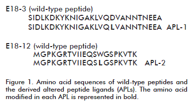
In contrast, with      previous reports, attention was put on the N-terminal region of human Hsp60.      In this region, the computer algorithm Propred [10] predicted two epitopes,      corresponding to amino acid 90-109 and 55-75, directly involved in the interaction      with MHC class II molecule. These peptides were called E18-3 and E18-12 and      the sequences are shown in <a href="#fig1">Figure 1</a>. An amino acid residue      involved in the interaction with the MHC II was changed in each peptide to      increase the affinity for this molecule. MHC II molecules frequently expressed      by RA patients were considered. The affinity improved new peptides were called      APL-1 and APL-2 and their sequences are shown in <a href="#fig1">figure 1</a>.      </font></P >       <P   align="center" ><img src="/img/revistas/bta/v30n2/f0111213.gif" width="388" height="210"><a name="fig1"></a></P >       
<P   ><font size="2" face="Verdana, Arial, Helvetica, sans-serif"><b>Evaluation of      regulatory T cells induced by APL-1 in PBMC from RA patients </b></font></P >       <P   align="justify" ><font size="2" face="Verdana, Arial, Helvetica, sans-serif">The induction of      regulatory T cells (Tregs) by the APL-1 peptide in <I>ex vivo</I> assays using      PBMCs or synovial fluid mononuclear cells (SFMCs) from RA patients was analyzed.      In total, 13 patients were included in this study. PBMCs were stimulated with      40 &micro;g/mL of APL-1 or wild-type peptide for 5 days.The samples were screened      for frequency of FoxP3+ cells among CD25<Sup>high</Sup>CD4+ T cells by flow      cytometry. For this purpose, T lymphocytes were first gated for CD4+, and      further gates were applied on that subset to select those coexpressing low      or high levels of CD25. FoxP3 was performed on CD25<Sup>high</Sup>CD4+ gated      T cells. A relative increase in FoxP3+ CD25<Sup>high</Sup>CD4+ T cells was      found in PBMCs treated with APL-1 in comparison to PBMCs stimulated either      with the wild-type peptide or without stimulation (<a href="#fig2">Figure      2</a>). </font></P >       <P   align="center" ><img src="/img/revistas/bta/v30n2/f0211213.gif" width="387" height="413"><a name="fig2"></a></P >   <FONT size="+1"><FONT size="+1"><FONT size="+1"><FONT size="+1"><FONT size="+1"><FONT size="+1">        
]]></body>
<body><![CDATA[<P   ><font size="2" face="Verdana, Arial, Helvetica, sans-serif"><b>Evaluation of      IL-17, IL-10 and TNF-&alpha; levels induced by APL-2 in PBMCs from RA patients</b>      </font></P >       <P   align="justify" ><font size="2" face="Verdana, Arial, Helvetica, sans-serif">Once demonstrated      that APL-2 induced proliferation of CD4+ T cells, the effect of this peptide      on cytokine secretion was further evaluated. PBMCs without peptide stimulation      were used as negative control. APL-2 suppressed the interleukin 17 (IL-17)      levels <I>in vitro</I> by more than 50 % and remarkably increased IL-10 levels      more than 3 times compared to negative controls. Tumor necrosis factor alpha      (TNF-&alpha;) levels remained steady regardless the treatment. </font></P >       <P   ><font size="2" face="Verdana, Arial, Helvetica, sans-serif"><b>Therapeutic evaluation      of APLs on AA model </b></font></P >       <P   align="justify" ><font size="2" face="Verdana, Arial, Helvetica, sans-serif">
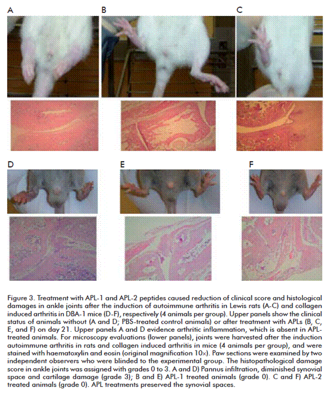
Therapeutic effects      of APLs on arthritis was evaluated and compared with wild-type peptides in      the AA model. The signs associated with the development of arthritis began      gradually in all animals inoculated with <I>Mycobacterium tuberculosis</I>.      These signs were evident on day 10, characterized by a slight redness and      swelling of the posterior joints. On day 11, the rats were randomly divided      in several groups (n = 12). The mean arthritis score on day 21 (the day of      maximum arthritis severity) was the main parameter used in this study to measure      clinical outcomes and evaluation of the effects of the peptides. A significant      reduction of AA means arthritis score on day 21 was observed with APL-1 and      APL-2 compared to rats treated with phosphate buffer saline (PBS). The improvement      induced by both APLs was comparable to healthy animals. Clinical improvement      of AA with the APLs is corresponded with a decrease of joint destruction by      the arthritic process. Four animals were sacrificed per group and ankle joints      were collected on day 21 after induction of AA and scored for severity. The      histological examination of the joints showed severe erosion of cartilage      and bone as well as massive inflammatory cell infiltration and obvious pannus      formation in all rats inoculated with PBS. In contrast, subcutaneous administration      of APL-1 or APL-2 resulted in a suppression of these histological signs characteristic      of AA. However, in all rats treated with wild-type peptides presented a histological      score of 3, with massive cellular infiltration, synovitis and moderate erosions      of bone and cartilage. These results indicate that both APLs suppressed AA      efficiently (<a href="/img/revistas/bta/v30n2/f0311213.gif">Figure 3A-C</a>). </font></P >       
<P   align="justify" ><font size="2" face="Verdana, Arial, Helvetica, sans-serif">In addition, the      production of TNF-&alpha; was investigated in the spleen from sacrificed rats      at day 21 after disease induction. Administration of APLs significantly reduced      TNF-&alpha; levels. </font></P >       <P   align="justify" ><font size="2" face="Verdana, Arial, Helvetica, sans-serif">On the other hand,      the capacity of APL-1 to induce regulatory T cells during the therapy with      this peptide in rats was confirmed by a significant increase in the percentages      of T cells with phenotype CD4+CD25<Sup>high</Sup>FoxP3+ in rats treated with      APL-1, compared to rats either without treatment or treated with the corresponding      wild-type peptide. </font></P >   <FONT size="+1"><FONT size="+1">        <P   ><font size="2" face="Verdana, Arial, Helvetica, sans-serif"><b>Therapeutic evaluation      of APL-2 on CIA model </b></font></P >       <P   align="justify" ><font size="2" face="Verdana, Arial, Helvetica, sans-serif">Therapeutic effect      of APLs were evaluated and compared to MTX, in another animal model where      arthritis is induced by a different autoantigen. CIA was elicited in DBA/1      mice by two subcutaneous immunizations with collagen type II. Thirty-two out      of thirty-six mice (88 %) developed arthritis between days 23 and 26, showing      evidences of clinical inflammation in one or two hind paws. CIA mice were      randomly allocated in several groups (n = 12 per group), and showed no differences      in the arthritis score among them. Then, the CIA mice were treated with APL-1,      APL-2 or MTX. PBS was injected as negative control. </font></P >       <P   align="justify" ><font size="2" face="Verdana, Arial, Helvetica, sans-serif">Treatment with APLs      reduced the severity of CIA compared to PBS treated mice from day 37 after      the first immunization until the end of protocol. Similar results were observed      in MTX treated animals. On day 46, 4 mice per group were sacrificed for histological      examination. A close correlation between the clinical signs and the histopathological      findings was found. All untreated mice developed a histological score of 3.      In contrast, mice inoculated with APLs only showed slight histological damages      in the joints (<a href="/img/revistas/bta/v30n2/f0311213.gif">Figure 3D-F</a>). </font></P >       
<P   align="justify" ><font size="2" face="Verdana, Arial, Helvetica, sans-serif">To investigate the      mechanism of action of both APLs in suppressing CIA, the levels of TNF-&alpha;,      IL-17 and IL-10 were measured in serum of mice sacrificed on day 46. The treatment      with APL-2 significantly reduced TNF-&alpha; and IL-17 secretion compared      to PBS inoculated mice. Levels of both cytokines in this group were similar      to those obtained in healthy animals. The administration of APL-1 or MTX significantly      reduced TNF-&alpha; secretion, but did not modify IL-17. Otherwise, only the      combination of APL-1 with MTX increased the percentages of T cells with CD4+CD25<Sup>high</Sup>FoxP3+      phenotype in mice. </font></P >       ]]></body>
<body><![CDATA[<P   align="justify" >&nbsp;</P >   <FONT size="+1"><FONT size="+1">        <P   ><font size="2" face="Verdana, Arial, Helvetica, sans-serif"><B><font size="3">DISCUSSION      </font></b></font></P >   <FONT size="+1">        <P   align="justify" ><font size="2" face="Verdana, Arial, Helvetica, sans-serif">Conceptually, the      therapeutic intervention presented here is based on modulating the T cell      function and therefore, higher specificity and lower toxicity is expected      [11]. In this study, two novel T cell epitopes from the hHsp60 were defined.      According to our prediction, these peptides represent strong epitopes directly      involved in the interaction with human MHC II </font></P >   <FONT size="+1">        <P   align="justify" ><font size="2" face="Verdana, Arial, Helvetica, sans-serif">molecules, particularly      those related to RA. We evaluated the potential of these wild-type peptides      to modify proinflammatory and immunoregulatory cytokine levels in <I>ex vivo</I>      assays using PBMCs from patients with RA. In theses assays, an increment of      TNF-&alpha; and INF-&gamma; levels was confirmed. </font></P >       <P   align="justify" ><font size="2" face="Verdana, Arial, Helvetica, sans-serif">Two APLs were designed      from these wild-type peptides, retaining the binding properties to MHC II      molecules, but carrying single mutations of an essential contact position      for its binding. All these modifications were aimed at transforming the T      cell response generated by the wild-type peptides towards the tolerance induction      in RA patients. </font></P >       <P   align="justify" ><font size="2" face="Verdana, Arial, Helvetica, sans-serif">Recent studies have      reported defects in the number and/or activity of Tregs in humans with RA,      similar to those observed in mouse models of arthritis [12]. Failures in the      function of the Tregs can therefore be responsible for the development of      autoimmune diseases, and enhancing their functions may represent a feasible      treatment strategy. We suggested that the modification in APL-1 could induce      Tregs in patients that may attenuate the pathogenic T cells with specificity,      without affecting other non-related T cells. To test this hypothesis, the      induction of Tregs by APL-1 was analyzed in <I>ex vivo</I> assays using PBMCs      or SFMCs from RA patients, and it was confirmed that APL-1 induced an increment      of Tregs with a CD4+CD25</font></P >       <P   align="justify" ><font size="2" face="Verdana, Arial, Helvetica, sans-serif">On the other hand,      APL-2 preferentially increased IL-10 and decreased IL-17 but do not have any      effect on TNF-&alpha; level in <I>ex vivo</I> assays with PBMCs isolated from      RA patients, suggesting a deviation from inflammatory to regulatory cytokine      profile. This peptide increased IL-10 levels in all patients&rsquo; PBMCs      irrespective of their HLA background; therefore APL-2 could bind to several      allelic variants of the human MHC II (DQ and DR). </font></P >       <P   align="justify" ><font size="2" face="Verdana, Arial, Helvetica, sans-serif">To explore the therapeutic      effect of the APLs, AA and CIA animal models were chosen. The treatment with      the APL-1 induced excellent clinical in both models. This effect was correlated      with improvement of the histological score of the joints induced by the peptide,      and it was comparable to healthy animals. In contrast, the wild-type peptide      (E18-3) did not induce any clinical or histological improvement in the animals.      According to these results, we can confirm that the modification carried out      in E18-3 was very effective through attenuation of the pathogenic inflammation      in these animal models. The clinical efficacy achieved by the treatment with      APL-1 was associated with an increment in Tregs in the spleen. The Tregs induced      by APL-1 in the periphery apparently could migrate to the joints and induce      suppression of the local self-reactive response. In addition, we found that      the therapy with the APL-1 reduces significantly the TNF-&alpha; level in      spleen. Given these facts, we think that probably the potent therapeutic effect      of the APL-1 in the reduction of AA and CIA would be due to the processing      and presentation of the peptide by the antigen presenting cells to the autoreactive      T lymphocytes in periphery. The recognition of this altered ligand may induce      the expansion of T cells with immunoregulatory phenotype like CD4+CD25<Sup>high</Sup>FoxP3+      Tregs. The activated cells migrate to the inflammation site and they could      cross-recognize the native epitope from the hHsp60, where it is highly expressed      due to the inflammation process. </font></P >   <FONT size="+1"><FONT size="+1">        <P   align="justify" ><font size="2" face="Verdana, Arial, Helvetica, sans-serif">This new contact      with the autoantigen may induce potent immunoregulatory effect, attenuating      the autoreactive T cells responsible for arthritis pathogenesis and inhibiting      the TNF-&alpha; expression. The pivotal role of TNF-&alpha; in the induction      and progression of rheumatoid synovitis is well established [13]. Consequently,      the result demonstrating that APL-1 inhibited the expression of TNF-&alpha;      represents a beneficial effect for the control of the inflammatory process.      </font></P >       <P   align="justify" ><font size="2" face="Verdana, Arial, Helvetica, sans-serif">Similarly, APL-2      was effective for down-regulating the inflammatory response in AA model with      a reduction of TNF-&alpha; level. Also, APL-2 reduced the TNF-&alpha; level      in sera of CIA mice to concentrations comparable to healthy animals. Additionally,      pannus formation and joint damage were not observed. Similar results were      obtained in MTX-treated mice. </font></P >       ]]></body>
<body><![CDATA[<P   align="justify" ><font size="2" face="Verdana, Arial, Helvetica, sans-serif">Many evidences support      the role of IL-17 in the pathogenesis of human RA and its animal models such      as CIA [14, 15]. The decrease of IL-17 and TNF-&alpha; secretion induced by      APL-2 could indicate that the therapeutic effect of this peptide is mediated      by downregulation of inflammatory cytokines rather than induction of Tregs.      Consistent with this, APL-2 treatment does not induce Tregs with a Foxp3+      CD4+ phenotype at day 46 in spleen of CIA mice. </font></P >       <P   align="justify" >&nbsp;</P >       <P   ><font size="2" face="Verdana, Arial, Helvetica, sans-serif"><B><font size="3">CONCLUSIONS      </font></b></font></P >   <FONT size="+1">        <P   align="justify" ><font size="2" face="Verdana, Arial, Helvetica, sans-serif">The therapeutic effect      induced by APLs in both animal models is mediated by different molecular mechanisms,      associated with immunologic tolerance. These results indicate a therapeutic      potential for these APLs and support further investigation of these candidate      drugs for RA treatment. </font></P >   <FONT size="+1">        <P   align="justify" ><font size="2" face="Verdana, Arial, Helvetica, sans-serif">Intervention on T      cell function in a specific manner as shown here would bring the possibility      of focusing on one or more antigens involved in the autoimmune pathogenesis,      thus avoiding the general immune suppression in patients as happens with anti-TNF-&alpha;      treatment. This study contributes to the knowledge of mechanisms and tools      needed for the induction of tolerance in humans using autoantigens or their      variants. </font></P >       <P   align="justify" >&nbsp;</P >   <FONT size="+1">        <P   align="justify" > </P >   <FONT size="+1"><B>        <P   align="justify" ><font size="3" face="Verdana, Arial, Helvetica, sans-serif">REFERENCES </font></P >   </B>        <!-- ref --><P   align="justify" ><font size="2" face="Verdana, Arial, Helvetica, sans-serif">1. Breedveld FC,      Weisman MH, Kavanaugh AF, Cohen SB, Pavelka K, van Vollenhoven R, <I>et al</I>.      The PREMIER study: A multicenter, randomized, double-blind clinical trial      of combination therapy with adalimumab plus methotrexate versus methotrexate      alone or adalimumab alone in patients with early, aggressive rheumatoid arthritis      who had not had previous methotrexate treatment. Arthritis Rheum. 2006;54(1):26-37.          </font></P >   <FONT size="+1">        ]]></body>
<body><![CDATA[<P   align="justify" ><font size="2" face="Verdana, Arial, Helvetica, sans-serif">2. Kooloos WM, de      Jong DJ, Huizinga TW, Guchelaar HJ. Potential role of pharmacogenetics in      anti-TNF treatment of rheumatoid arthritis and Crohn&rsquo;s disease. Drug      Discov Today. 2007;12(3-4):125-31. </font></P >       <!-- ref --><P   align="justify" ><font size="2" face="Verdana, Arial, Helvetica, sans-serif">3. van Vollenhoven      RF. Treatment of rheumatoid arthritis: state of the art 2009. Nat Rev Rheumatol<I>.</I>      2009;5(10):531-41.     </font></P >       <!-- ref --><P   align="justify" ><font size="2" face="Verdana, Arial, Helvetica, sans-serif">4. Satpute SR, Durai      M, Moudgil KD. Antigen-specific tolerogenic and immunomodulatory strategies      for the treatment of autoimmune arthritis. Semin Arthritis Rheum. 2008;38(3):195-207.          </font></P >       <!-- ref --><P   align="justify" ><font size="2" face="Verdana, Arial, Helvetica, sans-serif">5. van Eden W, van      der Zee R, Prakken B. Heat-shock proteins induce T-cell regulation of chronic      inflammation. Nat Rev Immunol. 2005;5(4):318-30.     </font></P >       <!-- ref --><P   align="justify" ><font size="2" face="Verdana, Arial, Helvetica, sans-serif">6. Sakaguchi S, Yamaguchi      T, Nomura T, Ono M. Regulatory T cells and immune tolerance. Cell. 2008;133(5):775-87.          </font></P >       <!-- ref --><P   align="justify" ><font size="2" face="Verdana, Arial, Helvetica, sans-serif">7. Prakken BJ, Roord      S, van Kooten PJ, Wagenaar JP, van Eden W, Albani S, <I>et al</I>. Inhibition      of adjuvant-induced arthritis by interleukin-10-driven regulatory cells induced      via nasal administration of a peptide analog of an arthritis-related heat-shock      protein 60 T cell epitope. Arthritis Rheum. 2002;46(7):1937-46.     </font></P >       <!-- ref --><P   align="justify" ><font size="2" face="Verdana, Arial, Helvetica, sans-serif">8. Huurman VA, van      der Meide PE, Duinkerken G, Willemen S, Cohen IR, Elias D, et al. Immunological      efficacy of heat shock protein 60 peptide DiaPep277 therapy in clinical type      I diabetes. Clin Exp Immunol. 2008;152(3):488-97.     </font></P >       <!-- ref --><P   align="justify" ><font size="2" face="Verdana, Arial, Helvetica, sans-serif">9. Bielekova B, Martin      R. Antigen-specific immunomodulation via altered peptide ligands. J Mol Med      (Berl). 2001;79(10):552-65.     </font></P >       <!-- ref --><P   align="justify" ><font size="2" face="Verdana, Arial, Helvetica, sans-serif">10. Lin HH, Zhang      GL, Tongchusak S, Reinherz EL, Brusic V. Evaluation of MHC-II peptide binding      prediction servers: applications for vaccine research. BMC. Bioinformatics.      2008;9 Suppl 12:S22.     </font></P >       <!-- ref --><P   align="justify" ><font size="2" face="Verdana, Arial, Helvetica, sans-serif">11. Garrood T, Pitzalis      C. Targeting the inflamed synovium: the quest for specificity. Arthritis Rheum.      2006;54(4):1055-60.     </font></P >       <!-- ref --><P   align="justify" ><font size="2" face="Verdana, Arial, Helvetica, sans-serif">12. van Amelsfort      JM, Jacobs KM, Bijlsma JW, Lafeber FP, Taams LS. CD4(+)CD25(+) regulatory      T cells in rheumatoid arthritis: differences in the presence, phenotype, and      function between peripheral blood and synovial fluid. Arthritis Rheum. 2004;50(9):2775-85.          </font></P >       <!-- ref --><P   align="justify" ><font size="2" face="Verdana, Arial, Helvetica, sans-serif">13. Choy EH, Panayi      GS. Cytokine pathways and joint inflammation in rheumatoid arthritis. N Engl      J Med. 2001;344(12):907-16.     </font></P >       <!-- ref --><P   align="justify" ><font size="2" face="Verdana, Arial, Helvetica, sans-serif">14. Park H, Li Z,      Yang XO, Chang SH, Nurieva R, Wang YH, <I>et al</I>. A distinct lineage of      CD4 T cells regulates tissue inflammation by producing interleukin 17. Nat      Immunol. 2005;6(11):1133-41.     </font></P >       <!-- ref --><P   align="justify" ><font size="2" face="Verdana, Arial, Helvetica, sans-serif">15. Nakae S, Nambu      A, Sudo K, Iwakura Y. Suppression of immune induction of collagen-induced      arthritis in IL-17-deficient mice. J Immunol. 2003;171(11):6173-7.     </font></P >       <P   align="justify" >&nbsp;</P >       <P   align="justify" >&nbsp;</P >       <P   align="justify" > </P >   <FONT size="+1">        ]]></body>
<body><![CDATA[<P   align="justify" > </P >       <P   align="justify" ><font size="2" face="Verdana, Arial, Helvetica, sans-serif"><I>Mar&iacute;a del      C Dom&iacute;nguez</I>. Centro de Ingenier&iacute;a Gen&eacute;tica y Biotecnolog&iacute;a,      CIGB. Ave 31 e/ 158 y 190, Cubanac&aacute;n, Playa, CP 11600, La Habana, Cuba.      E-mail: <A href="mailto:mcarmen.dominguez@cigb.edu.cu"> <U><U><FONT color="#0000FF">mcarmen.dominguez@cigb.edu.cu</font></U></U></A><FONT color="#0000FF"><FONT color="#000000">.      </font></font></font></P >   <FONT color="#0000FF"><FONT color="#000000">        <P   > </P >   </font></font></font></font></font></font></font></font></font></font></font></font></font></font></font></font></font></font></font></font></font></font></font></font></font></font></font></font></font></font></font></font></font></font></font></font></font></font></font></font></font></font></font></font></font></font></font></font></font></font></font></font></font></font></font></font></font></font></font></font></font></font></font></font></font></font></font></font></font></font></font></DIV >      ]]></body><back>
<ref-list>
<ref id="B1">
<label>1</label><nlm-citation citation-type="journal">
<person-group person-group-type="author">
<name>
<surname><![CDATA[Breedveld]]></surname>
<given-names><![CDATA[FC]]></given-names>
</name>
<name>
<surname><![CDATA[Weisman]]></surname>
<given-names><![CDATA[MH]]></given-names>
</name>
<name>
<surname><![CDATA[Kavanaugh]]></surname>
<given-names><![CDATA[AF]]></given-names>
</name>
<name>
<surname><![CDATA[Cohen]]></surname>
<given-names><![CDATA[SB]]></given-names>
</name>
<name>
<surname><![CDATA[Pavelka]]></surname>
<given-names><![CDATA[K]]></given-names>
</name>
<name>
<surname><![CDATA[van Vollenhoven]]></surname>
<given-names><![CDATA[R]]></given-names>
</name>
</person-group>
<article-title xml:lang="en"><![CDATA[The PREMIER study: A multicenter, randomized, double-blind clinical trial of combination therapy with adalimumab plus methotrexate versus methotrexate alone or adalimumab alone in patients with early, aggressive rheumatoid arthritis who had not had previous methotrexate treatment]]></article-title>
<source><![CDATA[Arthritis Rheum]]></source>
<year>2006</year>
<volume>54</volume>
<numero>1</numero>
<issue>1</issue>
<page-range>26-37</page-range></nlm-citation>
</ref>
<ref id="B2">
<label>2</label><nlm-citation citation-type="journal">
<person-group person-group-type="author">
<name>
<surname><![CDATA[Kooloos]]></surname>
<given-names><![CDATA[WM]]></given-names>
</name>
<name>
<surname><![CDATA[de Jong]]></surname>
<given-names><![CDATA[DJ]]></given-names>
</name>
<name>
<surname><![CDATA[Huizinga]]></surname>
<given-names><![CDATA[TW]]></given-names>
</name>
<name>
<surname><![CDATA[Guchelaar]]></surname>
<given-names><![CDATA[HJ]]></given-names>
</name>
</person-group>
<article-title xml:lang="en"><![CDATA[Potential role of pharmacogenetics in anti-TNF treatment of rheumatoid arthritis and Crohn's disease]]></article-title>
<source><![CDATA[Drug Discov Today]]></source>
<year>2007</year>
<volume>12</volume>
<numero>3-4</numero>
<issue>3-4</issue>
<page-range>125-31</page-range></nlm-citation>
</ref>
<ref id="B3">
<label>3</label><nlm-citation citation-type="journal">
<person-group person-group-type="author">
<name>
<surname><![CDATA[van Vollenhoven]]></surname>
<given-names><![CDATA[RF]]></given-names>
</name>
</person-group>
<article-title xml:lang="en"><![CDATA[Treatment of rheumatoid arthritis: state of the art 2009]]></article-title>
<source><![CDATA[Nat Rev Rheumatol]]></source>
<year>2009</year>
<volume>5</volume>
<numero>10</numero>
<issue>10</issue>
<page-range>531-41</page-range></nlm-citation>
</ref>
<ref id="B4">
<label>4</label><nlm-citation citation-type="journal">
<person-group person-group-type="author">
<name>
<surname><![CDATA[Satpute]]></surname>
<given-names><![CDATA[SR]]></given-names>
</name>
<name>
<surname><![CDATA[Durai]]></surname>
<given-names><![CDATA[M]]></given-names>
</name>
<name>
<surname><![CDATA[Moudgil]]></surname>
<given-names><![CDATA[KD]]></given-names>
</name>
</person-group>
<article-title xml:lang="en"><![CDATA[Antigen-specific tolerogenic and immunomodulatory strategies for the treatment of autoimmune arthritis]]></article-title>
<source><![CDATA[Semin Arthritis Rheum]]></source>
<year>2008</year>
<volume>38</volume>
<numero>3</numero>
<issue>3</issue>
<page-range>195-207</page-range></nlm-citation>
</ref>
<ref id="B5">
<label>5</label><nlm-citation citation-type="journal">
<person-group person-group-type="author">
<name>
<surname><![CDATA[van Eden]]></surname>
<given-names><![CDATA[W]]></given-names>
</name>
<name>
<surname><![CDATA[van der Zee]]></surname>
<given-names><![CDATA[R]]></given-names>
</name>
<name>
<surname><![CDATA[Prakken]]></surname>
<given-names><![CDATA[B]]></given-names>
</name>
</person-group>
<article-title xml:lang="en"><![CDATA[Heat-shock proteins induce T-cell regulation of chronic inflammation]]></article-title>
<source><![CDATA[Nat Rev Immunol]]></source>
<year>2005</year>
<volume>5</volume>
<numero>4</numero>
<issue>4</issue>
<page-range>318-30</page-range></nlm-citation>
</ref>
<ref id="B6">
<label>6</label><nlm-citation citation-type="journal">
<person-group person-group-type="author">
<name>
<surname><![CDATA[Sakaguchi]]></surname>
<given-names><![CDATA[S]]></given-names>
</name>
<name>
<surname><![CDATA[Yamaguchi]]></surname>
<given-names><![CDATA[T]]></given-names>
</name>
<name>
<surname><![CDATA[Nomura]]></surname>
<given-names><![CDATA[T]]></given-names>
</name>
<name>
<surname><![CDATA[Ono]]></surname>
<given-names><![CDATA[M]]></given-names>
</name>
</person-group>
<article-title xml:lang="en"><![CDATA[Regulatory T cells and immune tolerance]]></article-title>
<source><![CDATA[Cell]]></source>
<year>2008</year>
<volume>133</volume>
<numero>5</numero>
<issue>5</issue>
<page-range>775-87</page-range></nlm-citation>
</ref>
<ref id="B7">
<label>7</label><nlm-citation citation-type="journal">
<person-group person-group-type="author">
<name>
<surname><![CDATA[Prakken]]></surname>
<given-names><![CDATA[BJ]]></given-names>
</name>
<name>
<surname><![CDATA[Roord]]></surname>
<given-names><![CDATA[S]]></given-names>
</name>
<name>
<surname><![CDATA[van Kooten]]></surname>
<given-names><![CDATA[PJ]]></given-names>
</name>
<name>
<surname><![CDATA[Wagenaar]]></surname>
<given-names><![CDATA[JP]]></given-names>
</name>
<name>
<surname><![CDATA[van Eden]]></surname>
<given-names><![CDATA[W]]></given-names>
</name>
<name>
<surname><![CDATA[Albani]]></surname>
<given-names><![CDATA[S]]></given-names>
</name>
</person-group>
<article-title xml:lang="en"><![CDATA[Inhibition of adjuvant-induced arthritis by interleukin-10-driven regulatory cells induced via nasal administration of a peptide analog of an arthritis-related heat-shock protein 60 T cell epitope]]></article-title>
<source><![CDATA[Arthritis Rheum]]></source>
<year>2002</year>
<volume>46</volume>
<numero>7</numero>
<issue>7</issue>
<page-range>1937-46</page-range></nlm-citation>
</ref>
<ref id="B8">
<label>8</label><nlm-citation citation-type="journal">
<person-group person-group-type="author">
<name>
<surname><![CDATA[Huurman]]></surname>
<given-names><![CDATA[VA]]></given-names>
</name>
<name>
<surname><![CDATA[van der Meide]]></surname>
<given-names><![CDATA[PE]]></given-names>
</name>
<name>
<surname><![CDATA[Duinkerken]]></surname>
<given-names><![CDATA[G]]></given-names>
</name>
<name>
<surname><![CDATA[Willemen]]></surname>
<given-names><![CDATA[S]]></given-names>
</name>
<name>
<surname><![CDATA[Cohen]]></surname>
<given-names><![CDATA[IR]]></given-names>
</name>
<name>
<surname><![CDATA[Elias]]></surname>
<given-names><![CDATA[D]]></given-names>
</name>
</person-group>
<article-title xml:lang="en"><![CDATA[Immunological efficacy of heat shock protein 60 peptide DiaPep277 therapy in clinical type I diabetes]]></article-title>
<source><![CDATA[Clin Exp Immunol]]></source>
<year>2008</year>
<volume>152</volume>
<numero>3</numero>
<issue>3</issue>
<page-range>488-97</page-range></nlm-citation>
</ref>
<ref id="B9">
<label>9</label><nlm-citation citation-type="journal">
<person-group person-group-type="author">
<name>
<surname><![CDATA[Bielekova]]></surname>
<given-names><![CDATA[B]]></given-names>
</name>
<name>
<surname><![CDATA[Martin]]></surname>
<given-names><![CDATA[R]]></given-names>
</name>
</person-group>
<article-title xml:lang="en"><![CDATA[Antigen-specific immunomodulation via altered peptide ligands]]></article-title>
<source><![CDATA[J Mol Med (Berl)]]></source>
<year>2001</year>
<volume>79</volume>
<numero>10</numero>
<issue>10</issue>
<page-range>552-65</page-range></nlm-citation>
</ref>
<ref id="B10">
<label>10</label><nlm-citation citation-type="journal">
<person-group person-group-type="author">
<name>
<surname><![CDATA[Lin]]></surname>
<given-names><![CDATA[HH]]></given-names>
</name>
<name>
<surname><![CDATA[Zhang]]></surname>
<given-names><![CDATA[GL]]></given-names>
</name>
<name>
<surname><![CDATA[Tongchusak]]></surname>
<given-names><![CDATA[S]]></given-names>
</name>
<name>
<surname><![CDATA[Reinherz]]></surname>
<given-names><![CDATA[EL]]></given-names>
</name>
<name>
<surname><![CDATA[Brusic]]></surname>
<given-names><![CDATA[V]]></given-names>
</name>
</person-group>
<article-title xml:lang="en"><![CDATA[Evaluation of MHC-II peptide binding prediction servers: applications for vaccine research]]></article-title>
<source><![CDATA[BMC Bioinformatics]]></source>
<year>2008</year>
<volume>9</volume>
<numero>^s12</numero>
<issue>^s12</issue>
<supplement>12</supplement>
<page-range>S22</page-range></nlm-citation>
</ref>
<ref id="B11">
<label>11</label><nlm-citation citation-type="journal">
<person-group person-group-type="author">
<name>
<surname><![CDATA[Garrood]]></surname>
<given-names><![CDATA[T]]></given-names>
</name>
<name>
<surname><![CDATA[Pitzalis]]></surname>
<given-names><![CDATA[C]]></given-names>
</name>
</person-group>
<article-title xml:lang="en"><![CDATA[Targeting the inflamed synovium: the quest for specificity]]></article-title>
<source><![CDATA[Arthritis Rheum]]></source>
<year>2006</year>
<volume>54</volume>
<numero>4</numero>
<issue>4</issue>
<page-range>1055-60</page-range></nlm-citation>
</ref>
<ref id="B12">
<label>12</label><nlm-citation citation-type="journal">
<person-group person-group-type="author">
<name>
<surname><![CDATA[van Amelsfort]]></surname>
<given-names><![CDATA[JM]]></given-names>
</name>
<name>
<surname><![CDATA[Jacobs]]></surname>
<given-names><![CDATA[KM]]></given-names>
</name>
<name>
<surname><![CDATA[Bijlsma]]></surname>
<given-names><![CDATA[JW]]></given-names>
</name>
<name>
<surname><![CDATA[Lafeber]]></surname>
<given-names><![CDATA[FP]]></given-names>
</name>
<name>
<surname><![CDATA[Taams]]></surname>
<given-names><![CDATA[LS]]></given-names>
</name>
</person-group>
<article-title xml:lang="en"><![CDATA[CD4(+)CD25(+) regulatory T cells in rheumatoid arthritis: differences in the presence, phenotype, and function between peripheral blood and synovial fluid]]></article-title>
<source><![CDATA[Arthritis Rheum]]></source>
<year>2004</year>
<volume>50</volume>
<numero>9</numero>
<issue>9</issue>
<page-range>2775-85</page-range></nlm-citation>
</ref>
<ref id="B13">
<label>13</label><nlm-citation citation-type="journal">
<person-group person-group-type="author">
<name>
<surname><![CDATA[Choy]]></surname>
<given-names><![CDATA[EH]]></given-names>
</name>
<name>
<surname><![CDATA[Panayi]]></surname>
<given-names><![CDATA[GS]]></given-names>
</name>
</person-group>
<article-title xml:lang="en"><![CDATA[Cytokine pathways and joint inflammation in rheumatoid arthritis]]></article-title>
<source><![CDATA[N Engl J Med]]></source>
<year>2001</year>
<volume>344</volume>
<numero>12</numero>
<issue>12</issue>
<page-range>907-16</page-range></nlm-citation>
</ref>
<ref id="B14">
<label>14</label><nlm-citation citation-type="journal">
<person-group person-group-type="author">
<name>
<surname><![CDATA[Park]]></surname>
<given-names><![CDATA[H]]></given-names>
</name>
<name>
<surname><![CDATA[Li]]></surname>
<given-names><![CDATA[Z]]></given-names>
</name>
<name>
<surname><![CDATA[Yang]]></surname>
<given-names><![CDATA[XO]]></given-names>
</name>
<name>
<surname><![CDATA[Chang]]></surname>
<given-names><![CDATA[SH]]></given-names>
</name>
<name>
<surname><![CDATA[Nurieva]]></surname>
<given-names><![CDATA[R]]></given-names>
</name>
<name>
<surname><![CDATA[Wang]]></surname>
<given-names><![CDATA[YH]]></given-names>
</name>
</person-group>
<article-title xml:lang="en"><![CDATA[A distinct lineage of CD4 T cells regulates tissue inflammation by producing interleukin 17]]></article-title>
<source><![CDATA[Nat Immunol]]></source>
<year>2005</year>
<volume>6</volume>
<numero>11</numero>
<issue>11</issue>
<page-range>1133-41</page-range></nlm-citation>
</ref>
<ref id="B15">
<label>15</label><nlm-citation citation-type="journal">
<person-group person-group-type="author">
<name>
<surname><![CDATA[Nakae]]></surname>
<given-names><![CDATA[S]]></given-names>
</name>
<name>
<surname><![CDATA[Nambu]]></surname>
<given-names><![CDATA[A]]></given-names>
</name>
<name>
<surname><![CDATA[Sudo]]></surname>
<given-names><![CDATA[K]]></given-names>
</name>
<name>
<surname><![CDATA[Iwakura]]></surname>
<given-names><![CDATA[Y]]></given-names>
</name>
</person-group>
<article-title xml:lang="en"><![CDATA[Suppression of immune induction of collagen-induced arthritis in IL-17-deficient mice]]></article-title>
<source><![CDATA[J Immunol]]></source>
<year>2003</year>
<volume>171</volume>
<numero>11</numero>
<issue>11</issue>
<page-range>6173-7</page-range></nlm-citation>
</ref>
</ref-list>
</back>
</article>
