<?xml version="1.0" encoding="ISO-8859-1"?><article xmlns:mml="http://www.w3.org/1998/Math/MathML" xmlns:xlink="http://www.w3.org/1999/xlink" xmlns:xsi="http://www.w3.org/2001/XMLSchema-instance">
<front>
<journal-meta>
<journal-id>1029-3019</journal-id>
<journal-title><![CDATA[MEDISAN]]></journal-title>
<abbrev-journal-title><![CDATA[MEDISAN]]></abbrev-journal-title>
<issn>1029-3019</issn>
<publisher>
<publisher-name><![CDATA[Centro Provincial de Información de Ciencias Médicas]]></publisher-name>
</publisher>
</journal-meta>
<article-meta>
<article-id>S1029-30192009000300012</article-id>
<title-group>
<article-title xml:lang="es"><![CDATA[Presillamiento de aneurisma intracraneal en una niña mediante microcirugía y endoscopia asistida]]></article-title>
<article-title xml:lang="en"><![CDATA[Clipping of intracraneal aneurysm in a girl by means of microsurgery and assisted endoscopy]]></article-title>
</title-group>
<contrib-group>
<contrib contrib-type="author">
<name>
<surname><![CDATA[Brossard Alejo]]></surname>
<given-names><![CDATA[Julio S]]></given-names>
</name>
<xref ref-type="aff" rid="A01"/>
</contrib>
<contrib contrib-type="author">
<name>
<surname><![CDATA[Dearriba Romanidi]]></surname>
<given-names><![CDATA[Manuel]]></given-names>
</name>
<xref ref-type="aff" rid="A02"/>
</contrib>
<contrib contrib-type="author">
<name>
<surname><![CDATA[Gonzálvez Bertot]]></surname>
<given-names><![CDATA[Jaime]]></given-names>
</name>
<xref ref-type="aff" rid="A01"/>
</contrib>
<contrib contrib-type="author">
<name>
<surname><![CDATA[Vigueaux Esteve]]></surname>
<given-names><![CDATA[Adria]]></given-names>
</name>
<xref ref-type="aff" rid="A01"/>
</contrib>
<contrib contrib-type="author">
<name>
<surname><![CDATA[ardona Castillo]]></surname>
<given-names><![CDATA[Marianela]]></given-names>
</name>
<xref ref-type="aff" rid="A03"/>
</contrib>
</contrib-group>
<aff id="A01">
<institution><![CDATA[,Hospital Infantil Sur,Santiago de Cuba,Cuba  ]]></institution>
<addr-line><![CDATA[Santiago de Cuba ]]></addr-line>
<country>Cuba</country>
</aff>
<aff id="A02">
<institution><![CDATA[,Hospital Provincial Docente Saturnino Lora,Santiago de Cuba,Cuba  ]]></institution>
<addr-line><![CDATA[Santiago de Cuba ]]></addr-line>
<country>Cuba</country>
</aff>
<aff id="A03">
<institution><![CDATA[,Hospital Agostinho Neto,Guantánamo,Cuba  ]]></institution>
<addr-line><![CDATA[Guantánamo ]]></addr-line>
<country>Cuba</country>
</aff>
<pub-date pub-type="pub">
<day>00</day>
<month>06</month>
<year>2009</year>
</pub-date>
<pub-date pub-type="epub">
<day>00</day>
<month>06</month>
<year>2009</year>
</pub-date>
<volume>13</volume>
<numero>3</numero>
<fpage>0</fpage>
<lpage>0</lpage>
<copyright-statement/>
<copyright-year/>
<self-uri xlink:href="http://scielo.sld.cu/scielo.php?script=sci_arttext&amp;pid=S1029-30192009000300012&amp;lng=en&amp;nrm=iso"></self-uri><self-uri xlink:href="http://scielo.sld.cu/scielo.php?script=sci_abstract&amp;pid=S1029-30192009000300012&amp;lng=en&amp;nrm=iso"></self-uri><self-uri xlink:href="http://scielo.sld.cu/scielo.php?script=sci_pdf&amp;pid=S1029-30192009000300012&amp;lng=en&amp;nrm=iso"></self-uri><abstract abstract-type="short" xml:lang="es"><p><![CDATA[Se expone el caso clínico de una paciente de 12 años de edad, afectada por un aneurisma intracraneal de la bifurcación carótida derecha, que fue operada satisfactoriamente por medio de microcirugía con endoscopia asistida, puesto que el uso de esa técnica combinada disminuye las craneotomías y retracción del cerebro, permite una mejor visualización durante el acto quirúrgico y reduce la morbilidad operatoria. Este parece ser el primer informe en Cuba sobre el empleo de ambos procederes a la vez para el tratamiento de un aneurisma intracraneal en la infancia.]]></p></abstract>
<abstract abstract-type="short" xml:lang="en"><p><![CDATA[The clinical case of a 12 year-old patient, affected by an intracraneal aneurysm of the right carotid bifurcation who had a satisfactory surgery by means of microsurgery with assisted endoscopy is exposed, since the use of this combined technique diminishes the craneotomies and brain retraction, it allows a better visualization during the surgical intervention and reduces the surgical morbility. This seems to be the first report in Cuba on the use of both procedures at the same time for the treatment of an intracraneal aneurysm in the childhood.]]></p></abstract>
<kwd-group>
<kwd lng="es"><![CDATA[ANEURISMA INTRACRANEAL]]></kwd>
<kwd lng="es"><![CDATA[MICROCIRUGIA]]></kwd>
<kwd lng="es"><![CDATA[ENDOSCOPIA]]></kwd>
<kwd lng="en"><![CDATA[INTRACRANIAL ANEURYSM]]></kwd>
<kwd lng="en"><![CDATA[MICROSURGERY]]></kwd>
<kwd lng="en"><![CDATA[ENDOSCOPY]]></kwd>
</kwd-group>
</article-meta>
</front><body><![CDATA[ <div class=Section1>    <h2 align="right" class="Estilo24" ><span lang=ES-TRAD style=''>CASO CL&Iacute;NICO</span></h2>       <p style='line-height:150%; font-family: Arial, Helvetica, sans-serif; font-size: 10pt;'>Hospital Infantil Sur Santiago de Cuba</p>    <h2 style='text-align:justify; font-family: Arial, Helvetica, sans-serif; font-size: 14pt;'><b><span style=''>Presillamiento de aneurisma intracraneal en una niña mediante microcirugía y endoscopia asistida      <br>   </span></b>Clipping of intracraneal aneurysm in a girl by means of microsurgery and assisted endoscopy </h2>       <p><span style='font-size:9.0pt;font-family:Arial;color:black'> Julio S. Brossard Alejo, <sup>1</sup>   Manuel Dearriba Romanidi,  <sup>2  </sup> Jaime Gonzálvez Bertot,  <sup>3  </sup> Adria Vigueaux Esteve  <sup>4   </sup>y<sup>  </sup> Marianela Cardona Castillo <sup>5</sup> </span></p>    <hr>   <h4><b><span style='font-family:Arial'>RESUMEN</span></b></h4>        <p style='text-align:justify'><span style='font-size:10.0pt; font-family:Arial'>Se expone el caso clínico de una paciente de 12 años de edad, afectada por un aneurisma  intracraneal  de la  bifurcación carótida derecha, que fue operada satisfactoriamente por medio de microcirugía con endoscopia asistida, puesto que el uso de esa técnica combinada disminuye las craneotomías  y   retracción del  cerebro,  permite una mejor visualización  durante el acto quirúrgico y  reduce la morbilidad operatoria.  Este parece ser el primer informe en Cuba sobre el empleo de ambos procederes a la vez para el tratamiento de un aneurisma intracraneal en la infancia.</span></p>        <p style='text-align:justify;'><span style='font-size:10.0pt;font-family:Arial'>Descriptores:ANEURISMA<span style='color:black;'> INTRACRANEAL</span></span><span style='font-size:10.0pt;font-family:Verdana;color:black;'>/</span><span style='font-size:10.0pt;font-family:Arial;color:black; '>cirugía;MICROCIRUGIA;ENDOSCOPIA &nbsp; </span></p>        <p style='text-align:justify'><span style='font-size:10.0pt; font-family:Arial;color:black;'>Límites: </span><span style='font-size:10.0pt;font-family:Arial;'>HUMANO FEMENINO; HUMANO FEMENINO, NIÑO</span></p>    <hr>   <h4 class="Estilo9"><span style=''>ABSTRACT</span></h4>       <p align="justify" class="Estilo10">The clinical case of a 12 year-old patient, affected by an intracraneal aneurysm of the right carotid bifurcation who had a satisfactory surgery by means of microsurgery with assisted endoscopy is exposed, since the use of this combined technique diminishes the craneotomies and brain retraction, it allows a better visualization during the surgical intervention and reduces the surgical morbility. This seems to be the first report in Cuba on the use of both procedures at the same time for the treatment of an intracraneal aneurysm in the childhood. </p>       <p align="justify" style='text-align:justify; font-family: Arial, Helvetica, sans-serif; font-size: 10pt;'><span lang=EN-GB style=''>Subject headings:</span><span lang=EN-GB style='color:black;'>INTRACRANIAL ANEURYSM/surgery;MICROSURGERY;ENDOSCOPY&nbsp;</span></p>        <p align="justify" style='text-align:justify'><span lang=EN-GB style='font-size:10pt;font-family:Arial, Helvetica, sans-serif;'>Limits: <span style='color:black;'>HUMAN FEMALE; HUMAN FEMALE, CHILD </span></span></p>       ]]></body>
<body><![CDATA[<p align="justify" style='text-align:justify'><span style='font-size:10.0pt;font-family:Arial'>Recibido: 29 de enero del 2008    <br>   </span><span style='font-size:10.0pt;font-family:Arial'>Aprobado: 15 de mayo del 2008</span></p>   <hr>       <p style='text-align:justify;'><span style='font-size:10.0pt;font-family:Arial'>&nbsp;&nbsp;&nbsp;&nbsp;&nbsp;Los aneurismas intracraneales en la infancia y la adolescencia son raros. Según algunos autores su incidencia  se calcula entre 0,5 y 4,6 % del total de estos. <sup>1 - 3</sup>   </span></p>        <p style='text-align:justify;'><span style='font-size:10.0pt;font-family:Arial'>&nbsp;&nbsp;&nbsp;&nbsp;&nbsp;Los que ocurren en esta etapa difieren, en  varios aspectos, de los que suceden en los adultos:<sup> 4 - 6</sup></span></p>        <p style='text-align:justify;'><span style='font-size:10.0pt;font-family:Symbol;'>·<span style='font:7.0pt "Times New Roman"'>&nbsp;&nbsp;&nbsp;&nbsp;&nbsp;&nbsp;&nbsp;&nbsp; </span></span><span style='font-size:10.0pt;font-family:Arial'>Mayor frecuencia de aneurismas  largos y gigantes (mayores de </span><span  style='font-size:10.0pt;font-family:Arial'>25 mm</span><span style='font-size:10.0pt;font-family:Arial'>). </span></p>        <p style='text-align:justify;'><span style='font-size:10.0pt;font-family:Symbol;'>·<span style='font:7.0pt "Times New Roman"'>&nbsp;&nbsp;&nbsp;&nbsp;&nbsp;&nbsp;&nbsp;&nbsp; </span></span><span style='font-size:10.0pt;font-family:Arial'>Menor incidencia de  presentación con signos y síntomas de ruptura de aneurismas.</span></p>        <p style='text-align:justify;'><span style='font-size:10.0pt;font-family:Symbol;'>·<span style='font:7.0pt "Times New Roman"'>&nbsp;&nbsp;&nbsp;&nbsp;&nbsp;&nbsp;&nbsp;&nbsp; </span></span><span style='font-size:10.0pt;font-family:Arial'>Mayor tendencia a presentarse déficit neurológico debido al efecto de masa causado por aneurismas largos o gigantes. </span></p>        <p style='text-align:justify;'><span style='font-size:10.0pt;font-family:Symbol;'>·<span style='font:7.0pt "Times New Roman"'>&nbsp;&nbsp;&nbsp;&nbsp;&nbsp;&nbsp;&nbsp;&nbsp; </span></span><span style='font-size:10.0pt;font-family:Arial'>Predilección,  en cuanto a  localización,  en la bifurcación carótida, circulación posterior y en las porciones distales de la arteria anterior y media.</span></p>        <p style='text-align:justify;'><span style='font-size:10.0pt;font-family:Arial'>&nbsp;&nbsp;&nbsp;&nbsp;&nbsp;Estas características hacen que el tratamiento de un aneurisma en la infancia  tenga sus  particularidades. </span></p>    <h4 class="Estilo9"><span style=''>CASO CL&Iacute;NICO </span></h4>        <p style='text-align:justify;'><span style='font-size:10.0pt;font-family:Arial'>&nbsp;&nbsp;&nbsp;&nbsp;&nbsp;Paciente femenina  de 12 años de edad,  con antecedentes de salud, que  comenzó a presentar,  de forma brusca, convulsiones tónico- clónicas generalizadas, con salida de espuma por la boca, la cual fue asistida en su área de salud y una vez estabilizada clínicamente es ingresada en el Hospital Pediátrico de Guantánamo.  En el momento del examen físico se encontraba orientada, con los ojos abiertos espontáneamente,  conversaba, obedecía órdenes, sin defecto motor, pero tenía el cuello rígido.</span></p>        ]]></body>
<body><![CDATA[<p style='text-align:justify;'><span style='font-size:10.0pt;font-family:Arial'>&nbsp;&nbsp;&nbsp;&nbsp;&nbsp;Se le realizó  punción lumbar y se constató líquido cefalorraquídeo hemorrágico. </span></p>        <p style='text-align:justify;'><span style='font-size:10.0pt;font-family:Arial'>&nbsp;&nbsp;&nbsp;&nbsp;&nbsp;Teniendo en cuenta estos resultados fue trasladada a la provincia de Santiago de Cuba.</span></p>        <p style='text-align:justify'><b><span style='font-size:10.0pt;font-family:Arial'>Exámenes complementarios</span></b></p>        <p style='text-align:justify;'><span style='font-size:10.0pt;font-family:Arial;'>-<span style='font:7.0pt "Times New Roman"'>&nbsp;&nbsp;&nbsp;&nbsp;&nbsp;&nbsp;&nbsp;&nbsp;&nbsp; </span></span><span style='font-size:10.0pt;font-family:Arial'>Tomografía axial computarizada: Se  observa sangre en cisternas basales más prominente del lado derecho.  Escala de Fisher II.</span></p>        <p style='text-align:justify;'><span style='font-size:10.0pt;font-family:Arial;'>-<span style='font:7.0pt "Times New Roman"'>&nbsp;&nbsp;&nbsp;&nbsp;&nbsp;&nbsp;&nbsp;&nbsp;&nbsp;      </span></span><span style='font-size:10.0pt;font-family:Arial'>
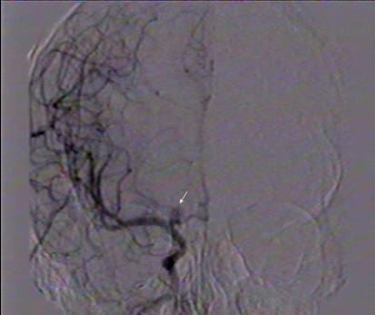
Angiografía      por sustracción digital por técnica de cateterismo femoral: Se aprecia un      aneurisma de bifurcación carótida derecha (<b><a href="#f1">figura 1</a></b>).       </span></p>        <p align=center style='text-align:center;'><a href="/img/revistas/san/v13n3/f0912309.jpg"><img src="/Img/revistas/san/v13n3/f0912309.jpg" width="141" height="107" border="0"></a><a name="f1"></a></p>        
<blockquote>         <blockquote>           <blockquote>             <p align="center"> <span class="Estilo10">&nbsp;&nbsp;&nbsp;Figura 1. <em>Aneurisma de la bifurcaci&oacute;n     ]]></body>
<body><![CDATA[<br> &nbsp;&nbsp;&nbsp;&nbsp;&nbsp;        car&oacute;tida intracraneal </em></span></p>       </blockquote>     </blockquote>   </blockquote>       <p style="font-size:10.0pt;font-family:Arial">&nbsp;&nbsp;&nbsp;&nbsp;&nbsp;Es      ingresada en el servicio de Neurocirugía del  Hospital Infantil Sur  de Santiago      de Cuba  e  intervenida quirúrgicamente diez días después, mediante craneotomía      pterional y presillamiento de aneurisma con microcirugía y endoscopia asistida      (<b><a href="#f2">figura 2</a></b>). </p>       <p align="center" style="font-size:10.0pt;font-family:Arial"><a href="/img/revista/san/v13n3/f1012309.jpg"><img src="/Img/revistas/san/v13n3/f1012309.jpg" width="140" height="105" border="0"></a><a name="f2"></a></p>       
<blockquote>         <blockquote>           <blockquote>             <blockquote>               <p align="center" style="font-size:10.0pt;font-family:Arial">Figura 2. <em>Vista endosc&oacute;pica de presilla en el cuello del aneurisma intracraneal </em></p>         </blockquote>       </blockquote>     </blockquote>   </blockquote>       <p align="left" style="font-size:10.0pt;font-family:Arial">&nbsp;&nbsp;&nbsp;&nbsp;&nbsp;<span style='font-size:10.0pt;font-family:Arial'>La evolución posquirúrgica fue favorable y la paciente es  seguida por consulta externa.  A los 34 meses de operada continúa  asintomática y está incorporada  a la escuela con buenos resultados académicos y vida social normal. </span></p>   <h4><b><span style='font-family:Arial'>Comentarios</span></b></h4>        <p style='text-align:justify;'><span style='font-size:10.0pt;font-family:Arial;'>&nbsp;&nbsp;&nbsp;&nbsp;&nbsp;Los aneurismas intracraneales durante la niñez están asociados con varias enfermedades: coartación de la aorta, riñón poliquístico, esclerosis tuberosa, síndrome de Ehler Danlos,  síndrome de Marfan y la enfermedad de Moyamoya, pero su patogénesis  en los niños es aún controvertida.</span></p>        ]]></body>
<body><![CDATA[<p style='text-align:justify;'><span style='font-size:10.0pt;font-family:Arial;'>&nbsp;&nbsp;&nbsp;&nbsp;&nbsp;Factores hemodinámicos  crónicos en determinados  puntos de las ramas arteriales o áreas donde las arterias cambian abruptamente la curvatura, han sido también invocados en la patogénesis de dichos aneurismas. <sup>7</sup> </span></p>        <p style='text-align:justify;'><span style='font-size:10.0pt;font-family:Arial'>&nbsp;&nbsp;&nbsp;&nbsp;&nbsp;Los actuales progresos en técnicas de diagnóstico, microcirugía, tratamientos endovasculares  y neuroanestesia, permiten tratar con  excelentes resultados esta enfermedad en los niños.  <sup> 5,  7</sup></span></p>        <p style='text-align:justify;'><span style='font-size:10.0pt;font-family:Arial'>&nbsp;&nbsp;&nbsp;&nbsp;&nbsp;Los aneurismas localizados en la bifurcación carotídea tienen  complejas relaciones arteriales que le confieren un riesgo particular, ya que el neurocirujano debe de  prestar atención a estructuras como: vasos perforantes originados  en la propia bifurcación, arteria recurrente de Heubner, arteria cerebral anterior, arteria cerebral media y sus perforantes, arteria coroidea anterior  y la arteria hipofisiaria superior. <sup>8</sup></span></p>        <p style='text-align:justify;'><span style='font-size:10.0pt;font-family:Arial'>&nbsp;&nbsp;&nbsp;&nbsp;&nbsp;Recientes avances  en cuanto a instrumentos ópticos permiten una clara visión a través de endoscopios de pequeño calibre y la introducción de estos en regiones estrechas y profundas del cerebro. Varios  estudios  han demostrado la utilidad de la endoscopia asistida en las operaciones microquirúrgicas de aneurismas cerebrales.   <sup>9, 10</sup>   </span></p>        <p style='text-align:justify;'><span style='font-size:10.0pt;font-family:Arial'>&nbsp;&nbsp;&nbsp;&nbsp;&nbsp;Los aneurismas  de la bifurcación carótida  se proyectan hacia arriba, pero también pueden dirigirse hacia delante  o hacia atrás. Estos sacos que se dirigen hacia arriba  y hacia delante  penetran en el espacio perforado anterior y la posición más medial o lateral  del saco imprime otras complejidades  a esta localización.  <sup>8</sup></span></p>        <p style='text-align:justify;'><span style='font-size:10.0pt;font-family:Arial'> &nbsp;&nbsp;&nbsp;&nbsp;Estas particularidades anatómicas hacen  imprescindible para el neurocirujano la exacta identificación de todos los elementos neuroanatómicos de esta región. </span></p>        <p style='text-align:justify;'><span style='font-size:10.0pt;font-family:Arial'> &nbsp;&nbsp;&nbsp;&nbsp;Con el uso  del microscopio quirúrgico  y  la ayuda  de la endoscopia asistida se logró la completa disección del saco aneurismático y se realizó  el presillamiento  de este  satisfactoriamente. </span></p>        <p style='text-align:justify;'><span style='font-size:10.0pt;font-family:Arial'>&nbsp;&nbsp;&nbsp;&nbsp;&nbsp;El endoscopio fue un instrumento eficaz en el proceder quirúrgico realizado.  Después de la apertura de la duramadre y la colocación de los retractores cerebrales  permitió la  identificación, desde diferentes ángulos,  de los elementos neurovasculares  necesarios para  la  disección y presillamiento del aneurisma, así como   la  exacta colocación de la presilla  en el cuello de este  en una sola aplicación, sin  riesgo de dejar  remanentes del cuello del aneurisma y de lesionar o comprimir con los extremos de dicha presilla  las estructuras anatómicas vecinas. </span></p>        <p style='text-align:justify;'><span style='font-size:10.0pt;font-family:Arial'>&nbsp;&nbsp;&nbsp;&nbsp;&nbsp;Mediante la  microcirugía con endoscopia asistida,  las craneotomías  y  la  retracción del  cerebro  son menores, de manera que se logra una mejor visualización  transquirúrgica  y  se reduce la morbilidad operatoria. </span></p>        <p><span style='font-size:10.0pt;font-family:Arial'> &nbsp;&nbsp;&nbsp;&nbsp;&nbsp;Este parece ser el primer informe en Cuba sobre el empleo de ambos procederes a la vez para el tratamiento de un aneurisma intracraneal en la infancia.</span></p>    <h4 class="Estilo9"><span style=''>REFERENCIAS BIBLIOGR&Aacute;FICAS </span></h4>        ]]></body>
<body><![CDATA[<!-- ref --><p style='text-align:justify;'><span lang=EN-US style='font-size:10.0pt;font-family:Arial;color:black;'>1.</span><span style='font-size:10.0pt; font-family:Arial; color: #000000;'>Sharma BS</span><span lang=PT-BR style='font-size:10.0pt; font-family:Arial; color: #000000;'>,</span> <span style='font-size:10.0pt; font-family:Arial; color: #000000;'>Sinha S</span><span lang=PT-BR style='font-size:10.0pt; font-family:Arial; color: #000000;'>, </span><span style='font-size:10.0pt; font-family:Arial; color: #000000;'>Mehta VS</span><span lang=PT-BR style='font-size:10.0pt; font-family:Arial; color: #000000;'>, </span><span style='font-size:10.0pt; font-family:Arial; color: #000000;'>Suri A</span><span lang=PT-BR style='font-size:10.0pt; font-family:Arial; color: #000000;'>, </span><span style='font-size:10.0pt; font-family:Arial; color: #000000;'>Gupta A</span><span lang=PT-BR style='font-size:10.0pt; font-family:Arial; color: #000000;'>, </span><span style='font-size:10.0pt; font-family:Arial; color: #000000;'>Mahapatra AK</span><span lang=PT-BR style='font-size:10.0pt;font-family:Arial;'>. </span><span lang=EN-GB style='font-size:10.0pt;font-family:Arial;'>Pediatric intracranial aneurysms clinical characteristics and outcome of surgical treatment. </span><span style='font-size:10.0pt;font-family:Arial'><span lang=EN-US style='color:windowtext;text-decoration: none;text-underline:none'>Childs Nerv Syst</span> </span><span style='font-size:10.0pt;font-family:Arial;'> <span lang=EN-US>2007; 23(3):327-33.</span></span><!-- ref --><p style='text-align:justify;'><span lang=EN-US style='font-size:10.0pt;font-family:Arial;color:black;'>2.</span><span style='font-size:10.0pt;font-family:Arial'><span lang=EN-US style='color:windowtext;text-decoration: none;text-underline:none'>Blount JP</span></span><span lang=EN-US style='font-size:10.0pt;font-family:Arial;'>, </span><span style='font-size:10.0pt;font-family:Arial'><span lang=EN-US style='color:windowtext;text-decoration: none;text-underline:none'>Oakes WJ</span></span><span lang=EN-US style='font-size:10.0pt;font-family:Arial;'>, </span><span style='font-size:10.0pt;font-family:Arial'><span lang=EN-US style='color:windowtext;text-decoration: none;text-underline:none'>Tubbs RS</span></span><span lang=EN-US style='font-size:10.0pt;font-family:Arial;'>, </span><span style='font-size:10.0pt;font-family:Arial'><span lang=EN-US style='color:windowtext;text-decoration: none;text-underline:none'>Humphreys RP</span></span><span lang=EN-US style='font-size:10.0pt;font-family:Arial;'>. History of surgery for cerebrovascular disease in children. </span><st2:Sn><span   lang=EN-US style='font-size:10.0pt;font-family:Arial;'>Part</span></st2:Sn><span  lang=EN-US style='font-size:10.0pt;font-family:Arial;'> </span><st2:Sn><span lang=EN-US style='font-size:10.0pt;font-family:Arial;   '>I.</span></st2:Sn><span lang=EN-US style='font-size:10.0pt;font-family:Arial;'> Intracranial arterial aneurysms. Neurosurg Focus 2006;</span><span style='font-size:10.0pt;font-family:Arial'><a href="javascript:AL_get(this,%20'jour',%20'Neurosurg%20Focus.');"></a></span><span lang=EN-US style='font-size:10.0pt;font-family:Arial;'>20(6):23-9.</span><!-- ref --><p style='text-align:justify;'><span style='font-size:10.0pt;font-family:Arial;color:black'>3.</span><span style='font-size:10.0pt;font-family:Arial'><span lang=EN-US style='color:windowtext;text-decoration: none;text-underline:none'>Huang J</span></span><span lang=EN-US style='font-size:10.0pt;font-family:Arial;'>, </span><span style='font-size:10.0pt;font-family:Arial'><span lang=EN-US style='color:windowtext;text-decoration: none;text-underline:none'>McGirt MJ</span></span><span lang=EN-US style='font-size:10.0pt;font-family:Arial;'>, </span><span style='font-size:10.0pt;font-family:Arial'><span lang=EN-US style='color:windowtext;text-decoration: none;text-underline:none'>Gailloud P</span></span><span lang=EN-US style='font-size:10.0pt;font-family:Arial;'>, </span><span style='font-size:10.0pt;font-family:Arial'><span lang=EN-US style='color:windowtext;text-decoration: none;text-underline:none'>Tamargo RJ</span></span><span lang=EN-US style='font-size:10.0pt;font-family:Arial;'>. Intracranial aneurysms in the pediatric population: case series and literature review. </span><span style='font-size:10.0pt;font-family:Arial'><span style='color:windowtext;text-decoration:none;text-underline:none'>Surg Neurol 2005;</span>63(5):424-32. </span><!-- ref --><p style='text-align:justify;'><span lang=EN-US style='font-size:10.0pt;font-family:Arial;color:black;'>4.</span><span style='font-size:10.0pt;font-family:Arial'>Khoo L, Wallace M, Gordon McComb J, Levy ML. Pediatric Aneurysmal Disease. </span><span lang=EN-GB style='font-size:10.0pt;font-family:Arial;'>In: Mclone D, Martin AE, Scot RM, Steinbok P, Reigel </span><span lang=EN-US style='font-size:10.0pt;font-family:Arial;'>DH, Walker ML.  Pediatric neurosurgery. Surgery of the developing nervous system. </span><span lang=EN-US   style='font-size:10.0pt;font-family:Arial;'>Philadelphia</span><span lang=EN-US style='font-size:10.0pt;font-family:Arial;'>: WB Saunders, 2001:1 133- 52. </span><!-- ref --><p style='text-align:justify;'><span lang=EN-US style='font-size:10.0pt;font-family:Arial;color:black;'>5.</span><span lang=PT-BR style='font-size:10.0pt;font-family:Arial;'>Heros RC. Pediatric intracranial aneurysm. </span><span lang=EN-US style='font-size:10.0pt;font-family:Arial;'>J Neurosurgery 2006; 104 (2 Suppl):77-8. </span><!-- ref --><p style='text-align:justify;'><span lang=EN-US style='font-size:10.0pt;font-family:Arial;color:black;'>6.<span style='font:7.0pt "Times New Roman"'> </span></span><span lang=EN-US style='font-size:10.0pt;font-family:Arial;'>Ventureyra EC. Pediatric intracranial aneurysm: a different perspective. J Neurosurgery 2006; 104 (2 Suppl):79-81. </span><!-- ref --><p style='text-align:justify;'><span style='font-size:10.0pt;font-family:Arial;color:black'>7.</span><span lang=EN-US style='font-size:10.0pt; font-family:Arial;'>Sanai N, Quiñones-Hinojosa A, Gupta N, Perry V, Sun PT,Wilson ChB, Lawton MT. Pediatric intracranial aneurysm: durability of treatment following microsurgical and endovascular management. </span><span style='font-size:10.0pt;font-family:Arial'>J Neurosurgery 2006; 104 (2 Suppl):82-9. </span><!-- ref --><p style='text-align:justify;'><span style='font-size:10.0pt;font-family:Arial;color:black'>8.<span style='font:7.0pt "Times New Roman"'> </span></span><span style='font-size:10.0pt;font-family:Arial'>Vega SD, Montejo J. Aneurisma de la bifurcación de la carótida intracraneal. Rev Neurol 2002;35 (12):1 106 -1 111. </span><!-- ref --><p style='text-align:justify;'><span class=ti><span style='font-size:10.0pt;font-family:Arial;color:black'>9.</span></span><span style='font-size:10.0pt;font-family: Arial'><span lang=FR style='color:windowtext;text-decoration:none; text-underline:none'>Kinouchi H</span></span><span lang=FR style='font-size:10.0pt;font-family:Arial;'>, </span><span style='font-size:10.0pt;font-family:Arial'><span lang=FR style='color:windowtext;text-decoration:none; text-underline:none'>Yanagisawa T</span></span><span lang=FR style='font-size:10.0pt;font-family:Arial;'>, </span><span style='font-size:10.0pt;font-family:Arial'><span lang=FR style='color:windowtext;text-decoration:none; text-underline:none'>Suzuki A</span></span><span lang=FR style='font-size: 10.0pt;font-family:Arial;'>, </span><span style='font-size: 10.0pt;font-family:Arial'><span lang=FR style='color:windowtext;text-decoration:none; text-underline:none'>Ohta T</span></span><span lang=FR style='font-size: 10.0pt;font-family:Arial;'>, </span><span style='font-size: 10.0pt;font-family:Arial'><span lang=FR style='color:windowtext;text-decoration:none; text-underline:none'>Hirano Y</span></span><span lang=FR style='font-size: 10.0pt;font-family:Arial;'>, </span><span style='font-size: 10.0pt;font-family:Arial'><span lang=FR style='color:windowtext;text-decoration:none; text-underline:none'>Sugawara T</span></span><span lang=FR style='font-size:10.0pt;font-family:Arial;'>, et al. </span><span lang=EN-GB style='font-size:10.0pt;font-family:Arial;'>Simultaneous microscopic and endoscopic monitoring during surgery for internal carotid artery aneurysms. </span><span style='font-size:10.0pt;font-family:Arial'><span style='color:windowtext;text-decoration:none;text-underline:none'>J Neurosurg</span><span class=ti> 2004; 6:989-95. </span></span><!-- ref --><p style='text-align:justify;'><span class=ti><span style='font-size:10.0pt;font-family:Arial;color:black'>10.</span></span><span style='font-size:10.0pt;font-family: Arial'><span style='color:windowtext;text-decoration:none;text-underline:none'>Zhao&nbsp;J</span>,<span style='color:windowtext;text-decoration:none;text-underline:none'>Wang&nbsp;Y</span>,&nbsp;<span style='color:windowtext;text-decoration:none;text-underline:none'>Zhao&nbsp;Y</span>,&nbsp;<span style='color:windowtext;text-decoration:none;text-underline:none'>Wang&nbsp;S</span>.&nbsp;Neuroendoscope&nbsp;-assisted&nbsp;minimally&nbsp;invasive&nbsp;microsurgery&nbsp;for&nbsp;clipping&nbsp;intracranial&nbsp;aneurysms. Minim Invasive Neurosurg 2006; <span class=ti>49(6):335-41. </span></span><p><span class=ti><span style='font-size:10.0pt;font-family: Arial'>Dr. Julio S.  Brossard Alejo.   Barnada nr. 302, apto 8-C, e/ San Germán y Trinidad, Santiago de Cuba  </span></span></p>        <p><span class=ti><span style='font-size:10.0pt;font-family: Arial'><span style='color:black'><a href="mailto:brossard@infomed.sld.cu"></a><a href="mailto:brossard@infomed.sld.cu">Dr. Julio S. Brossard Alejo</a></span></span></span></p>    <hr>       <p><sup><span style='font-size:9.0pt;font-family:Arial;color:black'><span class="Estilo2"><span class="Estilo3"><span class="Estilo5"><span class="Estilo10"><span class="Estilo11">1</span>&nbsp; </span></span></span></span></span></sup><span class="Estilo12"><span class="Estilo11">&nbsp;Especialista de I Grado en Neurocirugía.Instructor    <br>   &nbsp;&nbsp;Hospital Infantil Sur, Santiago de Cuba, Cuba</span></span><span class="Estilo4">    <br> <span class="Estilo26"><sup>2</sup></span> </span><span class="Estilo28">&nbsp;<span class="Estilo27">&nbsp;Especialista de I Grado en Neurocirugía.Instructor</span></span><span class="Estilo4"><a href="#auto">    <br>   </a></span><span class="Estilo8">&nbsp;<span class="Estilo27">&nbsp;&nbsp;&nbsp;</span></span><span class="Estilo12"><span class="Estilo11">Hospital Provincial Docente “Saturnino Lora”, Santiago de Cuba, Cuba    <br>   <sup>3</sup> &nbsp;&nbsp;Especialista de II Grado en Cirugía Pediátrica.Instructor</span>    <br> &nbsp;&nbsp;&nbsp;&nbsp;<span class="Estilo11">Hospital Infantil Sur, Santiago de Cuba, Cuba    <br>   <sup>4&nbsp; </sup>&nbsp;Especialista de I Grado en Anestesiología y  Reanimación     <br>    &nbsp;Hospital Infantil Sur, Santiago de Cuba, Cuba    ]]></body>
<body><![CDATA[<br>   <sup>5&nbsp; </sup>&nbsp;Especialista de I Grado de Neurocirugía    <br> &nbsp;&nbsp;&nbsp;&nbsp;Hospital <em>Agostinho</em> Neto, Guantánamo, Cuba</span></span></p>        <p><span class=ti><span style='font-size:10.0pt;line-height:150%;font-family:Arial'>CÓMO CITAR ESTE ARTÍCULO</span></span></p>        <p style='text-align:justify;text-autospace:none'><span style='font-size:10.0pt;font-family:Arial'>Brossard Alejo JS, Dearriba Romanidi M,  Gonzálvez Bertot J Vigueaux Esteve A, <sup>  </sup>Cardona Castillo M. Presillamiento de aneurisma intracraneal en una niña mediante microcirugía y endoscopia asistida </span><span lang=ES-DO style='font-size: 10.0pt;font-family:Arial;'>[</span><span style='font-size:10.0pt;font-family:Arial'>artículo en línea]</span><span lang=ES-DO style='font-size:10.0pt;font-family:Arial;'> MEDISAN 2009;13(3). &lt;http://bvs.sld.cu/revistas/san/vol13_3_09/san12309.htm&gt;</span><span lang=ES-DO style='font-size:10.0pt;font-family:Arial'> </span><span lang=ES-DO style='font-size:10.0pt;font-family:Arial;'>[consulta: fecha de acceso].</span></p> </div>       ]]></body><back>
<ref-list>
<ref id="B1">
<label>1</label><nlm-citation citation-type="journal">
<person-group person-group-type="author">
<name>
<surname><![CDATA[Sharma]]></surname>
<given-names><![CDATA[BS]]></given-names>
</name>
<name>
<surname><![CDATA[Sinha]]></surname>
<given-names><![CDATA[S]]></given-names>
</name>
<name>
<surname><![CDATA[Mehta]]></surname>
<given-names><![CDATA[VS]]></given-names>
</name>
<name>
<surname><![CDATA[Suri]]></surname>
<given-names><![CDATA[A]]></given-names>
</name>
<name>
<surname><![CDATA[Gupta]]></surname>
<given-names><![CDATA[A]]></given-names>
</name>
<name>
<surname><![CDATA[Mahapatra]]></surname>
<given-names><![CDATA[AK]]></given-names>
</name>
</person-group>
<article-title xml:lang="en"><![CDATA[Pediatric intracranial aneurysms clinical characteristics and outcome of surgical treatment]]></article-title>
<source><![CDATA[Childs Nerv Syst]]></source>
<year>2007</year>
<volume>23</volume>
<numero>3</numero>
<issue>3</issue>
<page-range>327-33</page-range></nlm-citation>
</ref>
<ref id="B2">
<label>2</label><nlm-citation citation-type="journal">
<person-group person-group-type="author">
<name>
<surname><![CDATA[Blount]]></surname>
<given-names><![CDATA[JP]]></given-names>
</name>
<name>
<surname><![CDATA[Oakes]]></surname>
<given-names><![CDATA[WJ]]></given-names>
</name>
<name>
<surname><![CDATA[Tubbs]]></surname>
<given-names><![CDATA[RS]]></given-names>
</name>
<name>
<surname><![CDATA[Humphreys]]></surname>
<given-names><![CDATA[RP]]></given-names>
</name>
</person-group>
<article-title xml:lang="en"><![CDATA[History of surgery for cerebrovascular disease in children: Part I. Intracranial arterial aneurysms]]></article-title>
<source><![CDATA[Neurosurg Focus]]></source>
<year>2006</year>
<volume>20</volume>
<numero>6</numero>
<issue>6</issue>
<page-range>23-9</page-range></nlm-citation>
</ref>
<ref id="B3">
<label>3</label><nlm-citation citation-type="journal">
<person-group person-group-type="author">
<name>
<surname><![CDATA[Huang]]></surname>
<given-names><![CDATA[J]]></given-names>
</name>
<name>
<surname><![CDATA[McGirt]]></surname>
<given-names><![CDATA[MJ]]></given-names>
</name>
<name>
<surname><![CDATA[Gailloud]]></surname>
<given-names><![CDATA[P]]></given-names>
</name>
<name>
<surname><![CDATA[Tamargo]]></surname>
<given-names><![CDATA[RJ]]></given-names>
</name>
</person-group>
<article-title xml:lang="en"><![CDATA[Intracranial aneurysms in the pediatric population: case series and literature review]]></article-title>
<source><![CDATA[Surg Neurol]]></source>
<year>2005</year>
<volume>63</volume>
<numero>5</numero>
<issue>5</issue>
<page-range>424-32</page-range></nlm-citation>
</ref>
<ref id="B4">
<label>4</label><nlm-citation citation-type="book">
<person-group person-group-type="author">
<name>
<surname><![CDATA[Khoo]]></surname>
<given-names><![CDATA[L]]></given-names>
</name>
<name>
<surname><![CDATA[Wallace]]></surname>
<given-names><![CDATA[M]]></given-names>
</name>
<name>
<surname><![CDATA[Gordon McComb]]></surname>
<given-names><![CDATA[J]]></given-names>
</name>
<name>
<surname><![CDATA[Levy]]></surname>
<given-names><![CDATA[ML]]></given-names>
</name>
</person-group>
<article-title xml:lang="en"><![CDATA[Pediatric Aneurysmal Disease]]></article-title>
<person-group person-group-type="editor">
<name>
<surname><![CDATA[Mclone]]></surname>
<given-names><![CDATA[D]]></given-names>
</name>
<name>
<surname><![CDATA[Martin]]></surname>
<given-names><![CDATA[AE]]></given-names>
</name>
<name>
<surname><![CDATA[Scot]]></surname>
<given-names><![CDATA[RM]]></given-names>
</name>
<name>
<surname><![CDATA[Steinbok]]></surname>
<given-names><![CDATA[P]]></given-names>
</name>
<name>
<surname><![CDATA[Reigel]]></surname>
<given-names><![CDATA[DH]]></given-names>
</name>
<name>
<surname><![CDATA[Walker]]></surname>
<given-names><![CDATA[ML]]></given-names>
</name>
</person-group>
<source><![CDATA[Pediatric neurosurgery: Surgery of the developing nervous system]]></source>
<year>2001</year>
<page-range>1 133-52</page-range><publisher-loc><![CDATA[Philadelphia ]]></publisher-loc>
<publisher-name><![CDATA[WB Saunders]]></publisher-name>
</nlm-citation>
</ref>
<ref id="B5">
<label>5</label><nlm-citation citation-type="journal">
<person-group person-group-type="author">
<name>
<surname><![CDATA[Heros]]></surname>
<given-names><![CDATA[RC]]></given-names>
</name>
</person-group>
<article-title xml:lang="en"><![CDATA[Pediatric intracranial aneurysm]]></article-title>
<source><![CDATA[J Neurosurgery]]></source>
<year>2006</year>
<volume>104</volume>
<numero>^s2 Suppl</numero>
<issue>^s2 Suppl</issue>
<supplement>2 Suppl</supplement>
<page-range>77-8</page-range></nlm-citation>
</ref>
<ref id="B6">
<label>6</label><nlm-citation citation-type="journal">
<person-group person-group-type="author">
<name>
<surname><![CDATA[Ventureyra]]></surname>
<given-names><![CDATA[EC]]></given-names>
</name>
</person-group>
<article-title xml:lang="en"><![CDATA[Pediatric intracranial aneurysm: a different perspective]]></article-title>
<source><![CDATA[J Neurosurgery]]></source>
<year>2006</year>
<volume>104</volume>
<numero>^s2 Suppl</numero>
<issue>^s2 Suppl</issue>
<supplement>2 Suppl</supplement>
<page-range>79-81</page-range></nlm-citation>
</ref>
<ref id="B7">
<label>7</label><nlm-citation citation-type="journal">
<person-group person-group-type="author">
<name>
<surname><![CDATA[Sanai]]></surname>
<given-names><![CDATA[N]]></given-names>
</name>
<name>
<surname><![CDATA[Quiñones-Hinojosa]]></surname>
<given-names><![CDATA[A]]></given-names>
</name>
<name>
<surname><![CDATA[Gupta]]></surname>
<given-names><![CDATA[N]]></given-names>
</name>
<name>
<surname><![CDATA[Perry]]></surname>
<given-names><![CDATA[V]]></given-names>
</name>
<name>
<surname><![CDATA[Sun]]></surname>
<given-names><![CDATA[PT]]></given-names>
</name>
<name>
<surname><![CDATA[Wilson]]></surname>
<given-names><![CDATA[ChB]]></given-names>
</name>
<name>
<surname><![CDATA[Lawton]]></surname>
<given-names><![CDATA[MT]]></given-names>
</name>
</person-group>
<article-title xml:lang="en"><![CDATA[Pediatric intracranial aneurysm: durability of treatment following microsurgical and endovascular management]]></article-title>
<source><![CDATA[J Neurosurgery]]></source>
<year>2006</year>
<volume>104</volume>
<numero>^s2 Suppl</numero>
<issue>^s2 Suppl</issue>
<supplement>2 Suppl</supplement>
<page-range>82-9</page-range></nlm-citation>
</ref>
<ref id="B8">
<label>8</label><nlm-citation citation-type="journal">
<person-group person-group-type="author">
<name>
<surname><![CDATA[Vega]]></surname>
<given-names><![CDATA[SD]]></given-names>
</name>
<name>
<surname><![CDATA[Montejo]]></surname>
<given-names><![CDATA[J]]></given-names>
</name>
</person-group>
<article-title xml:lang="es"><![CDATA[Aneurisma de la bifurcación de la carótida intracraneal]]></article-title>
<source><![CDATA[Rev Neurol]]></source>
<year>2002</year>
<volume>35</volume>
<numero>12</numero>
<issue>12</issue>
<page-range>1 106-1 111</page-range></nlm-citation>
</ref>
<ref id="B9">
<label>9</label><nlm-citation citation-type="journal">
<person-group person-group-type="author">
<name>
<surname><![CDATA[Kinouchi]]></surname>
<given-names><![CDATA[H]]></given-names>
</name>
<name>
<surname><![CDATA[Yanagisawa]]></surname>
<given-names><![CDATA[T]]></given-names>
</name>
<name>
<surname><![CDATA[Suzuki]]></surname>
<given-names><![CDATA[A]]></given-names>
</name>
<name>
<surname><![CDATA[Ohta]]></surname>
<given-names><![CDATA[T]]></given-names>
</name>
<name>
<surname><![CDATA[Hirano]]></surname>
<given-names><![CDATA[Y]]></given-names>
</name>
<name>
<surname><![CDATA[Sugawara]]></surname>
<given-names><![CDATA[T]]></given-names>
</name>
</person-group>
<article-title xml:lang="en"><![CDATA[Simultaneous microscopic and endoscopic monitoring during surgery for internal carotid artery aneurysms]]></article-title>
<source><![CDATA[J Neurosurg]]></source>
<year>2004</year>
<volume>6</volume>
<page-range>989-95</page-range></nlm-citation>
</ref>
<ref id="B10">
<label>10</label><nlm-citation citation-type="journal">
<person-group person-group-type="author">
<name>
<surname><![CDATA[Zhao]]></surname>
<given-names><![CDATA[J]]></given-names>
</name>
<name>
<surname><![CDATA[Wang]]></surname>
<given-names><![CDATA[Y]]></given-names>
</name>
<name>
<surname><![CDATA[Zhao]]></surname>
<given-names><![CDATA[Y]]></given-names>
</name>
<name>
<surname><![CDATA[Wang]]></surname>
<given-names><![CDATA[S]]></given-names>
</name>
</person-group>
<article-title xml:lang="en"><![CDATA[Neuroendoscope -assisted minimally invasive microsurgery for clipping intracranial aneurysms]]></article-title>
<source><![CDATA[Minim Invasive Neurosurg]]></source>
<year>2006</year>
<volume>49</volume>
<numero>6</numero>
<issue>6</issue>
<page-range>335-41</page-range></nlm-citation>
</ref>
</ref-list>
</back>
</article>
