CIENCIAS CLÍNICAS Y PATOLÓGICAS
Universidad de Ciencias Médicas de La Habana
Facultad de Estomatología "Raúl González Sánchez"
Lesiones blancas de la cavidad bucal. Concordancia Diagnóstica
]]>
Diagnostic concordance of white lesions of oral cavity
Orlando Guerra Cobián,I Humberto Sarracent Pérez,II Joaquin Urbizo VélezIII
I Especialista Primer Grado en Cirugía Maxilofacial. Asistente. Facultad de Estomatología "Raúl González Sánchez". e.mail: orlando.guerra@infomed.sld.cu
II Especialista Segundo Grado en Cirugía Maxilofacial. Auxiliar. Jefe de Departamento de Cirugía. Facultad de Estomatología "Raúl González Sánchez". e.mail: hsarracentp@infomed.sld.cu
III Especialista Segundo Grado en Anatomía Patológica. Profesor Titular. Facultad de Estomatología "Raúl González Sánchez". e.mail: joaquin.urbizo@infomed.sld.cu
]]>
RESUMEN
Introducción: las lesiones bucales resultan un fuerte indicador del estado de salud del individuo, y dentro de estas, las lesiones de apariencia blanquecina están sujetas a errores diagnósticos dada su similitud y variada forma de presentación.
Objetivo: determinar la concordancia existente entre el diagnóstico clínico e histopatológico de lesiones bucales blanquecinas, así como caracterizar dichas lesiones socio-demográficamente y distribuirlas topográficamente.
Material y Métodos: se realizó un estudio descriptivo retrospectivo de corte transversal en una muestra de 52 pacientes del total con lesiones de apariencia blanquecina en la cavidad bucal sometidos a biopsia para confirmar diagnóstico en el Departamento de Cirugía de la Facultad de Estomatología "Raúl González Sánchez", en el período comprendido de enero 2013 a diciembre 2013. Se tomaron datos sociodemográficos y diagnósticos de modelo de solicitud de biopsia, informe histopatológico y registro estadístico de la Institución. La información recogida fue sometida al estadígrafo Índice de Kappa para evaluar concordancia de diagnóstico clínico e histopatológico.
Resultados: pacientes con edades entre 40 y 49 años resultaron los más afectados (28,85%). Predominaron lesiones blanquecinas en pacientes de piel blanca (59,62%) y femeninas (61,54%). Un 34,33% de las lesiones se ubicaron en la mucosa del carrillo. Clínicamente, 50% de las lesiones clasificaron como leucoplasia e histopatológicamente 42,31%. Existió muy buena concordancia entre ambos diagnósticos para leucoplasia (k=0,84) liquen plano (k=0,83). La concordancia total del grupo resultó buena (k=0,79).
Conclusiones: la concordancia total de la muestra fue buena, aunque la concordancia diagnóstica de leucoplasia y liquen plano clasificó como muy buena.
]]>
Palabras clave: lesiones bucales blanquecinas, diagnóstico clínico, diagnóstico histopatológico, concordancia.
ABSTRACT
Introduction: the buccal lesions are a strong indicator of the state of the individual's health, and inside these the white lesions are subject to be misdiagnosed due to their similarity and varied presentation form.
Objective: to determine the concordance between the clinical and histopathological diagnosis of buccal white lesions, as well as to characterize these lesions social-demographically and to distribute them topographically.
Material and Methods: a cross sectional descriptive retrospective study was conducted in a sample of 52 patients of the total of patients with buccal white lesions to be biopsied in the Department of Surgery of Dentistry College "Raúl González Sánchez" in the period January 2013 to December 2013. The Sociodemographic and diagnostics data were obtained from the Biopsy request model and histopathological report .The information was subjected to kappa index to evaluate the concordance between clinical and histopathological diagnosis.
Results: patients with ages between 40-49 were more affected (28,85%).White lesions prevailed in female( 61,54%) and white race people (59,62%).34,33% was located in the cheek mucous. 50% was clinically diagnosed like leukoplakia and confirmed histologically in 42.31%. The concordance was very good in the diagnosis of leukoplakia (k=0,84) and lichen planus (K=0,83). The total concordance of the group was good (k=0,79).
Conclusions: the total concordance of the studied sample was good, although the diagnostic concordance to leukoplakia and lichen planus were very good.
Key words: buccal white lesions, clinical diagnosis, histopathological diagnosis, concordance.
]]>
INTRODUCCIÓN
Las lesiones de la cavidad bucal resultan un fuerte indicador del estado de salud general del individuo.1 Se ha señalado una prevalencia de lesiones bucales que oscila entre 45 a 65% de la población. 2-4 Reflejando dentro de estas, las lesiones de color blanquecino, unas de las más reportadas y asociadas a diversos factores pudiendo aparecer de forma hereditaria o adquirida. 5
Las lesiones blanquecinas, constituyen aproximadamente 24,8% de las lesiones bucales y dentro de estas la leucoplasia reporta tasas entre 0,3 a 3,6%. 6
Se ha señalado que cualquier condición que incremente el grosor epitelial de la mucosa distanciando la superficie del lecho vascular produce apariencia de blancura. La causa más frecuente aparece por ensanchamiento de la capa de queratina denominada hiperqueratosis.1,7-9
Las lesiones adquiridas caracterizan por su amplio espectro etiológico. Entre las de mayor frecuencia se ubica la leucoplasia, con un destacado pleomorfismo clínico, histológico y etiológico, y que en una pequeña proporción de casos podía experimentar una transformación maligna.10,11 ]]>
La O.M.S. (1978) la definió como una mancha o placa que no puede ser eliminada mediante el raspado, ni caracterizada clínica ni histopatológicamente como ninguna otra enfermedad. En 1984, la Conferencia de Malmo añadió que no estaba asociada a ningún otro agente o enfermedad que no sea el tabaco, mientras que en Uppsala (1996), incluyó en su definición su carácter cancerizable.10Entre 70-90% de las leucoplasias se han relacionado con hábitos tóxicos como el fumar, aunque muchos estudios han encontrado papiloma virus humano en estas lesiones no se ha evidenciado la relación directa con la aparición de las mismas o su potencial para cancerizarse.11,12
Otra de las lesiones blanquecinas que se observan con gran frecuencia en la mucosa bucal es el liquen plano. Esta entidad constituye una enfermedad inflamatoria que involucra piel y mucosas.13
El liquen plano tiene una prevalencia entre 1 y 2% de la población general, y su etiología se ha vinculado a numerosos factores que incluyen: procesos inmunológicos, stress, determinados grupos HLA e infecciones previas con virus de la hepatitis. 14
Se han identificado diferentes formas de presentación clínica del liquen siendo la reticular la más frecuente, también se han descrito formas puntiforme, dendríticas, anulares, plexiformes, en placas, y variantes atróficas y erosivas.13,14,15
Otras lesiones blancas en apariencia pueden observarse en la mucosa bucal. Las denominadas queratosis friccionales, constituyen placas blanquecinas con superficie rugosa o irregular asociada a una fuente de irritación mecánica especifica y que desaparecen al eliminarse esta última; usualmente son muy reversibles y no exhiben displasia. 3
Las infecciones por cándida, fundamentalmente la subespecie albicans en cavidad bucal pueden presentar dentro de sus manifestaciones clínicas lesiones de apariencia blanquecina, tales como la candidiasis pseudomebranosa aguda y la crónica hiperplásica.16,17
Por otra parte, marcados cambios neoplásicos de la mucosa bucal pueden presentar áreas blanquecinas homogéneas o no, los carcinomas espinocelulares bien diferenciados y moderadamente diferenciados.18
Otras entidades clínicas pueden presentar lesiones blancas bucales: la psoriasis, el lupus eritematoso discoide crónico, reacciones liquenoides con marcada similitud clínica e histopatológica al liquen plano pero con una bien identificada causa física o química y de carácter reversible. 3
Las condiciones y resultados del diagnóstico clínico de las lesiones sin un basamento histopatológico solo exhiben deducciones empíricas basadas en la experiencia clínica acumulada, que pueden concordar o no con la real naturaleza de la entidad clínicamente diagnosticada. El estudio de su concordancia permite evidenciar la fortaleza del diagnóstico clínico en un grupo de profesionales. ]]>
Antes tales problemáticas, específicamente ante un grupo de lesiones que, dado su mimetismo y similitud, pueden estar sujetas a errores diagnósticos, pretendemos dilucidar la siguiente hipótesis ¿existe concordancia entre el diagnóstico clínico e histopatológico de lesiones blanquecinas en nuestro departamento de cirugía?Por tanto, nos trazamos como objetivo determinar la concordancia existente entre el diagnóstico clínico e histopatológico de lesiones bucales blanquecinas, así como caracterizar dichas lesiones sociodemográficamente y distribuirlas topográficamente.
MATERIAL Y MÉTODOS
Se realizó un estudio descriptivo retrospectivo de corte transversal en un universo de 56 pacientes que constituyó la totalidad de pacientes diagnosticados con lesiones de apariencia blanquecina en la cavidad bucal, sometidos a biopsia para confirmar diagnóstico en el Departamento de Cirugía de la Facultad de Estomatología "Raúl González Sánchez", en el período comprendido de enero 2013 a diciembre 2013.
La muestra quedó conformada por 52 pacientes, quienes poseían los siguientes criterios de inclusión: pacientes con edades comprendidas entre 20 y 79 años, pacientes con adecuada y completa confección de modelo de solicitud de biopsia, pacientes con completo y certero diagnóstico histopatológico, y respuesta positiva al consentimiento informado.
Como criterios de exclusión de la muestra, se definieron a los pacientes que no cumplieran con los criterios de inclusión antes mencionados.
Cada paciente constó con un formulario que incluyó número de orden y datos problémicos. Los datos a operacionalizar fueron tomados de fuentes secundarias como solicitud de biopsia, modelo de informe histopatológico y registro estadístico de la Institución.
Las variables operacionalizadas incluyeron edad (distribuidos en grupos de edades de 20-29,30-39,40-49,50-59,60-69 y70-79), sexo en masculino y femenino, color de la piel (clasificado en blanca, negra, amarilla y mestiza), localización de la lesión (acorde a clasificación de Roed-Peterson, sin lateralidad), 9 que los agrupa según subregiones en bermellón, mucosa del carrillo, encía inferior, encía superior, espacio retromolar, comisura labial mucosa, mucosa dorso lingual, mucosa labial, reborde alveolar inferior, reborde alveolar superior, mucosa paladar blando, mucosa paladar duro y borde lateral lingual. Toda esta información se obtuvo de los modelos de solicitud de biopsia. ]]>
El diagnóstico clínico fue obtenido de la impresión diagnóstica referida en el modelo de solitud de biopsia, que en el caso de existir más de una se tomó la número uno como diagnóstico clínico. El diagnóstico histopatológico se obtuvo del modelo de informe histopatológico incluyendo solo los casos con completo y certero diagnóstico.Los modelos de solicitud de biopsia e informe final histopatológico fueron revisados en el laboratorio de patología por los investigadores mediante el sistema de pares para el mismo paciente.
Los datos fueron agrupados y presentados en tablas. Los resultados, sometidos al test estadístico Índice de Kappa (k) para determinar la concordancia del diagnóstico clínico y el histopatológico, determinando el porcentaje de discordancia y concordancia, además se incluyó la valorización cualitativa del nivel de concordancia para cada entidad específica y total del grupo estudiado.
Valoración del Índice de Kappa
| Valor de k | Fuerza de la concordancia |
| < 0.20 | Pobre |
| 0.21 _ 0.40 | ]]> Débil |
| 0.41 _ 0.60 | Moderada |
| 0.61 _ 0.80 | Buena |
| 0.81 _ 1.00 | Muy buena |
Se utilizaron además como medidas-resumen: porcentaje, promedio, desviación estándar y la modal.
El estudio en todo momento mantuvo el respeto y los parámetros éticos con los pacientes, así como para los profesionales sujetos a valoración prevaleció el carácter anónimo de los casos y los resultados individuales presentados acordes a los principios establecidos en Helsinki para la investigación científica.
]]>
RESULTADOS
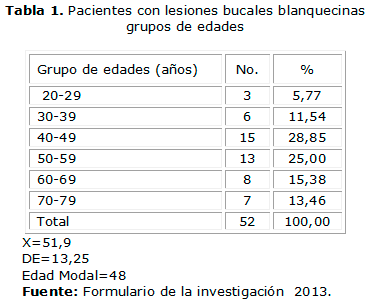
La Tabla 1 exhibe la distribución de pacientes con lesiones blancas bucales acorde a grupos de edades; se incluye además la edad promedio del grupo, la desviación estándar y la edad modal. Se aprecia que predominaron pacientes con edades comprendidas entre 40-49 años (28,85%). La edad promedio del grupo fue de 51,9 con una desviación estándar de 13,25. La edad modal fue 48.
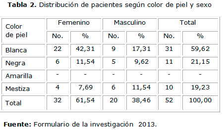
La distribución de pacientes según color de piel y sexo se aprecia en la Tabla 2. Se evidencia que las lesiones blanquecinas predominaron en pacientes del sexo femenino (61,74%) y pacientes de piel blanca (59,62%).
La distribución de lesiones blanquecinas acorde a su topografía se muestra en la Tabla 3. Se muestran 67 localizaciones debido a la presencia de lesiones que incluyeron en su extensión más de un área topográfica. Se observa que la mucosa del carrillo resultó el sitio de mayor asiento de lesiones blanquecinas en 34,33% de los casos, le siguió en frecuencia el reborde alveolar inferior (14,93%).
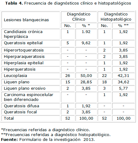
La Tabla 4 ilustra la distribución de pacientes acorde al diagnóstico clínico e histopatológico. Se evidencia que la leucoplasia resultó la lesión blanquecina más diagnosticada clínicamente (50%), seguida del liquen plano con 28,85%. También se exhiben resultados del examen histopatológico de los especímenes enviados al laboratorio; se evidencia que la leucoplasia también resultó la lesión blanquecina más encontrada en 42,31% de los casos. El carcinoma espinocelular bien diferenciado resultó 1,92% de la muestra.
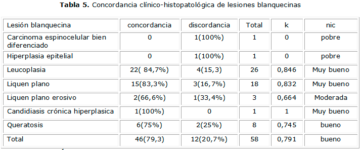
Finalmente, la Tabla 5 exhibe la concordancia entre diagnóstico clínico-histopatológico y su evaluación cualitativa según lesión y grupo. Se observa un nivel de concordancia muy bueno en leucoplasia (k=0,84), liquen plano (k=0,83) y en la candidiasis crónica hiperplásica (k=1) . Resultando la concordancia final de todo el grupo, bueno (k=0,791). Se exhibió una concordancia pobre para la hiperplasia epitelial y el carcinoma bien diferenciado.
DISCUSIÓN
En nuestro trabajo se estudió una muestra de 52 pacientes con lesiones blanquecinas de la cavidad bucal con una edad promedio que resulta superior a lo encontrado por Simi 1 en la India en una muestra semejante en un escenario hospitalario; también resultó superior a la media de 38,6 obtenida por Tatli 19 en un escenario docente en Turquía, pero en una muestra muy numerosa y que incluyó otras lesiones mucosas.
Numerosos estudios han expuesto la relación entre el sexo y el color de la piel con la aparición de lesiones bucales. En nuestro estudio, 61,54% de los sujetos resultó femenino, lo que resulta muy diferente de 88,9% de sujetos masculinos registrado en la India por Patil 3 en muestra de pacientes fumadores o mascadores de nuez de betel, pero concuerda con lo referido por Suliman15 en Sudán en una muestra de referencia hospitalaria dermatológica. Las referencias en relación con el color de la piel son menos frecuentes, pero la mayoría de los trabajos consultados concuerda con nuestro estudio, 5,13,19,20 predominando en sujetos de piel blanca, aunque es destacable que mucha de ellas se realizaron en países con predomino racial blanco. ]]>
Topográficamente, nuestro estudio se caracterizó por la prevalencia de lesiones en la mucosa del carillo (34,33%), este hallazgo concuerda con los resultados de Simi,1 quien incluso subdividió esta localización acorde a su lateralidad. Brouns5 también exhibió semejantes hallazgos en Amsterdam en estudios que profundizaron específicamente en leucoplasia. Gorski y Epstein,13 por su parte, en trabajo dedicado a liquen plano encontró 88% de esta localización en la Facultad Dental de Tel Aviv.En nuestro estudio, 42,31% de las lesiones blanquecinas resultaron leucoplasias al diagnóstico histopatológico; difiriendo de los escasos valores reportados en el Norte de la India por Bhatnagar,21 donde solo encontró leucoplasia en 2,83% de una extensa muestra poblacional, aunque solo reportó valoración clínica. Misra,4 también en India, encontró la leucoplasia como lesión premaligna más frecuente, reportando 10% en su muestra.
Las tasa de liquen plano de nuestro estudio, 34,62%, resultó relativamente alta en relación con otras series que analizan grupos poblacionales generales,3,14 donde la muestra no fue predominantemente femenina. Aunque Simi1 reportó en su serie 64% de liquen plano en registro de lesiones blanquecinas
La concordancia valorada de muy buena en nuestro estudio incluyó lesiones como leucoplasia, liquen plano y candidiasis crónica hiperplásica equiparándose acertadamente el diagnóstico clínico con el histopatológico; en general, nuestra muestra calificó en concordancia como buena (k=0,791), resultado que es algo inferior a Tatli,19 pero sí concuerdan con los resultados de largos años de observación de Suliman,14,15 en Sudán.
CONCLUSIONES
REFERENCIAS BIBLIOGRÁFICAS
1. Simi SM, Nandakumar G. White lesions in the oral cavity. A clinical histopathological study from a tertiary care dermatology centre in Kerala INDIA. Indian J Dermatol. [Serial on the Internet]. 2013 Jul.; 58(4):269-74. [Cited 2014 Mar 4]. Available from: http://dx.doi.org/10.4103%2F0019-5154.113933
2. Ali M, Joseph B. Prevalence of oral mucosal lesions in patients of the Kuwait University Dental Center. Saudi Dental J. [Serial on the Internet]. 2013 Jul.;25(3):111-118.[Cited 2014 Mar 5]. Available from: http://dx.doi.org/10.1016/j.sdentj.2013.05.003
]]>
3. Patil P, Bathi R. Prevalence of oral mucosal lesions in dental patients with tobacco smoking, chewing, and mixed habits: A cross-sectional study in South India. J Family Community Med. [Serial on the Internet]. 2013 May.; 20(2). 130-135. [Cited 2014 Mar 5]; Available from: http://dx.doi.org/10.4103%2F2230-8229.114777
4. Misra V, Singh P. Changing Pattern of Oral Cavity Lesions and Personal Habits Over a Decade: Hospital Based Record Analysis from Allahabad. Indian J Community Med. [Serial on the Internet].2009 Oct .; 34(4): 321-325. [Cited 2014 Mar 1]. Available from: http://dx.doi.org/10.4103%2F0970-0218.58391
5. Brouns E, Baart J. The relevance of uniform reporting in oral leukoplakia: Definition, certainty factor and staging based on experience with 275 patients. Med Oral Patol Oral Cir Bucal. [Serial on the Internet]. 2013 Jan.; 18(1): 19-26. [Cited 2014 Mar 11]. Available from: http://dx.doi.org/10.4317%2Fmedoral.18756
6. Issrani R, Prabhu N. Oral proliferative verrucous leukoplakia: A case report with an update. Contemp Clin Dent. [Serial on the Internet]. 2013 Jan.; 4(2): 258-262. [Cited 2014 Mar 13]. Available from: http://dx.doi.org/10.4103%2F0976-237X.114887
7. Lee L, Chen P. Quantitative physiology and immunohistochemistry of oral lesions. Biomed Opt Express. [Serial on the Internet]. 2013 Nov.; 4(11): 2696-2709. [Cited 2014 Mar 12]. Available from: http://dx.doi.org/10.1364%2FBOE.4.002696
]]>
8. Tamgadge S, Ganvir S. Oral leukoplakia: Transmission electron microscopic correlation with clinical types and light microscopy. Dent Res J (Isfahan). [Serial on the Internet]. 2012 Dec.; 9(1): 94-104. [Cited 2014 Mar 10]. Available from: http://www.ncbi.nlm.nih.gov/portal/utils/pageresolver.fcgi?recordid=1394927820919023
9. Pérez Torres L, Díaz Rojas P, Conde Mengana S, Rivero Manresa Y, Bello Díaz EA. Parámetros morfométricos de la mucosa en pacientes portadores de leucoplasia bucal con displasia epitelial. AMC. [Revista en la Internet]. 2013 Ago.; 17(4): 468-478. [Citado 2014 Mar 16]. Disponible en: http://scielo.sld.cu/scielo.php?script=sci_arttext&pid=S1025-02552013000400005&lng=es.
10. Martínez-Sahuquillo A, Gallardo I. La leucoplasia oral. Su implicación como lesión precancerosa. Avances en Odontoestomatología. Serial on the Internet].2008 Ene.; 24(1):21-25. [Citado 2014 Mar 16]. Disponible en: http://dx.doi.org/10.4321/S0213-12852008000100003
11. Feller L, Lemmer J. Oral Leukoplakia as It Relates to HPV Infection: A Review. Int J Dent. [Serial on the Internet]. 2012 Feb. [Cited 2014 Mar 10]. Available from: http://dx.doi.org/10.1155%2F2012%2F540561
12. Van der Waal I. Potentially malignant disorders of the oral and oropharyngeal mucosa; present concepts of management. Oral Oncology. [Serial on the Internet]. 2010 Feb.; 46(6):423-425. [Cited 2014 Mar 10]. Available from: http://www.ncbi.nlm.nih.gov/pubmed/20308005
13. Gorsky M, Epstein J. Smoking Habits Among Patients Diagnosed with Oral Lichen Planus. Tob Induc Dis. [Serial on the Internet]. 2004 Jun.;2(2): 103-108. [Cited 2014 Mar 9]. Available from: http://dx.doi.org/10.1186%2F1617-9625-2-2-103
14. Suliman NM, Johannessen A. Influence of oral mucosal lesions and oral symptoms on oral health related quality of life in dermatological patients: a cross sectional study in Sudan. BMC Oral Health. [Serial on the Internet]. 2012 Jul;12: 19. [Cited 2014 Mar 9]. Available from: http://dx.doi.org/10.1186%2F1472-6831-12-19
15. Suliman NM, Astrom AN, Ali RW, Salman H, Johannessen AC. Oral mucosal lesions in skin diseased patients attending a dermatologic clinic: a cross-sectional study in Sudan. BMC Oral Health. [Serial on the Internet]. 2011 March.; 11(1):24. [Cited 2014 Mar 9]. Avalaible from: http://www.ncbi.nlm.nih.gov/pmc/articles/PMC3187735/
16. Byadahally S, Rajappa S. Isolation and Identification of Candida from the Oral Cavity. ISRN Dent. [Serial on the Internet]. 2011 Oct.;2011: 487921.[Cited 2014 Mar 5]. Available from: http://dx.doi.org/10.5402%2F2011%2F487921
17. Nweze El. Oral Candida isolates among HIV infected subjects in Nigeria. J Microbiol Immunol Infect. [Serial on the Internet]. 2011 Jun.; 44(3):172-177. [Cited 2014 Mar 10]. Available from: http://www.ncbi.nlm.nih.gov/pubmed/21524610
18. Wei L, Shun chi L. Oral Cancer Development in Patients with Leukoplakia _ Clinicopathological Factors Affecting Outcome. PLoS One. [Serial on the Internet]. 2014; Apr.; 7(4): e34773. [Cited 2014 Mar 10]. Available from: http://dx.doi.org/10.1371%2Fjournal.pone.0034773
19. Tatli U, Erdogan. Diagnostic Concordance characteristics of oral cavity lesions. Scientific World Journal. [Serial on the Internet]. 2013 Jun. [Cited 2014 Mar 7] Available from: http://dx.doi.org/10.1155%2F2013%2F785929
20. Jahanbani J, Sandvik L. Evaluation of Oral Mucosal Lesions in 598 Referred Iranian Patients. Open Dent J. [Serial on the Internet]. 2009 Mar.; 3: 42-47. [ Cited 2014 Mar 5]. Available from: http://dx.doi.org/10.2174%2F1874210600903010042
21. Bhatnagar P. Prevalence study of oral mucosal lesions, mucosal variants, and treatment required for patients reporting to a dental school in North India: In accordance with WHO guidelines. J. Family Community Med. [Serial on the Internet]. 2013 Jan-Apr.; 20(1): 41-48. [Cited 2014 Mar 4]. Available from: http://dx.doi.org/10.4103%2F2230-8229.108183
]]>
Recibido: 25 de marzo de 2014.
Aprobado: 15 de septiembre de 2014. ]]>
 ]]>
]]>