<?xml version="1.0" encoding="ISO-8859-1"?><article xmlns:mml="http://www.w3.org/1998/Math/MathML" xmlns:xlink="http://www.w3.org/1999/xlink" xmlns:xsi="http://www.w3.org/2001/XMLSchema-instance">
<front>
<journal-meta>
<journal-id>0034-7523</journal-id>
<journal-title><![CDATA[Revista Cubana de Medicina]]></journal-title>
<abbrev-journal-title><![CDATA[Rev cubana med]]></abbrev-journal-title>
<issn>0034-7523</issn>
<publisher>
<publisher-name><![CDATA[Centro Nacional de Información de Ciencias MédicasEditorial Ciencias Médicas]]></publisher-name>
</publisher>
</journal-meta>
<article-meta>
<article-id>S0034-75232009000200006</article-id>
<title-group>
<article-title xml:lang="es"><![CDATA[Batería neurofisiológica para la neuromonitorización del coma y el diagnóstico de la muerte encefálica]]></article-title>
<article-title xml:lang="en"><![CDATA[Neurophysiologic test battery for neuromonitoring of comatose patients and brain death diagnosis]]></article-title>
</title-group>
<contrib-group>
<contrib contrib-type="author">
<name>
<surname><![CDATA[Machado Curbelo]]></surname>
<given-names><![CDATA[Calixto]]></given-names>
</name>
<xref ref-type="aff" rid="A01"/>
</contrib>
<contrib contrib-type="author">
<name>
<surname><![CDATA[Portela Hernández]]></surname>
<given-names><![CDATA[Liana]]></given-names>
</name>
<xref ref-type="aff" rid="A02"/>
</contrib>
<contrib contrib-type="author">
<name>
<surname><![CDATA[García Roca]]></surname>
<given-names><![CDATA[Maria G]]></given-names>
</name>
<xref ref-type="aff" rid="A02"/>
</contrib>
<contrib contrib-type="author">
<name>
<surname><![CDATA[Pérez Nellar]]></surname>
<given-names><![CDATA[Jesús]]></given-names>
</name>
<xref ref-type="aff" rid="A03"/>
</contrib>
<contrib contrib-type="author">
<name>
<surname><![CDATA[Scherle Matamoros]]></surname>
<given-names><![CDATA[Claudio E]]></given-names>
</name>
<xref ref-type="aff" rid="A03"/>
</contrib>
</contrib-group>
<aff id="A01">
<institution><![CDATA[,Instituto de Neurología y Neurocirugía.  ]]></institution>
<addr-line><![CDATA[La Habana ]]></addr-line>
<country>Cuba</country>
</aff>
<aff id="A02">
<institution><![CDATA[,Hospital Universitario Calixto Garcia  ]]></institution>
<addr-line><![CDATA[La Habana ]]></addr-line>
<country>Cuba</country>
</aff>
<aff id="A03">
<institution><![CDATA[,Hospital Clinicoquirúrgico Hermanos Ameijeiras  ]]></institution>
<addr-line><![CDATA[La Habana ]]></addr-line>
<country>Cuba</country>
</aff>
<pub-date pub-type="pub">
<day>00</day>
<month>06</month>
<year>2009</year>
</pub-date>
<pub-date pub-type="epub">
<day>00</day>
<month>06</month>
<year>2009</year>
</pub-date>
<volume>48</volume>
<numero>2</numero>
<fpage>0</fpage>
<lpage>0</lpage>
<copyright-statement/>
<copyright-year/>
<self-uri xlink:href="http://scielo.sld.cu/scielo.php?script=sci_arttext&amp;pid=S0034-75232009000200006&amp;lng=en&amp;nrm=iso"></self-uri><self-uri xlink:href="http://scielo.sld.cu/scielo.php?script=sci_abstract&amp;pid=S0034-75232009000200006&amp;lng=en&amp;nrm=iso"></self-uri><self-uri xlink:href="http://scielo.sld.cu/scielo.php?script=sci_pdf&amp;pid=S0034-75232009000200006&amp;lng=en&amp;nrm=iso"></self-uri><abstract abstract-type="short" xml:lang="es"><p><![CDATA[Se hizo una revisión sobre la aplicación de los potenciales evocados multimodales (PEM) para la neuromonitorización del coma, y como pruebas confirmatorias, en el diagnóstico de la muerte encefálica (ME). Los resultados presentados demuestran el valor de los PEM en la predicción del curso evolutivo del paciente comatoso, al predecir el deterioro de la función encefálica, lo que permite que el médico pueda tomar medidas terapéuticas tempranas, antes de que se establezcan lesiones encefálicas irreversibles. La alta resolución temporal de estas pruebas, capaces de detectar cambios funcionales del encéfalo en milisegundos, las hace idóneas para la neuromonitorización de pacientes críticos. En relación con la aplicación de los PEM y el electrorretinograma (ERG) como pruebas confirmatorias en el diagnóstico de la ME, se encontraron patrones electrofisiológicos característicos que indican la ausencia de la conducción sensorial en 3 vías diferentes, dentro de la cavidad craneana. No obstante, al considerarse una batería de pruebas confirmatorias y no como técnicas aisladas, permite optimizar el estudio electrofisiológico, y aumentar la confiabilidad diagnóstica. Por otro lado, por la resistencia de los PEM y del ERG a la hipotermia, al empleo de barbitúricos, anestésicos, a intoxicaciones por diferentes fármacos, a la anoxia, etc., permite aplicar dicha batería de pruebas para reducir el tiempo de observación requerido para establecer el diagnóstico definitivo de la ME, y confirmar dicho diagnóstico en situaciones que dificulten ese proceder.]]></p></abstract>
<abstract abstract-type="short" xml:lang="en"><p><![CDATA[Authors reviewed on application of multimodality evoked potentials (MEP) for coma neuromonitoring and as confirmatory tests in brain death (BD) diagnosis. Results presented demonstrate the MEPs value in forecast of evolution course of comatose patients, allowing prediction of brain function deterioration and thus, physician may take early therapeutic measures before establishment of irreversible brain lesions. The high time resolution of these tests allows an early detection of bran function changes, making them suitable for neuromonitoring of critical patients. In relation to MEPs and the electroretinogram (ERG) application, as confirmatory tests in the diagnosis of BD, characteristic electrophysiological patterns are found showing a lack of sensorial conduction in three different pathways within the skull. However, when considering a confirmatory test battery but not as isolated techniques, it is possible to optimize the electrophysiological study and to increase diagnostic reliability. By other hand, due to MEPs and ERG resistance to hypothermia, to use of barbiturates, to anesthetics, to intoxications from different drugs, to anoxia, etc, it is possible to apply such test battery in decreasing observation time required to establish the definite diagnosis of BD, and to confirm this diagnosis in clinical circumstances that make difficult this diagnostic procedure.]]></p></abstract>
<kwd-group>
<kwd lng="es"><![CDATA[Coma]]></kwd>
<kwd lng="es"><![CDATA[muerte encefálica]]></kwd>
<kwd lng="es"><![CDATA[neuromonitorización]]></kwd>
<kwd lng="es"><![CDATA[potenciales evocados multimodales]]></kwd>
<kwd lng="es"><![CDATA[electrorretinograma.]]></kwd>
<kwd lng="en"><![CDATA[Coma]]></kwd>
<kwd lng="en"><![CDATA[brain death]]></kwd>
<kwd lng="en"><![CDATA[neuromonitoring]]></kwd>
<kwd lng="en"><![CDATA[multimodality evoked potentials]]></kwd>
<kwd lng="en"><![CDATA[electroretinogram.]]></kwd>
</kwd-group>
</article-meta>
</front><body><![CDATA[ <P align="right">     <div align="right">       <p><font size="2" face="Verdana"><B>TEMAS ACTUALIZADOS</B></font></p>       <p>&nbsp;</p>       <p><B> </B></p> </div> <B>      <P>      <P>      <P><font size="4" face="Verdana">Bater&iacute;a neurofisiol&oacute;gica para la    neuromonitorizaci&oacute;n del coma y el diagn&oacute;stico de la muerte encef&aacute;lica    </font>      <P>&nbsp;      <P><font size="3" face="Verdana, Arial, Helvetica, sans-serif">Neurophysiologic    test battery for neuromonitoring of comatose patients and brain death diagnosis</font><font size="2" face="Verdana, Arial, Helvetica, sans-serif">    </font>  </B>      ]]></body>
<body><![CDATA[<P>&nbsp;      <P>&nbsp;  <B>     <P><font face="Verdana, Arial, Helvetica, sans-serif" size="2">Calixto Machado    Curbelo<SUP>I</SUP>; Liana Portela Hern&aacute;ndez<SUP>II</SUP>; Maria G. Garc&iacute;a Roca<SUP>III</SUP>;    Jes&uacute;s P&eacute;rez Nellar<SUP>IV</SUP>; Claudio E. Scherle Matamoros<SUP>V</SUP> </font> </B>      <p><font face="Verdana, Arial, Helvetica, sans-serif" size="2"><SUP>I</SUP> Doctor    en Ciencias. Especialista de II Grado en Neurolog&iacute;a y Neurofisiolog&iacute;a    Cl&iacute;nica. Investigador Titular. Instituto de Neurolog&iacute;a y Neurocirug&iacute;a.    La Habana, Cuba.    <br>   <SUP>II</SUP> Especialista de II Grado en Neurofisiolog&iacute;a Cl&iacute;nica.    Hospital Universitario &quot;Calixto Garcia&quot; La Habana, Cuba.    <br>   <SUP>III</SUP> Especialista de II Grado en Neurofisiolog&iacute;a Cl&iacute;nica.    Hospital Universitario &quot;Calixto Garcia&quot; La Habana, Cuba.    <br>   <SUP>IV</SUP> Doctor en Ciencias M&eacute;dicas. Especialista de II Grado en    Neurolog&iacute;a. Profesor Titular. Hospital Clinicoquir&uacute;rgico &quot;Hermanos    Ameijeiras&quot;, La Habana, Cuba.    <br>   <SUP>V</SUP>Especialista de II Grado en Neurolog&iacute;a. Hospital Clinicoquir&uacute;rgico    &quot;Hermanos Ameijeiras&quot;, La Habana, Cuba.     <br>   </font>     <p>&nbsp;      ]]></body>
<body><![CDATA[<p>&nbsp; <hr size="1" noshade>     <p><font face="Verdana, Arial, Helvetica, sans-serif" size="2"><b>RESUMEN</b></font></p> <font face="Verdana, Arial, Helvetica, sans-serif" size="2">    <br> </font><font face="Verdana" size="2">Se hizo una revisi&oacute;n sobre la aplicaci&oacute;n  de los potenciales evocados multimodales (PEM) para la neuromonitorizaci&oacute;n  del coma, y como pruebas confirmatorias, en el diagn&oacute;stico de la muerte  encef&aacute;lica (ME). Los resultados presentados demuestran el valor de los  PEM en la predicci&oacute;n del curso evolutivo del paciente comatoso, al predecir  el deterioro de la funci&oacute;n encef&aacute;lica, lo que permite que el m&eacute;dico  pueda tomar medidas terap&eacute;uticas tempranas, antes de que se establezcan  lesiones encef&aacute;licas irreversibles. La alta resoluci&oacute;n temporal  de estas pruebas, capaces de detectar cambios funcionales del enc&eacute;falo  en milisegundos, las hace id&oacute;neas para la neuromonitorizaci&oacute;n de  pacientes cr&iacute;ticos. En relaci&oacute;n con la aplicaci&oacute;n de los  PEM y el electrorretinograma (ERG) como pruebas confirmatorias en el diagn&oacute;stico  de la ME, se encontraron patrones electrofisiol&oacute;gicos caracter&iacute;sticos  que indican la ausencia de la conducci&oacute;n sensorial en 3 v&iacute;as diferentes,  dentro de la cavidad craneana. No obstante, al considerarse una bater&iacute;a  de pruebas confirmatorias y no como t&eacute;cnicas aisladas, permite optimizar  el estudio electrofisiol&oacute;gico, y aumentar la confiabilidad diagn&oacute;stica.  Por otro lado, por la resistencia de los PEM y del ERG a la hipotermia, al empleo  de barbit&uacute;ricos, anest&eacute;sicos, a intoxicaciones por diferentes f&aacute;rmacos,  a la anoxia, etc., permite aplicar dicha bater&iacute;a de pruebas para reducir  el tiempo de observaci&oacute;n requerido para establecer el diagn&oacute;stico  definitivo de la ME, y confirmar dicho diagn&oacute;stico en situaciones que dificulten  ese proceder. </font>      <p><font size="2" face="Verdana"><b>Palabras clave: </b>Coma, muerte encef&aacute;lica,    neuromonitorizaci&oacute;n, potenciales evocados multimodales, electrorretinograma.    <br>   </font><font size="2"> </font> </p> <hr size="1" noshade>     <P><font face="Verdana, Arial, Helvetica, sans-serif" size="2"><b>ABSTRACT</b></font>     <P><font face="Verdana" size="2">Authors reviewed on application of multimodality    evoked potentials (MEP) for coma neuromonitoring and as confirmatory tests in    brain death (BD) diagnosis. Results presented demonstrate the MEPs value in    forecast of evolution course of comatose patients, allowing prediction of brain    function deterioration and thus, physician may take early therapeutic measures    before establishment of irreversible brain lesions. The high time resolution    of these tests allows an early detection of bran function changes, making them    suitable for neuromonitoring of critical patients. In relation to MEPs and the    electroretinogram (ERG) application, as confirmatory tests in the diagnosis    of BD, characteristic electrophysiological patterns are found showing a lack    of sensorial conduction in three different pathways within the skull. However,    when considering a confirmatory test battery but not as isolated techniques,    it is possible to optimize the electrophysiological study and to increase diagnostic    reliability. By other hand, due to MEPs and ERG resistance to hypothermia, to    use of barbiturates, to anesthetics, to intoxications from different drugs,    to anoxia, etc, it is possible to apply such test battery in decreasing observation    time required to establish the definite diagnosis of BD, and to confirm this    diagnosis in clinical circumstances that make difficult this diagnostic procedure.    </font>     <P><font size="2" face="Verdana"><b>Key words:</b> Coma, brain death, neuromonitoring,    multimodality evoked potentials, electroretinogram.</font> <hr size="1" noshade>     <p>&nbsp;</p>    <P>&nbsp;     ]]></body>
<body><![CDATA[<p></p>     <P>      <P>      <P><font size="3" face="Verdana"><B>INTRODUCCI&Oacute;N</B> </font>     <P>      <P><font size="2" face="Verdana">Desde tiempos remotos, el hombre ha conocido    que el comportamiento consciente normal depende de un enc&eacute;falo sano y    que los trastornos de la conciencia son signos de insuficiencia del funcionamiento    encef&aacute;lico.<SUP>1-5</SUP> </font>     <P>      <P><font size="2" face="Verdana">Dos componentes fisiol&oacute;gicos rigen la    conducta consciente del ser humano: el <I>contenido</I> y el <I>despertar</I>,    conocido tambi&eacute;n como <I>capacidad</I> para la conciencia. El <I>contenido    de la conciencia</I> representa la suma de las funciones mentales cognitivas,    afectivas y otras funciones corticales superiores, mientras que el despertar    est&aacute; estrechamente vinculada a la aparici&oacute;n de la vigilia.<SUP>1,2,6-14</SUP>    </font>     <P>      <P><font size="2" face="Verdana">Por tanto, los mecanismos de la conciencia, reflejan    tanto del nivel de vigilia, que depende fundamentalmente del sistema reticular    activador ascendente (SRAA) y la suma de funciones cognitivas afectivas, que    se relaciona b&aacute;sicamente con el funcionamiento de la corteza cerebral,    as&iacute; como otras procesos funcionales complejos relacionados con la percepci&oacute;n,    la orientaci&oacute;n con respecto a s&iacute; y al medio, la actividad motora    y el planeamiento conductual, que dependen de complejos mecanismos f&iacute;sicos    y sicol&oacute;gicos con los cuales el sistema l&iacute;mbico y el cerebro (cerebrum)    individualizan y enriquecen la conciencia y la conducta.<SUP>11,13,15-19</SUP>    </font>      ]]></body>
<body><![CDATA[<P>      <P><font size="2" face="Verdana">Seg&uacute;n <I>Plum</I> y <I>Posner</I>, un    individuo consciente logra una total relaci&oacute;n con el medio externo e    interno, mientras que en el coma sucede lo opuesto, es decir, una carencia total    de la relaci&oacute;n consigo mismo y con el medio externo, a pesar de que el    sujeto sea estimulado fuertemente.<SUP>1,2</SUP> </font>     <P>      <P><font size="2" face="Verdana">La necesidad de mantener estrecha vigilancia    en pacientes con riesgos inminentes de morir ha hecho necesario el desarrollo    de una especialidad m&eacute;dica, la medicina intensiva, as&iacute; como de    salas especiales como las Unidades de Cuidados Intensivos (UCI). De este modo,    se ha podido lograr en las &uacute;ltimas d&eacute;cadas indudables avances    en el desarrollo de sistemas de ventilaci&oacute;n, en el monitoreo continuo    de distintas variables fisiol&oacute;gicas, en la estandarizaci&oacute;n de    nuevas terap&eacute;uticas, etc., de manera que los pacientes con trastornos    de la conciencia pueden permanecer con vida por tiempos pr&aacute;cticamente    ilimitados.<SUP>20-22</SUP> </font>      <P>      <P><font size="2" face="Verdana">Desde el punto de vista cl&iacute;nico ha sido    necesario desarrollar escalas para el seguimiento de los pacientes en coma,    de modo que se pueda objetivar la situaci&oacute;n cl&iacute;nica del enfermo    en un momento determinado, as&iacute; como durante su evoluci&oacute;n cl&iacute;nica.    <I>Teasdale</I> y <I>Jennett</I> introdujeron la <I>Escala de Glasgow para el    Coma </I>(EG), la cual ha sido ampliamente empleada en todo el mundo.<SUP>23-28</SUP>    Sin embargo, en la literatura se encuentran alrededor de 50 escalas para la    valoraci&oacute;n del coma. Por supuesto, la mayor&iacute;a de los autores plantean    determinados aspectos novedosos en sus escalas tomando casi siempre como punto    de referencia a la EG. No obstante, esta &uacute;ltima ha sido, sin lugar a    dudas, la m&aacute;s empleada en la mayor&iacute;a de los centros hospitalarios    del mundo. </font>     <P>      <P><font size="2" face="Verdana">Por otro lado, a pesar de que para el seguimiento    del paciente en coma la evaluaci&oacute;n neurol&oacute;gica es insustituible,    desde un punto de vista pr&aacute;ctico no es posible que el personal m&eacute;dico    y de enfermer&iacute;a puedan examinar al enfermo de manera continua; adem&aacute;s,    es frecuente que en ellos concurran situaciones que limitan los resultados del    examen cl&iacute;nico: en la anestesia, en la narcosis barbit&uacute;rica usualmente    empleada para prevenir el aumento de la presi&oacute;n intracraneana en el trauma    craneal, el uso de relajantes de la fibra muscular esquel&eacute;tica para favorecer    la ventilaci&oacute;n asistida, etc.<SUP>3,29-32</SUP> </font>     <P>      <P><font size="2" face="Verdana">En las &uacute;ltimas d&eacute;cadas se han desarrollado    t&eacute;cnicas neurofisiol&oacute;gicas que han mostrado su utilidad para la    evaluaci&oacute;n funcional del sistema nervioso: los potenciales evocados sensoriales    (PE), que permiten evaluar objetivamente diferentes v&iacute;as sensoriales,    y que al igual que el EEG, son t&eacute;cnicas no invasivas que pueden repetirse    tantas veces como se quiera sin riesgo alguno para el paciente. Por otro lado,    el desarrollo de equipos de prop&oacute;sito espec&iacute;fico ha permitido    obtener registros de calidad, a&uacute;n en las condiciones dif&iacute;ciles    de una unidad de cuidados intensivos.<SUP>33-43</SUP> </font>     ]]></body>
<body><![CDATA[<P>      <P><font size="2" face="Verdana">Los potenciales evocados, en contraposici&oacute;n    al examen neurol&oacute;gico, son altamente resistentes a la hipotermia, a la    intoxicaci&oacute;n por drogas, al uso de anest&eacute;sicos, a la narcosis    barbit&uacute;rica y por el contrario, el uso de agentes paralizantes de la    fibra muscular esquel&eacute;tica hace posible obtener registros a&uacute;n    mejores, libres de contaminaci&oacute;n electromiogr&aacute;fica. Esto ha dado    lugar a que en la literatura hayan aparecido en a&ntilde;os recientes numerosos    trabajos acerca de la aplicaci&oacute;n de los PE para el estudio del coma.<SUP>44-52</SUP>    </font>     <P>      <P><font size="2" face="Verdana">Dentro de las distintas modalidades de PE, los    potenciales evocados auditivos del tronco encef&aacute;lico (PEATC) y los potenciales    evocados somatosensoriales de corta latencia para la estimulaci&oacute;n del    nervio mediano (PES), han sido los que mejores resultados han mostrado en el    diagn&oacute;stico neurol&oacute;gico, ya que presentan poca variabilidad en    sujetos normales y se pueden obtener registros de calidad a&uacute;n en las    situaciones adversas que concurren en una UCI.<SUP>42,53-61</SUP> </font>     <P>      <P><font size="2" face="Verdana">Las pruebas neurofisiol&oacute;gicas pueden contribuir    en la evaluaci&oacute;n del coma en 3 aspectos fundamentales. La inmensa mayor&iacute;a    de las publicaciones sobre la aplicaci&oacute;n de estas pruebas para el estudio    de pacientes comatosos trata sobre los 2 primeros aspectos, o sea, el diagn&oacute;stico    y el pron&oacute;stico de los enfermos a largo plazo. &Eacute;stas facilitan    el diagn&oacute;stico diferencial entre comas metab&oacute;licos y estructurales,    as&iacute; como la localizaci&oacute;n topogr&aacute;fica de las lesiones, ya    sean supratentoriales o infratentoriales. Diferentes autores se&ntilde;alan    que un estudio neurofisiol&oacute;gico en las horas iniciales de la evoluci&oacute;n    de un paciente en coma, permite pronosticar con alta confiabilidad acerca de    la calidad de la vida, si el enfermo no muere.<SUP>62-65</SUP> </font>     <P>      <P><font size="2" face="Verdana">Sin embargo, el tercer aspecto, relacionado con    el neuromonitoreo, ha sido muy poco estudiado. La detecci&oacute;n precoz de    una arritmia card&iacute;aca puede permitir un tratamiento a tiempo y prevenir    un paro cardiocirculatorio. Tambi&eacute;n, la predicci&oacute;n de que en el    curso evolutivo de un enfermo se va a producir un deterioro de la funci&oacute;n    encef&aacute;lica, permitir&iacute;a tomar determinadas medidas terap&eacute;uticas,    antes de que se establecieran lesiones encef&aacute;licas irreversibles.<SUP>46,57,66-68</SUP>    </font>     <P>      <P><font size="2" face="Verdana">A pesar de la mejor atenci&oacute;n que se ofrezca    a un paciente en coma, en muchas ocasiones ocurre p&eacute;rdida total de las    funciones integradas en el enc&eacute;falo, a pesar de que otros &oacute;rganos    del cuerpo mantengan su integridad.<SUP>69-75</SUP> </font>     ]]></body>
<body><![CDATA[<P>      <P><font size="2" face="Verdana">Durante siglos, la ausencia irreversible de la    funci&oacute;n cardiorrespiratoria espont&aacute;nea se consider&oacute; como    determinante de la muerte del individuo. Sin embargo, con el desarrollo de la    terapia intensiva, sobre todo a partir de la segunda mitad del presente siglo,    fue posible suplir aquellas funciones reconocidas hasta ese momento como vitales.    Esto motiv&oacute; una verdadera revoluci&oacute;n en el concepto de la muerte,    cuando la atenci&oacute;n se desplaz&oacute; hacia definiciones basadas en considerar    la p&eacute;rdida definitiva de funciones integradas en el enc&eacute;falo.<SUP>62,71,76-79</SUP>    </font>     <P>      <P><font size="2" face="Verdana">El advenimiento de la cirug&iacute;a de los trasplantes    de &oacute;rganos, primero con el trasplante del ri&ntilde;&oacute;n en los    a&ntilde;os 50 y despu&eacute;s,<B><FONT  COLOR="#ff0000"> </FONT></B>de forma m&aacute;s sorprendente, con el trasplante    del coraz&oacute;n en los a&ntilde;os 60, provoc&oacute; un inter&eacute;s especial    en determinar la muerte basada en criterios neurol&oacute;gicos. No obstante,    es importante subrayar que el concepto de muerte encef&aacute;lica (ME), sin&oacute;nimo    de muerte del individuo, no surgi&oacute; para beneficiar la trasplantolog&iacute;a,    sino que fue una consecuencia del desarrollo de la terapia intensiva.<SUP>8,15,62,63,71,76,80,81</SUP>    </font>     <P>&nbsp;     <P><font face="Verdana" size="3"><b>DESARROLLO</b></font>     <P>      <P><font size="2" face="Verdana">El diagn&oacute;stico de la ME como muerte del    individuo ha dado lugar a que distintos pa&iacute;ses e instituciones hayan    elaborado sus criterios diagn&oacute;sticos. Con vistas a dar respuesta al C&oacute;digo    Civil Cubano, la Comisi&oacute;n Nacional para la Determinaci&oacute;n y Certificaci&oacute;n    de la Muerte elabor&oacute; un documento, la Resoluci&oacute;n # 90 de Salud    P&uacute;blica, que presenta la metodolog&iacute;a para diagnosticar la muerte,    agrupados en los llamados <I>signos ciertos de la muerte</I>, y que constituye    la base legal para el diagn&oacute;stico de la muerte en nuestro pa&iacute;s.<SUP>62,82,83</SUP>    </font>     <P>      <P><font size="2" face="Verdana">En dicha resoluci&oacute;n se discute que el    diagn&oacute;stico de la ME es eminentemente cl&iacute;nico, pero existen 2    grupos fundamentales de pruebas confirmatorias: aquellas que eval&uacute;an    la ausencia definitiva de circulaci&oacute;n intracraneal y las que eval&uacute;an    la ausencia de actividad bioel&eacute;ctrica cerebral. Dentro de las pruebas    para determinar la ausencia de actividad bioel&eacute;ctrica encef&aacute;lica,    la Comisi&oacute;n propuso el uso de una bater&iacute;a de pruebas confirmatorias    conformada por potenciales evocados multimodales (PEM) y el electrorretinograma    (ERG).<SUP>44,65,75,82-91</SUP> </font>     ]]></body>
<body><![CDATA[<P>      <P><font size="2" face="Verdana">El examen cl&iacute;nico neurol&oacute;gico es    sensible a condiciones potencialmente reversibles, que pueden aparentar el estado    de ME, como son: la hipotermia, la intoxicaci&oacute;n por drogas depresoras    del sistema nervioso central, el uso de anest&eacute;sicos, la narcosis barbit&uacute;rica,    la anoxia, el coma postraum&aacute;tico, etc. Debido a estas limitaciones del    examen cl&iacute;nico neurol&oacute;gico, la mayor&iacute;a de las escuelas    exigen un per&iacute;odo de observaci&oacute;n del enfermo una vez que se encuentre    supuestamente en el estado de ME, que var&iacute;a entre 6, 12, 24 h, etc. Sin    embargo, a medida que se prolonga el tiempo de permanencia del paciente en este    estado, los mecanismos de control circulatorios, tanto perif&eacute;ricos como    card&iacute;acos, se deterioran y ello puede provocar da&ntilde;os irreversibles    en los posibles &oacute;rganos a trasplantar.<SUP>47,74,92-94</SUP> </font>     <P>      <P><font size="2" face="Verdana">En contraposici&oacute;n al examen neurol&oacute;gico,    los PEM y el ERG son altamente resistentes a las condiciones potencialmente    reversibles que pueden aparentar la ME por lo que han aparecido diversas publicaciones    sobre la aplicaci&oacute;n los PEM y el ERG para diagnosticar la ME.<SUP>50,95-98</SUP>    Sin embargo, en la mayor&iacute;a de dichas investigaciones se aplica estas    pruebas de forma aislada, por lo que no existe un consenso general acerca de    su utilidad en este sentido y, por lo tanto, no se incluyen rutinariamente en    los criterios para dicho diagn&oacute;stico. La Comisi&oacute;n Nacional propuso    el uso de esta bater&iacute;a neurofisiol&oacute;gica como pruebas confirmatorias    para el diagn&oacute;stico de la ME.<SUP>65,75,82,83</SUP> Desde un punto de    vista cient&iacute;fico no son obligatorias, pero se preconiza su uso en las    siguientes situaciones: </font>     <P><font size="6" face="Verdana">.</font> <font size="2" face="Verdana"> En condiciones    que dificultan el diagn&oacute;stico de ME.    <br>   <font size="6">&#183;</font> Ausencia de lesi&oacute;n destructiva cerebral    demostrable por evidencia cl&iacute;nica o por neuroimagen.    <br>   </font>     <P><font size="6" face="Verdana">&#183;</font><font size="2" face="Verdana"> Con    el objetivo de complementar el diagn&oacute;stico y de acortar adem&aacute;s    el per&iacute;odo de observaci&oacute;n instrumental.    <br>   </font>     <P><font size="6" face="Verdana">&#183;</font><font size="2" face="Verdana"> En    el caso particular de que la etiolog&iacute;a causante del coma sea de localizaci&oacute;n    infratentorial. </font>     ]]></body>
<body><![CDATA[<P>    <br>     <P>      <P>      <P><font size="2" face="Verdana"><B>Neuromonitorizaci&oacute;n del paciente en    coma</B> </font>     <P>      <P><font size="2" face="Verdana">Para la<B> </B>neuromonitorizaci&oacute;n del    coma hemos propuesto los potenciales evocados auditivos de tronco encef&aacute;lico    (PEATC) y los potenciales evocados somatosensoriales de corta latencia (PES).<SUP>62,87,89,90,91,99</SUP>    Inmediatamente antes de estudiar a los enfermos, deben ser evaluados cl&iacute;nicamente    de forma conjunta por 2 especialistas. La evaluaci&oacute;n cl&iacute;nica se    llevar&aacute; a cabo mediante la aplicaci&oacute;n de la EG (<U><a href="/img/revistas/med/v48n2/f0106209.jpg" target="_blank">fig.    1</a></U>). La metodolog&iacute;a para ambas modalidades es la siguiente: </font>      
<P align="center"><a href="/img/revistas/med/v48n2/f0106209.jpg"><img src="/img/revistas/med/v48n2/f0106209.jpg" width="500" height="357" border="0"></a>      
<p>&nbsp;</p>     <p><font size="2" face="Verdana"><B>Potenciales evocados auditivos de tronco encef&aacute;lico    </B> </font> </p>     ]]></body>
<body><![CDATA[<P>      <P><font size="2" face="Verdana">Se aplican est&iacute;mulos auditivos en forma    de chasquidos o &quot;clicks&quot; de polaridad alterna de 0,1 milisegundos    (ms) de duraci&oacute;n, presentados monoauralmente a trav&eacute;s de aud&iacute;fonos    electromagn&eacute;ticos TDH, a una frecuencia de 20/s y a una intensidad de    70 dB (nPL). Se presenta en el o&iacute;do contralateral un ruido blanco de    -40 dB (nPL).<SUP>85,87,91</SUP> </font>     <P>      <P><font size="2" face="Verdana">Se conforma una derivaci&oacute;n a partir de    electrodos de aguja colocados subd&eacute;rmicamente en Cz, y en la mastoide    del o&iacute;do estimulado (A1 o A2) y un electrodo tierra en Fpz (Sistema Internacional    10-20). El ancho de banda de los amplificadores fue de 100-1500 Hz. Al menos    se obtiene de 2 a 3 repeticiones de cada potencial registrado con un tiempo    de an&aacute;lisis de 10 ms. </font>     <P>      <P>      <P><font size="2" face="Verdana"><B>Variables de los PEATC</B> </font>     <P>      <P><font size="6" face="Verdana">&#183;</font><font size="2" face="Verdana"> Latencia    de la onda I = LI     <br>   </font><font size="6" face="Verdana">&#183;</font><font size="2" face="Verdana">    Latencia de la onda II = LII     ]]></body>
<body><![CDATA[<br>   </font><font size="6" face="Verdana">&#183;</font><font size="2" face="Verdana">    Latencia de la onda III = LIII     <br>   </font><font size="6" face="Verdana">&#183;</font><font size="2" face="Verdana">    Latencia de la onda IV = LIV     <br>   </font><font size="6" face="Verdana">&#183; </font><font size="2" face="Verdana">Latencia    de la onda V = LV     <br>   </font><font size="6" face="Verdana">&#183; </font><font size="2" face="Verdana">Intervalo    interpico = I-V     <br>   </font><font size="6" face="Verdana">&#183; </font><font size="2" face="Verdana">Intervalo    interpico = I-III     <br>   </font><font size="6" face="Verdana">&#183; </font><font size="2" face="Verdana">Intervalo    interpico = III-V     <br>   </font><font size="6" face="Verdana">&#183; </font><font size="2" face="Verdana">Amplitud    de la onda I = AI     <br>   </font><font size="6" face="Verdana">&#183;</font><font size="2" face="Verdana">    Amplitud de la onda III = AIII     <br>   </font><font size="6" face="Verdana">&#183; </font><font size="2" face="Verdana">Amplitud    de la onda V = AV     <br>   </font><font size="6" face="Verdana">&#183; </font><font size="2" face="Verdana">Logaritmo    de AI= LnAI+1     ]]></body>
<body><![CDATA[<br>   </font><font size="6" face="Verdana">&#183;</font><font size="2" face="Verdana">    Logaritmo de AIII= LnAIII+1     <br>   </font><font size="6" face="Verdana">&#183;</font><font size="2" face="Verdana">    Logaritmo de AV= LnAV+1 </font>     <P>    <br>     <P>      <P>      <P><font size="2" face="Verdana"><B>Potenciales evocados somatosensoriales de    corta latencia</B> </font>     <P>      <P><font size="2" face="Verdana">Se aplican est&iacute;mulos el&eacute;ctricos    en forma de pulsos cuadrados de 0,1 ms de duraci&oacute;n a una frecuencia de    5/s, sobre el nervio mediano al nivel de la mu&ntilde;eca. La intensidad en    miliamperes se ajust&oacute; hasta lograr una contracci&oacute;n visible del    dedo pulgar. Se conformar&aacute; un montaje de 3 canales, tambi&eacute;n mediante    electrodos de aguja.<SUP>86,87,91,100</SUP> </font>     <P>      ]]></body>
<body><![CDATA[<P><font size="2" face="Verdana">a) C3' &oacute; C4', o sea, 2 cm por detr&aacute;s    de los puntos C3 y C4 (Sistema Internacional 10-20) referidos contra Fpz (derivaci&oacute;n    c&eacute;falo-cef&aacute;lica). C3' &oacute; C4' referidos contra punto de Erb    contralateral (Erbc) al est&iacute;mulo (derivaci&oacute;n c&eacute;falo-cef&aacute;lica).    <br>       <br>   </font><font size="2" face="Verdana">b) C3' &oacute; C4' contra Erbc (cef&aacute;lica-no    cef&aacute;lica).    <br>       <br>   </font>     <P>      <P><font size="2" face="Verdana">c) C7 (Sistema Internacional 10-20) referido    contra C7 (al nivel de la piel que cubre la ap&oacute;fisis de la s&eacute;ptima    v&eacute;rtebra cervical, o sea, derivaci&oacute;n c&eacute;rvico-cef&aacute;lica).    </font>     <P>      <P><font size="2" face="Verdana">d) Erbc contra Erb ipsilateral (Erbi). </font>     <P>      ]]></body>
<body><![CDATA[<P>      <P><font size="2" face="Verdana">El ancho de banda para la primera derivaci&oacute;n    fue de 0,5-1 000 Hz y para el resto, de 100-1 000 Hz. Se emple&oacute; un tiempo    de an&aacute;lisis de 50 ms y un retardo (<I>delay</I>) de 5 ms. </font>     <P>      <P>      <P><font size="2" face="Verdana"><B>Variables de los PESCL</B> </font>     <P>      <P><font size="6" face="Verdana">&#183;</font><font size="2" face="Verdana"> Latencia    del potencial de Erb = Erb    <br>   </font>     <P><font size="6" face="Verdana">&#183;</font><font size="2" face="Verdana"> Latencia    de N9 = LN9    <br>   </font>     ]]></body>
<body><![CDATA[<P><font size="6" face="Verdana">&#183;</font><font size="2" face="Verdana"> Latencia    de N13 = LN13    <br>   </font>     <P><font size="6" face="Verdana">&#183;</font><font size="2" face="Verdana"> Latencia    de P15 = LP15    <br>   </font>     <P><font size="6" face="Verdana">&#183; </font><font size="2" face="Verdana">Latencia    de N20 = LN20    <br>   </font>     <P><font size="6" face="Verdana">&#183;</font><font size="2" face="Verdana"> Latencia    de P25 = LP25    <br>   </font>     <P><font size="6" face="Verdana">&#183;</font><font size="2" face="Verdana"> Latencia    de N30 = LN30    <br>   </font>     ]]></body>
<body><![CDATA[<P><font size="6" face="Verdana">&#183;</font><font size="2" face="Verdana"> Intervalo    N13-N20= (TCC)    <br>   </font>     <P><font size="6" face="Verdana">&#183;</font><font size="2" face="Verdana"> Amplitud    P15-N20 = AN20    <br>   </font>     <P><font size="6" face="Verdana">&#183; </font><font size="2" face="Verdana">Amplitud    N20-P25 = AP25    <br>   </font>     <P><font size="6" face="Verdana">&#183;</font><font size="2" face="Verdana"> Amplitud    P25-N30 = AN30    <br>   </font>     <P><font size="6" face="Verdana">&#183; </font><font size="2" face="Verdana">Suma    de las 3 amplitudes = ACor    <br>   </font>     ]]></body>
<body><![CDATA[<P><font size="6" face="Verdana">&#183; </font><font size="2" face="Verdana">Logaritmo    de AN20= LnAN20    <br>   </font>     <P><font size="6" face="Verdana">&#183;</font><font size="2" face="Verdana"> Logaritmo    de AP25= LnAP25    <br>   </font>     <P><font size="6" face="Verdana">&#183;</font><font size="2" face="Verdana"> Logaritmo    de AN30= LnN30    <br>   </font>     <P><font size="6" face="Verdana">&#183; </font><font size="2" face="Verdana">Logaritmo    de Acort= LnACor </font>     <P>      <P>      <P><font size="2" face="Verdana">Tanto para los PEATC como para los PESCL se calcularon    las variables para la estimulaci&oacute;n de uno y otro lado, no obstante, para    el an&aacute;lisis, s&oacute;lo se tiene en cuenta el promedio de las variables    para ambos lados de estimulaci&oacute;n. </font>     ]]></body>
<body><![CDATA[<P>      <P><font size="2" face="Verdana"><B>An&aacute;lisis estad&iacute;stico</B>.<SUP>62,66,89,99,101</SUP>    </font>     <P>      <P><font size="2" face="Verdana">Se calcula la correlaci&oacute;n estad&iacute;stica    existente entre las variables cl&iacute;nicas y las electrofisiol&oacute;gicas.    Para esto se aplica la prueba de correlaci&oacute;n no param&eacute;trica de    Spearman para una p&lt;0,05. Se seleccionan entonces las variables electrofisiol&oacute;gicas    que mejor se correlacionen con las variables cl&iacute;nicas (p&lt;0,05), para    aplicar un m&eacute;todo de regresi&oacute;n log&iacute;stica y definir el valor    predictivo de las variables neurofisiol&oacute;gicas en relaci&oacute;n con    las escalas cl&iacute;nicas. Para lograr esto se deben seguir varios pasos.    En primer lugar, definir 2 ficheros: uno constituido con las variables neurofisiol&oacute;gicas    del primer d&iacute;a de estudio con las EG del primero y del &uacute;ltimo    d&iacute;a (fichero D&iacute;as primero y final) y otro fichero en el que se    consideren las variables neurofisiol&oacute;gicas del primer d&iacute;a y las    escalas cl&iacute;nicas del primero y el segundo d&iacute;a de estudio (fichero    D&iacute;as consecutivos). El segundo paso es crear las variables diferencia    de Glasgow, calculadas a partir de la diferencia entre el valor de la escalas    cl&iacute;nicas del &uacute;ltimo d&iacute;a de estudio y del primero (fichero    D&iacute;as primero y final) y entre el segundo y el primer d&iacute;a de estudio    (fichero D&iacute;as consecutivos). Obviamente, estas diferencias arrojar&aacute;n    resultados mayores, menores o iguales que cero. En tercer lugar, se definir&aacute;    la variable Categor&iacute;a Glasgow, a la que se le asignar&aacute;n valores    de 0 y 1, seg&uacute;n los resultados de esta diferencia. Como el modelo de    regresi&oacute;n log&iacute;stica utilizado permite trabajar con 2 categor&iacute;as    (0 y 1), se aparear&aacute;n las posibilidades: </font>     <P>      <blockquote>        <p><font size="2" face="Verdana"><B>mejor&iacute;a= 0 &nbsp;&nbsp;&nbsp;&nbsp;&nbsp;&nbsp;&nbsp;&nbsp;&nbsp;&nbsp;&nbsp;&nbsp;&nbsp;&nbsp;&nbsp;&nbsp;empeoramiento=1</B>      </font></p> </blockquote>     <P>      <P>      <P><font size="2" face="Verdana">Este an&aacute;lisis permitir&aacute; definir    las mejores variables neurofisiol&oacute;gicas para predecir el curso cl&iacute;nico    de los enfermos. La aplicaci&oacute;n del modelo de regresi&oacute;n log&iacute;stica    permite demostrar que tanto los PEATC, como los PES son poderosos para predecir    un deterioro o una mejor&iacute;a del estado cl&iacute;nico, evaluado por la    EG. </font>     ]]></body>
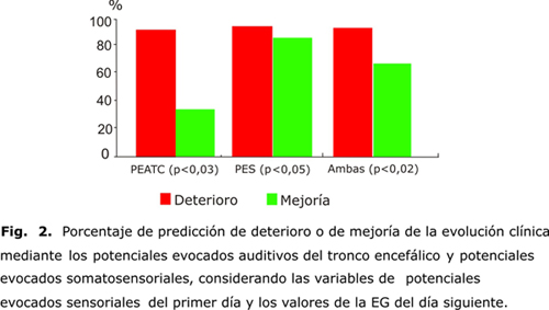
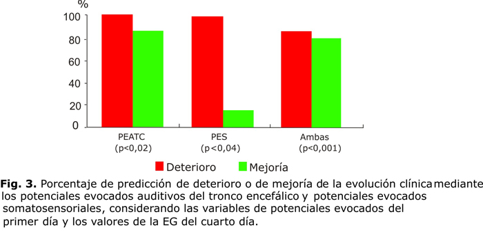
<body><![CDATA[<P>      <P><font size="2" face="Verdana">En la <U><a href="/img/revistas/med/v48n2/f0206209.jpg" target="_blank">figura    2</a></U> se presenta el porcentaje de predicci&oacute;n para ambas modalidades    de PE (PEATC, y PES) considerando las variables de PE para el primer d&iacute;a    y el valor de la EG el d&iacute;a siguiente.<SUP>62,64,99</SUP> En la <U><a href="/img/revistas/med/v48n2/f0306209.jpg" target="_blank">figura    3</a></U> se presenta el mismo an&aacute;lisis para las variables de PE para    el primer d&iacute;a de evoluci&oacute;n cl&iacute;nica y la EG para el cuarto    d&iacute;a. El modelo de regresi&oacute;n demostr&oacute; que los PEATC fueron    m&aacute;s potentes en la predicci&oacute;n, no obstante, al considerar ambas    modalidades de PE en conjunto, se logr&oacute; mejor valor predictivo. </font>      
<P align="center">&nbsp;     <P align="center"><a href="/img/revistas/med/v48n2/f0206209.jpg"><img src="/img/revistas/med/v48n2/f0206209.jpg" width="500" height="283" border="0"></a>      
<P align="center">&nbsp;     <P align="center">&nbsp;     <P align="center"><a href="/img/revistas/med/v48n2/f0306209.jpg"><img src="/img/revistas/med/v48n2/f0306209.jpg" width="700" height="363" border="0"></a>      
<P><font size="2" face="Verdana"><B>Potenciales evocados multimodales y electrorretinograma    como pruebas confirmatorias en el diagn&oacute;stico de la muerte encef&aacute;lica</B>    </font>      <P>      <P><font size="2" face="Verdana">La metodolog&iacute;a para los PEATC y los PES    es la misma que se describi&oacute; previamente.<SUP>85,86,91</SUP> Los potenciales    evocados visuales (PEV) y el ERG se obtuvieron seg&uacute;n la siguiente metodolog&iacute;a:    los PEV y el ERG para la estimulaci&oacute;n por LEDs se obtienen en un mismo    montaje. Se aplican est&iacute;mulos monoculares a una frecuencia de 2/s. Para    los PEV se conforma una derivaci&oacute;n Fz-Oz (Sistema Internacional 10-20)    y para el ERG, un lente de contacto corneal (<I>Work &amp; Shop</I>) se refiri&oacute;    a Fz. Tanto para los PEV como para el ERG se forma una derivaci&oacute;n adicional,    pero con una referencia no cef&aacute;lica, o sea al nivel de la piel que cubre    la ap&oacute;fisis de la s&eacute;ptima v&eacute;rtebra cervical (C7). El ojo    estimulado se mantiene abierto por la presencia del lente corneal. Las se&ntilde;ales    registradas se amplifican (x 100 000) y se filtran entre 0,5 y 100 Hz. Se comprueba    la repetibilidad de los potenciales mediante al menos 2 registros de 200 est&iacute;mulos    cada uno.<SUP>90,91,102-104</SUP> </font>     ]]></body>
<body><![CDATA[<P>      <P>      <P><font size="2" face="Verdana"><B>Patrones neurofisiol&oacute;gicos encontrados    en la ME</B><SUP>44-48,65,75,84,85,87-91,99,102-109</SUP> </font>     <P>      <P>      <P><font size="2" face="Verdana"><B>Potenciales evocados auditivos de tronco encef&aacute;lico    </B> </font>     <P>      <P><font size="2" face="Verdana">Se identificaron 3 patrones caracter&iacute;sticos    para los PEATC, como se muestran respectivamente en las <U><a href="/img/revistas/med/v48n2/f0406209.jpg" target="_blank">figuras    4</a></U>,<a href="/img/revistas/med/v48n2/f0506209.jpg" target="_blank">    <U>5</U></a>, y<U><a href="/img/revistas/med/v48n2/f0606209.jpg" target="_blank">    6</a>.</U> </font>      
<P>      <P><font size="2" face="Verdana">a) Ausencia bilateral de respuestas 73,34 % (<U>fig.    4</U>). </font>     ]]></body>
<body><![CDATA[<P><font size="2" face="Verdana">b) Onda I bilateral aislada 16,66 % (<U>fig.    5</U>). </font>     <P><font size="2" face="Verdana">c) Onda I unilateral aislada 10,00 % (<U>fig.    6</U>). </font>     <P>      <P><font size="2" face="Verdana">La ausencia bilateral de respuestas (<U>fig.    4</U>) fue el patr&oacute;n de los PEATC m&aacute;s frecuentemente encontrado    en los enfermos. </font>     <P>      <P><font size="2" face="Verdana">En todos los pacientes con presencia de onda    I, unilateral o bilateral, la latencia de dicha onda estuvo siempre significativamente    prolongada. Las ondas II, III, IV y V de los PEATC no se registraron en ning&uacute;n    enfermo. </font>     <P>&nbsp;     <P align="center"><a href="/img/revistas/med/v48n2/f0406209.jpg"><img src="/img/revistas/med/v48n2/f0406209.jpg" width="500" height="409" border="0"></a>      
<P align="center"><a href="/img/revistas/med/v48n2/f0506209.jpg"><img src="/img/revistas/med/v48n2/f0506209.jpg" width="500" height="437" border="0"></a>      
<P align="center"><a href="/img/revistas/med/v48n2/f0606209.jpg"><img src="/img/revistas/med/v48n2/f0606209.jpg" width="500" height="455" border="0"></a>      
]]></body>
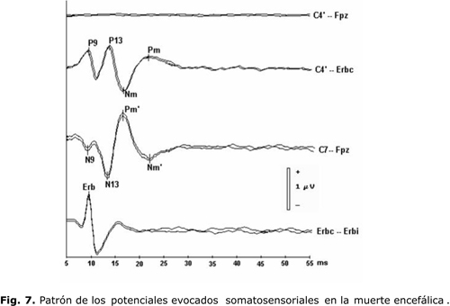
<body><![CDATA[<p>&nbsp;</p>     <P><font size="2" face="Verdana"><B>Potenciales somatosensoriales de corta latencia    </B> </font>      <P>      <P><font size="2" face="Verdana">El patr&oacute;n de los PESCL en la ME se ejemplifica    en la<a href="/img/revistas/med/v48n2/f0706209.jpg" target="_blank"> <U>figura    7</U></a>: ausencia de la onda N20 y de los potenciales corticales m&aacute;s    tard&iacute;os en la derivaci&oacute;n C4'-Fpz, y preservaci&oacute;n de los    denominados potenciales subcorticales en el resto de los canales. </font>      
<P>      <P>      <P>      <P>&nbsp;     <P align="center"><a href="/img/revistas/med/v48n2/f0706209.jpg"><img src="/img/revistas/med/v48n2/f0706209.jpg" width="500" height="340" border="0"></a>      
<P>      ]]></body>
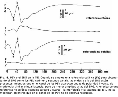
<body><![CDATA[<P>      <P><font size="2" face="Verdana"><B>Potenciales evocados visuales y electrorretinograma</B>    </font>     <P>      <P><font size="2" face="Verdana">En todos los casos se encontr&oacute; un patr&oacute;n    electrofisiol&oacute;gico caracter&iacute;stico (<U><a href="/img/revistas/med/v48n2/f0806209.jpg" target="_blank">fig.    8</a></U>). Cuando se emple&oacute; una referencia cef&aacute;lica (Fz) para    obtener tanto el ERG como los PEV (primer y segundo canal), las ondas a y b    del ERG estaban presentes, mientras que en el canal de los PEV aparec&iacute;an    ondas de polaridad inversa, de morfolog&iacute;a similar e igual latencia, pero    de menor amplitud a las del ERG. Al emplearse una referencia no cef&aacute;lica    (canales tercero y cuarto), la morfolog&iacute;a y la latencia del ERG no se    modificaban, mientras que en el canal de los PEV no se observaba respuesta.    </font>      
<p>&nbsp;</p>     <p align="center"><a href="/img/revistas/med/v48n2/f0806209.jpg"><img src="/img/revistas/med/v48n2/f0806209.jpg" width="500" height="412" border="0"></a></p>     
<P>&nbsp;     <P>      <P>      <P><font size="3" face="Verdana"><B>CONSIDERACIONES FINALES</B> </font>     ]]></body>
<body><![CDATA[<P>      <P><font size="2" face="Verdana">Los resultados presentados en este trabajo demuestran    el valor de los PE en la predicci&oacute;n en el curso evolutivo de un enfermo    en coma, pues permite valorar que se va a producir un deterioro de la funci&oacute;n    encef&aacute;lica, de modo que el m&eacute;dico pueda tomar en la UCI tempranas    medidas terap&eacute;uticas, antes de que se establecieran lesiones encef&aacute;licas    irreversibles.<SUP>64,99,108</SUP> La alta resoluci&oacute;n temporal de estas    pruebas, es decir, que son capaces de detectar cambios funcionales del enc&eacute;falo    en milisegundos, las hace id&oacute;neas para la neuromonitorizaci&oacute;n    de los pacientes cr&iacute;ticos.<SUP>64,66,99,108,110,111</SUP> Se hace necesario    adem&aacute;s desarrollar sistemas automatizados que permitan aplicar estas    t&eacute;cnicas en monitores junto a la cama de los enfermos en las UCI, como    se monitorizan el electrocardiograma y otras variables fisiol&oacute;gicas.<SUP>57,67,112-118</SUP>    </font>     <P>      <P><font size="2" face="Verdana">En relaci&oacute;n con la aplicaci&oacute;n de    la bater&iacute;a neurofisiol&oacute;gica como prueba confirmatoria en el diagn&oacute;stico    de la ME, se encontraron patrones caracter&iacute;sticos que indican ausencia    de conducci&oacute;n sensorial en 3 v&iacute;as diferentes, dentro de la cavidad    craneana.<SUP>44,89-91,118</SUP> </font>     <P>      <P><font size="2" face="Verdana">En relaci&oacute;n con los PEATC, la mayor&iacute;a    de los autores han encontrado patrones similares a los del presente trabajo:    ausencia bilateral de respuestas y onda I aislada (unilateral o bilateral).    La ausencia bilateral de respuestas es el patr&oacute;n m&aacute;s frecuentemente    hallado por diferentes autores al igual que por nosotros.<SUP>42,44,53,60,85,91,108,119-124</SUP>    </font>     <P>      <P><font size="2" face="Verdana">De acuerdo con los resultados de los PES, en    todos los enfermos se encontr&oacute; un patr&oacute;n caracter&iacute;stico    en la MIE, consistente en la desaparici&oacute;n de los componentes corticales    y la preservaci&oacute;n parcial o completa de los denominados componentes subcorticales.<SUP>86,87,90,91,100</SUP>    </font>     <P>      <P><font size="2" face="Verdana">En este estudio, el registro simult&aacute;neo    de los PEV y del ERG nos permiti&oacute; comprobar que con el empleo de una    referencia no cef&aacute;lica no se produc&iacute;an cambios ni en la morfolog&iacute;a    ni en la latencia del ERG, mientras que en el canal de los PEV no se observaba    respuesta. Seg&uacute;n este hecho, podemos afirmar que en las v&iacute;as visuales    de pacientes en el estado de ME s&oacute;lo persiste actividad el&eacute;ctrica    en la retina, lo cual constituye por tanto un patr&oacute;n electrofisiol&oacute;gico    caracter&iacute;stico de este estadio. En otras modalidades de potenciales evocados    se ha sugerido el empleo de ambas referencias para dilucidar los generadores    de los diferentes componentes.<SUP>90,91,102-104</SUP> </font>     ]]></body>
<body><![CDATA[<P>      <P><font size="2" face="Verdana">En general, se encontraron patrones caracter&iacute;sticos    para los PEM y el ERG en nuestros pacientes con el diagn&oacute;stico de ME.    Para los PEATC, una ausencia de respuestas o la presencia unilateral o bilateral    de las ondas I y II puede ser un indicador de la ME. Con respecto a los     <BR>   PESCL, la ausencia de los potenciales corticales con la preservaci&oacute;n    total o parcial de los denominados potenciales subcorticales fue un patr&oacute;n    estable en todos los casos. En los PEV y el ERG, el uso de referencias cef&aacute;lica    y no cef&aacute;lica permiti&oacute; demostrar que en las v&iacute;as visuales    de enfermos en ME, la actividad bioel&eacute;ctrica est&aacute; confinada a    la retina, lo que a su vez constituy&oacute; un patr&oacute;n caracter&iacute;stico.<SUP>85-87,91,102</SUP>    </font>     <P>      <P><font size="2" face="Verdana">Sin embargo, la aplicaci&oacute;n de una bater&iacute;a    de pruebas y no de t&eacute;cnicas aisladas indudablemente permite optimizar    el estudio electrofisiol&oacute;gico y aumentar la confiabilidad diagn&oacute;stica.    Por ejemplo, si se encuentra una ausencia bilateral de respuestas para los PEATC,    que ha sido el patr&oacute;n m&aacute;s frecuentemente hallado por diferentes    autores, en un enfermo cuya anamnesis es pobre este hecho puede inspirar dudas    sobre si dicho enfermo padec&iacute;a previamente de una sordera o si presentaba    lesiones de la c&oacute;clea y/o el nervio estatoac&uacute;stico o un hemot&iacute;mpano.    No obstante, si en este paciente se obtienen adem&aacute;s los patrones descritos    para los PES, as&iacute; como para los PEV y el ERG, entonces podemos aceptar    que la ausencia bilateral de respuestas de los PEATC es un indicador de la ME.<SUP>44,45,85,87</SUP>    </font>     <P>      <P><font size="2" face="Verdana">Se puede hacer un an&aacute;lisis similar con    el resto de las pruebas, pues en el canal de los PEV se registraban ondas que    demostramos se deb&iacute;an a la contaminaci&oacute;n por la actividad electrorretinogr&aacute;fica,    pero s&oacute;lo se pudo comprobar combinando los PEV con el ERG. Tampoco la    aplicaci&oacute;n del ERG aisladamente es &uacute;til para el diagn&oacute;stico    de la ME, pues sus componentes persisten aun despu&eacute;s de eliminar todo    soporte ventilatorio mec&aacute;nico; sin embargo, en conjunto, con los PEV    brinda un patr&oacute;n caracter&iacute;stico de la ME. Si bien hay autores    que niegan la utilidad de los PES para este diagn&oacute;stico, ya que a&uacute;n    se discute el origen de algunos de sus componentes, los patrones encontrados    en todos nuestros casos son un indicadores de la ME, lo cual se reafirma al    hallarse adem&aacute;s los otros patrones descritos para el resto de las pruebas    de la bater&iacute;a.<SUP>44,45,87,90,91,102,103,125</SUP> </font>     <P>      <P><font size="2" face="Verdana">Las ventajas de considerar una bater&iacute;a    de pruebas se incrementan indudablemente por la resistencia de los PEM y el    ERG a la hipotermia, al empleo de barbit&uacute;ricos, anest&eacute;sicos, a    intoxicaciones por diferentes f&aacute;rmacos, a la anoxia, etc. Este hecho    es de vital importancia para eliminar los tiempos de observaci&oacute;n requeridos    para establecer el diagn&oacute;stico definitivo de la ME (6 a 24 h), como es    exigido por la mayor&iacute;a de las escuelas para evitar errores diagn&oacute;sticos,    principalmente cuando no se pueden excluir las condiciones antes se&ntilde;aladas.<SUP>51,52,95-97,126</SUP>    </font>     <P>      ]]></body>
<body><![CDATA[<P><font size="2" face="Verdana">Por todo lo anteriormente expuesto, consideramos    que la aplicaci&oacute;n de una bater&iacute;a de pruebas compuesta por los    PEM y el ERG, como apoyo de la evaluaci&oacute;n cl&iacute;nica y de otros ex&aacute;menes    confirmatorios, permite eliminar o reducir per&iacute;odos de observaci&oacute;n    requeridos para establecer un diagn&oacute;stico definitivo de la ME, as&iacute;    como facilitar dicho diagn&oacute;stico en condiciones en las que el examen    cl&iacute;nico se vea limitado. Este hecho es un requisito indispensable para    la obtener &oacute;rganos viables para la trasplantolog&iacute;a y, por otro    lado, est&aacute; vinculado estrechamente a la dignidad misma del hombre, su    derecho a vivir y a morir dignamente.<SUP>64</SUP> </font>     <P>      <P><font size="2" face="Verdana">Queremos expresar, adem&aacute;s, que al diagnosticar    la ME de una forma responsable y certera, con los recursos que nos proporciona    el desarrollo tecnol&oacute;gico actual, se puede lograr que esos enfermos,    con lesiones encef&aacute;licas irreversibles, fallecidos, se conviertan en    donantes de &oacute;rganos viables para otros pacientes a quienes la trasplantolog&iacute;a    s&iacute; puede brindar esperanzas reales de vida. </font>     <P>&nbsp;     <P>      <P>      <P>      <P>      <P><font size="3" face="Verdana"><B>REFERENCIAS BIBLIOGR&Aacute;FICAS</B></font>     <P>      ]]></body>
<body><![CDATA[<!-- ref --><P><font size="2" face="Verdana">1. Plum F, Posner JB. The diagnosis of stupor    and coma. Philadelphia: FA Davis, 1980. </font>    <!-- ref --><P><font size="2" face="Verdana">2. Plum P. Coma and related global disturbances    of the human conscious state. En: Peters A, editor. Cerebral cortex. New York:    Plenum; 1991.p. 359-425. </font>    <!-- ref --><P><font size="2" face="Verdana">3. Jouvet M. Diagnostic &eacute;lectro-sous-cortico-graphique    de la mort du syst&egrave;me nerveux central au cours de certains comas. Electroenceph    Clin Neurophysiol. 1959;11(4):805-8. </font>    <!-- ref --><P><font size="2" face="Verdana">4. Daltrozzo J, Wioland N, Mutschler V, Kotchoubey    B. Predicting coma and other low responsive patients outcome using event-related    brain potentials: A meta-analysis. Clin Neurophysiol. 2007;118(3):606-14. </font>    <!-- ref --><P><font size="2" face="Verdana">5. Horn J, Zandbergen EGJ, Koelman JHTM, Hijdra    A. Prognosis for patients in a coma following cardiopulmonary resuscitation.    Ned Tijdschr Geneeskd. 2008;152(6):308-13. </font>    <!-- ref --><P><font size="2" face="Verdana">6. Moruzzi G, Magoun HW. Brain stem reticular    formation and activation of the EEG. Electroenceph Clin Neurophysiol. 1949;1(1-4):455-73.    </font>    <!-- ref --><P><font size="2" face="Verdana">7. Steriade M. Brain activation, then (1949)    and now: coherent fast rhythms in corticothalamic networks. Arch Ital Biol.    1995;134(1):5-20. </font>    <!-- ref --><P><font size="2" face="Verdana">8. Machado C. Consciousness as a definition of    death: its appeal and complexity. Clin Electroenceph. 1999;30(4):156-64. </font>    <!-- ref --><P><font size="2" face="Verdana">9. Machado-Curbelo C. Do we defend a brain oriented    view of death? Rev Neurol. 2002;35(4):387-96. </font>    <!-- ref --><P><font size="2" face="Verdana">10. Vaitl D, Birbaumer N, Gruzelier J, Jamieson    GA, Kotchoubey B, Kubler A et al. Psychobiology of altered states of consciousness.    Psychol Bull. 2005;131(1):98-127. </font>    <!-- ref --><P><font size="2" face="Verdana">11. Nunez A, Amzica F, Steriade M. Voltage-dependent    fast (20-40 Hz) oscillations in long-axoned neocortical neurons. Neuroscience.    1992;51(1):7-10. </font>    <!-- ref --><P><font size="2" face="Verdana">12. Steriade M, Amzica F, Contreras D. Synchronization    of fast (30-40 Hz) spontaneous cortical rhythms during brain activation. J Neurosci.    1996;16(1):392-417. </font>    <!-- ref --><P><font size="2" face="Verdana">13. Steriade M. Arousal: revisiting the reticular    activating system. Science. 1996;272(5259):225-6. </font>    <!-- ref --><P><font size="2" face="Verdana">14. Steriade M, Timofeev I, Durmuller N, Grenier    F. Dynamic properties of corticothalamic neurons and local cortical interneurons    generating fast rhythmic (30-40 Hz) spike bursts. J Neurophysiol. 1998;79(1):483-90.    </font>    <!-- ref --><P><font size="2" face="Verdana">15. Machado C. Death on neurological grounds.    J Neurosurg Sci. 1994;38(4):209-22. </font>    <!-- ref --><P><font size="2" face="Verdana">16. Korein J, Machado C. Brain death: updating    a valid concept for 2004. Adv Exp Med Biol. 2004;550:1-14. </font>    <!-- ref --><P><font size="2" face="Verdana">17. Steriade M, Contreras D, Amzica F, Timofeev    I. Synchronization of fast (30-40 Hz) spontaneous oscillations in intrathalamic    and thalamocortical networks. J Neurosci. 1996;16(8):2788-808. </font>    <!-- ref --><P><font size="2" face="Verdana">18. Steriade M. Local gating of information processing    through the thalamus. Neuron. 2004;41(4):493-4. </font>    <!-- ref --><P><font size="2" face="Verdana">19. Steriade M. Grouping of brain rhythms in    corticothalamic systems. Neuroscience. 2006;137(4):1087-106. </font>    <!-- ref --><P><font size="2" face="Verdana">20. Machado-Curbelo C. Do we defend a brain oriented    view of death? Rev Neurol. 2002;35(4):387-96. </font>    <!-- ref --><P><font size="2" face="Verdana">21. Guidelines for the diagnosis of brain death.    Canadian Neurocritical Care Group. Can J Neurol Sci. 1999;26(1):64-6. </font>    <!-- ref --><P><font size="2" face="Verdana">22. Haupt WF, Hansen HC. Neurophysiological diagnostics    in neurocritical care. Aktuelle Neurologie. 2008;35(3):124-30. </font>    <!-- ref --><P><font size="2" face="Verdana">23. Teasdale GM. Follow-up computerized tomography    (CT) scans in moderate and severe head injuries: Correlation with Glasgow coma    scores (GCS), and complication rate - Comments. Acta Neurochirurgica. 1997;139(11):1047-8.    </font>    <!-- ref --><P><font size="2" face="Verdana">24. Wilson JTL, Pettigrew LEL, Teasdale GM. Structured    interviews for the Glasgow Outcome Scale and the extended Glasgow Outcome Scale:    Guidelines for their use. J Neurotr. 1998;15(8):573-85. </font>    <!-- ref --><P><font size="2" face="Verdana">25. Nissen JJ, Jones PA, Signorini DF, Murray    LS, Teasdale GM, Miller JD. Glasgow Head Injury Outcome Prediction Program:    an independent assessment. J Neurol Neurosurg Psychiatr. 1999;67(6):796-9. </font>    <!-- ref --><P><font size="2" face="Verdana">26. Teasdale GM, Murray L. Revisiting the Glasgow    Coma Scale and Coma Score. Int Care Med. 2000;26(2):153-4. </font>    <!-- ref --><P><font size="2" face="Verdana">27. Wilson JTL, Pettigrew LEL, Teasdale GM. Emotional    and cognitive consequences of head injury in relation to the Glasgow outcome    scale. J Neurol Neurosurg Psychiatr. 2000;69(2):204-9. </font>    <!-- ref --><P><font size="2" face="Verdana">28. Bullock MR, Merchant RE, Choi SC, Gilman    CB, Kreutzer JS, Marmarou A et al. Outcome measures for clinical trials in neurotrauma.    Neurosurg Focus. 2002;13(1):ECP1. </font>    <!-- ref --><P><font size="2" face="Verdana">29. Morgalla MH, Bauer J, Ritz R, Tatagiba M.    Coma. Anaesthesist. 2006;55(7):760-8. </font>    <!-- ref --><P><font size="2" face="Verdana">30. Robinson LR. A very bad prognosis for awakening    from coma. Am J Phys Med &amp; Rehab. 2007;86(6):512. </font>    <!-- ref --><P><font size="2" face="Verdana">31. Zandbergen EGJ. Postanoxic coma: how (long)    should we treat? Eur J Anaesthesiol Suppl. 2008;42:39-42. </font>    <!-- ref --><P><font size="2" face="Verdana">32. Gagliardo A, Fossi S, Carrai R, Mazzeschi    E, Scarpelli S, Cianchi G et al. Predicting disability in post-traumatic coma:    prognostic value of somatosensory-evoked potentials. J Neurol. 2008;255:379.    </font>    <!-- ref --><P><font size="2" face="Verdana">33. Cobb WA, Dawson GD. The latency and form    in man of the occipital potentials evoked by bright flashes. J Physiol. 1960;152:108-21.    </font>    <!-- ref --><P><font size="2" face="Verdana">34. Arfel G, be-Fessard D, Walter S. Evoked potentials    in coma. Electroenceph Clin Neurophysiol. 1968;25(1):93-4. </font>    <!-- ref --><P><font size="2" face="Verdana">35. Anziska BJ, Cracco RQ. Short latency somatosensory    evoked potentials in brain dead patients. Arch Neurol. 1980; 37(4):222-5. </font>    <!-- ref --><P><font size="2" face="Verdana">36. Loiselle DL, Nuwer MR. When should we warn    the surgeon? Diagnosis-based warning criteria for BAEP monitoring. Neurol. 2005;65(10):1522-3.    </font>    <!-- ref --><P><font size="2" face="Verdana">37. Lew HL, Poole JH, Castaneda A, Salerno RM,    Gray M. Prognostic value of evoked and event-related potentials in moderate    to severe brain injury. J Head Traum Rehab. 2006;21(4):350-60. </font>    <!-- ref --><P><font size="2" face="Verdana">38. Gonzalez-Garcia E, Vilela-Soler C, Roma-Ambrosio    J, Fenollosa-Entrena B. The use of evoked potentials in the follow-up and prognosis    of patients in coma following severe traumatic brain injury. Rev Neurol. 2007;44(7):404-10.    </font>    <!-- ref --><P><font size="2" face="Verdana">39. Wang K, Yuan Y, Xu ZQ, Wu XL, Luo BY. Benefits    of combination of electroencephalography, short latency somatosensory evoked    potentials, and transcranial Doppler techniques for confirming brain death.    J Zhejiang University-Science B. 2008; 9(11):916-20. </font>    <!-- ref --><P><font size="2" face="Verdana">40. Schorl M. Giant somatosensory evoked potentials    as indicator of nonconvulsive status epilepticus. Clin Neurophysiol. 2008;119(3):726-8.    </font>    <!-- ref --><P><font size="2" face="Verdana">41. Amantini A, Amadori A, Fossi S. Evoked potentials    in the ICU. Eur J Anaesthesiol Suppl. 2008;42:196-202. </font>    <!-- ref --><P><font size="2" face="Verdana">42. Jardim M, Person OC, Rapoport PB. Brainstem    auditory evoked potentials as a method to assist the diagnosis of brain death.    Pro Fono. 2008;20(2):123-8. </font>    <!-- ref --><P><font size="2" face="Verdana">43. Daubin C, Guillotin D, Etard O, Gaillard    C, du Cheyron D, Ramakers M et al. A clinical and EEG scoring system that predicts    early cortical response (N20) to somatosensory evoked potentials and outcome    after cardiac arrest. BMC Cardiovasc Disord. 2008;8:35. </font>    <!-- ref --><P><font size="2" face="Verdana">44. Machado C, Pumariega J, Garc&iacute;a-Tigera    J, Miranda J, Coutin P, Antelo J et al. A multimodal evoked potential and electroretinography    test battery for the early diagnosis of brain death. Int J Neurosciences. 1989;49:241-22.    Ref Type: Journal (Full) </font>    <!-- ref --><P><font size="2" face="Verdana">45. Machado C. An early approach to brain death    diagnosis using multimodality evoked potentials and electroretinography. Minerva    Anestesiol. 1994;60(10):573-7. </font>    <!-- ref --><P><font size="2" face="Verdana">46. Gu&eacute;rit JM, Tourtchaninoff M De, Hantson    P, Mahieu P. Miltimodality evoked potentials in the differential diagnosis of    brain death. In: Machado C, editor. Brain Death (Proceedings of the Second International    Symposium on Brain Death). Amsterdam: Elsevier Science, BV; 1995.p.119-26. </font>    <!-- ref --><P><font size="2" face="Verdana">47. Machado-Curbelo C. A new formulation of death:    definition, criteria and diagnostic tests. Rev Neurol. 1998;26(154):1040-7.    </font>    <!-- ref --><P><font size="2" face="Verdana">48. Facco E, Machado C. Evoked potentials in    the diagnosis of brain death. Adv Exp Med Biol. 2004;550:175-87. </font>    <!-- ref --><P><font size="2" face="Verdana">49. Bricolo A, Ore GD, Da PR, Faccioli F. Local    cooling in spinal cord injury. Surg Neurol. 1976;6(2):101-6. </font>    <!-- ref --><P><font size="2" face="Verdana">50. Markand ON, Warren C, Mallik GS, Williams    CJ. Temperature-dependent hysteresis in somatosensory and auditory evoked potentials.    Electroenceph Clin Neurophysiol. 1990;77(6):425-35. </font>    <!-- ref --><P><font size="2" face="Verdana">51. Kadoi Y, Fujita N, Saito S, Fukura H, Fujita    T. Effects of anesthetic drugs and temperature on brain stem and mid-latency    evoked potentials. Masui. 1995;44(9):1213-7. </font>    <!-- ref --><P><font size="2" face="Verdana">52. Zhou L, Shao Z, Ou S. Cryoanalgesia: electrophysiology    at different temperatures. Cryobiol. 2003;46(1):26-32. </font>    <!-- ref --><P><font size="2" face="Verdana">53. Goldie WD, Chiappa KH, Young RR, Brooks EB.    Brainstem auditory and short-latency somatosensory evoked responses in brain    death. Neurol. 1981;31(3):248-56. </font>    <!-- ref --><P><font size="2" face="Verdana">54. Rodin E, Tahir S, Austin D, Andaya L. Brainstem    death. Clin Electroenceph. 1985;16(2):63-71. </font>    <!-- ref --><P><font size="2" face="Verdana">55. Tomita Y, Fukuda C, Maegaki Y, Hanaki K,    Kitagawa K, Sanpei M. Re-evaluation of short latency somatosensory evoked potentials    (P13, P14 and N18) for brainstem function in children who once suffered from    deep coma. Brain Dev. 2003;25(5):352-6. </font>    <!-- ref --><P><font size="2" face="Verdana">56. Trojaborg W, Jorgensen EO. Evoked cortical    potentials in patients with &quot;isoelectric&quot; EEGs. Electroenceph Clin    Neurophysiol. 1973;35(3):301-9. </font>    <!-- ref --><P><font size="2" face="Verdana">57. Morgalla MH, Deininger M, Bitzer M, Seifert    HC. Neuromonitoring: The value of somatosensory, acoustic and multimodal evoked    potentials in comatose patients in the intensive care unit. Neurol Psychiatr    Brain Res. 2004;11(1):7-12. </font>    <!-- ref --><P><font size="2" face="Verdana">58. Fukuda S. Somatosensory evoked potential.    Masui. 2006;55(3):280-93. </font>    <!-- ref --><P><font size="2" face="Verdana">59. Cruccu G, Aminoff MJ, Curio G, Guerit JM,    Kakigi R, Mauguiere F et al. Recommendations for the clinical use of somatosensory-evoked    potentials. Clin Neurophysiol. 2008;119(8):1705-19. </font>    <!-- ref --><P><font size="2" face="Verdana">60. Starr A, Achor J. Auditory brain stem responses    in neurological disease. Arch Neurol. 1975;32(11):761-8. </font>    <!-- ref --><P><font size="2" face="Verdana">61. Arulprakash S, Verma SP, Bhardwaj VK, Mishra    SS, Chansoria M. Brain stem auditory evoked-responses and visual evoked responses    in children with tubercular meningitis. Indian Pediatr. 2006;43(7):631-4. </font>    <!-- ref --><P><font size="2" face="Verdana">62. Machado C, Wagner A, Coutin P, Diaz G, Cant&oacute;n    M, Hern&aacute;ndez O et al. Potenciales evocados somatosensoriales de corta    latencia. II- Tiempo de conducci&oacute;n central. Rev Hospital Psiq Habana.    1988.p.211-21. </font>    <!-- ref --><P><font size="2" face="Verdana">63. Machado C. Resolution for the determination    and certification of death in Cuba. Rev Neurol. 2003;36(8):763-70. </font>    <!-- ref --><P><font size="2" face="Verdana">64. Machado C, Shewmon DL. Brain death and disorders    of consciousness. New York: Kluwer Academics/Plenum Publishers; 2004. </font>    <!-- ref --><P><font size="2" face="Verdana">65. Machado C, Abeledo M, &Aacute;lvarez C, Aroche    RM, Barrios I, Lasanta AM et al. Cuba has passed a law for the determination    and certification of death. Adv Exp Med Biol. 2004; 550:139-42. </font>    <!-- ref --><P><font size="2" face="Verdana">66. Cuspineda E, Machado C, Aubert E, Galan L,    Llopis F, Avila Y. Predicting outcome in acute stroke: a comparison between    QEEG and the Canadian Neurological Scale. Clin Electroenceph. 2003;34(1):1-4.    </font>    <!-- ref --><P><font size="2" face="Verdana">67. Hacke W. Neuromonitoring. J Neurol. 1985;232(3):125-33.    </font>    <!-- ref --><P><font size="2" face="Verdana">68. Fukuda S. Progress in perioperative neuromonitoring:    preface and comments. Masui. 2006;55(3):264-8. </font>    <!-- ref --><P><font size="2" face="Verdana">69. Walker AE. The death of a brain. Johns Hopkins    Med J. 1969;124(4):190-201. </font>    <!-- ref --><P><font size="2" face="Verdana">70. Walker AE. Pathology of brain death. Ann    N Y Acad Sci. 1978;315:272-80. </font>    <!-- ref --><P><font size="2" face="Verdana">71. Pallis C. Brain stem death&#151;the evolution    of a concept. Med Leg J. 1987;55 (Pt 2):84-107. </font>    <!-- ref --><P><font size="2" face="Verdana">72. Muller HR. On the problem of a shallow cerebral    current curve and the diagnosis of &quot;cerebral death&quot; following acute    cerebral anoxia. Med Klin. 1966;61(49):1955-9. </font>    <!-- ref --><P><font size="2" face="Verdana">73. Machado C, Korein J, Ferrer Y, Portela L,    De la C Garcia M, Manero JM. The concept of brain death did not evolve to benefit    organ transplants. J Med Ethics. 2007;33:197-200. </font>    <!-- ref --><P><font size="2" face="Verdana">74. Jan MM. Brain death criteria The neurological    determination of death. Neurosciences. 2008;13(4):350-5. </font>    <!-- ref --><P><font size="2" face="Verdana">75. Machado C. Variability of brain death determination    guidelines in leading US neurologic institutions. Neurol. 2008;71(14):1125.    </font>    <!-- ref --><P><font size="2" face="Verdana">76. Kretschmer E. Das apallische syndrom. Z Ges    Neurol Psychiat. 1940;169:576-9. </font>    <!-- ref --><P><font size="2" face="Verdana">77. Pallis C. Brainstem death. Handbook of Clinical    Neurology: Head Injury. Amsterdam: Elsevier Science Publisher BV, 1990.p.441-96.    </font>    <!-- ref --><P><font size="2" face="Verdana">78. Machado C. The first organ transplant from    a brain-dead donor. Neurol. 2005;64(11):1938-42. </font>    <!-- ref --><P><font size="2" face="Verdana">79. Machado C. The declaration of Sydney on death.    Neurol. 2007;68(12):A6. </font>    <!-- ref --><P><font size="2" face="Verdana">80. Machado C. The concept of brain death did    not evolve to benefit transplantation. Neurol. 2006;66(5):A2. </font>    <!-- ref --><P><font size="2" face="Verdana">81. Machado C, Korein J, Ferrer Y, Portela L,    Garcia MD, Chinchilla M et al. The declaration of Sydney on human death. J Medl    Ethics. 2007;33(12):699-703. </font>    <!-- ref --><P><font size="2" face="Verdana">82. Machado C. Resolution for the determination    and certification of death in Cuba. Rev Neurol. 2003;36(8):763-70. </font>    <!-- ref --><P><font size="2" face="Verdana">83. _____. Determination of death. Acta Anaesthesiol    Scand. 2005;49(4):592-3. </font>    <!-- ref --><P><font size="2" face="Verdana">84. Machado C, Garc&iacute;a-Tigera J, Coutin    P. Multimodality evoked potentials and electroretinography in a test battery    for the early diagnosis of brain death. Electroenceph Clin Neurophys. 1991;79(5):S19.    Ref Type: Journal (Full) </font>    <!-- ref --><P><font size="2" face="Verdana">85. Machado C, Valdes P, Garcia-Tigera J, Virues    T, Biscay R, Miranda J et al. Brain-stem auditory evoked potentials and brain    death. Electroenceph Clin Neurophys. 1991;80(5):392-8. </font>    <!-- ref --><P><font size="2" face="Verdana">86. Machado C, Valdes P, Garcia O, Coutin P,    Miranda J, Roman J. Short latency somatosensory evoked potentials in brain-dead    patients using restricted low cut filter setting. J Neurosurg Sci. 1993;37(3):133-40.    </font>    <!-- ref --><P><font size="2" face="Verdana">87. Machado C. Multimodality evoked potentials    and electroretinography in a test battery for an early diagnosis of brain death.    J Neurosurg Sci. 1993;37(3):125-31. </font>    <!-- ref --><P><font size="2" face="Verdana">88. Machado C, Santiesteban R, Garcia O, Coutin    P, Beurgo MA, Roman J et al. Visual-Evoked Potentials and Electroretinography    in Brain-Dead Patients. Documenta Ophthalmologica. 1993;84(1):89-96. </font>    <!-- ref --><P><font size="2" face="Verdana">89. Machado C. Early prediction of the clinical    course in comatose patients by evoked potentials. Neurol. 1996;46(2):2049. </font>    <!-- ref --><P><font size="2" face="Verdana">90. Machado-Curbelo C, Roman-Murga JM. Usefulness    of multimodal evoked potentials and the electroretinogram in the early diagnosis    of brain death. Rev Neurol. 1998;27(159):809-17. </font>    <!-- ref --><P><font size="2" face="Verdana">91. Machado C. Evoked potentials in brain death.    Clin Neurophys. 2004;115(1):238-9. </font>    <!-- ref --><P><font size="2" face="Verdana">92. Gros C, Vlahovitch B, Frerebeau P, Kuhner    A, Billet M, Sahut G et al. [Arteriographic criteria of irreversible coma in    neurosurgery]. Neurochirurg. 1969;15(6):477-86. </font>    <!-- ref --><P><font size="2" face="Verdana">93. ten Velden GH, van Huffelen AC. Brain death    criteria; guidelines by the Public Health Council. Ned Tijdschr Geneeskd. 1997;141(2):77-9.    </font>    <!-- ref --><P><font size="2" face="Verdana">94. Morenski JD, Oro JJ, Tobias JD, Singh A.    Determination of death by neurological criteria. J Intens Care Med. 2003;18(4):211-21.    </font>    <!-- ref --><P><font size="2" face="Verdana">95. Hughes JR. Limitations of the EEG in coma    and brain death. Ann N Y Acad Sci. 1978;315:121-36. </font>    <!-- ref --><P><font size="2" face="Verdana">96. Guerit JM. Evoked potentials: a safe brain-death    confirmatory tool? Eur J Med. 1992;1(4):233-43. </font>    <!-- ref --><P><font size="2" face="Verdana">97. de TM, Hantson P, Mahieu P, Guerit JM. Brain    death diagnosis in misleading conditions. QJM. 1999;92(7):407-14. </font>    <!-- ref --><P><font size="2" face="Verdana">98. Romani A, Bergamaschi R, Versino M, Zilioli    A, Callieco R, Cosi V. Circadian and hypothermia-induced effects on visual and    auditory evoked potentials in multiple sclerosis. Clin Neurophys. 2000;111(9):1602-6.    </font>    <!-- ref --><P><font size="2" face="Verdana">99. Machado C, Rom&aacute;n JM, Garc&iacute;a-Tigera    J, Garc&iacute;a O, Miranda J. Utilidad de los potenciales auditivos de tallo    encef&aacute;lico y somato- sensoriales corta latencia en el neuromonitoreo.    Acta M&eacute;dica. 1990;1:95-108. </font>    <!-- ref --><P><font size="2" face="Verdana">100. Shiogai T, Takeuchi K. Diagnostic reliability    in loss of brainstem function evaluated by brainstem auditory evoked potentials    and somatosensory evoked potentials in impending brain death. In: Machado C,    editor. Brain Death (Proceedings of the Second International Symposium on Brain    Death). Amsterdam: Elsevier Science, BV; 1995.p.111-8. </font>    <!-- ref --><P><font size="2" face="Verdana">101. Machado C, Cuspineda E, Valdes P, Virues    T, Llopis F, Bosch J et al. Assessing acute middle cerebral artery ischemic    stroke by quantitative electric tomography. Clin Eeg Neurosc. 2004;35(3):116-24.    </font>    <!-- ref --><P><font size="2" face="Verdana">102. Machado C. Visual evoked potentials and    electroretinography in brain-dead patients. Neurophysiol Clin. 1990;20(Suppl.):18s.    </font>    <!-- ref --><P><font size="2" face="Verdana">103. Machado C, Santiesteban R, Garc&iacute;a-Tigera    J, Garc&iacute;a O. Potenciales evocados visuales y el electrorretinograma en    la muerte encef&aacute;lica. Rev Cubana Oftalmol. 1991;4[2]:117-24. </font>    <!-- ref --><P><font size="2" face="Verdana">104. Machado C, Santiesteban R, Garcia O, Coutin    P, Beurgo MA, Roman J et al. Visual evoked potentials and electroretinography    in brain-dead patients. Doc Ophthalmol. 1993;84(1):89-96. </font>    <!-- ref --><P><font size="2" face="Verdana">105. Machado C, Garc&iacute;a-Tigera J, Garc&iacute;a    O, Garc&iacute;a-Pumariega J, Rom&aacute;n J. Muerte encef&aacute;lica. Criterios    diagn&oacute;sticos. Rev Cubana Med. 1991;30(3):181-206. </font>    <!-- ref --><P><font size="2" face="Verdana">106. Machado C. Early diagnosis of brain death.    Clinical Intensive care (Supp). 1992;3(2):116. </font>    <!-- ref --><P><font size="2" face="Verdana">107. Machado C, Vald&eacute;s P, Garc&iacute;a    O, Coutin P, Miranda J, Roman J. Short latency somatosensory evoked potentials    in brain-dead patients using restricted low cut filter setting. J Neurosurg    Sci. 1993;37(3):133-40. </font>    <!-- ref --><P><font size="2" face="Verdana">108. Facco E, Machado C. Evoked potentials in    the diagnosis of brain death. Adv Exp Med Biol. 2004;550:175-87. </font>    <!-- ref --><P><font size="2" face="Verdana">109. Machado C. Determination of death. Acta    Anaesthesiologica Scand. 2005;49(4):592-3. </font>    <!-- ref --><P><font size="2" face="Verdana">110. Machado C, Cuspineda E, Valdes P, Virues    T, Llopis F, Bosch J et al. Assessing acute middle cerebral artery ischemic    stroke by quantitative electric tomography. Clin EEG Neurosci. 2004;35(3):116-24.    </font>    <!-- ref --><P><font size="2" face="Verdana">111. Cuspineda E, Machado C, Galan L, Aubert    E, Alvarez MA, Llopis F et al. QEEG prognostic value in acute stroke. Clin Eeg    Neurosc. 2007;38(3):155-60. </font>    <!-- ref --><P><font size="2" face="Verdana">112. Shiogai T. Evaluation of electrophysiological    neuromonitoring in the diagnosis of brain death: Part III. Compressed spectral    arrays. No To Shinkei. 1989;41(3):309-18. </font>    <!-- ref --><P><font size="2" face="Verdana">113.Shiogai T. Evaluation of electrophysiological    neuromonitoring in the diagnosis of brain death: Part I. Brainstem auditory    evoked potentials. No To Shinkei. 1989;41(1):73-83. </font>    <!-- ref --><P><font size="2" face="Verdana">114. Nuwer MR, Daube J, Fischer C, Schramm J,    Yingling CD. Neuromonitoring during surgery. Report of an IFCN Committee. Electroenceph    Clin Neurophysiol. 1993;87(5):263-76. </font>    <!-- ref --><P><font size="2" face="Verdana">115. Shiogai T, Takeuchi K. Multimodal neuromonitoring    in impending brain death. Min Anestesiol. 1994;60(10):583-8. </font>    <!-- ref --><P><font size="2" face="Verdana">116. Meixensberger J, Jager A, Dings J, Baunach    S, Roosen K. Multimodal hemodynamic neuromonitoring - Quality and consequences    for therapy of severely head injured patients. Intracran Press Neuromonit Brain    Inj. 1998;71:260-2. </font>    <!-- ref --><P><font size="2" face="Verdana">117. Granry JC, Dube L, Terminassian A, Frebet    E, Le Rolle T. Multimodal neuromonitoring in children with severe head injury.    Ann Franca D Anesth Rean. 2002;21(2):148-56. </font>    <!-- ref --><P><font size="2" face="Verdana">118. </font>Machado C, Abeledo M, Alvarez C,    Aroche RM, Barrios I, Lasanta AM et al. Cuba has passed a law for the determination    and certification of death. En: Machado C, Shewmon DL, ed. Brain death and disorders    of consciousness. New York: Springer 2004;pp;139-42.    <!-- ref --><P><font size="2" face="Verdana">119. Chiappa KH, Ropper AH. Evoked potentials    in clinical medicine (first of two parts). N Engl J Med. 1982;306(19):1140-50.    </font>    <!-- ref --><P><font size="2" face="Verdana">120. Cohen SN, Potvin A, Syndulko K, Pettler-Jennings    P, Potvin JH, Tourtellotte WW. Multimodality evoked potentials: clinical applications    and assessment of utility. Bull Los Angeles Neurol Soc. 1982;47:55-61. </font>    <!-- ref --><P><font size="2" face="Verdana">121. Sasaki M, Sakamoto T, Yamashita M, Tsutsumi    H, Aruga T, Toyooka H et al. Auditory evoked brain-stem responses (ABRs) in    brain dead status. No To Shinkei. 1984;36(9):917-24. </font>    <!-- ref --><P><font size="2" face="Verdana">122. Ozgirgin ON, Ozcelik T, Sevimli NK. Auditory    brain stem responses in the detection of brain death. Kulak Burun Bogaz Ihtis    Derg. 2003;10(1):1-7. </font>    <!-- ref --><P><font size="2" face="Verdana">123. Su YY, Yang QL, Pang Y, Lv XP. Evaluation    of coma patients after cardiopulmonary resuscitation. Chin Med J (Engl). 2005;118(21):1808-11.    </font>    <!-- ref --><P><font size="2" face="Verdana">124. Avlonitis VS, Wigfield CH, Golledge HDR,    Rostron AJ, Kirby JA, Dark JH. Brain stem auditory evoked response for confirmation    of brain death in the rat. Transplant. 2008;86(5):745-6. </font>    <!-- ref --><P><font size="2" face="Verdana">125. Machado C, Santiesteban R, Garcia O, Coutin    P, Beurgo MA, Roman J et al. Visual evoked potentials and electroretinography    in brain-dead patients. Doc Ophthalmol. 1993;84(1):89-96. </font>    <!-- ref --><P><font size="2" face="Verdana">126. Freye E. Cerebral monitoring in the operating    room and the intensive care unit - an introductory for the clinician and a guide    for the novice wanting to open a window to the brain. Part II: Sensory-evoked    potentials (SSEP, AEP, VEP). J Clin Monit Comput. 2005;19(1-2):77-168. </font>    <P>&nbsp;     <P>&nbsp;     <P>      <P>      ]]></body>
<body><![CDATA[<P><font size="2" face="Verdana">Recibido: 9 de marzo de 32009.    <br>   </font><font size="2" face="Verdana">Aprobado: 8 de mayo de 2009. </font>     <P>&nbsp;     <P>&nbsp;     <P><font size="2" face="Verdana">M.D. Ph. D. <I>Calixto Machado Curbelo</I>. Instituto    de Neurolog&iacute;a y Neurocirug&iacute;a, 29 y D, El Vedado, Ciudad de La    Habana, Cuba. CP 10400 Correo electr&oacute;nico: <a href="mailto:braind@infomed.sld.cu" target="_blank">braind@infomed.sld.cu</a></FONT>       ]]></body><back>
<ref-list>
<ref id="B1">
<label>1</label><nlm-citation citation-type="book">
<person-group person-group-type="author">
<name>
<surname><![CDATA[Plum]]></surname>
<given-names><![CDATA[F]]></given-names>
</name>
<name>
<surname><![CDATA[Posner]]></surname>
<given-names><![CDATA[JB]]></given-names>
</name>
</person-group>
<source><![CDATA[The diagnosis of stupor and coma]]></source>
<year>1980</year>
<publisher-loc><![CDATA[Philadelphia ]]></publisher-loc>
<publisher-name><![CDATA[FA Davis]]></publisher-name>
</nlm-citation>
</ref>
<ref id="B2">
<label>2</label><nlm-citation citation-type="book">
<person-group person-group-type="author">
<name>
<surname><![CDATA[Plum]]></surname>
<given-names><![CDATA[P]]></given-names>
</name>
</person-group>
<article-title xml:lang="en"><![CDATA[Coma and related global disturbances of the human conscious state]]></article-title>
<person-group person-group-type="editor">
<name>
<surname><![CDATA[Peters]]></surname>
<given-names><![CDATA[A]]></given-names>
</name>
</person-group>
<source><![CDATA[Cerebral cortex]]></source>
<year>1991</year>
<page-range>359-425</page-range><publisher-loc><![CDATA[New York ]]></publisher-loc>
<publisher-name><![CDATA[Plenum]]></publisher-name>
</nlm-citation>
</ref>
<ref id="B3">
<label>3</label><nlm-citation citation-type="journal">
<person-group person-group-type="author">
<name>
<surname><![CDATA[Jouvet]]></surname>
<given-names><![CDATA[M]]></given-names>
</name>
</person-group>
<article-title xml:lang="en"><![CDATA[Diagnostic électro-sous-cortico-graphique de la mort du système nerveux central au cours de certains comas]]></article-title>
<source><![CDATA[Electroenceph Clin Neurophysiol.]]></source>
<year>1959</year>
<volume>11</volume>
<numero>4</numero>
<issue>4</issue>
<page-range>805-8</page-range></nlm-citation>
</ref>
<ref id="B4">
<label>4</label><nlm-citation citation-type="journal">
<person-group person-group-type="author">
<name>
<surname><![CDATA[Daltrozzo]]></surname>
<given-names><![CDATA[J]]></given-names>
</name>
<name>
<surname><![CDATA[Wioland]]></surname>
<given-names><![CDATA[N]]></given-names>
</name>
<name>
<surname><![CDATA[Mutschler]]></surname>
<given-names><![CDATA[V]]></given-names>
</name>
<name>
<surname><![CDATA[Kotchoubey]]></surname>
<given-names><![CDATA[B]]></given-names>
</name>
</person-group>
<article-title xml:lang="en"><![CDATA[Predicting coma and other low responsive patients outcome using event-related brain potentials: A meta-analysis]]></article-title>
<source><![CDATA[Clin Neurophysiol.]]></source>
<year>2007</year>
<volume>118</volume>
<numero>3</numero>
<issue>3</issue>
<page-range>606-14</page-range></nlm-citation>
</ref>
<ref id="B5">
<label>5</label><nlm-citation citation-type="journal">
<person-group person-group-type="author">
<name>
<surname><![CDATA[Horn]]></surname>
<given-names><![CDATA[J]]></given-names>
</name>
<name>
<surname><![CDATA[Zandbergen]]></surname>
<given-names><![CDATA[EGJ]]></given-names>
</name>
<name>
<surname><![CDATA[Koelman]]></surname>
<given-names><![CDATA[JHTM]]></given-names>
</name>
<name>
<surname><![CDATA[Hijdra]]></surname>
<given-names><![CDATA[A]]></given-names>
</name>
</person-group>
<article-title xml:lang="en"><![CDATA[Prognosis for patients in a coma following cardiopulmonary resuscitation]]></article-title>
<source><![CDATA[Ned Tijdschr Geneeskd.]]></source>
<year>2008</year>
<volume>152</volume>
<numero>6</numero>
<issue>6</issue>
<page-range>308-13</page-range></nlm-citation>
</ref>
<ref id="B6">
<label>6</label><nlm-citation citation-type="journal">
<person-group person-group-type="author">
<name>
<surname><![CDATA[Moruzzi]]></surname>
<given-names><![CDATA[G]]></given-names>
</name>
<name>
<surname><![CDATA[Magoun]]></surname>
<given-names><![CDATA[HW]]></given-names>
</name>
</person-group>
<article-title xml:lang="en"><![CDATA[Brain stem reticular formation and activation of the EEG]]></article-title>
<source><![CDATA[Electroenceph Clin Neurophysiol]]></source>
<year>1949</year>
<volume>1</volume>
<numero>1-4</numero>
<issue>1-4</issue>
<page-range>455-73</page-range></nlm-citation>
</ref>
<ref id="B7">
<label>7</label><nlm-citation citation-type="journal">
<person-group person-group-type="author">
<name>
<surname><![CDATA[Steriade]]></surname>
<given-names><![CDATA[M]]></given-names>
</name>
</person-group>
<article-title xml:lang="en"><![CDATA[Brain activation, then (1949) and now: coherent fast rhythms in corticothalamic networks]]></article-title>
<source><![CDATA[Arch Ital Biol.]]></source>
<year>1995</year>
<volume>134</volume>
<numero>1</numero>
<issue>1</issue>
<page-range>5-20</page-range></nlm-citation>
</ref>
<ref id="B8">
<label>8</label><nlm-citation citation-type="journal">
<person-group person-group-type="author">
<name>
<surname><![CDATA[Machado]]></surname>
<given-names><![CDATA[C]]></given-names>
</name>
</person-group>
<article-title xml:lang="en"><![CDATA[Consciousness as a definition of death: its appeal and complexity]]></article-title>
<source><![CDATA[Clin Electroenceph.]]></source>
<year>1999</year>
<volume>30</volume>
<numero>4</numero>
<issue>4</issue>
<page-range>156-64</page-range></nlm-citation>
</ref>
<ref id="B9">
<label>9</label><nlm-citation citation-type="journal">
<person-group person-group-type="author">
<name>
<surname><![CDATA[Machado-Curbelo]]></surname>
<given-names><![CDATA[C]]></given-names>
</name>
</person-group>
<article-title xml:lang="en"><![CDATA[Do we defend a brain oriented view of death]]></article-title>
<source><![CDATA[Rev Neurol.]]></source>
<year>2002</year>
<volume>35</volume>
<numero>4</numero>
<issue>4</issue>
<page-range>387-96</page-range></nlm-citation>
</ref>
<ref id="B10">
<label>10</label><nlm-citation citation-type="journal">
<person-group person-group-type="author">
<name>
<surname><![CDATA[Vaitl]]></surname>
<given-names><![CDATA[D]]></given-names>
</name>
<name>
<surname><![CDATA[Birbaumer]]></surname>
<given-names><![CDATA[N]]></given-names>
</name>
<name>
<surname><![CDATA[Gruzelier]]></surname>
<given-names><![CDATA[J]]></given-names>
</name>
<name>
<surname><![CDATA[Jamieson]]></surname>
<given-names><![CDATA[GA]]></given-names>
</name>
<name>
<surname><![CDATA[Kotchoubey]]></surname>
<given-names><![CDATA[B]]></given-names>
</name>
</person-group>
<article-title xml:lang="en"><![CDATA[Psychobiology of altered states of consciousness]]></article-title>
<source><![CDATA[Psychol Bull.]]></source>
<year>2005</year>
<volume>131</volume>
<numero>1</numero>
<issue>1</issue>
<page-range>98-127</page-range></nlm-citation>
</ref>
<ref id="B11">
<label>11</label><nlm-citation citation-type="journal">
<person-group person-group-type="author">
<name>
<surname><![CDATA[Nunez]]></surname>
<given-names><![CDATA[A]]></given-names>
</name>
<name>
<surname><![CDATA[Amzica]]></surname>
<given-names><![CDATA[F]]></given-names>
</name>
<name>
<surname><![CDATA[Steriade]]></surname>
<given-names><![CDATA[M]]></given-names>
</name>
</person-group>
<article-title xml:lang="en"><![CDATA[Voltage-dependent fast (20-40 Hz) oscillations in long-axoned neocortical neurons]]></article-title>
<source><![CDATA[Neuroscience.]]></source>
<year>1992</year>
<volume>51</volume>
<numero>1</numero>
<issue>1</issue>
<page-range>7-10</page-range></nlm-citation>
</ref>
<ref id="B12">
<label>12</label><nlm-citation citation-type="journal">
<person-group person-group-type="author">
<name>
<surname><![CDATA[Steriade]]></surname>
<given-names><![CDATA[M]]></given-names>
</name>
<name>
<surname><![CDATA[Amzica]]></surname>
<given-names><![CDATA[F]]></given-names>
</name>
<name>
<surname><![CDATA[Contreras]]></surname>
<given-names><![CDATA[D]]></given-names>
</name>
</person-group>
<article-title xml:lang="en"><![CDATA[Synchronization of fast (30-40 Hz) spontaneous cortical rhythms during brain activation]]></article-title>
<source><![CDATA[J Neurosci.]]></source>
<year>1996</year>
<volume>16</volume>
<numero>1</numero>
<issue>1</issue>
<page-range>392-417</page-range></nlm-citation>
</ref>
<ref id="B13">
<label>13</label><nlm-citation citation-type="journal">
<person-group person-group-type="author">
<name>
<surname><![CDATA[Steriade]]></surname>
<given-names><![CDATA[M]]></given-names>
</name>
</person-group>
<article-title xml:lang="en"><![CDATA[Arousal: revisiting the reticular activating system]]></article-title>
<source><![CDATA[Science.]]></source>
<year>1996</year>
<volume>272</volume>
<numero>5259</numero>
<issue>5259</issue>
<page-range>225-6</page-range></nlm-citation>
</ref>
<ref id="B14">
<label>14</label><nlm-citation citation-type="journal">
<person-group person-group-type="author">
<name>
<surname><![CDATA[Steriade]]></surname>
<given-names><![CDATA[M]]></given-names>
</name>
<name>
<surname><![CDATA[Timofeev]]></surname>
<given-names><![CDATA[I]]></given-names>
</name>
<name>
<surname><![CDATA[Durmuller]]></surname>
<given-names><![CDATA[N]]></given-names>
</name>
<name>
<surname><![CDATA[Grenier]]></surname>
<given-names><![CDATA[F]]></given-names>
</name>
</person-group>
<article-title xml:lang="en"><![CDATA[Dynamic properties of corticothalamic neurons and local cortical interneurons generating fast rhythmic (30-40 Hz) spike bursts]]></article-title>
<source><![CDATA[J Neurophysiol.]]></source>
<year>1998</year>
<volume>79</volume>
<numero>1</numero>
<issue>1</issue>
<page-range>483-90</page-range></nlm-citation>
</ref>
<ref id="B15">
<label>15</label><nlm-citation citation-type="journal">
<person-group person-group-type="author">
<name>
<surname><![CDATA[Machado]]></surname>
<given-names><![CDATA[C]]></given-names>
</name>
</person-group>
<article-title xml:lang="en"><![CDATA[Death on neurological grounds]]></article-title>
<source><![CDATA[J Neurosurg Sci.]]></source>
<year>1994</year>
<volume>38</volume>
<numero>4</numero>
<issue>4</issue>
<page-range>209-22</page-range></nlm-citation>
</ref>
<ref id="B16">
<label>16</label><nlm-citation citation-type="journal">
<person-group person-group-type="author">
<name>
<surname><![CDATA[Korein]]></surname>
<given-names><![CDATA[J]]></given-names>
</name>
<name>
<surname><![CDATA[Machado]]></surname>
<given-names><![CDATA[C]]></given-names>
</name>
</person-group>
<article-title xml:lang="en"><![CDATA[Brain death: updating a valid concept for 2004]]></article-title>
<source><![CDATA[Adv Exp Med Biol.]]></source>
<year>2004</year>
<volume>550</volume>
<page-range>1-14</page-range></nlm-citation>
</ref>
<ref id="B17">
<label>17</label><nlm-citation citation-type="journal">
<person-group person-group-type="author">
<name>
<surname><![CDATA[Steriade]]></surname>
<given-names><![CDATA[M]]></given-names>
</name>
<name>
<surname><![CDATA[Contreras]]></surname>
<given-names><![CDATA[D]]></given-names>
</name>
<name>
<surname><![CDATA[Amzica]]></surname>
<given-names><![CDATA[F]]></given-names>
</name>
<name>
<surname><![CDATA[Timofeev]]></surname>
<given-names><![CDATA[I]]></given-names>
</name>
</person-group>
<article-title xml:lang="en"><![CDATA[Synchronization of fast (30-40 Hz) spontaneous oscillations in intrathalamic and thalamocortical networks]]></article-title>
<source><![CDATA[J Neurosci.]]></source>
<year>1996</year>
<volume>16</volume>
<numero>8</numero>
<issue>8</issue>
<page-range>2788-808</page-range></nlm-citation>
</ref>
<ref id="B18">
<label>18</label><nlm-citation citation-type="journal">
<person-group person-group-type="author">
<name>
<surname><![CDATA[Steriade]]></surname>
<given-names><![CDATA[M]]></given-names>
</name>
</person-group>
<article-title xml:lang="en"><![CDATA[Local gating of information processing through the thalamus]]></article-title>
<source><![CDATA[Neuron.]]></source>
<year>2004</year>
<volume>41</volume>
<numero>4</numero>
<issue>4</issue>
<page-range>493-4</page-range></nlm-citation>
</ref>
<ref id="B19">
<label>19</label><nlm-citation citation-type="journal">
<person-group person-group-type="author">
<name>
<surname><![CDATA[Steriade]]></surname>
<given-names><![CDATA[M]]></given-names>
</name>
</person-group>
<article-title xml:lang="en"><![CDATA[Grouping of brain rhythms in corticothalamic systems]]></article-title>
<source><![CDATA[Neuroscience.]]></source>
<year>2006</year>
<volume>137</volume>
<numero>4</numero>
<issue>4</issue>
<page-range>1087-106</page-range></nlm-citation>
</ref>
<ref id="B20">
<label>20</label><nlm-citation citation-type="journal">
<person-group person-group-type="author">
<name>
<surname><![CDATA[Machado-Curbelo]]></surname>
<given-names><![CDATA[C]]></given-names>
</name>
</person-group>
<article-title xml:lang="en"><![CDATA[Do we defend a brain oriented view of death]]></article-title>
<source><![CDATA[Rev Neurol.]]></source>
<year>2002</year>
<volume>35</volume>
<numero>4</numero>
<issue>4</issue>
<page-range>387-96</page-range></nlm-citation>
</ref>
<ref id="B21">
<label>21</label><nlm-citation citation-type="journal">
<collab>Guidelines for the diagnosis of brain death</collab>
<article-title xml:lang="en"><![CDATA[Canadian Neurocritical Care Group]]></article-title>
<source><![CDATA[Can J Neurol Sci]]></source>
<year>1999</year>
<volume>26</volume>
<numero>1</numero>
<issue>1</issue>
<page-range>64-6</page-range></nlm-citation>
</ref>
<ref id="B22">
<label>22</label><nlm-citation citation-type="journal">
<person-group person-group-type="author">
<name>
<surname><![CDATA[Haupt]]></surname>
<given-names><![CDATA[WF]]></given-names>
</name>
<name>
<surname><![CDATA[Hansen]]></surname>
<given-names><![CDATA[HC]]></given-names>
</name>
</person-group>
<article-title xml:lang="en"><![CDATA[Neurophysiological diagnostics in neurocritical care]]></article-title>
<source><![CDATA[Aktuelle Neurologie.]]></source>
<year>2008</year>
<volume>35</volume>
<numero>3</numero>
<issue>3</issue>
<page-range>124-30</page-range></nlm-citation>
</ref>
<ref id="B23">
<label>23</label><nlm-citation citation-type="journal">
<person-group person-group-type="author">
<name>
<surname><![CDATA[Teasdale]]></surname>
<given-names><![CDATA[GM]]></given-names>
</name>
</person-group>
<article-title xml:lang="en"><![CDATA[Follow-up computerized tomography (CT) scans in moderate and severe head injuries: Correlation with Glasgow coma scores (GCS), and complication rate - Comments]]></article-title>
<source><![CDATA[Acta Neurochirurgica.]]></source>
<year>1997</year>
<volume>139</volume>
<numero>11</numero>
<issue>11</issue>
<page-range>1047-8</page-range></nlm-citation>
</ref>
<ref id="B24">
<label>24</label><nlm-citation citation-type="journal">
<person-group person-group-type="author">
<name>
<surname><![CDATA[Wilson]]></surname>
<given-names><![CDATA[JTL]]></given-names>
</name>
<name>
<surname><![CDATA[Pettigrew]]></surname>
<given-names><![CDATA[LEL]]></given-names>
</name>
<name>
<surname><![CDATA[Teasdale]]></surname>
<given-names><![CDATA[GM]]></given-names>
</name>
</person-group>
<article-title xml:lang="en"><![CDATA[Structured interviews for the Glasgow Outcome Scale and the extended Glasgow Outcome Scale: Guidelines for their use]]></article-title>
<source><![CDATA[J Neurotr.]]></source>
<year>1998</year>
<volume>15</volume>
<numero>8</numero>
<issue>8</issue>
<page-range>573-85</page-range></nlm-citation>
</ref>
<ref id="B25">
<label>25</label><nlm-citation citation-type="journal">
<person-group person-group-type="author">
<name>
<surname><![CDATA[Nissen]]></surname>
<given-names><![CDATA[JJ]]></given-names>
</name>
<name>
<surname><![CDATA[Jones]]></surname>
<given-names><![CDATA[PA]]></given-names>
</name>
<name>
<surname><![CDATA[Signorini]]></surname>
<given-names><![CDATA[DF]]></given-names>
</name>
<name>
<surname><![CDATA[Murray]]></surname>
<given-names><![CDATA[LS]]></given-names>
</name>
<name>
<surname><![CDATA[Teasdale]]></surname>
<given-names><![CDATA[GM]]></given-names>
</name>
<name>
<surname><![CDATA[Miller]]></surname>
<given-names><![CDATA[JD]]></given-names>
</name>
</person-group>
<article-title xml:lang="en"><![CDATA[Glasgow Head Injury Outcome Prediction Program: an independent assessment]]></article-title>
<source><![CDATA[J Neurol Neurosurg Psychiatr.]]></source>
<year>1999</year>
<volume>67</volume>
<numero>6</numero>
<issue>6</issue>
<page-range>796-9</page-range></nlm-citation>
</ref>
<ref id="B26">
<label>26</label><nlm-citation citation-type="journal">
<person-group person-group-type="author">
<name>
<surname><![CDATA[Teasdale]]></surname>
<given-names><![CDATA[GM]]></given-names>
</name>
<name>
<surname><![CDATA[Murray]]></surname>
<given-names><![CDATA[L]]></given-names>
</name>
</person-group>
<article-title xml:lang="en"><![CDATA[Revisiting the Glasgow Coma Scale and Coma Score]]></article-title>
<source><![CDATA[Int Care Med.]]></source>
<year>2000</year>
<volume>26</volume>
<numero>2</numero>
<issue>2</issue>
<page-range>153-4</page-range></nlm-citation>
</ref>
<ref id="B27">
<label>27</label><nlm-citation citation-type="journal">
<person-group person-group-type="author">
<name>
<surname><![CDATA[Wilson]]></surname>
<given-names><![CDATA[JTL]]></given-names>
</name>
<name>
<surname><![CDATA[Pettigrew]]></surname>
<given-names><![CDATA[LEL]]></given-names>
</name>
<name>
<surname><![CDATA[Teasdale]]></surname>
<given-names><![CDATA[GM]]></given-names>
</name>
</person-group>
<article-title xml:lang="en"><![CDATA[Emotional and cognitive consequences of head injury in relation to the Glasgow outcome scale]]></article-title>
<source><![CDATA[J Neurol Neurosurg Psychiatr.]]></source>
<year>2000</year>
<volume>69</volume>
<numero>2</numero>
<issue>2</issue>
<page-range>204-9</page-range></nlm-citation>
</ref>
<ref id="B28">
<label>28</label><nlm-citation citation-type="journal">
<person-group person-group-type="author">
<name>
<surname><![CDATA[Bullock]]></surname>
<given-names><![CDATA[MR]]></given-names>
</name>
<name>
<surname><![CDATA[Merchant]]></surname>
<given-names><![CDATA[RE]]></given-names>
</name>
<name>
<surname><![CDATA[Choi]]></surname>
<given-names><![CDATA[SC]]></given-names>
</name>
<name>
<surname><![CDATA[Gilman]]></surname>
<given-names><![CDATA[CB]]></given-names>
</name>
<name>
<surname><![CDATA[Kreutzer]]></surname>
<given-names><![CDATA[JS]]></given-names>
</name>
<name>
<surname><![CDATA[Marmarou]]></surname>
<given-names><![CDATA[A]]></given-names>
</name>
</person-group>
<article-title xml:lang="en"><![CDATA[Outcome measures for clinical trials in neurotrauma]]></article-title>
<source><![CDATA[Neurosurg Focus]]></source>
<year>2002</year>
<volume>13</volume>
<numero>1</numero>
<issue>1</issue>
</nlm-citation>
</ref>
<ref id="B29">
<label>29</label><nlm-citation citation-type="journal">
<person-group person-group-type="author">
<name>
<surname><![CDATA[Morgalla]]></surname>
<given-names><![CDATA[MH]]></given-names>
</name>
<name>
<surname><![CDATA[Bauer]]></surname>
<given-names><![CDATA[J]]></given-names>
</name>
<name>
<surname><![CDATA[Ritz]]></surname>
<given-names><![CDATA[R]]></given-names>
</name>
<name>
<surname><![CDATA[Tatagiba]]></surname>
<given-names><![CDATA[M]]></given-names>
</name>
</person-group>
<article-title xml:lang="en"><![CDATA[Coma]]></article-title>
<source><![CDATA[Anaesthesist.]]></source>
<year>2006</year>
<volume>55</volume>
<numero>7</numero>
<issue>7</issue>
<page-range>760-8</page-range></nlm-citation>
</ref>
<ref id="B30">
<label>30</label><nlm-citation citation-type="journal">
<person-group person-group-type="author">
<name>
<surname><![CDATA[Robinson]]></surname>
<given-names><![CDATA[LR]]></given-names>
</name>
</person-group>
<article-title xml:lang="en"><![CDATA[A very bad prognosis for awakening from coma]]></article-title>
<source><![CDATA[Am J Phys Med & Rehab]]></source>
<year>2007</year>
<volume>86</volume>
<numero>6</numero>
<issue>6</issue>
<page-range>512</page-range></nlm-citation>
</ref>
<ref id="B31">
<label>31</label><nlm-citation citation-type="journal">
<person-group person-group-type="author">
<name>
<surname><![CDATA[Zandbergen]]></surname>
<given-names><![CDATA[EGJ]]></given-names>
</name>
</person-group>
<article-title xml:lang="en"><![CDATA[Postanoxic coma: how (long) should we treat]]></article-title>
<source><![CDATA[Eur J Anaesthesiol]]></source>
<year>2008</year>
<volume>42</volume>
<numero>^sSuppl</numero>
<issue>^sSuppl</issue>
<supplement>Suppl</supplement>
<page-range>39-42</page-range></nlm-citation>
</ref>
<ref id="B32">
<label>32</label><nlm-citation citation-type="journal">
<person-group person-group-type="author">
<name>
<surname><![CDATA[Gagliardo]]></surname>
<given-names><![CDATA[A]]></given-names>
</name>
<name>
<surname><![CDATA[Fossi]]></surname>
<given-names><![CDATA[S]]></given-names>
</name>
<name>
<surname><![CDATA[Carrai]]></surname>
<given-names><![CDATA[R]]></given-names>
</name>
<name>
<surname><![CDATA[Mazzeschi]]></surname>
<given-names><![CDATA[E]]></given-names>
</name>
<name>
<surname><![CDATA[Scarpelli]]></surname>
<given-names><![CDATA[S]]></given-names>
</name>
</person-group>
<article-title xml:lang="en"><![CDATA[Predicting disability in post-traumatic coma: prognostic value of somatosensory-evoked potentials]]></article-title>
<source><![CDATA[J Neurol.]]></source>
<year>2008</year>
<volume>255</volume>
<page-range>379</page-range></nlm-citation>
</ref>
<ref id="B33">
<label>33</label><nlm-citation citation-type="journal">
<person-group person-group-type="author">
<name>
<surname><![CDATA[Cobb]]></surname>
<given-names><![CDATA[WA]]></given-names>
</name>
<name>
<surname><![CDATA[Dawson]]></surname>
<given-names><![CDATA[GD]]></given-names>
</name>
</person-group>
<article-title xml:lang="en"><![CDATA[The latency and form in man of the occipital potentials evoked by bright flashes]]></article-title>
<source><![CDATA[J Physiol.]]></source>
<year>1960</year>
<volume>152</volume>
<page-range>108-21</page-range></nlm-citation>
</ref>
<ref id="B34">
<label>34</label><nlm-citation citation-type="journal">
<person-group person-group-type="author">
<name>
<surname><![CDATA[Arfel]]></surname>
<given-names><![CDATA[G]]></given-names>
</name>
<name>
<surname><![CDATA[be-Fessard]]></surname>
<given-names><![CDATA[D]]></given-names>
</name>
<name>
<surname><![CDATA[Walter]]></surname>
<given-names><![CDATA[S]]></given-names>
</name>
</person-group>
<article-title xml:lang="en"><![CDATA[Evoked potentials in coma]]></article-title>
<source><![CDATA[Electroenceph Clin Neurophysiol.]]></source>
<year>1968</year>
<volume>25</volume>
<numero>1</numero>
<issue>1</issue>
<page-range>93-4</page-range></nlm-citation>
</ref>
<ref id="B35">
<label>35</label><nlm-citation citation-type="journal">
<person-group person-group-type="author">
<name>
<surname><![CDATA[Anziska]]></surname>
<given-names><![CDATA[BJ]]></given-names>
</name>
<name>
<surname><![CDATA[Cracco]]></surname>
<given-names><![CDATA[RQ]]></given-names>
</name>
</person-group>
<article-title xml:lang="en"><![CDATA[Short latency somatosensory evoked potentials in brain dead patients]]></article-title>
<source><![CDATA[Arch Neurol.]]></source>
<year>1980</year>
<volume>37</volume>
<numero>4</numero>
<issue>4</issue>
<page-range>222-5</page-range></nlm-citation>
</ref>
<ref id="B36">
<label>36</label><nlm-citation citation-type="journal">
<person-group person-group-type="author">
<name>
<surname><![CDATA[Loiselle]]></surname>
<given-names><![CDATA[DL]]></given-names>
</name>
<name>
<surname><![CDATA[Nuwer]]></surname>
<given-names><![CDATA[MR]]></given-names>
</name>
</person-group>
<article-title xml:lang="en"><![CDATA[When should we warn the surgeon? Diagnosis-based warning criteria for BAEP monitoring]]></article-title>
<source><![CDATA[Neurol.]]></source>
<year>2005</year>
<volume>65</volume>
<numero>10</numero>
<issue>10</issue>
<page-range>1522-3</page-range></nlm-citation>
</ref>
<ref id="B37">
<label>37</label><nlm-citation citation-type="journal">
<person-group person-group-type="author">
<name>
<surname><![CDATA[Lew]]></surname>
<given-names><![CDATA[HL]]></given-names>
</name>
<name>
<surname><![CDATA[Poole]]></surname>
<given-names><![CDATA[JH]]></given-names>
</name>
<name>
<surname><![CDATA[Castaneda]]></surname>
<given-names><![CDATA[A]]></given-names>
</name>
<name>
<surname><![CDATA[Salerno]]></surname>
<given-names><![CDATA[RM]]></given-names>
</name>
<name>
<surname><![CDATA[Gray]]></surname>
<given-names><![CDATA[M]]></given-names>
</name>
</person-group>
<article-title xml:lang="en"><![CDATA[Prognostic value of evoked and event-related potentials in moderate to severe brain injury]]></article-title>
<source><![CDATA[J Head Traum Rehab.]]></source>
<year>2006</year>
<volume>21</volume>
<numero>4</numero>
<issue>4</issue>
<page-range>350-60</page-range></nlm-citation>
</ref>
<ref id="B38">
<label>38</label><nlm-citation citation-type="journal">
<person-group person-group-type="author">
<name>
<surname><![CDATA[Gonzalez-Garcia]]></surname>
<given-names><![CDATA[E]]></given-names>
</name>
<name>
<surname><![CDATA[Vilela-Soler]]></surname>
<given-names><![CDATA[C]]></given-names>
</name>
<name>
<surname><![CDATA[Roma-Ambrosio]]></surname>
<given-names><![CDATA[J]]></given-names>
</name>
<name>
<surname><![CDATA[Fenollosa-Entrena]]></surname>
<given-names><![CDATA[B]]></given-names>
</name>
</person-group>
<article-title xml:lang="en"><![CDATA[The use of evoked potentials in the follow-up and prognosis of patients in coma following severe traumatic brain injury]]></article-title>
<source><![CDATA[Rev Neurol.]]></source>
<year>2007</year>
<volume>44</volume>
<numero>7</numero>
<issue>7</issue>
<page-range>404-10</page-range></nlm-citation>
</ref>
<ref id="B39">
<label>39</label><nlm-citation citation-type="journal">
<person-group person-group-type="author">
<name>
<surname><![CDATA[Wang]]></surname>
<given-names><![CDATA[K]]></given-names>
</name>
<name>
<surname><![CDATA[Yuan]]></surname>
<given-names><![CDATA[Y]]></given-names>
</name>
<name>
<surname><![CDATA[Xu]]></surname>
<given-names><![CDATA[ZQ]]></given-names>
</name>
<name>
<surname><![CDATA[Wu]]></surname>
<given-names><![CDATA[XL]]></given-names>
</name>
<name>
<surname><![CDATA[Luo]]></surname>
<given-names><![CDATA[BY]]></given-names>
</name>
</person-group>
<article-title xml:lang="en"><![CDATA[Benefits of combination of electroencephalography, short latency somatosensory evoked potentials, and transcranial Doppler techniques for confirming brain death]]></article-title>
<source><![CDATA[J Zhejiang University-Science B.]]></source>
<year>2008</year>
<volume>9</volume>
<numero>11</numero>
<issue>11</issue>
<page-range>916-20</page-range></nlm-citation>
</ref>
<ref id="B40">
<label>40</label><nlm-citation citation-type="journal">
<person-group person-group-type="author">
<name>
<surname><![CDATA[Schorl]]></surname>
<given-names><![CDATA[M]]></given-names>
</name>
</person-group>
<article-title xml:lang="en"><![CDATA[Giant somatosensory evoked potentials as indicator of nonconvulsive status epilepticus]]></article-title>
<source><![CDATA[Clin Neurophysiol.]]></source>
<year>2008</year>
<volume>119</volume>
<numero>3</numero>
<issue>3</issue>
<page-range>726-8</page-range></nlm-citation>
</ref>
<ref id="B41">
<label>41</label><nlm-citation citation-type="journal">
<person-group person-group-type="author">
<name>
<surname><![CDATA[Amantini]]></surname>
<given-names><![CDATA[A]]></given-names>
</name>
<name>
<surname><![CDATA[Amadori]]></surname>
<given-names><![CDATA[A]]></given-names>
</name>
<name>
<surname><![CDATA[Fossi]]></surname>
<given-names><![CDATA[S]]></given-names>
</name>
</person-group>
<article-title xml:lang="en"><![CDATA[Evoked potentials in the ICU]]></article-title>
<source><![CDATA[Eur J Anaesthesiol Suppl.]]></source>
<year>2008</year>
<volume>42</volume>
<page-range>196-202</page-range></nlm-citation>
</ref>
<ref id="B42">
<label>42</label><nlm-citation citation-type="journal">
<person-group person-group-type="author">
<name>
<surname><![CDATA[Jardim]]></surname>
<given-names><![CDATA[M]]></given-names>
</name>
<name>
<surname><![CDATA[Person]]></surname>
<given-names><![CDATA[OC]]></given-names>
</name>
<name>
<surname><![CDATA[Rapoport]]></surname>
<given-names><![CDATA[PB]]></given-names>
</name>
</person-group>
<article-title xml:lang="en"><![CDATA[Brainstem auditory evoked potentials as a method to assist the diagnosis of brain death]]></article-title>
<source><![CDATA[Pro Fono.]]></source>
<year>2008</year>
<volume>20</volume>
<numero>2</numero>
<issue>2</issue>
<page-range>123-8</page-range></nlm-citation>
</ref>
<ref id="B43">
<label>43</label><nlm-citation citation-type="journal">
<person-group person-group-type="author">
<name>
<surname><![CDATA[Daubin]]></surname>
<given-names><![CDATA[C]]></given-names>
</name>
<name>
<surname><![CDATA[Guillotin]]></surname>
<given-names><![CDATA[D]]></given-names>
</name>
<name>
<surname><![CDATA[Etard]]></surname>
<given-names><![CDATA[O]]></given-names>
</name>
<name>
<surname><![CDATA[Gaillard]]></surname>
<given-names><![CDATA[C]]></given-names>
</name>
<name>
<surname><![CDATA[du Cheyron]]></surname>
<given-names><![CDATA[D]]></given-names>
</name>
</person-group>
<article-title xml:lang="en"><![CDATA[A clinical and EEG scoring system that predicts early cortical response (N20) to somatosensory evoked potentials and outcome after cardiac arrest]]></article-title>
<source><![CDATA[BMC Cardiovasc Disord.]]></source>
<year>2008</year>
<volume>8</volume>
<page-range>35</page-range></nlm-citation>
</ref>
<ref id="B44">
<label>44</label><nlm-citation citation-type="journal">
<person-group person-group-type="author">
<name>
<surname><![CDATA[Machado]]></surname>
<given-names><![CDATA[C]]></given-names>
</name>
<name>
<surname><![CDATA[Pumariega]]></surname>
<given-names><![CDATA[J]]></given-names>
</name>
<name>
<surname><![CDATA[García-Tigera]]></surname>
<given-names><![CDATA[J]]></given-names>
</name>
<name>
<surname><![CDATA[Miranda]]></surname>
<given-names><![CDATA[J]]></given-names>
</name>
<name>
<surname><![CDATA[Coutin]]></surname>
<given-names><![CDATA[P]]></given-names>
</name>
<name>
<surname><![CDATA[Antelo]]></surname>
<given-names><![CDATA[J]]></given-names>
</name>
</person-group>
<article-title xml:lang="en"><![CDATA[A multimodal evoked potential and electroretinography test battery for the early diagnosis of brain death]]></article-title>
<source><![CDATA[Int J Neurosciences]]></source>
<year>1989</year>
<volume>49</volume>
<page-range>241-22</page-range></nlm-citation>
</ref>
<ref id="B45">
<label>45</label><nlm-citation citation-type="journal">
<person-group person-group-type="author">
<name>
<surname><![CDATA[Machado]]></surname>
<given-names><![CDATA[C]]></given-names>
</name>
</person-group>
<article-title xml:lang="en"><![CDATA[An early approach to brain death diagnosis using multimodality evoked potentials and electroretinography]]></article-title>
<source><![CDATA[Minerva Anestesiol.]]></source>
<year>1994</year>
<volume>60</volume>
<numero>10</numero>
<issue>10</issue>
<page-range>573-7</page-range></nlm-citation>
</ref>
<ref id="B46">
<label>46</label><nlm-citation citation-type="book">
<person-group person-group-type="author">
<name>
<surname><![CDATA[Guérit]]></surname>
<given-names><![CDATA[JM]]></given-names>
</name>
<name>
<surname><![CDATA[Tourtchaninoff]]></surname>
<given-names><![CDATA[M]]></given-names>
</name>
<name>
<surname><![CDATA[De, Hantson]]></surname>
<given-names><![CDATA[P]]></given-names>
</name>
<name>
<surname><![CDATA[Mahieu]]></surname>
<given-names><![CDATA[P]]></given-names>
</name>
</person-group>
<article-title xml:lang="en"><![CDATA[Miltimodality evoked potentials in the differential diagnosis of brain death]]></article-title>
<person-group person-group-type="editor">
<name>
<surname><![CDATA[C]]></surname>
<given-names><![CDATA[Machado]]></given-names>
</name>
</person-group>
<source><![CDATA[Brain Death (Proceedings of the Second International Symposium on Brain Death)]]></source>
<year>1995</year>
<page-range>119-26</page-range><publisher-loc><![CDATA[Amsterdam ]]></publisher-loc>
<publisher-name><![CDATA[Elsevier Science]]></publisher-name>
</nlm-citation>
</ref>
<ref id="B47">
<label>47</label><nlm-citation citation-type="journal">
<person-group person-group-type="author">
<name>
<surname><![CDATA[Machado-Curbelo]]></surname>
<given-names><![CDATA[C]]></given-names>
</name>
</person-group>
<article-title xml:lang="en"><![CDATA[A new formulation of death: definition, criteria and diagnostic tests]]></article-title>
<source><![CDATA[Rev Neurol.]]></source>
<year>1998</year>
<volume>26</volume>
<numero>154</numero>
<issue>154</issue>
<page-range>1040-7</page-range></nlm-citation>
</ref>
<ref id="B48">
<label>48</label><nlm-citation citation-type="journal">
<person-group person-group-type="author">
<name>
<surname><![CDATA[Facco]]></surname>
<given-names><![CDATA[E]]></given-names>
</name>
<name>
<surname><![CDATA[Machado]]></surname>
<given-names><![CDATA[C]]></given-names>
</name>
</person-group>
<article-title xml:lang="en"><![CDATA[Evoked potentials in the diagnosis of brain death]]></article-title>
<source><![CDATA[Adv Exp Med Biol.]]></source>
<year>2004</year>
<volume>550</volume>
<page-range>175-87</page-range></nlm-citation>
</ref>
<ref id="B49">
<label>49</label><nlm-citation citation-type="journal">
<person-group person-group-type="author">
<name>
<surname><![CDATA[Bricolo]]></surname>
<given-names><![CDATA[A]]></given-names>
</name>
<name>
<surname><![CDATA[Ore]]></surname>
<given-names><![CDATA[GD]]></given-names>
</name>
<name>
<surname><![CDATA[Da]]></surname>
<given-names><![CDATA[PR]]></given-names>
</name>
<name>
<surname><![CDATA[Faccioli]]></surname>
<given-names><![CDATA[F]]></given-names>
</name>
</person-group>
<article-title xml:lang="en"><![CDATA[Local cooling in spinal cord injury]]></article-title>
<source><![CDATA[Surg Neurol.]]></source>
<year>1976</year>
<volume>6</volume>
<numero>2</numero>
<issue>2</issue>
<page-range>101-6</page-range></nlm-citation>
</ref>
<ref id="B50">
<label>50</label><nlm-citation citation-type="journal">
<person-group person-group-type="author">
<name>
<surname><![CDATA[Markand]]></surname>
<given-names><![CDATA[ON]]></given-names>
</name>
<name>
<surname><![CDATA[Warren]]></surname>
<given-names><![CDATA[C]]></given-names>
</name>
<name>
<surname><![CDATA[Mallik]]></surname>
<given-names><![CDATA[GS]]></given-names>
</name>
<name>
<surname><![CDATA[Williams]]></surname>
<given-names><![CDATA[CJ]]></given-names>
</name>
</person-group>
<article-title xml:lang="en"><![CDATA[Temperature-dependent hysteresis in somatosensory and auditory evoked potentials]]></article-title>
<source><![CDATA[Electroenceph Clin Neurophysiol.]]></source>
<year>1990</year>
<volume>77</volume>
<numero>6</numero>
<issue>6</issue>
<page-range>425-35</page-range></nlm-citation>
</ref>
<ref id="B51">
<label>51</label><nlm-citation citation-type="journal">
<person-group person-group-type="author">
<name>
<surname><![CDATA[Kadoi]]></surname>
<given-names><![CDATA[Y]]></given-names>
</name>
<name>
<surname><![CDATA[Fujita]]></surname>
<given-names><![CDATA[N]]></given-names>
</name>
<name>
<surname><![CDATA[Saito]]></surname>
<given-names><![CDATA[S]]></given-names>
</name>
<name>
<surname><![CDATA[Fukura]]></surname>
<given-names><![CDATA[H]]></given-names>
</name>
<name>
<surname><![CDATA[Fujita]]></surname>
<given-names><![CDATA[T]]></given-names>
</name>
</person-group>
<article-title xml:lang="en"><![CDATA[Effects of anesthetic drugs and temperature on brain stem and mid-latency evoked potentials]]></article-title>
<source><![CDATA[Masui.]]></source>
<year>1995</year>
<volume>44</volume>
<numero>9</numero>
<issue>9</issue>
<page-range>1213-7</page-range></nlm-citation>
</ref>
<ref id="B52">
<label>52</label><nlm-citation citation-type="journal">
<person-group person-group-type="author">
<name>
<surname><![CDATA[Zhou]]></surname>
<given-names><![CDATA[L]]></given-names>
</name>
<name>
<surname><![CDATA[Shao]]></surname>
<given-names><![CDATA[Z]]></given-names>
</name>
<name>
<surname><![CDATA[Ou]]></surname>
<given-names><![CDATA[S]]></given-names>
</name>
</person-group>
<article-title xml:lang="en"><![CDATA[Cryoanalgesia: electrophysiology at different temperatures]]></article-title>
<source><![CDATA[Cryobiol.]]></source>
<year>2003</year>
<volume>46</volume>
<numero>1</numero>
<issue>1</issue>
<page-range>26-32</page-range></nlm-citation>
</ref>
<ref id="B53">
<label>53</label><nlm-citation citation-type="journal">
<person-group person-group-type="author">
<name>
<surname><![CDATA[Goldie]]></surname>
<given-names><![CDATA[WD]]></given-names>
</name>
<name>
<surname><![CDATA[Chiappa]]></surname>
<given-names><![CDATA[KH]]></given-names>
</name>
<name>
<surname><![CDATA[Young]]></surname>
<given-names><![CDATA[RR]]></given-names>
</name>
<name>
<surname><![CDATA[Brooks]]></surname>
<given-names><![CDATA[EB]]></given-names>
</name>
</person-group>
<article-title xml:lang="en"><![CDATA[Brainstem auditory and short-latency somatosensory evoked responses in brain death]]></article-title>
<source><![CDATA[Neurol.]]></source>
<year>1981</year>
<volume>31</volume>
<numero>3</numero>
<issue>3</issue>
<page-range>248-56</page-range></nlm-citation>
</ref>
<ref id="B54">
<label>54</label><nlm-citation citation-type="journal">
<person-group person-group-type="author">
<name>
<surname><![CDATA[Rodin]]></surname>
<given-names><![CDATA[E]]></given-names>
</name>
<name>
<surname><![CDATA[Tahir]]></surname>
<given-names><![CDATA[S]]></given-names>
</name>
<name>
<surname><![CDATA[Austin]]></surname>
<given-names><![CDATA[D]]></given-names>
</name>
<name>
<surname><![CDATA[Andaya]]></surname>
<given-names><![CDATA[L]]></given-names>
</name>
</person-group>
<article-title xml:lang="en"><![CDATA[Brainstem death]]></article-title>
<source><![CDATA[Clin Electroenceph.]]></source>
<year>1985</year>
<volume>16</volume>
<numero>2</numero>
<issue>2</issue>
<page-range>63-71</page-range></nlm-citation>
</ref>
<ref id="B55">
<label>55</label><nlm-citation citation-type="journal">
<person-group person-group-type="author">
<name>
<surname><![CDATA[Tomita]]></surname>
<given-names><![CDATA[Y]]></given-names>
</name>
<name>
<surname><![CDATA[Fukuda]]></surname>
<given-names><![CDATA[C]]></given-names>
</name>
<name>
<surname><![CDATA[Maegaki]]></surname>
<given-names><![CDATA[Y]]></given-names>
</name>
<name>
<surname><![CDATA[Hanaki]]></surname>
<given-names><![CDATA[K]]></given-names>
</name>
<name>
<surname><![CDATA[Kitagawa]]></surname>
<given-names><![CDATA[K]]></given-names>
</name>
<name>
<surname><![CDATA[Sanpei]]></surname>
<given-names><![CDATA[M]]></given-names>
</name>
</person-group>
<article-title xml:lang="en"><![CDATA[Re-evaluation of short latency somatosensory evoked potentials (P13, P14 and N18) for brainstem function in children who once suffered from deep coma]]></article-title>
<source><![CDATA[Brain Dev.]]></source>
<year>2003</year>
<volume>25</volume>
<numero>5</numero>
<issue>5</issue>
<page-range>352-6</page-range></nlm-citation>
</ref>
<ref id="B56">
<label>56</label><nlm-citation citation-type="journal">
<person-group person-group-type="author">
<name>
<surname><![CDATA[Trojaborg]]></surname>
<given-names><![CDATA[W]]></given-names>
</name>
<name>
<surname><![CDATA[Jorgensen]]></surname>
<given-names><![CDATA[EO]]></given-names>
</name>
</person-group>
<article-title xml:lang="en"><![CDATA[Evoked cortical potentials in patients with "isoelectric" EEGs]]></article-title>
<source><![CDATA[Electroenceph Clin Neurophysiol.]]></source>
<year>1973</year>
<volume>35</volume>
<numero>3</numero>
<issue>3</issue>
<page-range>301-9</page-range></nlm-citation>
</ref>
<ref id="B57">
<label>57</label><nlm-citation citation-type="journal">
<person-group person-group-type="author">
<name>
<surname><![CDATA[Morgalla]]></surname>
<given-names><![CDATA[MH]]></given-names>
</name>
<name>
<surname><![CDATA[Deininger]]></surname>
<given-names><![CDATA[M]]></given-names>
</name>
<name>
<surname><![CDATA[Bitzer]]></surname>
<given-names><![CDATA[M]]></given-names>
</name>
<name>
<surname><![CDATA[Seifert]]></surname>
<given-names><![CDATA[HC]]></given-names>
</name>
</person-group>
<article-title xml:lang="en"><![CDATA[Neuromonitoring: The value of somatosensory, acoustic and multimodal evoked potentials in comatose patients in the intensive care unit]]></article-title>
<source><![CDATA[Neurol Psychiatr Brain Res.]]></source>
<year>2004</year>
<volume>11</volume>
<numero>1</numero>
<issue>1</issue>
<page-range>7-12</page-range></nlm-citation>
</ref>
<ref id="B58">
<label>58</label><nlm-citation citation-type="journal">
<person-group person-group-type="author">
<name>
<surname><![CDATA[Fukuda]]></surname>
<given-names><![CDATA[S]]></given-names>
</name>
</person-group>
<article-title xml:lang="en"><![CDATA[Somatosensory evoked potential]]></article-title>
<source><![CDATA[Masui.]]></source>
<year>2006</year>
<volume>55</volume>
<numero>3</numero>
<issue>3</issue>
<page-range>280-93</page-range></nlm-citation>
</ref>
<ref id="B59">
<label>59</label><nlm-citation citation-type="journal">
<person-group person-group-type="author">
<name>
<surname><![CDATA[Cruccu]]></surname>
<given-names><![CDATA[G]]></given-names>
</name>
<name>
<surname><![CDATA[Aminoff]]></surname>
<given-names><![CDATA[MJ]]></given-names>
</name>
<name>
<surname><![CDATA[Curio]]></surname>
<given-names><![CDATA[G]]></given-names>
</name>
<name>
<surname><![CDATA[Guerit]]></surname>
<given-names><![CDATA[JM]]></given-names>
</name>
<name>
<surname><![CDATA[Kakigi]]></surname>
<given-names><![CDATA[R]]></given-names>
</name>
</person-group>
<article-title xml:lang="en"><![CDATA[Recommendations for the clinical use of somatosensory-evoked potentials]]></article-title>
<source><![CDATA[Clin Neurophysiol.]]></source>
<year>2008</year>
<volume>119</volume>
<numero>8</numero>
<issue>8</issue>
<page-range>1705-19</page-range></nlm-citation>
</ref>
<ref id="B60">
<label>60</label><nlm-citation citation-type="journal">
<person-group person-group-type="author">
<name>
<surname><![CDATA[Starr]]></surname>
<given-names><![CDATA[A]]></given-names>
</name>
<name>
<surname><![CDATA[Achor]]></surname>
<given-names><![CDATA[J]]></given-names>
</name>
</person-group>
<article-title xml:lang="en"><![CDATA[Auditory brain stem responses in neurological disease]]></article-title>
<source><![CDATA[Arch Neurol.]]></source>
<year>1975</year>
<volume>32</volume>
<numero>11</numero>
<issue>11</issue>
<page-range>761-8</page-range></nlm-citation>
</ref>
<ref id="B61">
<label>61</label><nlm-citation citation-type="journal">
<person-group person-group-type="author">
<name>
<surname><![CDATA[Arulprakash]]></surname>
<given-names><![CDATA[S]]></given-names>
</name>
<name>
<surname><![CDATA[Verma]]></surname>
<given-names><![CDATA[SP]]></given-names>
</name>
<name>
<surname><![CDATA[Bhardwaj]]></surname>
<given-names><![CDATA[VK]]></given-names>
</name>
<name>
<surname><![CDATA[Mishra]]></surname>
<given-names><![CDATA[SS]]></given-names>
</name>
<name>
<surname><![CDATA[Chansoria]]></surname>
<given-names><![CDATA[M]]></given-names>
</name>
</person-group>
<article-title xml:lang="en"><![CDATA[Brain stem auditory evoked-responses and visual evoked responses in children with tubercular meningitis]]></article-title>
<source><![CDATA[Indian Pediatr.]]></source>
<year>2006</year>
<volume>43</volume>
<numero>7</numero>
<issue>7</issue>
<page-range>631-4</page-range></nlm-citation>
</ref>
<ref id="B62">
<label>62</label><nlm-citation citation-type="journal">
<person-group person-group-type="author">
<name>
<surname><![CDATA[Machado]]></surname>
<given-names><![CDATA[C]]></given-names>
</name>
<name>
<surname><![CDATA[Wagner]]></surname>
<given-names><![CDATA[A]]></given-names>
</name>
<name>
<surname><![CDATA[Coutin]]></surname>
<given-names><![CDATA[P]]></given-names>
</name>
<name>
<surname><![CDATA[Diaz]]></surname>
<given-names><![CDATA[G]]></given-names>
</name>
<name>
<surname><![CDATA[Cantón]]></surname>
<given-names><![CDATA[M]]></given-names>
</name>
<name>
<surname><![CDATA[Hernández]]></surname>
<given-names><![CDATA[O]]></given-names>
</name>
</person-group>
<article-title xml:lang="es"><![CDATA[Potenciales evocados somatosensoriales de corta latencia: II- Tiempo de conducción central]]></article-title>
<source><![CDATA[Rev Hospital Psiq Habana]]></source>
<year>1988</year>
<page-range>211-21</page-range></nlm-citation>
</ref>
<ref id="B63">
<label>63</label><nlm-citation citation-type="journal">
<person-group person-group-type="author">
<name>
<surname><![CDATA[Machado]]></surname>
<given-names><![CDATA[C]]></given-names>
</name>
</person-group>
<article-title xml:lang="en"><![CDATA[Resolution for the determination and certification of death in Cuba]]></article-title>
<source><![CDATA[Rev Neurol.]]></source>
<year>2003</year>
<volume>36</volume>
<numero>8</numero>
<issue>8</issue>
<page-range>763-70</page-range></nlm-citation>
</ref>
<ref id="B64">
<label>64</label><nlm-citation citation-type="book">
<person-group person-group-type="author">
<name>
<surname><![CDATA[Machado]]></surname>
<given-names><![CDATA[C]]></given-names>
</name>
<name>
<surname><![CDATA[Shewmon]]></surname>
<given-names><![CDATA[DL]]></given-names>
</name>
</person-group>
<source><![CDATA[Brain death and disorders of consciousness]]></source>
<year>2004</year>
<publisher-loc><![CDATA[New York ]]></publisher-loc>
<publisher-name><![CDATA[Kluwer Academics/Plenum Publishers]]></publisher-name>
</nlm-citation>
</ref>
<ref id="B65">
<label>65</label><nlm-citation citation-type="journal">
<person-group person-group-type="author">
<name>
<surname><![CDATA[Machado]]></surname>
<given-names><![CDATA[C]]></given-names>
</name>
<name>
<surname><![CDATA[Abeledo]]></surname>
<given-names><![CDATA[M]]></given-names>
</name>
<name>
<surname><![CDATA[Álvarez]]></surname>
<given-names><![CDATA[C]]></given-names>
</name>
<name>
<surname><![CDATA[Aroche]]></surname>
<given-names><![CDATA[RM]]></given-names>
</name>
<name>
<surname><![CDATA[Barrios]]></surname>
<given-names><![CDATA[I]]></given-names>
</name>
</person-group>
<article-title xml:lang="en"><![CDATA[Cuba has passed a law for the determination and certification of death]]></article-title>
<source><![CDATA[Adv Exp Med Biol.]]></source>
<year>2004</year>
<volume>550</volume>
<page-range>139-42</page-range></nlm-citation>
</ref>
<ref id="B66">
<label>66</label><nlm-citation citation-type="journal">
<person-group person-group-type="author">
<name>
<surname><![CDATA[Cuspineda]]></surname>
<given-names><![CDATA[E]]></given-names>
</name>
<name>
<surname><![CDATA[Machado]]></surname>
<given-names><![CDATA[C]]></given-names>
</name>
<name>
<surname><![CDATA[Aubert]]></surname>
<given-names><![CDATA[E]]></given-names>
</name>
<name>
<surname><![CDATA[Galan]]></surname>
<given-names><![CDATA[L]]></given-names>
</name>
<name>
<surname><![CDATA[Llopis]]></surname>
<given-names><![CDATA[F]]></given-names>
</name>
<name>
<surname><![CDATA[Avila]]></surname>
<given-names><![CDATA[Y]]></given-names>
</name>
</person-group>
<article-title xml:lang="en"><![CDATA[Predicting outcome in acute stroke: a comparison between QEEG and the Canadian Neurological Scale]]></article-title>
<source><![CDATA[Clin Electroenceph.]]></source>
<year>2003</year>
<volume>34</volume>
<numero>1</numero>
<issue>1</issue>
<page-range>1-4</page-range></nlm-citation>
</ref>
<ref id="B67">
<label>67</label><nlm-citation citation-type="journal">
<person-group person-group-type="author">
<name>
<surname><![CDATA[Hacke]]></surname>
<given-names><![CDATA[W]]></given-names>
</name>
</person-group>
<article-title xml:lang="en"><![CDATA[Neuromonitoring]]></article-title>
<source><![CDATA[J Neurol.]]></source>
<year>1985</year>
<volume>232</volume>
<numero>3</numero>
<issue>3</issue>
<page-range>125-33</page-range></nlm-citation>
</ref>
<ref id="B68">
<label>68</label><nlm-citation citation-type="journal">
<person-group person-group-type="author">
<name>
<surname><![CDATA[Fukuda]]></surname>
<given-names><![CDATA[S]]></given-names>
</name>
</person-group>
<article-title xml:lang="en"><![CDATA[Progress in perioperative neuromonitoring: preface and comments]]></article-title>
<source><![CDATA[Masui.]]></source>
<year>2006</year>
<volume>55</volume>
<numero>3</numero>
<issue>3</issue>
<page-range>264-8</page-range></nlm-citation>
</ref>
<ref id="B69">
<label>69</label><nlm-citation citation-type="journal">
<person-group person-group-type="author">
<name>
<surname><![CDATA[Walker]]></surname>
<given-names><![CDATA[AE]]></given-names>
</name>
</person-group>
<article-title xml:lang="en"><![CDATA[The death of a brain]]></article-title>
<source><![CDATA[Johns Hopkins Med J.]]></source>
<year>1969</year>
<volume>124</volume>
<numero>4</numero>
<issue>4</issue>
<page-range>190-201</page-range></nlm-citation>
</ref>
<ref id="B70">
<label>70</label><nlm-citation citation-type="journal">
<person-group person-group-type="author">
<name>
<surname><![CDATA[Walker]]></surname>
<given-names><![CDATA[AE]]></given-names>
</name>
</person-group>
<article-title xml:lang="en"><![CDATA[Pathology of brain death]]></article-title>
<source><![CDATA[Ann N Y Acad Sci.]]></source>
<year>1978</year>
<volume>315</volume>
<page-range>272-80</page-range></nlm-citation>
</ref>
<ref id="B71">
<label>71</label><nlm-citation citation-type="journal">
<person-group person-group-type="author">
<name>
<surname><![CDATA[Pallis]]></surname>
<given-names><![CDATA[C]]></given-names>
</name>
</person-group>
<article-title xml:lang="en"><![CDATA[Brain stem death-the evolution of a concept]]></article-title>
<source><![CDATA[Med Leg J]]></source>
<year>1987</year>
<volume>55</volume>
<page-range>84-107</page-range></nlm-citation>
</ref>
<ref id="B72">
<label>72</label><nlm-citation citation-type="journal">
<person-group person-group-type="author">
<name>
<surname><![CDATA[Muller]]></surname>
<given-names><![CDATA[HR]]></given-names>
</name>
</person-group>
<article-title xml:lang="en"><![CDATA[On the problem of a shallow cerebral current curve and the diagnosis of "cerebral death" following acute cerebral anoxia]]></article-title>
<source><![CDATA[Med Klin.]]></source>
<year>1966</year>
<volume>61</volume>
<numero>49</numero>
<issue>49</issue>
<page-range>1955-9</page-range></nlm-citation>
</ref>
<ref id="B73">
<label>73</label><nlm-citation citation-type="journal">
<person-group person-group-type="author">
<name>
<surname><![CDATA[Machado]]></surname>
<given-names><![CDATA[C]]></given-names>
</name>
<name>
<surname><![CDATA[Korein]]></surname>
<given-names><![CDATA[J]]></given-names>
</name>
<name>
<surname><![CDATA[Ferrer]]></surname>
<given-names><![CDATA[Y]]></given-names>
</name>
<name>
<surname><![CDATA[Portela]]></surname>
<given-names><![CDATA[L]]></given-names>
</name>
<name>
<surname><![CDATA[De la C Garcia]]></surname>
<given-names><![CDATA[M]]></given-names>
</name>
<name>
<surname><![CDATA[Manero]]></surname>
<given-names><![CDATA[JM]]></given-names>
</name>
</person-group>
<article-title xml:lang="en"><![CDATA[The concept of brain death did not evolve to benefit organ transplants]]></article-title>
<source><![CDATA[J Med Ethics]]></source>
<year>2007</year>
<volume>33</volume>
<page-range>197-200</page-range></nlm-citation>
</ref>
<ref id="B74">
<label>74</label><nlm-citation citation-type="journal">
<person-group person-group-type="author">
<name>
<surname><![CDATA[Jan]]></surname>
<given-names><![CDATA[MM]]></given-names>
</name>
</person-group>
<article-title xml:lang="en"><![CDATA[Brain death criteria The neurological determination of death]]></article-title>
<source><![CDATA[Neurosciences.]]></source>
<year>2008</year>
<volume>13</volume>
<numero>4</numero>
<issue>4</issue>
<page-range>350-5</page-range></nlm-citation>
</ref>
<ref id="B75">
<label>75</label><nlm-citation citation-type="journal">
<person-group person-group-type="author">
<name>
<surname><![CDATA[Machado]]></surname>
<given-names><![CDATA[C]]></given-names>
</name>
</person-group>
<article-title xml:lang="en"><![CDATA[Variability of brain death determination guidelines in leading US neurologic institutions]]></article-title>
<source><![CDATA[Neurol.]]></source>
<year>2008</year>
<volume>71</volume>
<numero>14</numero>
<issue>14</issue>
<page-range>1125</page-range></nlm-citation>
</ref>
<ref id="B76">
<label>76</label><nlm-citation citation-type="journal">
<person-group person-group-type="author">
<name>
<surname><![CDATA[Kretschmer]]></surname>
<given-names><![CDATA[E]]></given-names>
</name>
</person-group>
<article-title xml:lang="en"><![CDATA[Das apallische syndrom]]></article-title>
<source><![CDATA[Z Ges Neurol Psychiat.]]></source>
<year>1940</year>
<volume>169</volume>
<page-range>576-9</page-range></nlm-citation>
</ref>
<ref id="B77">
<label>77</label><nlm-citation citation-type="book">
<person-group person-group-type="author">
<name>
<surname><![CDATA[Pallis]]></surname>
<given-names><![CDATA[C]]></given-names>
</name>
</person-group>
<source><![CDATA[Brainstem death: Handbook of Clinical Neurology: Head Injury]]></source>
<year>1990</year>
<page-range>441-96</page-range><publisher-loc><![CDATA[Amsterdam ]]></publisher-loc>
<publisher-name><![CDATA[Elsevier Science Publisher BV]]></publisher-name>
</nlm-citation>
</ref>
<ref id="B78">
<label>78</label><nlm-citation citation-type="journal">
<person-group person-group-type="author">
<name>
<surname><![CDATA[Machado]]></surname>
<given-names><![CDATA[C]]></given-names>
</name>
</person-group>
<article-title xml:lang="en"><![CDATA[The first organ transplant from a brain-dead donor]]></article-title>
<source><![CDATA[Neurol.]]></source>
<year>2005</year>
<volume>64</volume>
<numero>11</numero>
<issue>11</issue>
<page-range>1938-42</page-range></nlm-citation>
</ref>
<ref id="B79">
<label>79</label><nlm-citation citation-type="journal">
<person-group person-group-type="author">
<name>
<surname><![CDATA[Machado]]></surname>
<given-names><![CDATA[C]]></given-names>
</name>
</person-group>
<article-title xml:lang="en"><![CDATA[The declaration of Sydney on death]]></article-title>
<source><![CDATA[Neurol]]></source>
<year>2007</year>
<volume>68</volume>
<numero>12</numero>
<issue>12</issue>
</nlm-citation>
</ref>
<ref id="B80">
<label>80</label><nlm-citation citation-type="journal">
<person-group person-group-type="author">
<name>
<surname><![CDATA[Machado]]></surname>
<given-names><![CDATA[C]]></given-names>
</name>
</person-group>
<article-title xml:lang="en"><![CDATA[The concept of brain death did not evolve to benefit transplantation]]></article-title>
<source><![CDATA[Neurol]]></source>
<year>2006</year>
<volume>66</volume>
<numero>5</numero>
<issue>5</issue>
</nlm-citation>
</ref>
<ref id="B81">
<label>81</label><nlm-citation citation-type="journal">
<person-group person-group-type="author">
<name>
<surname><![CDATA[Machado]]></surname>
<given-names><![CDATA[C]]></given-names>
</name>
<name>
<surname><![CDATA[Korein]]></surname>
<given-names><![CDATA[J]]></given-names>
</name>
<name>
<surname><![CDATA[Ferrer]]></surname>
<given-names><![CDATA[Y]]></given-names>
</name>
<name>
<surname><![CDATA[Portela]]></surname>
<given-names><![CDATA[L]]></given-names>
</name>
<name>
<surname><![CDATA[Garcia]]></surname>
<given-names><![CDATA[MD]]></given-names>
</name>
<name>
<surname><![CDATA[Chinchilla]]></surname>
<given-names><![CDATA[M]]></given-names>
</name>
</person-group>
<article-title xml:lang="en"><![CDATA[The declaration of Sydney on human death]]></article-title>
<source><![CDATA[J Medl Ethics.]]></source>
<year>2007</year>
<volume>33</volume>
<numero>12</numero>
<issue>12</issue>
<page-range>699-703</page-range></nlm-citation>
</ref>
<ref id="B82">
<label>82</label><nlm-citation citation-type="journal">
<person-group person-group-type="author">
<name>
<surname><![CDATA[Machado]]></surname>
<given-names><![CDATA[C]]></given-names>
</name>
</person-group>
<article-title xml:lang="en"><![CDATA[Resolution for the determination and certification of death in Cuba]]></article-title>
<source><![CDATA[Rev Neurol.]]></source>
<year>2003</year>
<volume>36</volume>
<numero>8</numero>
<issue>8</issue>
<page-range>763-70</page-range></nlm-citation>
</ref>
<ref id="B83">
<label>83</label><nlm-citation citation-type="journal">
<article-title xml:lang="en"><![CDATA[Determination of death]]></article-title>
<source><![CDATA[Acta Anaesthesiol Scand]]></source>
<year>2005</year>
<volume>49</volume>
<numero>4</numero>
<issue>4</issue>
<page-range>592-3</page-range></nlm-citation>
</ref>
<ref id="B84">
<label>84</label><nlm-citation citation-type="journal">
<person-group person-group-type="author">
<name>
<surname><![CDATA[Machado]]></surname>
<given-names><![CDATA[C]]></given-names>
</name>
<name>
<surname><![CDATA[García-Tigera]]></surname>
<given-names><![CDATA[J]]></given-names>
</name>
<name>
<surname><![CDATA[Coutin]]></surname>
<given-names><![CDATA[P]]></given-names>
</name>
</person-group>
<article-title xml:lang="en"><![CDATA[Multimodality evoked potentials and electroretinography in a test battery for the early diagnosis of brain death]]></article-title>
<source><![CDATA[Electroenceph Clin Neurophys]]></source>
<year>1991</year>
<volume>79</volume>
<numero>5</numero>
<issue>5</issue>
</nlm-citation>
</ref>
<ref id="B85">
<label>85</label><nlm-citation citation-type="journal">
<person-group person-group-type="author">
<name>
<surname><![CDATA[Machado]]></surname>
<given-names><![CDATA[C]]></given-names>
</name>
<name>
<surname><![CDATA[Valdes]]></surname>
<given-names><![CDATA[P]]></given-names>
</name>
<name>
<surname><![CDATA[Garcia-Tigera]]></surname>
<given-names><![CDATA[J]]></given-names>
</name>
<name>
<surname><![CDATA[Virues]]></surname>
<given-names><![CDATA[T]]></given-names>
</name>
<name>
<surname><![CDATA[Biscay]]></surname>
<given-names><![CDATA[R]]></given-names>
</name>
<name>
<surname><![CDATA[Miranda]]></surname>
<given-names><![CDATA[J]]></given-names>
</name>
</person-group>
<article-title xml:lang="en"><![CDATA[Brain-stem auditory evoked potentials and brain death]]></article-title>
<source><![CDATA[Electroenceph Clin Neurophys.]]></source>
<year>1991</year>
<volume>80</volume>
<numero>5</numero>
<issue>5</issue>
<page-range>392-8</page-range></nlm-citation>
</ref>
<ref id="B86">
<label>86</label><nlm-citation citation-type="journal">
<person-group person-group-type="author">
<name>
<surname><![CDATA[Machado]]></surname>
<given-names><![CDATA[C]]></given-names>
</name>
<name>
<surname><![CDATA[Valdes]]></surname>
<given-names><![CDATA[P]]></given-names>
</name>
<name>
<surname><![CDATA[Garcia]]></surname>
<given-names><![CDATA[O]]></given-names>
</name>
<name>
<surname><![CDATA[Coutin]]></surname>
<given-names><![CDATA[P]]></given-names>
</name>
<name>
<surname><![CDATA[Miranda]]></surname>
<given-names><![CDATA[J]]></given-names>
</name>
<name>
<surname><![CDATA[Roman]]></surname>
<given-names><![CDATA[J]]></given-names>
</name>
</person-group>
<article-title xml:lang="en"><![CDATA[Short latency somatosensory evoked potentials in brain-dead patients using restricted low cut filter setting]]></article-title>
<source><![CDATA[J Neurosurg Sci.]]></source>
<year>1993</year>
<volume>37</volume>
<numero>3</numero>
<issue>3</issue>
<page-range>133-40</page-range></nlm-citation>
</ref>
<ref id="B87">
<label>87</label><nlm-citation citation-type="journal">
<person-group person-group-type="author">
<name>
<surname><![CDATA[Machado]]></surname>
<given-names><![CDATA[C]]></given-names>
</name>
</person-group>
<article-title xml:lang="en"><![CDATA[Multimodality evoked potentials and electroretinography in a test battery for an early diagnosis of brain death]]></article-title>
<source><![CDATA[J Neurosurg Sci.]]></source>
<year>1993</year>
<volume>37</volume>
<numero>3</numero>
<issue>3</issue>
<page-range>125-31</page-range></nlm-citation>
</ref>
<ref id="B88">
<label>88</label><nlm-citation citation-type="journal">
<person-group person-group-type="author">
<name>
<surname><![CDATA[Machado]]></surname>
<given-names><![CDATA[C]]></given-names>
</name>
<name>
<surname><![CDATA[Santiesteban]]></surname>
<given-names><![CDATA[R]]></given-names>
</name>
<name>
<surname><![CDATA[Garcia]]></surname>
<given-names><![CDATA[O]]></given-names>
</name>
<name>
<surname><![CDATA[Coutin]]></surname>
<given-names><![CDATA[P]]></given-names>
</name>
<name>
<surname><![CDATA[Beurgo]]></surname>
<given-names><![CDATA[MA]]></given-names>
</name>
</person-group>
<article-title xml:lang="en"><![CDATA[Visual-Evoked Potentials and Electroretinography in Brain-Dead Patients]]></article-title>
<source><![CDATA[Documenta Ophthalmologica.]]></source>
<year>1993</year>
<volume>84</volume>
<numero>1</numero>
<issue>1</issue>
<page-range>89-96</page-range></nlm-citation>
</ref>
<ref id="B89">
<label>89</label><nlm-citation citation-type="journal">
<person-group person-group-type="author">
<name>
<surname><![CDATA[Machado]]></surname>
<given-names><![CDATA[C]]></given-names>
</name>
</person-group>
<article-title xml:lang="en"><![CDATA[Early prediction of the clinical course in comatose patients by evoked potentials]]></article-title>
<source><![CDATA[Neurol.]]></source>
<year>1996</year>
<volume>46</volume>
<numero>2</numero>
<issue>2</issue>
<page-range>2049</page-range></nlm-citation>
</ref>
<ref id="B90">
<label>90</label><nlm-citation citation-type="journal">
<person-group person-group-type="author">
<name>
<surname><![CDATA[Machado-Curbelo]]></surname>
<given-names><![CDATA[C]]></given-names>
</name>
<name>
<surname><![CDATA[Roman-Murga]]></surname>
<given-names><![CDATA[JM]]></given-names>
</name>
</person-group>
<article-title xml:lang="en"><![CDATA[Usefulness of multimodal evoked potentials and the electroretinogram in the early diagnosis of brain death]]></article-title>
<source><![CDATA[Rev Neurol.]]></source>
<year>1998</year>
<volume>27</volume>
<numero>159</numero>
<issue>159</issue>
<page-range>809-17</page-range></nlm-citation>
</ref>
<ref id="B91">
<label>91</label><nlm-citation citation-type="journal">
<person-group person-group-type="author">
<name>
<surname><![CDATA[Machado]]></surname>
<given-names><![CDATA[C]]></given-names>
</name>
</person-group>
<article-title xml:lang="en"><![CDATA[Evoked potentials in brain death]]></article-title>
<source><![CDATA[Clin Neurophys.]]></source>
<year>2004</year>
<volume>115</volume>
<numero>1</numero>
<issue>1</issue>
<page-range>238-9</page-range></nlm-citation>
</ref>
<ref id="B92">
<label>92</label><nlm-citation citation-type="journal">
<person-group person-group-type="author">
<name>
<surname><![CDATA[Gros]]></surname>
<given-names><![CDATA[C]]></given-names>
</name>
<name>
<surname><![CDATA[Vlahovitch]]></surname>
<given-names><![CDATA[B]]></given-names>
</name>
<name>
<surname><![CDATA[Frerebeau]]></surname>
<given-names><![CDATA[P]]></given-names>
</name>
<name>
<surname><![CDATA[Kuhner]]></surname>
<given-names><![CDATA[A]]></given-names>
</name>
<name>
<surname><![CDATA[Billet]]></surname>
<given-names><![CDATA[M]]></given-names>
</name>
</person-group>
<article-title xml:lang="en"><![CDATA[Arteriographic criteria of irreversible coma in neurosurgery]]></article-title>
<source><![CDATA[Neurochirurg.]]></source>
<year>1969</year>
<volume>15</volume>
<numero>6</numero>
<issue>6</issue>
<page-range>477-86</page-range></nlm-citation>
</ref>
<ref id="B93">
<label>93</label><nlm-citation citation-type="journal">
<person-group person-group-type="author">
<name>
<surname><![CDATA[ten Velden]]></surname>
<given-names><![CDATA[GH]]></given-names>
</name>
<name>
<surname><![CDATA[van Huffelen]]></surname>
<given-names><![CDATA[AC]]></given-names>
</name>
</person-group>
<article-title xml:lang="en"><![CDATA[Brain death criteria; guidelines by the Public Health Council]]></article-title>
<source><![CDATA[Ned Tijdschr Geneeskd.]]></source>
<year>1997</year>
<volume>141</volume>
<numero>2</numero>
<issue>2</issue>
<page-range>77-9</page-range></nlm-citation>
</ref>
<ref id="B94">
<label>94</label><nlm-citation citation-type="journal">
<person-group person-group-type="author">
<name>
<surname><![CDATA[Morenski]]></surname>
<given-names><![CDATA[JD]]></given-names>
</name>
<name>
<surname><![CDATA[Oro]]></surname>
<given-names><![CDATA[JJ]]></given-names>
</name>
<name>
<surname><![CDATA[Tobias]]></surname>
<given-names><![CDATA[JD]]></given-names>
</name>
<name>
<surname><![CDATA[Singh]]></surname>
<given-names><![CDATA[A]]></given-names>
</name>
</person-group>
<article-title xml:lang="en"><![CDATA[Determination of death by neurological criteria]]></article-title>
<source><![CDATA[J Intens Care Med.]]></source>
<year>2003</year>
<volume>18</volume>
<numero>4</numero>
<issue>4</issue>
<page-range>211-21</page-range></nlm-citation>
</ref>
<ref id="B95">
<label>95</label><nlm-citation citation-type="journal">
<person-group person-group-type="author">
<name>
<surname><![CDATA[Hughes]]></surname>
<given-names><![CDATA[JR]]></given-names>
</name>
</person-group>
<article-title xml:lang="en"><![CDATA[Limitations of the EEG in coma and brain death]]></article-title>
<source><![CDATA[Ann N Y Acad Sci]]></source>
<year>1978</year>
<volume>315</volume>
<page-range>121-36</page-range></nlm-citation>
</ref>
<ref id="B96">
<label>96</label><nlm-citation citation-type="journal">
<person-group person-group-type="author">
<name>
<surname><![CDATA[Guerit]]></surname>
<given-names><![CDATA[JM]]></given-names>
</name>
</person-group>
<article-title xml:lang="en"><![CDATA[Evoked potentials: a safe brain-death confirmatory tool]]></article-title>
<source><![CDATA[Eur J Med]]></source>
<year>1992</year>
<volume>1</volume>
<numero>4</numero>
<issue>4</issue>
<page-range>233-43</page-range></nlm-citation>
</ref>
<ref id="B97">
<label>97</label><nlm-citation citation-type="journal">
<person-group person-group-type="author">
<name>
<surname><![CDATA[de]]></surname>
<given-names><![CDATA[TM]]></given-names>
</name>
<name>
<surname><![CDATA[Hantson]]></surname>
<given-names><![CDATA[P]]></given-names>
</name>
<name>
<surname><![CDATA[Mahieu]]></surname>
<given-names><![CDATA[P]]></given-names>
</name>
<name>
<surname><![CDATA[Guerit]]></surname>
<given-names><![CDATA[JM]]></given-names>
</name>
</person-group>
<article-title xml:lang="en"><![CDATA[Brain death diagnosis in misleading conditions]]></article-title>
<source><![CDATA[QJM.]]></source>
<year>1999</year>
<volume>92</volume>
<numero>7</numero>
<issue>7</issue>
<page-range>407-14</page-range></nlm-citation>
</ref>
<ref id="B98">
<label>98</label><nlm-citation citation-type="journal">
<person-group person-group-type="author">
<name>
<surname><![CDATA[Romani]]></surname>
<given-names><![CDATA[A]]></given-names>
</name>
<name>
<surname><![CDATA[Bergamaschi]]></surname>
<given-names><![CDATA[R]]></given-names>
</name>
<name>
<surname><![CDATA[Versino]]></surname>
<given-names><![CDATA[M]]></given-names>
</name>
<name>
<surname><![CDATA[Zilioli]]></surname>
<given-names><![CDATA[A]]></given-names>
</name>
<name>
<surname><![CDATA[Callieco]]></surname>
<given-names><![CDATA[R]]></given-names>
</name>
<name>
<surname><![CDATA[Cosi]]></surname>
<given-names><![CDATA[V]]></given-names>
</name>
</person-group>
<article-title xml:lang="en"><![CDATA[Circadian and hypothermia-induced effects on visual and auditory evoked potentials in multiple sclerosis]]></article-title>
<source><![CDATA[Clin Neurophys.]]></source>
<year>2000</year>
<volume>111</volume>
<numero>9</numero>
<issue>9</issue>
<page-range>1602-6</page-range></nlm-citation>
</ref>
<ref id="B99">
<label>99</label><nlm-citation citation-type="journal">
<person-group person-group-type="author">
<name>
<surname><![CDATA[Machado]]></surname>
<given-names><![CDATA[C]]></given-names>
</name>
<name>
<surname><![CDATA[Román]]></surname>
<given-names><![CDATA[JM]]></given-names>
</name>
<name>
<surname><![CDATA[García-Tigera]]></surname>
<given-names><![CDATA[J]]></given-names>
</name>
<name>
<surname><![CDATA[García]]></surname>
<given-names><![CDATA[O]]></given-names>
</name>
<name>
<surname><![CDATA[Miranda]]></surname>
<given-names><![CDATA[J]]></given-names>
</name>
</person-group>
<article-title xml:lang="es"><![CDATA[Utilidad de los potenciales auditivos de tallo encefálico y somato- sensoriales corta latencia en el neuromonitoreo]]></article-title>
<source><![CDATA[Acta Médica.]]></source>
<year>1990</year>
<volume>1</volume>
<page-range>95-108</page-range></nlm-citation>
</ref>
<ref id="B100">
<label>100</label><nlm-citation citation-type="book">
<person-group person-group-type="author">
<name>
<surname><![CDATA[Shiogai]]></surname>
<given-names><![CDATA[T]]></given-names>
</name>
<name>
<surname><![CDATA[Takeuchi]]></surname>
<given-names><![CDATA[K]]></given-names>
</name>
</person-group>
<article-title xml:lang="en"><![CDATA[Diagnostic reliability in loss of brainstem function evaluated by brainstem auditory evoked potentials and somatosensory evoked potentials in impending brain death]]></article-title>
<person-group person-group-type="editor">
<name>
<surname><![CDATA[Machado]]></surname>
<given-names><![CDATA[C]]></given-names>
</name>
</person-group>
<source><![CDATA[Brain Death (Proceedings of the Second International Symposium on Brain Death)]]></source>
<year>1995</year>
<page-range>111-8</page-range><publisher-loc><![CDATA[Amsterdam ]]></publisher-loc>
<publisher-name><![CDATA[Elsevier Science, BV]]></publisher-name>
</nlm-citation>
</ref>
<ref id="B101">
<label>101</label><nlm-citation citation-type="journal">
<person-group person-group-type="author">
<name>
<surname><![CDATA[Machado]]></surname>
<given-names><![CDATA[C]]></given-names>
</name>
<name>
<surname><![CDATA[Cuspineda]]></surname>
<given-names><![CDATA[E]]></given-names>
</name>
<name>
<surname><![CDATA[Valdes]]></surname>
<given-names><![CDATA[P]]></given-names>
</name>
<name>
<surname><![CDATA[Virues]]></surname>
<given-names><![CDATA[T]]></given-names>
</name>
<name>
<surname><![CDATA[Llopis]]></surname>
<given-names><![CDATA[F]]></given-names>
</name>
</person-group>
<article-title xml:lang="en"><![CDATA[Assessing acute middle cerebral artery ischemic stroke by quantitative electric tomography]]></article-title>
<source><![CDATA[Clin Eeg Neurosc.]]></source>
<year>2004</year>
<volume>35</volume>
<numero>3</numero>
<issue>3</issue>
<page-range>116-24</page-range></nlm-citation>
</ref>
<ref id="B102">
<label>102</label><nlm-citation citation-type="journal">
<person-group person-group-type="author">
<name>
<surname><![CDATA[Machado]]></surname>
<given-names><![CDATA[C]]></given-names>
</name>
</person-group>
<article-title xml:lang="en"><![CDATA[Visual evoked potentials and electroretinography in brain-dead patients]]></article-title>
<source><![CDATA[Neurophysiol Clin]]></source>
<year>1990</year>
<volume>20</volume>
<numero>^sSuppl</numero>
<issue>^sSuppl</issue>
<supplement>Suppl</supplement>
<page-range>18</page-range></nlm-citation>
</ref>
<ref id="B103">
<label>103</label><nlm-citation citation-type="journal">
<person-group person-group-type="author">
<name>
<surname><![CDATA[Machado]]></surname>
<given-names><![CDATA[C]]></given-names>
</name>
<name>
<surname><![CDATA[Santiesteban]]></surname>
<given-names><![CDATA[R]]></given-names>
</name>
<name>
<surname><![CDATA[García-Tigera]]></surname>
<given-names><![CDATA[J]]></given-names>
</name>
<name>
<surname><![CDATA[García]]></surname>
<given-names><![CDATA[O]]></given-names>
</name>
</person-group>
<article-title xml:lang="es"><![CDATA[Potenciales evocados visuales y el electrorretinograma en la muerte encefálica]]></article-title>
<source><![CDATA[Rev Cubana Oftalmol.]]></source>
<year>1991</year>
<volume>4;2</volume>
<page-range>117-24</page-range></nlm-citation>
</ref>
<ref id="B104">
<label>104</label><nlm-citation citation-type="journal">
<person-group person-group-type="author">
<name>
<surname><![CDATA[Machado]]></surname>
<given-names><![CDATA[C]]></given-names>
</name>
<name>
<surname><![CDATA[Santiesteban]]></surname>
<given-names><![CDATA[R]]></given-names>
</name>
<name>
<surname><![CDATA[Garcia]]></surname>
<given-names><![CDATA[O]]></given-names>
</name>
<name>
<surname><![CDATA[Coutin]]></surname>
<given-names><![CDATA[P]]></given-names>
</name>
<name>
<surname><![CDATA[Beurgo]]></surname>
<given-names><![CDATA[MA]]></given-names>
</name>
</person-group>
<article-title xml:lang="en"><![CDATA[Visual evoked potentials and electroretinography in brain-dead patients]]></article-title>
<source><![CDATA[Doc Ophthalmol.]]></source>
<year>1993</year>
<volume>84</volume>
<numero>1</numero>
<issue>1</issue>
<page-range>89-96</page-range></nlm-citation>
</ref>
<ref id="B105">
<label>105</label><nlm-citation citation-type="journal">
<person-group person-group-type="author">
<name>
<surname><![CDATA[Machado]]></surname>
<given-names><![CDATA[C]]></given-names>
</name>
<name>
<surname><![CDATA[García-Tigera]]></surname>
<given-names><![CDATA[J]]></given-names>
</name>
<name>
<surname><![CDATA[García]]></surname>
<given-names><![CDATA[O]]></given-names>
</name>
<name>
<surname><![CDATA[García-Pumariega]]></surname>
<given-names><![CDATA[J]]></given-names>
</name>
<name>
<surname><![CDATA[Román]]></surname>
<given-names><![CDATA[J]]></given-names>
</name>
</person-group>
<article-title xml:lang="es"><![CDATA[Muerte encefálica: Criterios diagnósticos]]></article-title>
<source><![CDATA[Rev Cubana Med.]]></source>
<year>1991</year>
<volume>30</volume>
<numero>3</numero>
<issue>3</issue>
<page-range>181-206</page-range></nlm-citation>
</ref>
<ref id="B106">
<label>106</label><nlm-citation citation-type="journal">
<person-group person-group-type="author">
<name>
<surname><![CDATA[Machado]]></surname>
<given-names><![CDATA[C]]></given-names>
</name>
</person-group>
<article-title xml:lang="en"><![CDATA[Early diagnosis of brain death]]></article-title>
<source><![CDATA[Clinical Intensive care]]></source>
<year>1992</year>
<volume>3</volume>
<numero>2^sSupp</numero>
<issue>2^sSupp</issue>
<supplement>Supp</supplement>
<page-range>116</page-range></nlm-citation>
</ref>
<ref id="B107">
<label>107</label><nlm-citation citation-type="journal">
<person-group person-group-type="author">
<name>
<surname><![CDATA[Machado]]></surname>
<given-names><![CDATA[C]]></given-names>
</name>
<name>
<surname><![CDATA[Valdés]]></surname>
<given-names><![CDATA[P]]></given-names>
</name>
<name>
<surname><![CDATA[García]]></surname>
<given-names><![CDATA[O]]></given-names>
</name>
<name>
<surname><![CDATA[Coutin]]></surname>
<given-names><![CDATA[P]]></given-names>
</name>
<name>
<surname><![CDATA[Miranda]]></surname>
<given-names><![CDATA[J]]></given-names>
</name>
<name>
<surname><![CDATA[Roman]]></surname>
<given-names><![CDATA[J]]></given-names>
</name>
</person-group>
<article-title xml:lang="en"><![CDATA[Short latency somatosensory evoked potentials in brain-dead patients using restricted low cut filter setting]]></article-title>
<source><![CDATA[J Neurosurg Sci.]]></source>
<year>1993</year>
<volume>37</volume>
<numero>3</numero>
<issue>3</issue>
<page-range>133-40</page-range></nlm-citation>
</ref>
<ref id="B108">
<label>108</label><nlm-citation citation-type="journal">
<person-group person-group-type="author">
<name>
<surname><![CDATA[Facco]]></surname>
<given-names><![CDATA[E]]></given-names>
</name>
<name>
<surname><![CDATA[Machado]]></surname>
<given-names><![CDATA[C]]></given-names>
</name>
</person-group>
<article-title xml:lang="en"><![CDATA[Evoked potentials in the diagnosis of brain death]]></article-title>
<source><![CDATA[Adv Exp Med Biol.]]></source>
<year>2004</year>
<volume>550</volume>
<page-range>175-87</page-range></nlm-citation>
</ref>
<ref id="B109">
<label>109</label><nlm-citation citation-type="journal">
<person-group person-group-type="author">
<name>
<surname><![CDATA[Machado]]></surname>
<given-names><![CDATA[C]]></given-names>
</name>
</person-group>
<article-title xml:lang="en"><![CDATA[Determination of death]]></article-title>
<source><![CDATA[Acta Anaesthesiologica Scand.]]></source>
<year>2005</year>
<volume>49</volume>
<numero>4</numero>
<issue>4</issue>
<page-range>592-3</page-range></nlm-citation>
</ref>
<ref id="B110">
<label>110</label><nlm-citation citation-type="journal">
<person-group person-group-type="author">
<name>
<surname><![CDATA[Machado]]></surname>
<given-names><![CDATA[C]]></given-names>
</name>
<name>
<surname><![CDATA[Cuspineda]]></surname>
<given-names><![CDATA[E]]></given-names>
</name>
<name>
<surname><![CDATA[Valdes]]></surname>
<given-names><![CDATA[P]]></given-names>
</name>
<name>
<surname><![CDATA[Virues]]></surname>
<given-names><![CDATA[T]]></given-names>
</name>
<name>
<surname><![CDATA[Llopis]]></surname>
<given-names><![CDATA[F]]></given-names>
</name>
<name>
<surname><![CDATA[Bosch]]></surname>
<given-names><![CDATA[J]]></given-names>
</name>
</person-group>
<article-title xml:lang="en"><![CDATA[Assessing acute middle cerebral artery ischemic stroke by quantitative electric tomography]]></article-title>
<source><![CDATA[Clin EEG Neurosci.]]></source>
<year>2004</year>
<volume>35</volume>
<numero>3</numero>
<issue>3</issue>
<page-range>116-24</page-range></nlm-citation>
</ref>
<ref id="B111">
<label>111</label><nlm-citation citation-type="journal">
<person-group person-group-type="author">
<name>
<surname><![CDATA[Cuspineda]]></surname>
<given-names><![CDATA[E]]></given-names>
</name>
<name>
<surname><![CDATA[Machado]]></surname>
<given-names><![CDATA[C]]></given-names>
</name>
<name>
<surname><![CDATA[Galan]]></surname>
<given-names><![CDATA[L]]></given-names>
</name>
<name>
<surname><![CDATA[Aubert]]></surname>
<given-names><![CDATA[E]]></given-names>
</name>
<name>
<surname><![CDATA[Alvarez]]></surname>
<given-names><![CDATA[MA]]></given-names>
</name>
</person-group>
<article-title xml:lang="en"><![CDATA[QEEG prognostic value in acute stroke]]></article-title>
<source><![CDATA[Clin Eeg Neurosc.]]></source>
<year>2007</year>
<volume>38</volume>
<numero>3</numero>
<issue>3</issue>
<page-range>155-60</page-range></nlm-citation>
</ref>
<ref id="B112">
<label>112</label><nlm-citation citation-type="journal">
<person-group person-group-type="author">
<name>
<surname><![CDATA[Shiogai]]></surname>
<given-names><![CDATA[T]]></given-names>
</name>
</person-group>
<article-title xml:lang="en"><![CDATA[Evaluation of electrophysiological neuromonitoring in the diagnosis of brain death: Part III. Compressed spectral arrays]]></article-title>
<source><![CDATA[No To Shinkei]]></source>
<year>1989</year>
<volume>41</volume>
<numero>3</numero>
<issue>3</issue>
<page-range>309-18</page-range></nlm-citation>
</ref>
<ref id="B113">
<label>113</label><nlm-citation citation-type="journal">
<person-group person-group-type="author">
<name>
<surname><![CDATA[Shiogai]]></surname>
<given-names><![CDATA[T]]></given-names>
</name>
</person-group>
<article-title xml:lang="en"><![CDATA[Evaluation of electrophysiological neuromonitoring in the diagnosis of brain death: Part I. Brainstem auditory evoked potentials]]></article-title>
<source><![CDATA[No To Shinkei]]></source>
<year>1989</year>
<volume>41</volume>
<numero>1</numero>
<issue>1</issue>
<page-range>73-83</page-range></nlm-citation>
</ref>
<ref id="B114">
<label>114</label><nlm-citation citation-type="journal">
<person-group person-group-type="author">
<name>
<surname><![CDATA[Nuwer]]></surname>
<given-names><![CDATA[MR]]></given-names>
</name>
<name>
<surname><![CDATA[Daube]]></surname>
<given-names><![CDATA[J]]></given-names>
</name>
<name>
<surname><![CDATA[Fischer]]></surname>
<given-names><![CDATA[C]]></given-names>
</name>
<name>
<surname><![CDATA[Schramm]]></surname>
<given-names><![CDATA[J]]></given-names>
</name>
<name>
<surname><![CDATA[Yingling]]></surname>
<given-names><![CDATA[CD]]></given-names>
</name>
</person-group>
<article-title xml:lang="en"><![CDATA[Neuromonitoring during surgery: Report of an IFCN Committee]]></article-title>
<source><![CDATA[Electroenceph Clin Neurophysiol.]]></source>
<year>1993</year>
<volume>87</volume>
<numero>5</numero>
<issue>5</issue>
<page-range>263-76</page-range></nlm-citation>
</ref>
<ref id="B115">
<label>115</label><nlm-citation citation-type="journal">
<person-group person-group-type="author">
<name>
<surname><![CDATA[Shiogai]]></surname>
<given-names><![CDATA[T]]></given-names>
</name>
<name>
<surname><![CDATA[Takeuchi]]></surname>
<given-names><![CDATA[K]]></given-names>
</name>
</person-group>
<article-title xml:lang="en"><![CDATA[Multimodal neuromonitoring in impending brain death]]></article-title>
<source><![CDATA[Min Anestesiol.]]></source>
<year>1994</year>
<volume>60</volume>
<numero>10</numero>
<issue>10</issue>
<page-range>583-8</page-range></nlm-citation>
</ref>
<ref id="B116">
<label>116</label><nlm-citation citation-type="journal">
<person-group person-group-type="author">
<name>
<surname><![CDATA[Meixensberger]]></surname>
<given-names><![CDATA[J]]></given-names>
</name>
<name>
<surname><![CDATA[Jager]]></surname>
<given-names><![CDATA[A]]></given-names>
</name>
<name>
<surname><![CDATA[Dings]]></surname>
<given-names><![CDATA[J]]></given-names>
</name>
<name>
<surname><![CDATA[Baunach]]></surname>
<given-names><![CDATA[S]]></given-names>
</name>
<name>
<surname><![CDATA[Roosen]]></surname>
<given-names><![CDATA[K]]></given-names>
</name>
</person-group>
<article-title xml:lang="en"><![CDATA[Multimodal hemodynamic neuromonitoring - Quality and consequences for therapy of severely head injured patients]]></article-title>
<source><![CDATA[Intracran Press Neuromonit Brain Inj.]]></source>
<year>1998</year>
<volume>71</volume>
<page-range>260-2</page-range></nlm-citation>
</ref>
<ref id="B117">
<label>117</label><nlm-citation citation-type="journal">
<person-group person-group-type="author">
<name>
<surname><![CDATA[Granry]]></surname>
<given-names><![CDATA[JC]]></given-names>
</name>
<name>
<surname><![CDATA[Dube]]></surname>
<given-names><![CDATA[L]]></given-names>
</name>
<name>
<surname><![CDATA[Terminassian]]></surname>
<given-names><![CDATA[A]]></given-names>
</name>
<name>
<surname><![CDATA[Frebet]]></surname>
<given-names><![CDATA[E]]></given-names>
</name>
<name>
<surname><![CDATA[Le Rolle]]></surname>
<given-names><![CDATA[T]]></given-names>
</name>
</person-group>
<article-title xml:lang="en"><![CDATA[Multimodal neuromonitoring in children with severe head injury]]></article-title>
<source><![CDATA[Ann Franca D Anesth Rean.]]></source>
<year>2002</year>
<volume>21</volume>
<numero>2</numero>
<issue>2</issue>
<page-range>148-56</page-range></nlm-citation>
</ref>
<ref id="B118">
<label>118</label><nlm-citation citation-type="">
<person-group person-group-type="author">
<name>
<surname><![CDATA[Machado]]></surname>
<given-names><![CDATA[C]]></given-names>
</name>
<name>
<surname><![CDATA[Abeledo]]></surname>
<given-names><![CDATA[M]]></given-names>
</name>
<name>
<surname><![CDATA[Alvarez]]></surname>
<given-names><![CDATA[C]]></given-names>
</name>
<name>
<surname><![CDATA[Aroche]]></surname>
<given-names><![CDATA[RM]]></given-names>
</name>
<name>
<surname><![CDATA[Barrios]]></surname>
<given-names><![CDATA[I]]></given-names>
</name>
<name>
<surname><![CDATA[Lasanta]]></surname>
<given-names><![CDATA[AM]]></given-names>
</name>
</person-group>
<article-title xml:lang="en"><![CDATA[Cuba has passed a law for the determination and certification of death]]></article-title>
<person-group person-group-type="editor">
<name>
<surname><![CDATA[Machado]]></surname>
<given-names><![CDATA[C]]></given-names>
</name>
<name>
<surname><![CDATA[Shewmon]]></surname>
<given-names><![CDATA[DL]]></given-names>
</name>
</person-group>
<source><![CDATA[Brain death and disorders of consciousness]]></source>
<year>2004</year>
<page-range>139-42</page-range><publisher-loc><![CDATA[New York: Springer ]]></publisher-loc>
</nlm-citation>
</ref>
<ref id="B119">
<label>119</label><nlm-citation citation-type="journal">
<person-group person-group-type="author">
<name>
<surname><![CDATA[Chiappa]]></surname>
<given-names><![CDATA[KH]]></given-names>
</name>
<name>
<surname><![CDATA[Ropper]]></surname>
<given-names><![CDATA[AH]]></given-names>
</name>
</person-group>
<article-title xml:lang="en"><![CDATA[Evoked potentials in clinical medicine (first of two parts)]]></article-title>
<source><![CDATA[N Engl J Med.]]></source>
<year>1982</year>
<volume>306</volume>
<numero>19</numero>
<issue>19</issue>
<page-range>1140-50</page-range></nlm-citation>
</ref>
<ref id="B120">
<label>120</label><nlm-citation citation-type="journal">
<person-group person-group-type="author">
<name>
<surname><![CDATA[Cohen]]></surname>
<given-names><![CDATA[SN]]></given-names>
</name>
<name>
<surname><![CDATA[Potvin]]></surname>
<given-names><![CDATA[A]]></given-names>
</name>
<name>
<surname><![CDATA[Syndulko]]></surname>
<given-names><![CDATA[K]]></given-names>
</name>
<name>
<surname><![CDATA[Pettler-Jennings]]></surname>
<given-names><![CDATA[P]]></given-names>
</name>
<name>
<surname><![CDATA[Potvin]]></surname>
<given-names><![CDATA[JH]]></given-names>
</name>
<name>
<surname><![CDATA[Tourtellotte]]></surname>
<given-names><![CDATA[WW]]></given-names>
</name>
</person-group>
<article-title xml:lang="en"><![CDATA[Multimodality evoked potentials: clinical applications and assessment of utility]]></article-title>
<source><![CDATA[Bull Los Angeles Neurol Soc.]]></source>
<year>1982</year>
<volume>47</volume>
<page-range>55-61</page-range></nlm-citation>
</ref>
<ref id="B121">
<label>121</label><nlm-citation citation-type="journal">
<person-group person-group-type="author">
<name>
<surname><![CDATA[Sasaki]]></surname>
<given-names><![CDATA[M]]></given-names>
</name>
<name>
<surname><![CDATA[Sakamoto]]></surname>
<given-names><![CDATA[T]]></given-names>
</name>
<name>
<surname><![CDATA[Yamashita]]></surname>
<given-names><![CDATA[M]]></given-names>
</name>
<name>
<surname><![CDATA[Tsutsumi]]></surname>
<given-names><![CDATA[H]]></given-names>
</name>
<name>
<surname><![CDATA[Aruga]]></surname>
<given-names><![CDATA[T]]></given-names>
</name>
</person-group>
<article-title xml:lang="en"><![CDATA[Auditory evoked brain-stem responses (ABRs) in brain dead status]]></article-title>
<source><![CDATA[No To Shinkei.]]></source>
<year>1984</year>
<volume>36</volume>
<numero>9</numero>
<issue>9</issue>
<page-range>917-24</page-range></nlm-citation>
</ref>
<ref id="B122">
<label>122</label><nlm-citation citation-type="journal">
<person-group person-group-type="author">
<name>
<surname><![CDATA[Ozgirgin]]></surname>
<given-names><![CDATA[ON]]></given-names>
</name>
<name>
<surname><![CDATA[Ozcelik]]></surname>
<given-names><![CDATA[T]]></given-names>
</name>
<name>
<surname><![CDATA[Sevimli]]></surname>
<given-names><![CDATA[NK]]></given-names>
</name>
</person-group>
<article-title xml:lang="en"><![CDATA[Auditory brain stem responses in the detection of brain death]]></article-title>
<source><![CDATA[Kulak Burun Bogaz Ihtis Derg.]]></source>
<year>2003</year>
<volume>10</volume>
<numero>1</numero>
<issue>1</issue>
<page-range>1-7</page-range></nlm-citation>
</ref>
<ref id="B123">
<label>123</label><nlm-citation citation-type="journal">
<person-group person-group-type="author">
<name>
<surname><![CDATA[Su]]></surname>
<given-names><![CDATA[YY]]></given-names>
</name>
<name>
<surname><![CDATA[Yang]]></surname>
<given-names><![CDATA[QL]]></given-names>
</name>
<name>
<surname><![CDATA[Pang]]></surname>
<given-names><![CDATA[Y]]></given-names>
</name>
<name>
<surname><![CDATA[Lv]]></surname>
<given-names><![CDATA[XP]]></given-names>
</name>
</person-group>
<article-title xml:lang="en"><![CDATA[Evaluation of coma patients after cardiopulmonary resuscitation]]></article-title>
<source><![CDATA[Chin Med J (Engl).]]></source>
<year>2005</year>
<volume>118</volume>
<numero>21</numero>
<issue>21</issue>
<page-range>1808-11</page-range></nlm-citation>
</ref>
<ref id="B124">
<label>124</label><nlm-citation citation-type="journal">
<person-group person-group-type="author">
<name>
<surname><![CDATA[Avlonitis]]></surname>
<given-names><![CDATA[VS]]></given-names>
</name>
<name>
<surname><![CDATA[Wigfield]]></surname>
<given-names><![CDATA[CH]]></given-names>
</name>
<name>
<surname><![CDATA[Golledge]]></surname>
<given-names><![CDATA[HDR]]></given-names>
</name>
<name>
<surname><![CDATA[Rostron]]></surname>
<given-names><![CDATA[AJ]]></given-names>
</name>
<name>
<surname><![CDATA[Kirby]]></surname>
<given-names><![CDATA[JA]]></given-names>
</name>
<name>
<surname><![CDATA[Dark]]></surname>
<given-names><![CDATA[JH]]></given-names>
</name>
</person-group>
<article-title xml:lang="en"><![CDATA[Brain stem auditory evoked response for confirmation of brain death in the rat]]></article-title>
<source><![CDATA[Transplant.]]></source>
<year>2008</year>
<volume>86</volume>
<numero>5</numero>
<issue>5</issue>
<page-range>745-6</page-range></nlm-citation>
</ref>
<ref id="B125">
<label>125</label><nlm-citation citation-type="journal">
<person-group person-group-type="author">
<name>
<surname><![CDATA[Machado]]></surname>
<given-names><![CDATA[C]]></given-names>
</name>
<name>
<surname><![CDATA[Santiesteban]]></surname>
<given-names><![CDATA[R]]></given-names>
</name>
<name>
<surname><![CDATA[Garcia]]></surname>
<given-names><![CDATA[O]]></given-names>
</name>
<name>
<surname><![CDATA[Coutin]]></surname>
<given-names><![CDATA[P]]></given-names>
</name>
<name>
<surname><![CDATA[Beurgo]]></surname>
<given-names><![CDATA[MA]]></given-names>
</name>
</person-group>
<article-title xml:lang="en"><![CDATA[Visual evoked potentials and electroretinography in brain-dead patients]]></article-title>
<source><![CDATA[Doc Ophthalmol.]]></source>
<year>1993</year>
<volume>84</volume>
<numero>1</numero>
<issue>1</issue>
<page-range>89-96</page-range></nlm-citation>
</ref>
<ref id="B126">
<label>126</label><nlm-citation citation-type="journal">
<person-group person-group-type="author">
<name>
<surname><![CDATA[Freye]]></surname>
<given-names><![CDATA[E]]></given-names>
</name>
</person-group>
<article-title xml:lang="en"><![CDATA[Cerebral monitoring in the operating room and the intensive care unit - an introductory for the clinician and a guide for the novice wanting to open a window to the brain. Part II: Sensory-evoked potentials (SSEP, AEP, VEP)]]></article-title>
<source><![CDATA[J Clin Monit Comput]]></source>
<year>2005</year>
<volume>19</volume>
<numero>1-2</numero>
<issue>1-2</issue>
<page-range>77-168</page-range></nlm-citation>
</ref>
</ref-list>
</back>
</article>