PRESENTACIONES DE CASOS
Meningioma quístico
Cystic meningioma
]]>
Dra. Lesly Solís AlfonsoI; Dr. Ariel González LópezI; My. Jorge Luis Rojas ManresaII; Dra. Bárbara Mercedes Paula PiñeraIII
IEspecialista de I Grado en Imagenología. Instructor. Hospital Militar Central "Dr. Carlos J. Finlay". La Habana, Cuba.
IIEspecialista de I Grado de Neurocirugía. Hospital Militar Central "Dr. Carlos J. Finlay". La Habana, Cuba.
IIIEspecialista de I Grado en Anatomía Patológica. Instructora. Militar Central "Dr. Carlos J. Finlay". La Habana, Cuba.
]]>
RESUMEN
OBJETIVO: presentar un paciente con un meningioma quístico, hallazgo infrecuente en la práctica neuroquirúrgica.
DESCRIPCIÓN: paciente de sexo femenino, de 66 años de edad, con antecedentes de salud, quien fue llevada al cuerpo de guardia por presentar convulsiones. El examen físico resultó negativo. Los hallazgos obtenidos en la tomografía simple y contrastada hicieron pensar en el diagnóstico de tumor cerebral primario, probable astrocitoma de bajo grado vs. hemangioblastoma, que por su localización parietal alta podría justificar el cuadro clínico de la paciente.
INTERVENCIÓN: se realizó tratamiento quirúrgico, con posterior examen histopatológico, y se detectó un meningioma angiomatoso.
CONCLUSIONES: los meningiomas quísticos constituyen un hallazgo infrecuente en la práctica neuroquirúrgica, no obstante, no se debe olvidar que existen varios signos imagenológicos que orientan al diagnóstico preoperatorio como la existencia del signo de la cola, la irrigación procedente de la carótida externa o la ubicación en sitios de asentamiento frecuente de meningiomas.
Palabras claves: Meningioma quístico, meningioma angiomatoso, tomografía.
]]>
ABSTRACTOBJECTIVE: the presentation of a patient with cystic meningioma an uncommon finding in the neurosurgical practice.
DESCRIPTION: a female patient aged 66 with health history seen in emergency department due to convulsions. The physical examination was negative. The findings obtained in single and contrasted tomography to bring about the diagnosis of primary cerebral tumor, a low grade probable astrocytoma versus hemangioblastoma which due to its parietal location could to justify the clinical picture of the patient.
INTERVENTION: surgical treatment with a subsequent histological-pathological examination detecting the presence of an angiomatous meningioma.
CONCLUSIONS: the cystic meningiomas are a uncommon finding in the neurosurgical practice, however, we must to take into account the there are some imaging signs leading to the preoperative diagnosis as the presence of tail sign, the irrigation of the external carotid or the location in sites of frequent settlement of meningiomas.
Key words: Cystic meningioma, angiomatous meningioma, tomography.
]]>
INTRODUCCIÓNEl meningioma es la neoplasia extraaxial más frecuente del adulto. Representa entre el 15-20 % de todos los tumores intracraneales. El pico de frecuencia está entre los 50 y 60 años de edad. La localización más frecuente (40-50 %) es en la hoz del cerebro, la convexidad y alas del esfenoides. La mayoría son benignos (95 %), de crecimiento lento, no infiltrantes, proceden de la transformación de las células aracnoideas de las meninges y más raramente de los fibroblastos y los vasos sanguíneos presentes en ellas.1-5 Se clasifican según su histología en:1,2,5
o WHO grupo l (90 %): meningoendotelial, fibroso (fibroblástico), transicional, psamomatoso, angiomatoso, microquístico, secretor, linfoplasmocítico, metaplásico.
o WHO grupo II (5-7 %): de células claras, cordoide, meningioma atípico.
o WHO grupo III (1-5 %): meningioma papilar, meningioma anaplásico, rabdoide.
Los meningiomas poseen algunos de los hallazgos radiológicos más clásicos de todas las lesiones del sistema nervioso. Por tomografía axial computadorizada (TAC) simple, la imagen más frecuente es la lesión de bordes bien definidos y homogéneamente hiperdenso (74 %), isodensos (14 %) y raramente hipodensos (0,8 %). Menos frecuentemente se observan con densidad mixta, cuando contienen quistes que se pueden alojar dentro o alrededor de ellos, por lo que resulta difícil diferenciarlo del edema cerebral. Puede verse además, edema perilesional, calcificaciones, ensanchamiento de los surcos vasculares de la calota (arteria meníngea media), hiperostosis (15-20 %), osteolisis y erosión en el sitio de la inserción dural. Por TAC con contraste endovenoso se visualiza realce precoz y homogéneo de la lesión, que se hace muy densa en la fase venosa (blush). La gran duración de la opacificación lo hace característico y lo diferencia de otros tumores hipervasculares.3,4
Este trabajo tiene como objetivo presentar un paciente con un meningioma quístico, hallazgo infrecuente en la práctica neuroquirúrgica.
]]>
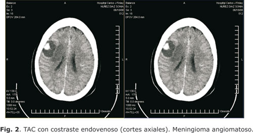
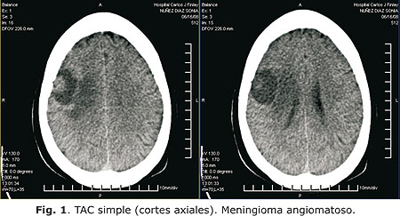
CASO CLÍNICOPaciente del sexo femenino, de 66 años de edad, con antecedentes de salud que fue llevada al cuerpo de guardia por presentar convulsiones. El examen físico fue negativo. Se le realizó TAC simple y contrastada, visualizándose una imagen mixta de contornos bien definidos, con nódulo hiperdenso (30-36 UH) en su interior de ± 22 x 15 x 19 mm; el resto de la lesión se observó hipodensa (14-19UH) midiendo en su conjunto 36 x 28 x 35 mm, se localizó en la región parietal anterior derecha, adyacente a la tabla interna, acompañándose de edema perilesional y colapso parcial del ventrículo lateral homolateral sin desplazamiento de las estructuras de la línea media (fig. 1).
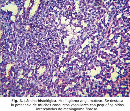
En el TAC con contrate endovenoso, el nódulo se realzó intensamente, de forma homogénea y precoz, la periferia de la lesión no captó (fig. 2). Estos hallazgos permitieron realizar el diagnóstico presuntivo de tumor cerebral primario, probable astrocitoma de bajo grado vs. hemangioblastoma, que por su localización parietal alta podría justificar el cuadro clínico, lo que motivó el tratamiento quirúrgico, con posterior examen histopatológico, encontrándose un meningioma angiomatoso (fig.3).
COMENTARIOS
El diagnóstico preoperatorio del meningioma quístico constituye un hecho difícil ya que por una parte los cambios quísticos son infrecuentes en estas neoplasias y por otro lado muy frecuentes en otras, por lo que son habitualmente confundidos con gliomas quísticos o con áreas de necrosis, abscesos intracraneales, metástasis y linfomas.6 ]]>
La bibliografía revisada sugiere que la existencia de un meningioma quístico debe ser sospechada cuando la porción quística del tumor es de similares proporciones que la sólida, cuando el componente sólido asienta en los sitios clásicos en que se encuentran los meningiomas, cuando la apariencia de la porción sólida sea lobulada y se realce uniformemente con el medio de contraste; especialmente si existen calcificaciones, reacción meníngea u ósea así como afluencia vascular por el sistema carotideo externo (angiografía cerebral). La resonancia magnética nuclear es más sensible y específica para demostrar la existencia de unión del tumor a las meninges (signo de la cola); no obstante, en la actualidad el diagnóstico preoperatorio de esta entidad solo se logra en el 65 % de los casos.5,7,8Un 8 % de estos tumores son de variedad maligna y un 12 % angioblásticos, pudiendo estar ausente por completo el sitio de unión a la duramadre. Hasta el momento no se ha demostrado que esta infrecuente variedad de meningioma lleve implícito mayor riesgo de malignidad o recidiva.5,9
En conclusión, los meningiomas quísticos constituyen un hallazgo infrecuente en la práctica neuroquirúrgica, no obstante, se debe recordar que existen varios signos imagenológicos que orientan al diagnóstico preoperatorio, como la existencia del signo de la cola, la irrigación procedente de la carótida externa o la ubicación en sitios de asentamiento frecuente de meningiomas.
REFERENCIAS BIBLIOGRÁFICAS
1. Sales Llopis J. Meningioma [Internet]. Servicio de Neurocirugía del Hospital General Universitario de Alicante. 2006. [Actualizado 17/2/2006; citado octubre de 2009]. Disponible en: http://www.neurocirugia.com/diagnostico/meningioma/index.htm.
2. De Girolani V. El sistema nervioso central. En: Robbinns, Cotran RS, Kumar V, Collins T. Patología estructural y funcional. 6ta. ed. Madrid: McGraw Hill; 2000. p.1339-404.
3. Gaensler HL, Barakos JA, Barr RM, Gean AD, Helms CA, Koeller K, et al. Neurorradiología Fundamental. Madrid: MARBÁN; 1998. p. 100-34.
4. Ugarte Suárez JC, Ugarte Moreno D, Jordán González J, Gaspar Obregón A, Quevedo Sotolongo L, Fermín Hernández E, et al. Manual de tomografía axial computarizada multicorte. 3ra ed. La Habana: CIMEQ; 2006. p. 80-2.
5. Varela Hernández A, Sánchez Rodríguez H, Rosales Torres P, Zayas Alba E. Presentación de un caso de meningioma quístico. Revisión de la literatura. [Internet] II Congreso Virtual Neurocirugía, 2002. Disponible en:
http://www.uninet.edu/neuroc2002/papers/TL-MeningQuist.htm
6. El- Fiki M, El-Henawy Y, Abdel-Rahman N. Cystic Meningioma. Acta Neurochirurgica [Internet]; julio de 1996 [citado octubre de 2009]; 138(7): [6 pantallas]. Disponible en: http://www.springerlink.com/content/ux606m1511637620/fulltext.pdf
7. McDermott MW, Wilson BC. Meningiomas. In: Winn RH. Youman Neurological Surgery. Philadelphia: Ed. Saunders; 1996. p. 2782-825.
8. Chen CT, Zee Ch, Miller AC, Weiss HM, Tang G, Chin L, et al. Magnetic resonance imaging and pathological correlates of meningioma. Neurosurgery. 1992 Diciembre;31(6):1015-21.
9. Suzuki Y, Sugimoto T, Shibuya M, Sugita K, Patel JS. Meningioma: Correlation between MRI characteristics and operative findings including consistency. Acta Neurochirurgica. 1994;129:29-46.
]]>
Recibido: 15 de octubre de 2010.
Dra. Lesly Solís Alfonso. Hospital Militar Central "Dr. Carlos J. Finlay". Avenida 114 y 31, Marianao, La Habana, Cuba. Correo electrónico: vicky@ida.cu ]]>