ANATOMY AND HISTOLOGY OF THE FEMALE REPRODUCTIVE SYSTEM OF Boophilus microplus (ACARI: IXODIDAE)
ANATOMIA E HISTOLOGIA DEL SISTEMA REPRODUCTOR FEMENINO DE Boophilus microplus (ACARI: IXODIDAE)
R. de la Vega*, G. Díaz**, M. Galán**, C. Fernández**
*LABIOFAM, Ave. Independencia Km16 ½, Boyeros, Ciudad Habana, Cuba, FAX: (537) 334857, E-mail: delavega@infomed.sld.cu; **Dpto. Biología Animal y Humana, Facultad de Biología, Universidad de La Habana, Cuba ]]>
ABSTRACT
The knowledge of the anatomy and histology of the female genital system in ticks is very important as a basic tool in all species in relation to morphological sciences and evolution, physiological events like ovulation, sexual behavior, maturation and mating, and also to research the interrelation tick-hemoparasite. The anatomy and histology of the female genital system in Boophilus microplus are scarcely studied. In the present paper, the female genital system of this tick has been considered as a whole and morphological and functional interrelations between different structures and also some other related items has been presented. Unfed and partially engorged females were employed in the study. The quality and resolution of images are remarkable because of the improvement by authors of a histological technique specially for arthropods.
Key words: ticks; Ixodidae; Boophilus microplus; anatomy and histology; female genital system.
RESUMEN
El estudio de la anatomía y la histología del sistema genital femenino en las garrapatas es de gran importancia como una herramienta básica para el conocimiento de las diferentes especies en relación con las ciencias morfológicas y la evolución, eventos fisiológicos como la ovulación, conducta sexual, maduración y apareamiento, y además para investigar la interrelación garrapata-hemoparásito. La anatomía e histología del sistema genital femenino de Boophilus microplus están escasamente estudiadas. En el presente artículo se describe de forma integral el sistema genital femenino en esta especie y se presentan interrelaciones morfológicas y funcionales entre las diferentes estructuras, así como otros aspectos relacionados. Se emplearon hembras no alimentadas y parcialmente ingurgitadas. La calidad y resolución de las imágenes son notables como resultado del perfeccionamiento de una técnica histológica específica para artrópodos, por parte de los autores.
Palabras clave: garrapatas; Ixodidae; Boophilus microplus; anatomía e histología; sistema genital femenino.
]]>
INTRODUCTIONThe anatomy and histology of the female genital system in ticks have been studied only in some species by different authors (1,2,3,4,5,6). The knowledge of these subjects is very important as a basic tool in all species in relation to morphological sciences and evolution, physiological events like ovulation, sexual behavior, maturation and mating, and also to research the interrelation tick-hemoparasite. The midgut, salivary glands and ovary in ticks, are organs of the great interest for their role in tick physiology and/or pathogen transmission (7).
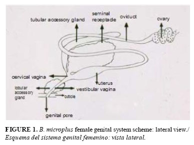
The structure of female genital system in Ixodidae consists of a single U-shaped tubular ovary in the posterior region of the body, paired and folded oviducts are in both extremes of the ovary joint into a common oviduct or uterus. The uterus opens into the vagina, which is divided in a cervical vagina and a vestibular vagina. A seminal receptacle, absent in argasids (3,4) as well as the uterus, open also in the cervical vagina. A pair of tubular accessory glands pours out their secretions at the level of the union of both cervical and vestibular vagina. Surrounding the vestibular vagina there is a glandular epithelium, the lobular accessory gland, found only in ixodid ticks (4).
The anatomy and histology of the female genital system in Boophilus microplus are scarcely studied; only it is possible to refer to articles about oviducts and fertilization (8,9) and studies of the ovary by Saito et al. (10). In the present paper, the general objective is to consider the female genital system of this tick as a whole and to analyze the morphological and functional interrelations between different structures. Also, the mating process in the species is approached and the semen transport inside the female´s tract is shown. The quality and resolution of images are remarkable because of the improvement by authors of a histological technique specially for arthropods and for the employ of an original procedure for feminine tract dissection that makes it easier and saves time avoiding technical complications.
MATERIALS AND METHODS
Procedures: Histological and dissection Techniques:
The histological technique of Schneider and Rudinsky (11) modified by de la Vega and Fernández (12) was used, to accomplish good results the inicial technique needs from 8 to 9 days, whereas that present demands only from 8 to 9 hours and images retain the same quality. Coloration was done with hematoxylin-eosine. A vertical microtome was used; tissue thickness of 3-5ì was achieved. Some serial slicing were performed transversely and other longitudinally in horizontal or in sagittal sense, this procedure permits a better comprehension of three-dimensional disposition of organs inside the arthropod. Dissection of tick specimens was performed with an original method (13) allowing a faster and secure procedure that could be done in 10 to 15 minutes per tick with a great percentage of success and without loss of genital structure parts.
Ticks: Unfed and partially engorged females of B. microplus, before or after mating, were employed in the study.
]]>
RESULTS AND DISCUSSIONGENERAL SCHEME OF FEMALE GENITAL STRUCTURE
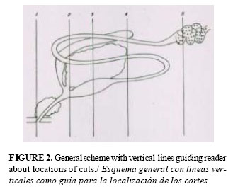
Figure 1 shows a general scheme of the principal organs that form the female genital system of B. microplus, in a lateral view, based on the mentioned papers and present results. As all schemes, it is a generalization from a number of individuals; reader ought to keep in mind that variations in each item from different specimens are actually a very frequent fact. Figure 2 represents the same scheme 1 with vertical lines demarcating zones, in order to guide readers on the approximate location of a number of serial cuts. So, next photographs will represent an approach of the anatomical location of these shown structures in relation to these conventional lines.
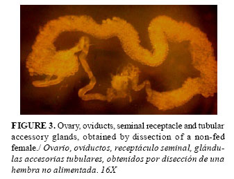
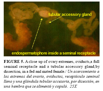
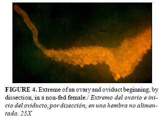
Ovary, oviducts, seminal receptacle and tubular accessory glands obtained by dissection of non-fed and fed, unmated and mated females are shown in Figures 3, 4 and 5.
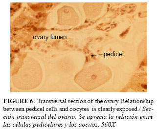
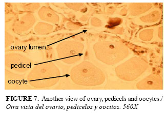
Ovary: The histological structure of ovary with oocytes and pedicel cells is shown in figures 6 and 7. The ovary is composed by a layer of small epithelial cells with rounded nuclei surrounding the lumen, and oocytes in different developmental stages: I, II, III, IV and V (10), attached through the pedicel cells to the ovarian wall. Also, the degenerative stage VI has been described (5,10). Cells very similar to the epithelial ones form the pedicels. Pedicel cells holding oocytes I and II stages are cylindric with large elliptic nuclei occupying most of the cytoplasm, there are numerous round mitochondria as well as rough endoplasmic reticulum, instead, pedicel cells attaching oocytes IV and V show signs of degeneration (14). In Rhipicephalus sanguineus, de Oliveira et al. (15) considered that there are three probable sources of elements for the oocytes' growth: an endogenous one, through synthesis processes inside the oocyte, and two exogenous ones: the haemolymph and the pedicel cells. Results of studies in Amblyomma triste by de Oliveira et al. (16) suggest that besides the exogenous production of vitellogenic elements, endogenous production can take place simultaneously contributing to the development and growth of the oocytes.
Central structures of female genital system
Oviducts: in both endings of the ovary there are two thin folded tubes, in B.microplus the diameter of oviducts oscillated between 75 and 140µ in females feed up to 48 hours; they fuse under the seminal receptacle to form the common oviduct or uterus.
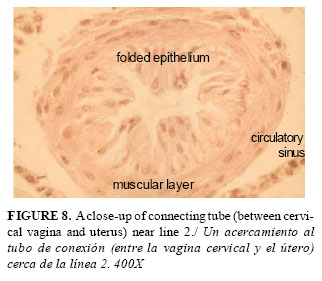
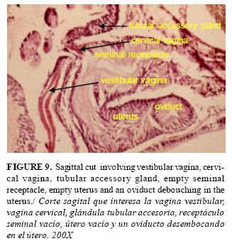
Uterus: It is in communication with the cervical vagina by means of an epithelial folded muscular tube: the connecting tube (Fig. 8). In the cervical vagina (Fig. 9), besides the uterus, the seminal receptacle, tubular accessory glands (TAG) and the vestibular vagina converge. There are some anatomical particularities in B. microplus that are evident when sections obtained in this tick (Fig. 9) are compared with schemes of other species like Rhipicephalus appendiculatus and Hyalomma asiaticum (1,3). In R. appendiculatus and H. asiaticum, the cervical vagina is very developed, the debouchment of the seminal receptacle and the uterus form an angle of 180° and are relatively distant one from the other. On the contrary, in B. microplus, the cervical vagina is very little, the debouchment of the seminal receptacle and the uterus in the mentioned vagina form an angle less than 90° and the seminal receptacle is completely above the uterus. This fact could facilitate the way for spermiophores to found the course toward the oviducts and ovary.
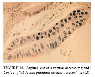
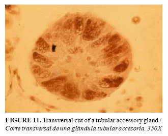
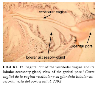
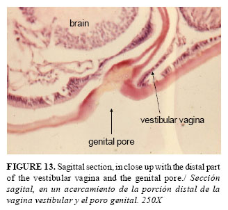
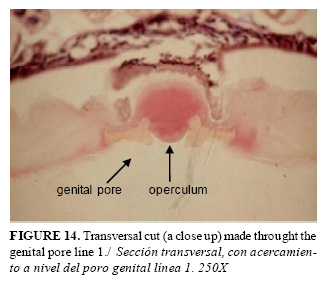
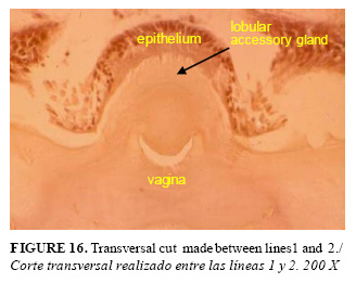
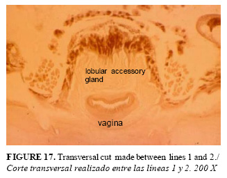
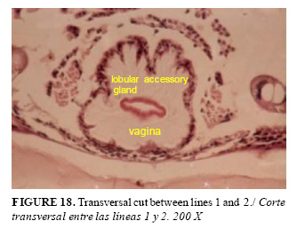
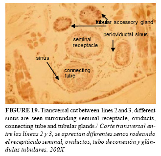
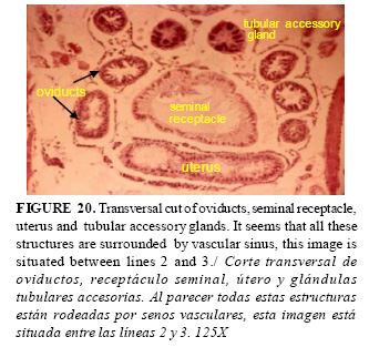
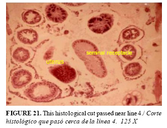
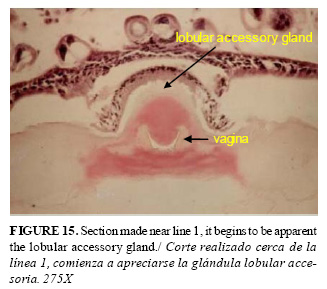
An anatomical view of one of TAG is shown in Fig. 5, and longitudinal and transversal histological views of one of these glands are presented in Fig. 10 and 11. In the female fixed for six days to the host and with 24 hours with a male near it, these glands have a length of 540µ and 90µ of wide. The vestibular vagina: in a sagittal histological cut (Fig. 12), it is seen that it extends from the genital pore to the cervical vagina. Also, the sagittal sections, and in close up of the genital pore and the final part of the vestibular vagina can be seen in Fig. 13, 14, 15. The lobular accessory gland is also shown in Fig 12; it is an enlargement of the vestibular vaginal epithelium which produces a secretion for the initial waterproofing layer for the eggs and also it is the most likely source of the genital sex pheromone (4, 17). Transversal sections of vestibular vagina appear in Fig. 16, 17, 18, they show that this structure is irregular in shape along it. Next transversal cuts (Fig. 19, 20, 21, 22) show images of the central part of the genital organs. In these images, all structures seem to be surrounded by circulatory sinus (in fact they were never shown in images of B. microplus). It is conspicuous the likeness of the images in all photographs and this fact discards the possibility of being a histological technic artifact.
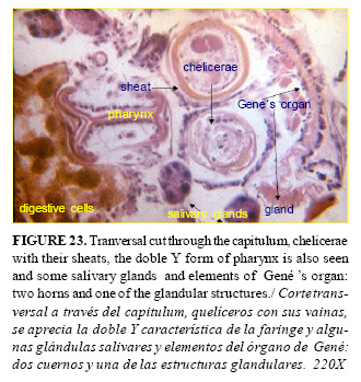
 Gené's organ: It is an organ only observed in ticks, it is located in the interior of the body cavity near the anterior end of the scutum (2,4). It is a paired structure formed by globular or finger like sacs sometimes called horns and glandular tissue. Different authors (1,2,3,4) have made a full description of this organ. When not in use, the organ is retracted within the anterior region of the body near the capitulum. In Fig. 23, a transversal cut can be appreciated at the cheliceral level, a horn and glandular tissue of the Gené's organ can be seen.
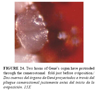
When oviposition begins, the swollen structure is projected forward by hydrostatic pressure, the finger-like processes are everted from the body (4). In Fig. 24, it can be appreciated an everted Gené's organ through the camerostomal fold just before oviposition begins. Gené's organ deposits wax on each egg as it is delivered from the vagina. The wax coat waterproofs the eggs and causes them to stick together (17) forming huge masses that also contribute to retard desiccation.
Copula and semen transport
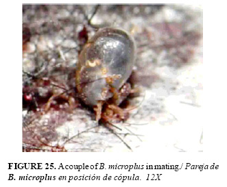
Male climbs over the back of the female and then slide down beneath the couple looking for the genital pore (18,19) and put itself venter to venter with the female, grasping it with its legs (Fig. 25). The male bends its capitulum toward the female genital aperture and then introduces its chelicerae which protrude out of its shafts into the female genital pore (18,19, 20); both hypostome and palps briefly touch the surface of the female, receptors on the palps mediate pheromone perception, these sensilla are concentrated on the fourth segment of the palp on hard ticks (20). A detail study of mating and sexual tick behavior could be read in Kiszewski et al. (20). ]]>
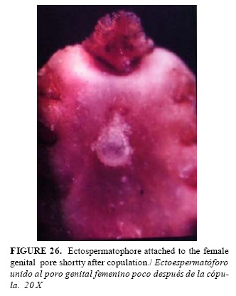
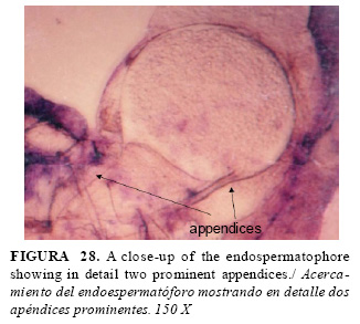
Male introduces its chelicerae inside the female genital pore and then ejaculates into a sac, named ectospermatophore; inside it , there is another sac the endospermatophore full of inmature seminal cells named spermatids. The male takes the ectospermatophore with its chelicerae by the neck of the sac and introduces it into the female genital pore (18). In Fig. 26, a female with the ectospermatophore hanguing from the genital pore can be seen, it is a weak union, and for this reason, it is not frequent to see it. The ectospermatophore is pear-like in shape and the maximum diameter oscillates between 400-500µ in this tick. The endospermatophore has a rounded shape (350-400 µ) with two appendices, one greater than the other one. These two appendices have been also described in other ticks and are characteristic of Ixodidae, whereas in Argasidae, there is only one (18).
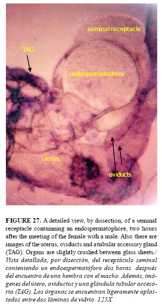
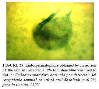
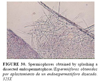
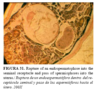
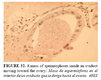
Spermatids suffer a capacitation inside the seminal receptacle in order to complete maturation; already mature cells are called spermiophores (Fig. 27, 28, 29, 30). The minimum time required after the copula is performed is seven days. When sexual cells are already mature, the endospermatophore´s wall breaks down and spermiophores form a compact packet passing into the uterus (Fig. 31). Finally in Fig. 32, it can be seen how spermiophores are released inside the feminine tract. It has been suggested (9) that fertilization takes place in the internal cylinder which extends from the uterus to the ovary itself.
REFERENCES
1. Till WM. A contribution to the anatomy and histology of the brown ear tick Rhipicephalus appendiculatus Newman. Mem Entomol Soc South Africa. 1961;6:124.
2. Arthur DR. Ticks and Disease. Pergamon Press. Oxford, 1962; 445 pp.
3. Balashov YuS. Bloodsucking ticks (Ixodoidea)-Vectors of Disease of Man and Animals. (English translation) Misc Publ Entomol Soc Amer. 1972;8(5):163-376.
4. Sonenshine DE. Biology of Ticks. Oxford University Press. 1991; 447 pp. ]]>
5. Denardi SE, Bechara GH, de Oliveira PR, Nunes ET, Saito KC, Camargo Mathias MI. Morphological characterization of the ovary and vitellogenesis dynamics in the tick Amblyomma cajennense (Acari: Ixodidae). Vet Parasitol. 2004;125:379-395.
6. de Oliveira PR, Camargo Mathias MI, Bechara GH. Amblyomma triste (Koch, 1844) (Acari: Ixodidae): Morphological description of the ovary and of vitellogenesis. Exp Parasitol. 2007;116:407-413.
7. Hajdušek O. Functional analysis of metabolic and imnune proteins in the tick Ixodes ricinus by RNA interference. University of South Bohemia, Faculty of Science, Department of Parasitology, Ph. D.Thesis. 2009; 62 pp.
8. García-Fernández C, García SML, Schneider FL, Severino AG, Winkelmann EC. Regionalization of oviducts in Boophilus microplus (Canestrini, 1887) (Acari: Ixodidae) and its potential significance for fertilization. Rev Brasil Biol. 1999;59(4):653-661.
9. García-Fernández C, García SML, Nunes García R, da Silva Valente VL. New histochemical and morphological findings in the female genital tract of Boophilus microplus (Acari, Ixodidae): an attempt toward the elucidation of fertilization in ticks. Iheringia, Sér. Zool., Porto Alegre. 2005;95(3):295-303. ]]>
10.Saito KC, Bechara GH, Nunes ET, de Oliveira PR, Denardi SE, Camargo Mathias MI. Morphological, histological, and ultrastructural studies of the ovary of the cattle-tick Boophilus microplus (Canestrini, 1887) (Acari: Ixodidae). Vet Parasitol. 2005;129:299-311.
11.Schneider I, Rudinsky JA. Anatomical and histological changes in internal organs of adult Tripodendron lineatum; Gnathotrichus retusus and G. sulcatus (Coleoptera: Scolytidae). Ann Entomol Soc Amer. 1969;62(5):995-1003.
12.de la Vega R, Fernández C. Método rápido de cortes de artrópodos en parafina. Rev Cub Med Trop. 1980;32:31-34.
13.de la Vega R. Una nota sobre un método para la disección de genitales femeninos en garrapatas. Rev cub Cienc agric. 1980;14:293-294.
14.de Oliveira PR, Camargo Mathias MI, Bechara GH. Vitellogenesis in the tick Amblyomma triste (Koch, 1844) (Acari: Ixodidae) Role for pedicel cells. Vet Parasitol. 2007;4:134-139. ]]>
15.de Oliveira PR, Bechara GH, Denardi SE, Nunes ET, Camargo Mathias MI. Morphological characterization of the ovary and oocytes vitellogenesis of the tick Rhipicephalus sanguineus (Latreille, 1806) (Acari:Ixodidae). Exp Parasitol. 2005;110:146-156.
16.de Oliveira PR, Bechara GH, Camargo Mathias MI. Amblyomma triste (Koch, 1844) (Acari: Ixodidae) Ovaries: An ultrastructural analysis. Exp Parasitol. 2007;116:407-413.
17.Kaufman WR. Assuring paternity in a promiscuous world: are there lessons for ticks among the insects? Parasitology. 2004;129:S145-S160.
18.Feldman-Musham B, Borut S. Copulation in ixodid ticks. J Parasitol. 1971;57(3):630-634.
19.Oliver JHJr. Symposium on reproduction of arthropods of medical and veterinary importance. IV Reproduction in ticks (Ixodoidea). J Med Entomol. 1974;11(1):26-34. ]]>
20.Kiszewski AE, Matuschka FR, Spielman A. Mating strategies and spermiogenesis in ixodid ticks. Annu Rev Entomol. 2001;46:167-182.
(Recibido 10-8-2011; Aceptado 15-11-2011) ]]>