<?xml version="1.0" encoding="ISO-8859-1"?><article xmlns:mml="http://www.w3.org/1998/Math/MathML" xmlns:xlink="http://www.w3.org/1999/xlink" xmlns:xsi="http://www.w3.org/2001/XMLSchema-instance">
<front>
<journal-meta>
<journal-id>0253-570X</journal-id>
<journal-title><![CDATA[Revista de Salud Animal]]></journal-title>
<abbrev-journal-title><![CDATA[Rev Salud Anim.]]></abbrev-journal-title>
<issn>0253-570X</issn>
<publisher>
<publisher-name><![CDATA[Centro Nacional de Sanidad Agropecuaria]]></publisher-name>
</publisher>
</journal-meta>
<article-meta>
<article-id>S0253-570X2016000200005</article-id>
<title-group>
<article-title xml:lang="en"><![CDATA[Prevalence of Mollicutes in Cell Cultures: experience in Cuba]]></article-title>
<article-title xml:lang="es"><![CDATA[Prevalencia de Mollicutes en cultivos celulares: experiencia en Cuba]]></article-title>
</title-group>
<contrib-group>
<contrib contrib-type="author">
<name>
<surname><![CDATA[Lobo Rivero]]></surname>
<given-names><![CDATA[Evelyn]]></given-names>
</name>
<xref ref-type="aff" rid="A01"/>
</contrib>
<contrib contrib-type="author">
<name>
<surname><![CDATA[Pérez Castillo]]></surname>
<given-names><![CDATA[Anisleidy]]></given-names>
</name>
<xref ref-type="aff" rid="A01"/>
</contrib>
<contrib contrib-type="author">
<name>
<surname><![CDATA[Duque Ortiz]]></surname>
<given-names><![CDATA[Arianna]]></given-names>
</name>
<xref ref-type="aff" rid="A01"/>
</contrib>
<contrib contrib-type="author">
<name>
<surname><![CDATA[Burgher Pulgarón]]></surname>
<given-names><![CDATA[Yaima]]></given-names>
</name>
<xref ref-type="aff" rid="A01"/>
</contrib>
<contrib contrib-type="author">
<name>
<surname><![CDATA[Miranda Marques]]></surname>
<given-names><![CDATA[Lucas]]></given-names>
</name>
<xref ref-type="aff" rid="A02"/>
</contrib>
<contrib contrib-type="author">
<name>
<surname><![CDATA[Timenetsky]]></surname>
<given-names><![CDATA[Jorge]]></given-names>
</name>
<xref ref-type="aff" rid="A02"/>
</contrib>
</contrib-group>
<aff id="A01">
<institution><![CDATA[,Centro Nacional de Sanidad Agropecuaria (CENSA) Departamento de Microbiología Laboratorio para el diagnóstico de micoplasmas]]></institution>
<addr-line><![CDATA[San José de las Lajas Mayabeque]]></addr-line>
<country>Cuba</country>
</aff>
<aff id="A02">
<institution><![CDATA[,Universidade de São Paulo Instituto de Ciências Biomédicas Departamento de Microbiología]]></institution>
<addr-line><![CDATA[ ]]></addr-line>
<country>Brazil</country>
</aff>
<pub-date pub-type="pub">
<day>00</day>
<month>08</month>
<year>2016</year>
</pub-date>
<pub-date pub-type="epub">
<day>00</day>
<month>08</month>
<year>2016</year>
</pub-date>
<volume>38</volume>
<numero>2</numero>
<fpage>100</fpage>
<lpage>104</lpage>
<copyright-statement/>
<copyright-year/>
<self-uri xlink:href="http://scielo.sld.cu/scielo.php?script=sci_arttext&amp;pid=S0253-570X2016000200005&amp;lng=en&amp;nrm=iso"></self-uri><self-uri xlink:href="http://scielo.sld.cu/scielo.php?script=sci_abstract&amp;pid=S0253-570X2016000200005&amp;lng=en&amp;nrm=iso"></self-uri><self-uri xlink:href="http://scielo.sld.cu/scielo.php?script=sci_pdf&amp;pid=S0253-570X2016000200005&amp;lng=en&amp;nrm=iso"></self-uri><abstract abstract-type="short" xml:lang="en"><p><![CDATA[Different mollicutes species of the Mycoplasma and Acholeplasma genera can contaminate the cell cultures and raw materials commonly used in the manufacture of a variety of biological and therapeutical products. In this study, the presence of mollicutes was detected in 19 of 50 cell culture samples analyzed using PCR and microbiological cultures. The species most frequently detected was M. fermentans (31.5%), followed by A. laidlawii (26.3%), M. orale (15.7%), M. hyorhinis (15.7%), and M. salivarium (10, 5%). A. laidlawii was present as the infecting agent only in 10.5% of the samples. These results showed the main species of contaminants, which suggested mollicutes multiple origin in the source of infection.]]></p></abstract>
<abstract abstract-type="short" xml:lang="es"><p><![CDATA[Diferentes especies de mollicutes, de los géneros Mycoplasma y Acholeplasma, pueden contaminar los cultivos celulares y las materias primas que se utilizan comúnmente en la fabricación de una variedad de productos biológicos y terapéuticos. En el presente estudio se detectó la presencia de mollicutes en 19 de 50 muestras analizadas de cultivo celular a través de PCR y cultivo microbiológico. Las especies detectadas con más frecuencia fueron M. fermentans (31,5%), seguido por A. laidlawii (26,3%), M. orale (15,7%), M. hyorhinis (15,7%) y M. salivarium (10,5%). A. laidlawii estaba presente como el agente infectante solo en 10,5% de las muestras. Estos resultados muestran las principales especies de mollicutes contaminantes, lo que sugiere un origen múltiple en la fuente de infección.]]></p></abstract>
<kwd-group>
<kwd lng="en"><![CDATA[mollicutes]]></kwd>
<kwd lng="en"><![CDATA[Mycoplasmas]]></kwd>
<kwd lng="en"><![CDATA[Acholeplasma]]></kwd>
<kwd lng="en"><![CDATA[cell cultures]]></kwd>
<kwd lng="en"><![CDATA[diagnostic]]></kwd>
<kwd lng="es"><![CDATA[mollicutes]]></kwd>
<kwd lng="es"><![CDATA[Mycoplasmas]]></kwd>
<kwd lng="es"><![CDATA[Acholeplasmas]]></kwd>
<kwd lng="es"><![CDATA[cultivos celulares]]></kwd>
<kwd lng="es"><![CDATA[diagnóstico]]></kwd>
</kwd-group>
</article-meta>
</front><body><![CDATA[ <p align="right"><font size="2" face="Verdana, Arial, Helvetica, sans-serif"><B>ORIGINAL    ARTICLE</B></font></p>     <p>&nbsp;</p>     <p><font size="2" face="Verdana, Arial, Helvetica, sans-serif"><b><font size="4">Prevalence    of <i>Mollicutes</i> in Cell Cultures: experience in Cuba</font></b></font></p>     <p>&nbsp;</p> <h1> <font size="2" face="Verdana, Arial, Helvetica, sans-serif"><b><font size="3">Prevalencia    de <i>Mollicutes</i> en cultivos celulares: experiencia en Cuba</font> </b></font></h1>     <p>&nbsp;</p>     <p>&nbsp; </p> <H1> <font size="2" face="Verdana, Arial, Helvetica, sans-serif"></font><B>        <P><font size="2" face="Verdana, Arial, Helvetica, sans-serif">Evelyn Lobo Rivero<SUP>I</SUP><a href="#autor">*</a><a name="pie"></a>,      Anisleidy P&eacute;rez Castillo<SUP>I</SUP>, Arianna Duque Ortiz<SUP>I</SUP>,      Yaima Burgher Pulgar&oacute;n<SUP>I</SUP>, Lucas Miranda Marques<SUP>II</SUP>,      Jorge Timenetsky<SUP>II</SUP> </font>    </B> </H1>     <P><font size="2" face="Verdana, Arial, Helvetica, sans-serif"><SUP>I</SUP>MYCOLAB,    Laboratorio para el diagn&oacute;stico de micoplasmas, Departamento de Microbiolog&iacute;a,    Centro Nacional de Sanidad Agropecuaria (CENSA), Carretera de Tapaste y Autopista    Nacional, San Jos&eacute; de las Lajas, Mayabeque, Cuba.    <br>   <SUP>II</SUP>Departamento de Microbiolog&iacute;a, Instituto de Ci&ecirc;ncias    Biom&eacute;dicas, Universidade de S&atilde;o Paulo, Brazil.</font>      <P>&nbsp;     ]]></body>
<body><![CDATA[<P>&nbsp; <hr noshade size="1">     <P><font size="2" face="Verdana, Arial, Helvetica, sans-serif"><B>ABSTRACT</B></font>     <P><font size="2" face="Verdana, Arial, Helvetica, sans-serif">Different mollicutes    species of the <I>Mycoplasma</I> and <I>Acholeplasma</I> genera can contaminate    the cell cultures and raw materials commonly used in the manufacture of a variety    of biological and therapeutical products. In this study, the presence of mollicutes    was detected in 19 of 50 cell culture samples analyzed using PCR and microbiological    cultures. The species most frequently detected was <I>M. fermentans </I>(31.5%),    followed by <I>A. laidlawii</I> (26.3%), <I>M. orale </I>(15.7%)<I>, M. hyorhinis</I>    (15.7%), and <I>M. salivarium</I> (10, 5%). <I>A. laidlawii</I> was present    as the infecting agent only in 10.5% of the samples. These results showed the    main species of contaminants, which suggested mollicutes multiple origin in    the source of infection. </font>     <P><font size="2" face="Verdana, Arial, Helvetica, sans-serif"><B>Key words:</B>    mollicutes, <I>Mycoplasmas</I>, <I>Acholeplasma</I>, cell cultures, diagnostic.</font> <hr noshade size="1">     <P><font size="2" face="Verdana, Arial, Helvetica, sans-serif"><B>RESUMEN</b></font>     <P><font size="2" face="Verdana, Arial, Helvetica, sans-serif">Diferentes especies    de mollicutes, de los g&eacute;neros <I>Mycoplasma</I> y <I>Acholeplasma,</I>    pueden contaminar los cultivos celulares y las materias primas que se utilizan    com&uacute;nmente en la fabricaci&oacute;n de una variedad de productos biol&oacute;gicos    y terap&eacute;uticos. En el presente estudio se detect&oacute; la presencia    de mollicutes en 19 de 50 muestras analizadas de cultivo celular a trav&eacute;s    de PCR y cultivo microbiol&oacute;gico. Las especies detectadas con m&aacute;s    frecuencia fueron <I>M. fermentans</I> (31,5%), seguido por <I>A. laidlawii</I>    (26,3%), <I>M. orale</I> (15,7%), <I>M. hyorhinis</I> (15,7%) y <I>M. salivarium</I>    (10,5%). <I>A. laidlawii</I> estaba presente como el agente infectante solo    en 10,5% de las muestras. Estos resultados muestran las principales especies    de mollicutes contaminantes, lo que sugiere un origen m&uacute;ltiple en la    fuente de infecci&oacute;n.</font>     <P><font size="2" face="Verdana, Arial, Helvetica, sans-serif"><B>Palabras clave:    </B>mollicutes, <I>Mycoplasmas, Acholeplasmas</I>, cultivos celulares, diagn&oacute;stico.</font> <hr noshade size="1">     <P>&nbsp;     <P>&nbsp;     <P><font size="2" face="Verdana, Arial, Helvetica, sans-serif"><B><font size="3">INTRODUCTION</font></B>    </font>     ]]></body>
<body><![CDATA[<P><font size="2" face="Verdana, Arial, Helvetica, sans-serif">Mycoplasmas (Mollicutes)    remain the smallest free-living bacteria found in animals including humans,    plants and insects (1). <I>Mycoplasma</I> and <I>Acholeplasma</I> are the main    genera that contaminate cell cultures and their substrates, as well as the raw    materials commonly used for manufacturing a variety of biological and therapeutic    products (2). Mycoplasma contamination, even at low load, at the initial steps    of manufacture, generally results in the loss of the whole batch of the product    due to poor cell growth or regulatory safety concerns. </font>     <P><font size="2" face="Verdana, Arial, Helvetica, sans-serif">The first isolation    of mycoplasma from a cell culture was reported in 1956 (3). At present, about    20 species have been identified (4). <I>Acholeplasma laidlawii</I>, <I>Mycoplasma    arginini</I>, <I>M.orale</I>, <I>M. salivarium</I>, <I>M.fermentans</I>, and    <I>M. hyorhins </I>have been detected in about 95% of cell cultures. Species    such as <I>Ureaplasma urealyticum</I>, <I>M. pneumoniae </I>and <I>M. pirum    </I>are rarely present in cell cultures and some of them have been isolated    only once (1, 5). </font>     <P><font size="2" face="Verdana, Arial, Helvetica, sans-serif">In most cases,    mycoplasma infection originates from contaminated animal serum, but contaminated    aerosols produced at the laboratory helps their spread. The frequency of mycoplasmas    in cell cultures depends on sampling, institution and the time for testing (6).    <I>Mycoplasma </I>contamination cause many cytogenetic effects in cell cultures,    leading to unreliable experimental results and potential harmful biological    products (7). Therefore, the result for mycoplasma<I> </I>contamination testing    must be strongly confident for the quality of biotechnological products (8).    In addition, as consequence of a wrong result, there are economic and labor    time losses. </font>     <P><font size="2" face="Verdana, Arial, Helvetica, sans-serif">In Cuba, the biotechnology    industry is increasingly being developed. Therefore, quality standards for detection    of mollicutes as contaminants of biotech products have been established for    biomedical application. However, the identification of mollicutes species in    cell cultures must also be added to the regular testing procedures. This approach    will help to a better control of the origin of such contaminations (2). The    aim of this study was to explore the status of mycoplasma contaminations in    cell cultures sent to our laboratory to be tested.</font>     <P>&nbsp;     <P><font size="2" face="Verdana, Arial, Helvetica, sans-serif"><B><font size="3">MATERIAL    AND METHODS</font></B> </font>     <P><font size="2" face="Verdana, Arial, Helvetica, sans-serif"><B>Samples</B>    </font>     <P><font size="2" face="Verdana, Arial, Helvetica, sans-serif">The following mycoplasma    ATCC strains were used as controls: <I>U. urealyticum </I>T960, <I>A. laidlawii    </I>P8, <I>M. pneumoniae</I>-FH, <I>M.genitalium </I>G37, <I>M. hominis </I>PG-21,    <I>M.hyorhinis </I>BTS7, <I>M. orale </I>CH19299, <I>M.salivarium </I>PG-20,    <I>M. buccale </I>CH202247, <I>M. fermentans </I>PG-18, <I>M. pulmonis </I>PG-34,    and <I>M. arginini </I>G230. </font>     <P><font size="2" face="Verdana, Arial, Helvetica, sans-serif"><B>Sample</B> </font>     <P><font size="2" face="Verdana, Arial, Helvetica, sans-serif">Fifty cell culture    samples were tested in 2014-2015; some cell cultures did not show morphological    alterations and were monitored for the first time. The samples were from five    laboratories in Havana, Cuba, </font>     ]]></body>
<body><![CDATA[<P><font size="2" face="Verdana, Arial, Helvetica, sans-serif"><B>Culture</B>    </font>     <P><font size="2" face="Verdana, Arial, Helvetica, sans-serif">Each cell sample    was inoculated in liquid and solid Hyflick&#180;s medium. The cultures were    incubated for 15 days at 37&#186;C under aerobic and anaerobic conditions (2).    The microorganism was presumptively identified based on pH shifts of the broth    without turbidity, production of &#171;fried egg&#187; colonies on Hyflick&#180;s    medium agar plates and positive subculture after filtration of the initial culture    through 0.22-mm membranes (9). </font>     <P><font size="2" face="Verdana, Arial, Helvetica, sans-serif"><B>Polymerase chain    reaction</B> </font>     <P><font size="2" face="Verdana, Arial, Helvetica, sans-serif">The technique used    was that described by Timenetsky <I>et al.</I> (6). The targeted DNA was extracted    from the cell culture samples and from cultures of the reference strains by    boiling 1 ml of each sample.
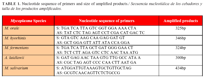
 </font>     <P><font size="2" face="Verdana, Arial, Helvetica, sans-serif">Twenty-five pmoles    of each primer (<a href="/img/revistas/rsa/v38n2/t0105216.jpg">Table 1</a>),    1U <I>Taq </I>DNA polymerase (Biotools), 1.4; 1.8 and 1.6 mM MgCl<SUB>2</SUB>    (Promega) for <I>M. salivarium</I>, <I>M. arginini </I>and <I>M. hyorhinis</I>,    respectively, 200 mM of each dNTP (Promega), 1 ml DNA extracted from the cell    culture sample and ultrapure water to a final volume of 50 mL. The amplification    was made in a thermocycler (Eppendorf). It was programmed for 40 cycles at 94&#186;C    for 30 s, 55&#186;C for 30 s, and 72&#186;C for 60 s and a final step at 72&#186;C    for 5 min.For the detection of <I>M. fermentans</I>, <I>M.orale </I>and <I>A.    laidlawii</I>, the reaction mixture contained 40 pmol of each primer, 1 U <I>Taq    </I>DNA polymerase (Biotools), 1.2 mM MgCl<SUB>2 </SUB>(Promega), 200 mM of    each dNTP (Promega), 1 mL DNA extracted from the cell culture sample, and ultrapure    water to a final volume of 50 mL. The amplification was made in a thermocycler    (Eppendorf). It was programmed for one cycle at 95&#186;C for 15 min, 30 cycles    at 95&#186;C for 30 s, 64&#186;C for 90 s, and 72&#186;C for 90 s, and a final    step at 72&#186;C for 10 min. </font>      
<P><font size="2" face="Verdana, Arial, Helvetica, sans-serif">A positive control    (DNA of each reference strains) and a negative control (ultrapure water) were    added to all amplifications.</font>     <P>&nbsp;     <P><font size="2" face="Verdana, Arial, Helvetica, sans-serif"><B><font size="3">RESULTS    AND DISCUSSION</font></B> </font>     <P><font size="2" face="Verdana, Arial, Helvetica, sans-serif">Mycoplasma-infected    cell lines are themselves the single most important source for further spreading    of contamination. This is due to the high concentration of mycoplasmas in infected    cultures, and the prolonged survival of dried mycoplasmas (5). Operator-induced    contamination is also a potential issue. Mycoplasmas spread by using laboratory    equipment, media, or reagents that have been contaminated (8). </font>     <P><font size="2" face="Verdana, Arial, Helvetica, sans-serif">In the present    study, 19 (38%) cell culture samples out of 50 were positive for mycoplasma    culturing; 15 samples (78.9%) presented pH shifts in the broths at 48 hours,    no pH shifts were observed in the remaining samples. However, when the samples    were subcultured from the broths to the agar plates, all 19 produced &#171;fried    egg&#187; colonies. Interestingly, 75.8% of the infected cell cultures presented    at least two mycoplasma species, and 15.78% of the samples were infected with    three species according to the biochemical test results. </font>     ]]></body>
<body><![CDATA[<P><font size="2" face="Verdana, Arial, Helvetica, sans-serif">According to Neto    <I>et al.</I> (8), up to 87% of the cellular cultures could be contaminated    by mycoplasmas. The contamination percentage variation found in the literature    was related to the size of the sample population studied, contamination control    practices, and efficiency of the detection assays used; in our case, the percentage    was in agreement with international reports. </font>     <P><font size="2" face="Verdana, Arial, Helvetica, sans-serif">The mollicutes    species detected in the present study derived from animals, humans, or both,    and the diversity of mycoplasma species in the same cell culture indicated the    occurrence of different initial infection sources. In our case, we agree with    Kazemiha <I>et al</I>. (4) about the subculturing of a cell culture among laboratories    over time, which, due to successive sharing, may explain the detection of multiple    mycoplasma species. Mycoplasma diversity accumulates over time mainly due to    failure to control the infection. </font>     <P><font size="2" face="Verdana, Arial, Helvetica, sans-serif">On the other hand,    because many infectious agents are not easily cultivable, PCR has been shown    to be an efficient methodology for detecting biological contaminants in cell    culture and its supplies (10). Moreover, the PCR technique has attracted much    attention in the detection of cell culture contaminants because it is fast,    robust, highly sensitive and specific compared with traditional techniques.    </font>     <P><font size="2" face="Verdana, Arial, Helvetica, sans-serif">
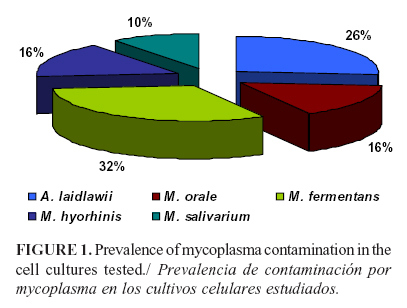
The results by    PCR showed that the most frequent species was <I>M. fermentans</I> in 31.5%    samples, followed by <I>A. laidlawii </I>in 26.3%, <I>M. orale </I>in 15.7%,    <I>M. hyorhinis</I> in 15.7%, and <I>M. salivarium </I>in 10.5%.<I> A. laidlawii    </I>was detected as the single mollicutes in 10.5% samples (<a href="#f1">Figure    1</a>).</font>      <P align="center"><img src="/img/revistas/rsa/v38n2/f0105216.jpg" width="407" height="308">    <a name="f1"></a>     
<P><font size="2" face="Verdana, Arial, Helvetica, sans-serif">In fact,<I> M.    fermentans </I>and<I> A. laidlawii</I> were also identified by Uphoff and Drexler    (5)<I>. </I> For<I> A. laidlawii, </I>the result<I> </I>was similar to that    mentioned by Timenetsky et al. (6), and in the case of <I>M. orale,</I> the    percentage obtained in our study was similar to that reported by Kazemiha <I>et    al.</I> (4), who indicated values of 12.5%. Moreover, <I>M. hyorhinis</I> percentage    in our case was 15.7, which was different from the percentages between 42 and    32 reported by other authors (11, 12). In this case, this percentage differences    could be explained by the type of sample worked because previous studies reported    trypsin and no cell cultures as the major source of contamination of <I>M. hyorhinis</I>    (9). </font>     <P><font size="2" face="Verdana, Arial, Helvetica, sans-serif">In most cases,    the contamination was through mycoplasmas derived from animal sera, mainly from    contaminated cattle, as well as from aerosols derived from humans due to non-aseptic    practices in the laboratory environments (13), justifying why such inputs were    important to choose. </font>     <P><font size="2" face="Verdana, Arial, Helvetica, sans-serif">Technical procedures    in a laboratory are the main sources of <I>M. orale</I>, <I>M. fermentans,</I>    and <I>M. hominis</I> (3). These mycoplasmas account for more than half of all    mycoplasma infections in cell cultures, and physiologically they are found in    the human oropharyngeal tract (11). <I>M. arginini </I>and <I>A. laidlawii </I>are    species originated from fetal bovine serum (FBS) or newborn bovine serum (NBS).    Although <I>M hyorhynis</I> has a swine origin, mycoplasmas have never been    isolated from this solution and their DNA has been rarely detected. Trypsin    has mycoplasmicide activity (9). </font>     <P><font size="2" face="Verdana, Arial, Helvetica, sans-serif">There is a number    of different sources for mycoplasma contamination in cell cultures associated    with human, bovine, and swine species (14). <I>M. fermentans </I>was considered    a normal inhabitant of the human urogenital tract and it is a fastidious species,    a fact that impaired its isolation in the past. In 1986, <I>M. fermentans </I>was    considered as a co-factor for the development of AIDS in HIV-positive individuals,    a fact that, in turn, attracted the interest of the scientific community (15).    Subsequently, this microorganism<I> </I>has been detected in or associated most    frequently with tissues and blood of individuals with diseases poorly studied    or with unknown etiology (14). In our case, the increase in the frequency of    <I>M. fermentans </I>in cell cultures can be explained by the increasing use    of human blood cells or tissues for primary culture. </font>     <P><font size="2" face="Verdana, Arial, Helvetica, sans-serif">Regarding<I> A.    laidlawii,</I> it is a commensal of the mucosa in the upper respiratory and    urogenital tract of many animal and bird species. Its potential pathogenicity    has been the subject of investigation particularly with regard to bovine mastitis    and spontaneous abortion in farm animals (9). Generally, it is regarded as non-pathogenic    but may cause opportunistic, often transient, infections; the only reported    incidence of human isolation was from an infected burn wound (14). The high    incidence of <I>A. laidlawii</I> in cell cultures in this work seems,,to be    in direct correlation with the use of fetal or newborn bovine serum. </font>     ]]></body>
<body><![CDATA[<P><font size="2" face="Verdana, Arial, Helvetica, sans-serif">Potential undetected    contamination of these products or process intermediates with mycoplasmas presents    a potential safety risk for patients and a business risk for producers of biopharmaceuticals    (1). To minimize these risks, monitoring for adventitious agents, such as mycoplasmas,    is performed during the manufacture of biologics produced in cell culture substrates.    </font>     <P><font size="2" face="Verdana, Arial, Helvetica, sans-serif">The results of    this study show the importance of mollicutes diagnosis in cell cultures, as    they remain one of the most common contaminations. In addition, it is also important    to know the possible sources of infection to help control and taking action    to prevent the spread to other cell lines and biological products.</font>     <P>&nbsp;     <P><font size="2" face="Verdana, Arial, Helvetica, sans-serif"><B><font size="3">ACKNOWLEDGMENTS    </font> </B> </font>     <P><font size="2" face="Verdana, Arial, Helvetica, sans-serif">Special thanks    are due to CAPES-Brasil and to Dr. Jorge Timenetsky from the University of S&atilde;o    Paulo in the make this work.</font>     <P>&nbsp;     <P><font size="2" face="Verdana, Arial, Helvetica, sans-serif"><B><font size="3">REFERENCES</font></B>    </font>          <!-- ref --><P><font size="2" face="Verdana, Arial, Helvetica, sans-serif">1. Volokhov DV,    Laurie J, Graham KA, Brorson E, Chizhikov V. Mycoplasma testing of cell substrates    and biologics: Review of alternative non-microbiological techniques. Mol Cell    Probe. 2011;25:69-77.     </font>      <!-- ref --><P><font size="2" face="Verdana, Arial, Helvetica, sans-serif">2. Lozada Y,      Betancourt A, Lobo E. Detecci&oacute;n confiable de micoplasmas como contaminantes      en cultivos biol&oacute;gicos. Tesis en opci&oacute;n al grado de Master en      Microbiolog&iacute;a veterinaria (MSc), Mayabeque, Cuba. 2013.     </font>        <!-- ref --><P><font size="2" face="Verdana, Arial, Helvetica, sans-serif">3. Robinson LB,      Wichelhausen RH. Contamination of human cell cultures by pleuropneumonialike      organisms. Science. 1956;124:1147-1148.     </font>         <!-- ref --><P><font size="2" face="Verdana, Arial, Helvetica, sans-serif">4. Kazemiha VM,    Shokrgozar MA, Arabestani MR, Moghadam MS, Azari S, Shokri F. PCR-based detection    and eradication of mycoplasmal infections from various mammalian cell lines:    a local experience. Cytotechnology. 2009;61:117-124. DOI 10.1007/s10616-010-9252-6.        </font>      <!-- ref --><P><font size="2" face="Verdana, Arial, Helvetica, sans-serif">5. Uphoff CC, Drexler    HG. Comparative PCR analysis for detection of mycoplasma infections in continuous    cell lines. Cancer Cell Culture: Methods and Protocols, 2nd Edition, Methods    in Molecular Biology. 2006:731.     </font>      <!-- ref --><P><font size="2" face="Verdana, Arial, Helvetica, sans-serif">6. Timenetsky      J, Santos LM, Buzinhani M, Mettifogo E. Detection of multiple mycoplasma infection      in cell cultures by PCR. Brazilian Journal of Medical and Biological Research.      2006;39:907-914.     </font>        <!-- ref --><P><font size="2" face="Verdana, Arial, Helvetica, sans-serif">7. Schmitt M,      Pawlita M. High-throughput detection and multiplex identification of cell      contaminations. Nucleic Acids Res.<I> </I>2004;37:119-126.     </font>         <!-- ref --><P><font size="2" face="Verdana, Arial, Helvetica, sans-serif">8. Netto C, Thomaz    V, Sepulveda L, Oliveira GH, Timenetsky J. Quality Control of Biotechnological    Inputs Detecting <I>Mycoplasma.<B> </B></I>Braz Arch Biol Technol. 2015;58(2):239-243.        </font>      <!-- ref --><P><font size="2" face="Verdana, Arial, Helvetica, sans-serif">9. Windsor HM,      Windsor GD, Noordergraaf JH. The growth and long-term survival of Acholeplasma      laidlawii in media products used in biopharmaceutical manufacturing. Biologicals.      2010;38:204-210.     </font>         <!-- ref --><P><font size="2" face="Verdana, Arial, Helvetica, sans-serif">10.Van Kuppeveld    FJM, Johansson KE, Galama JMD, Kissing J, B&ouml;lske G, et al. Detection of    <I>Mycoplasma </I>contamination in cell cultures by mycoplasma group-specific    PCR. Appl Environ Microbiol. 1994;60:149-152.     </font>      <!-- ref --><P><font size="2" face="Verdana, Arial, Helvetica, sans-serif">11.Camargos MF,      Oliveira AM, Fonseca Junior AA, Rivetti AV, Motta PMC, Assis RA. Aplica&ccedil;&atilde;o      da rea&ccedil;&atilde;o em cadeia da polimerase para detec&ccedil;&atilde;o      de Mycoplasma spp na rotina de cultivos celulares. Ci&ecirc;nc Anim Bras.      2008;9:786-790.     </font>         <!-- ref --><P><font size="2" face="Verdana, Arial, Helvetica, sans-serif">12.Rivera-Tapia    JA, Mu&ntilde;oz GZ, Rom&aacute;n C. Contaminaci&oacute;n por micoplasmas en    cultivos celulares. Anales M&eacute;dicos. 2006;51(3):109-112.     </font>      <!-- ref --><P><font size="2" face="Verdana, Arial, Helvetica, sans-serif">13.Rottem S.      Interaction of mycoplasmas with host cells. Physiol Rev. 2003;83:413-432.          </font>        <!-- ref --><P><font size="2" face="Verdana, Arial, Helvetica, sans-serif">14.Pinheiro de      Oliveira TF, Fonseca AJr, Fernandes M, Camargos M, Macedo de Oliveira A, Pinto      AC, et al. Detection of contaminants in cell cultures, sera and trypsin. Biologicals.      2013;41:407-414.     </font>         <!-- ref --><P><font size="2" face="Verdana, Arial, Helvetica, sans-serif">15.Shu H-W, Liu    T-T, Chan H-I, Liu Y-M, Wu K-M, et al. Complexity of the Mycoplasma fermentans    M64 Genome and Metabolic Essentiality and Diversity among Mycoplasmas. PLoS    ONE. 2012;7(4): e32940. doi:10.1371/journal.pone.0032940.    </font>     <P>&nbsp;     <P>&nbsp;     <P><font size="2" face="Verdana, Arial, Helvetica, sans-serif">Recibido: 4-11-2015.    ]]></body>
<body><![CDATA[<br>   </font><font size="2" face="Verdana, Arial, Helvetica, sans-serif">Aceptado:    9-5-2016.</font>     <P>&nbsp;     <P>&nbsp;     <P><font size="2" face="Verdana, Arial, Helvetica, sans-serif"><B><a href="#pie">*</a><a name="autor"></a>    </B>Corresponding author: <I>Evelyn Lobo Rivero.</I> E-mail: <U><a href="mailto:elobo@censa.edu.cu">elobo@censa.edu.cu</a></U></font>       ]]></body><back>
<ref-list>
<ref id="B1">
<label>1</label><nlm-citation citation-type="journal">
<person-group person-group-type="author">
<name>
<surname><![CDATA[Volokhov]]></surname>
<given-names><![CDATA[DV]]></given-names>
</name>
<name>
<surname><![CDATA[Laurie]]></surname>
<given-names><![CDATA[J]]></given-names>
</name>
<name>
<surname><![CDATA[Graham]]></surname>
<given-names><![CDATA[KA]]></given-names>
</name>
<name>
<surname><![CDATA[Brorson]]></surname>
<given-names><![CDATA[E]]></given-names>
</name>
<name>
<surname><![CDATA[Chizhikov]]></surname>
<given-names><![CDATA[V]]></given-names>
</name>
</person-group>
<article-title xml:lang="en"><![CDATA[Mycoplasma testing of cell substrates and biologics: Review of alternative non-microbiological techniques]]></article-title>
<source><![CDATA[Mol Cell Probe]]></source>
<year>2011</year>
<volume>25</volume>
<page-range>69-77</page-range></nlm-citation>
</ref>
<ref id="B2">
<label>2</label><nlm-citation citation-type="">
<person-group person-group-type="author">
<name>
<surname><![CDATA[Lozada]]></surname>
<given-names><![CDATA[Y]]></given-names>
</name>
<name>
<surname><![CDATA[Betancourt]]></surname>
<given-names><![CDATA[A]]></given-names>
</name>
<name>
<surname><![CDATA[Lobo]]></surname>
<given-names><![CDATA[E]]></given-names>
</name>
</person-group>
<article-title xml:lang="es"><![CDATA[Detección confiable de micoplasmas como contaminantes en cultivos biológicos]]></article-title>
<source><![CDATA[]]></source>
<year></year>
</nlm-citation>
</ref>
<ref id="B3">
<label>3</label><nlm-citation citation-type="journal">
<person-group person-group-type="author">
<name>
<surname><![CDATA[Robinson]]></surname>
<given-names><![CDATA[LB]]></given-names>
</name>
<name>
<surname><![CDATA[Wichelhausen]]></surname>
<given-names><![CDATA[RH]]></given-names>
</name>
</person-group>
<article-title xml:lang="en"><![CDATA[Contamination of human cell cultures by pleuropneumonialike organisms]]></article-title>
<source><![CDATA[Science]]></source>
<year>1956</year>
<volume>124</volume>
<page-range>1147-1148</page-range></nlm-citation>
</ref>
<ref id="B4">
<label>4</label><nlm-citation citation-type="journal">
<person-group person-group-type="author">
<name>
<surname><![CDATA[Kazemiha]]></surname>
<given-names><![CDATA[VM]]></given-names>
</name>
<name>
<surname><![CDATA[Shokrgozar]]></surname>
<given-names><![CDATA[MA]]></given-names>
</name>
<name>
<surname><![CDATA[Arabestani]]></surname>
<given-names><![CDATA[MR]]></given-names>
</name>
<name>
<surname><![CDATA[Moghadam]]></surname>
<given-names><![CDATA[MS]]></given-names>
</name>
<name>
<surname><![CDATA[Azari]]></surname>
<given-names><![CDATA[S]]></given-names>
</name>
<name>
<surname><![CDATA[Shokri]]></surname>
<given-names><![CDATA[F]]></given-names>
</name>
</person-group>
<article-title xml:lang="en"><![CDATA[PCR-based detection and eradication of mycoplasmal infections from various mammalian cell lines: a local experience]]></article-title>
<source><![CDATA[Cytotechnology]]></source>
<year>2009</year>
<volume>61</volume>
<page-range>117-124</page-range></nlm-citation>
</ref>
<ref id="B5">
<label>5</label><nlm-citation citation-type="">
<person-group person-group-type="author">
<name>
<surname><![CDATA[Uphoff]]></surname>
<given-names><![CDATA[CC]]></given-names>
</name>
<name>
<surname><![CDATA[Drexler]]></surname>
<given-names><![CDATA[HG]]></given-names>
</name>
</person-group>
<article-title xml:lang="en"><![CDATA[Comparative PCR analysis for detection of mycoplasma infections in continuous cell lines]]></article-title>
<source><![CDATA[Cancer Cell Culture: Methods and Protocols]]></source>
<year>2006</year>
<edition>2nd</edition>
<page-range>731</page-range></nlm-citation>
</ref>
<ref id="B6">
<label>6</label><nlm-citation citation-type="journal">
<person-group person-group-type="author">
<name>
<surname><![CDATA[Timenetsky]]></surname>
<given-names><![CDATA[J]]></given-names>
</name>
<name>
<surname><![CDATA[Santos]]></surname>
<given-names><![CDATA[LM]]></given-names>
</name>
<name>
<surname><![CDATA[Buzinhani]]></surname>
<given-names><![CDATA[M]]></given-names>
</name>
<name>
<surname><![CDATA[Mettifogo]]></surname>
<given-names><![CDATA[E]]></given-names>
</name>
</person-group>
<article-title xml:lang="en"><![CDATA[Detection of multiple mycoplasma infection in cell cultures by PCR]]></article-title>
<source><![CDATA[Brazilian Journal of Medical and Biological Research]]></source>
<year>2006</year>
<volume>39</volume>
<page-range>907-914</page-range></nlm-citation>
</ref>
<ref id="B7">
<label>7</label><nlm-citation citation-type="journal">
<person-group person-group-type="author">
<name>
<surname><![CDATA[Schmitt]]></surname>
<given-names><![CDATA[M]]></given-names>
</name>
<name>
<surname><![CDATA[Pawlita]]></surname>
<given-names><![CDATA[M]]></given-names>
</name>
</person-group>
<article-title xml:lang="en"><![CDATA[çHigh-throughput detection and multiplex identification of cell contaminations]]></article-title>
<source><![CDATA[Nucleic Acids Res]]></source>
<year>2004</year>
<volume>37</volume>
<page-range>119-126</page-range></nlm-citation>
</ref>
<ref id="B8">
<label>8</label><nlm-citation citation-type="journal">
<person-group person-group-type="author">
<name>
<surname><![CDATA[Netto]]></surname>
<given-names><![CDATA[C]]></given-names>
</name>
<name>
<surname><![CDATA[Thomaz]]></surname>
<given-names><![CDATA[V]]></given-names>
</name>
<name>
<surname><![CDATA[Sepulveda]]></surname>
<given-names><![CDATA[L]]></given-names>
</name>
<name>
<surname><![CDATA[Oliveira]]></surname>
<given-names><![CDATA[GH]]></given-names>
</name>
<name>
<surname><![CDATA[Timenetsky]]></surname>
<given-names><![CDATA[J]]></given-names>
</name>
</person-group>
<article-title xml:lang="en"><![CDATA[Quality Control of Biotechnological Inputs Detecting Mycoplasma]]></article-title>
<source><![CDATA[Braz Arch Biol Technol]]></source>
<year>2015</year>
<volume>58</volume>
<numero>2</numero>
<issue>2</issue>
<page-range>239-243</page-range></nlm-citation>
</ref>
<ref id="B9">
<label>9</label><nlm-citation citation-type="journal">
<person-group person-group-type="author">
<name>
<surname><![CDATA[Windsor]]></surname>
<given-names><![CDATA[HM]]></given-names>
</name>
<name>
<surname><![CDATA[Windsor]]></surname>
<given-names><![CDATA[GD]]></given-names>
</name>
<name>
<surname><![CDATA[Noordergraaf]]></surname>
<given-names><![CDATA[JH]]></given-names>
</name>
</person-group>
<article-title xml:lang="en"><![CDATA[The growth and long-term survival of Acholeplasma laidlawii in media products used in biopharmaceutical manufacturing]]></article-title>
<source><![CDATA[Biologicals]]></source>
<year>2010</year>
<volume>38</volume>
<page-range>204-210</page-range></nlm-citation>
</ref>
<ref id="B10">
<label>10</label><nlm-citation citation-type="journal">
<person-group person-group-type="author">
<name>
<surname><![CDATA[Van Kuppeveld]]></surname>
<given-names><![CDATA[FJM]]></given-names>
</name>
<name>
<surname><![CDATA[Johansson]]></surname>
<given-names><![CDATA[KE]]></given-names>
</name>
<name>
<surname><![CDATA[Galama]]></surname>
<given-names><![CDATA[JMD]]></given-names>
</name>
<name>
<surname><![CDATA[Kissing]]></surname>
<given-names><![CDATA[J]]></given-names>
</name>
<name>
<surname><![CDATA[Bölske]]></surname>
<given-names><![CDATA[G]]></given-names>
</name>
</person-group>
<article-title xml:lang="en"><![CDATA[Detection of Mycoplasma contamination in cell cultures by mycoplasma group-specific PCR]]></article-title>
<source><![CDATA[Appl Environ Microbiol]]></source>
<year>1994</year>
<volume>60</volume>
<page-range>149-152</page-range></nlm-citation>
</ref>
<ref id="B11">
<label>11</label><nlm-citation citation-type="journal">
<person-group person-group-type="author">
<name>
<surname><![CDATA[Camargos]]></surname>
<given-names><![CDATA[MF]]></given-names>
</name>
<name>
<surname><![CDATA[Oliveira]]></surname>
<given-names><![CDATA[AM]]></given-names>
</name>
<name>
<surname><![CDATA[Fonseca Junior]]></surname>
<given-names><![CDATA[AA]]></given-names>
</name>
<name>
<surname><![CDATA[Rivetti]]></surname>
<given-names><![CDATA[AV]]></given-names>
</name>
<name>
<surname><![CDATA[Motta]]></surname>
<given-names><![CDATA[PMC]]></given-names>
</name>
<name>
<surname><![CDATA[Assis]]></surname>
<given-names><![CDATA[RA]]></given-names>
</name>
</person-group>
<article-title xml:lang="pt"><![CDATA[Aplicação da reação em cadeia da polimerase para detecção de Mycoplasma spp na rotina de cultivos celulares]]></article-title>
<source><![CDATA[Ciênc Anim Bras]]></source>
<year>2008</year>
<volume>9</volume>
<page-range>786-790</page-range></nlm-citation>
</ref>
<ref id="B12">
<label>12</label><nlm-citation citation-type="journal">
<person-group person-group-type="author">
<name>
<surname><![CDATA[Rivera-Tapia]]></surname>
<given-names><![CDATA[JA]]></given-names>
</name>
<name>
<surname><![CDATA[Muñoz]]></surname>
<given-names><![CDATA[GZ]]></given-names>
</name>
<name>
<surname><![CDATA[Román]]></surname>
<given-names><![CDATA[C]]></given-names>
</name>
</person-group>
<article-title xml:lang="es"><![CDATA[Contaminación por micoplasmas en cultivos celulares]]></article-title>
<source><![CDATA[Anales Médicos]]></source>
<year>2006</year>
<volume>51</volume>
<numero>3</numero>
<issue>3</issue>
<page-range>109-112</page-range></nlm-citation>
</ref>
<ref id="B13">
<label>13</label><nlm-citation citation-type="journal">
<person-group person-group-type="author">
<name>
<surname><![CDATA[Rottem]]></surname>
<given-names><![CDATA[S]]></given-names>
</name>
</person-group>
<article-title xml:lang="en"><![CDATA[Interaction of mycoplasmas with host cells]]></article-title>
<source><![CDATA[Physiol Rev]]></source>
<year>2003</year>
<volume>83</volume>
<page-range>413-432</page-range></nlm-citation>
</ref>
<ref id="B14">
<label>14</label><nlm-citation citation-type="journal">
<person-group person-group-type="author">
<name>
<surname><![CDATA[Pinheiro de Oliveira]]></surname>
<given-names><![CDATA[TF]]></given-names>
</name>
<name>
<surname><![CDATA[Fonseca]]></surname>
<given-names><![CDATA[AJr]]></given-names>
</name>
<name>
<surname><![CDATA[Fernandes]]></surname>
<given-names><![CDATA[M]]></given-names>
</name>
<name>
<surname><![CDATA[Camargos]]></surname>
<given-names><![CDATA[M]]></given-names>
</name>
<name>
<surname><![CDATA[Macedo de Oliveira]]></surname>
<given-names><![CDATA[A]]></given-names>
</name>
<name>
<surname><![CDATA[Pinto]]></surname>
<given-names><![CDATA[AC]]></given-names>
</name>
</person-group>
<article-title xml:lang="es"><![CDATA[Detection of contaminants in cell cultures, sera and trypsin]]></article-title>
<source><![CDATA[Biologicals]]></source>
<year>2013</year>
<volume>41</volume>
<page-range>407-414</page-range></nlm-citation>
</ref>
<ref id="B15">
<label>15</label><nlm-citation citation-type="journal">
<person-group person-group-type="author">
<name>
<surname><![CDATA[Shu]]></surname>
<given-names><![CDATA[H-W]]></given-names>
</name>
<name>
<surname><![CDATA[Liu]]></surname>
<given-names><![CDATA[T-T]]></given-names>
</name>
<name>
<surname><![CDATA[Chan]]></surname>
<given-names><![CDATA[H-I]]></given-names>
</name>
<name>
<surname><![CDATA[Liu]]></surname>
<given-names><![CDATA[Y-M]]></given-names>
</name>
<name>
<surname><![CDATA[Wu]]></surname>
<given-names><![CDATA[K-M]]></given-names>
</name>
</person-group>
<article-title xml:lang="en"><![CDATA[Complexity of the Mycoplasma fermentans M64 Genome and Metabolic Essentiality and Diversity among Mycoplasmas]]></article-title>
<source><![CDATA[PLoS ONE]]></source>
<year>2012</year>
<volume>7</volume>
<numero>4</numero>
<issue>4</issue>
<page-range>e32940</page-range></nlm-citation>
</ref>
</ref-list>
</back>
</article>
