The mile-stone of PET radiopharmaceuticals development
Hitos en el desarrollo de los radiofármacos PET
Tatsuo Ido1, Tania Valdés2
1 Theragnostic Compound R&D Center, Neuroscience Research Institute, Gachon University, Incheon, South Korea Tohoku University, Sendai, Japan
2 Centro de Aplicaciones Tecnológicas y Desarrollo Nuclear, (Ceaden), Cuba
]]>
tidojapan@yahoo.com
ABSTRACT
The positron emitting nuclides were already tried in 1940's as in vivo radio-tracers in the research field of medical biology. In 1976, the discovery of 18FDG with the developing of a positron imaging device, allowed to obtain the image of the human brain by PET technology. Today, 18FDG is widely used in tumour diagnosis by a metabolic trapping mechanism, which is a new concept for functional imaging and makes possible the monitoring of the therapy process. This is first milestone of PET radiopharmaceutical development. The second milestone is the establishment of a molecular imaging method in nuclear medicine and third, is the development of the theragnostic concept of radiopharmaceuticals. At present highlight works are focused in tau protein imaging for Alzheimer disease diagnosis and inflammation imaging.
Key words: positron computed tomography, radiopharmaceuticals, radiotherapy, single photon emission computed tomography, biological functions, molecules, images.
RESUMEN
Los núclidos emisores de positrones fueron tratados en 1940 como radiotrazadores in vivo en el campo de las investigaciones biomédicas. En 1976, el descubrimiento de 18FDG, con el desarrollo de un equipo de imagenología positrónica, facilitó la obtención de imágenes del cerebro humano mediante la aplicación de la tecnología PET. En la actualidad, el 18FDG tiene amplia utilización en el diagnóstico de tumores mediante el mecanismo de captura metabólica. Este mecanismo es un concepto nuevo para la obtención de imágenes funcionales lo que permite realizar el monitoreo de los procesos terapéuticos. Este es el primer hito del desarrollo de radiofármacos PET. El segundo hito lo constituye el establecimiento del método de imagen molecular en la medicina nuclear. El tercer hito es el desarrollo del concepto teragnóstico de los radiofármacos. En el momento actual los trabajos principales están enfocados a la imagen de proteínas tau para el diagnóstico de la enfermedad de Alzheimer y las imágenes de inflamaciones.
Palabras claves: tomografía computarizada con positrón, radiofármacos, radioterapia, tomografía de emisión computarizada de fotón único, funciones biológicas, moléculas, imágenes.
INTRODUCTION
]]> The radioactive nuclides used for radiopharmaceuticals are detected with very high sensitivity and are suitable as in-vivo biomarkers tracers. These properties make them useful for in-vitro radioimmunoassay and in-vivo imaging in Nuclear Medicine.In the 40's, several radionuclides were tried for animal and human behavior research. In the 1950's, the nuclides ![]() ,
, ![]() and
and ![]() were used for diagnosis and treatment of diseases in nuclear medicine. In the 1960's -1970's,
were used for diagnosis and treatment of diseases in nuclear medicine. In the 1960's -1970's, ![]() (daughter of
(daughter of ![]() ) with the gamma camera was introduced as a practical method for nuclear medicine and was applied in various chemical forms to visualize organ degeneration in the body. The development of the single photon emission computed tomography (SPECT) in the 1980's led to the wide use of
) with the gamma camera was introduced as a practical method for nuclear medicine and was applied in various chemical forms to visualize organ degeneration in the body. The development of the single photon emission computed tomography (SPECT) in the 1980's led to the wide use of ![]() - radiopharmaceuticals. At present, the most commonly used single photon emitting nuclides are
- radiopharmaceuticals. At present, the most commonly used single photon emitting nuclides are ![]() ,
, ![]() ,
, ![]() ,
, ![]() ,
, ![]() ,
, ![]() and
and ![]() .
.
Positron emitting short half-life nuclides (![]() ,
, ![]() ,
, ![]() and
and ![]() ) were introduced in nuclear medicine in the late 1960's and used to study functional imaging with labeled biological organic compounds. Positron emission computed tomography (PET), developed in the late 1970's-1980's allowed to obtain better quantitative images than SPECT. In August 1976, the firsthuman brain functional image had been successfully obtained with
) were introduced in nuclear medicine in the late 1960's and used to study functional imaging with labeled biological organic compounds. Positron emission computed tomography (PET), developed in the late 1970's-1980's allowed to obtain better quantitative images than SPECT. In August 1976, the firsthuman brain functional image had been successfully obtained with ![]() -FDG (2-deoxy-2-fluoo -D-glucose) in a collaboration among of BNL, NIH and Pen-Univ. This was a big epic in nuclear medicine and the society of nuclear medicine in USA has just celebrated 40 anniversary of FDG at the 2016 annual conference in San Diego.
-FDG (2-deoxy-2-fluoo -D-glucose) in a collaboration among of BNL, NIH and Pen-Univ. This was a big epic in nuclear medicine and the society of nuclear medicine in USA has just celebrated 40 anniversary of FDG at the 2016 annual conference in San Diego.
FDG discover: First milestone
FDG is the molecule of glucose analogue to which fluorine is introduced at 2 position and is synthesized by the ![]() insertion reaction to triacetyl glucal and the later hydrolysis.
insertion reaction to triacetyl glucal and the later hydrolysis. ![]() is produced by
is produced by ![]() (d,
(d,![]() )
)![]() on Ne-
on Ne-![]() gas mixture target ( 0,1%
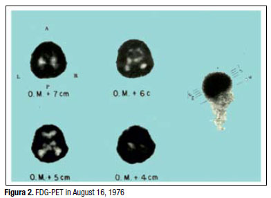
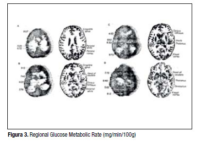
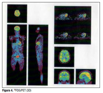
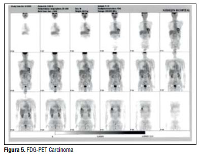
gas mixture target ( 0,1% ![]() in Ne) [1,2]. The bio-behaviour of FDG showed it as good substitute for glucose transporters and hexokinase activity to convert FDG-6-phosphate, but it is a very poor substitute for further metabolic pathways (metabolic trapping) and cannot be reuptaked by active transport at kidney. These parameters are simplified for the calculation of the regional cerebral glucose metabolic rate by the three compartment mathematic model (radioactivity of FDG in blood, radioactivity of FDG and radioactivity of FDG-6 phosphate in brain). In Figure 1 to Figure 3 the first trial images of human regional glucose metabolic rate [3] are shown. This result is a great successful example of collaboration between different institutes and different research fields (Brookhaven National Laboratory- chemistry, National Institute of Mental Health- neuro-physiology, Pennsylvania University- nuclear medicine). This methodology was applied to brain functional analysis. The motor cortex, the sensory cortex and the frontal cortex showed relatively high uptake. The tumour tissue also showed high uptake of FDG in contrast to normal tissue because of its active glucose consumptions. This is different from the inflamations due to a quick accumulation pattern in case of infammation (activated macrophage also uptake FDG). At present, FDG is mostly used for the imaging diagnosis of various tumours (lung, coronal, blood, bone, etc.) and to validate the effectiveness of a treatment (Figures 4, 5).
in Ne) [1,2]. The bio-behaviour of FDG showed it as good substitute for glucose transporters and hexokinase activity to convert FDG-6-phosphate, but it is a very poor substitute for further metabolic pathways (metabolic trapping) and cannot be reuptaked by active transport at kidney. These parameters are simplified for the calculation of the regional cerebral glucose metabolic rate by the three compartment mathematic model (radioactivity of FDG in blood, radioactivity of FDG and radioactivity of FDG-6 phosphate in brain). In Figure 1 to Figure 3 the first trial images of human regional glucose metabolic rate [3] are shown. This result is a great successful example of collaboration between different institutes and different research fields (Brookhaven National Laboratory- chemistry, National Institute of Mental Health- neuro-physiology, Pennsylvania University- nuclear medicine). This methodology was applied to brain functional analysis. The motor cortex, the sensory cortex and the frontal cortex showed relatively high uptake. The tumour tissue also showed high uptake of FDG in contrast to normal tissue because of its active glucose consumptions. This is different from the inflamations due to a quick accumulation pattern in case of infammation (activated macrophage also uptake FDG). At present, FDG is mostly used for the imaging diagnosis of various tumours (lung, coronal, blood, bone, etc.) and to validate the effectiveness of a treatment (Figures 4, 5).
The metabolic trapping method of PET functional imaging has been applied to amino acids and fatty acids as well. The ![]() -2-fluoo-4-boron-L-phenylalanine has been developed as a tracer for amino acid transporter to be used on BNCT (Boron Neutron Capture Therapy) for cancer therapy [4, 5]. The
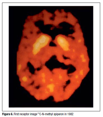
-2-fluoo-4-boron-L-phenylalanine has been developed as a tracer for amino acid transporter to be used on BNCT (Boron Neutron Capture Therapy) for cancer therapy [4, 5]. The ![]() -betamethyl fatty acids (against beta oxidation of fatty acids) has been used for tumour imaging and imaging analysis of myocardial diseases [6, 7]. In 1982, the first neuroreceptor imaging of human brain with
-betamethyl fatty acids (against beta oxidation of fatty acids) has been used for tumour imaging and imaging analysis of myocardial diseases [6, 7]. In 1982, the first neuroreceptor imaging of human brain with ![]() -Methyl spiperon succeeded in the collaborative work between Johns Hopkins Medical School and Uppsala University (Figure 6). This was the beginning of molecular imaging.
-Methyl spiperon succeeded in the collaborative work between Johns Hopkins Medical School and Uppsala University (Figure 6). This was the beginning of molecular imaging.
Molecular imaging: Second milestone
The successful result stimulated the research of receptor and transporter imaging and a tremendous number of radiopharmaceuticals (more than 100) were synthesized for imaging analysis of receptor density and affinity(dopamine [8], serotonin, acetylcholine, benzodiazepine [9], histamine [10,11], etc.), and not only neu-roreceptors, but also hormone receptors.
The results led to a new concept within the imaging analysis research, the molecular imaging, which is based on the analysis of molecular /molecular interactions by an imaging technique, and made a great contribution to bio-organic chemistry and drug discovery research fields.
The synthesis of labelled compounds by ![]() -alkyla-tion or
-alkyla-tion or ![]() -fluooalkylation requires very high specific radioactivity because of the limitation of receptor density.
-fluooalkylation requires very high specific radioactivity because of the limitation of receptor density. ![]() labelling is undergoing 10 000 times lower than theoretical specific activity by the influenceof
labelling is undergoing 10 000 times lower than theoretical specific activity by the influenceof ![]() contaminations while
contaminations while ![]() labelling is 100-1 000 times lower. A champion data on specificactivity of
labelling is 100-1 000 times lower. A champion data on specificactivity of ![]() -compounds has been reported by NIRS group (Chiba, Japan), only 1 000 times lower than theoretical, which made possible to get receptor PET image of
-compounds has been reported by NIRS group (Chiba, Japan), only 1 000 times lower than theoretical, which made possible to get receptor PET image of ![]() n/
n/![]() density.
density.
The success of FDG and molecular imaging by PET stimulated also ![]() chemistry and led to the development of bi-functional chelate agents which have affinity to biomarker and chelate binding to metal ion such as
chemistry and led to the development of bi-functional chelate agents which have affinity to biomarker and chelate binding to metal ion such as ![]() ,
, ![]() . This new technique is applied to the labelling of monoclonal antibody for tumour imaging. In a feedback to PET,
. This new technique is applied to the labelling of monoclonal antibody for tumour imaging. In a feedback to PET, ![]() (daughter of
(daughter of ![]() ) has been used in combination to
) has been used in combination to ![]() for tumour detection and internal radiation therapy purposes. This new concept defineda new word as “theragnostic or theranostic” in oncology.
for tumour detection and internal radiation therapy purposes. This new concept defineda new word as “theragnostic or theranostic” in oncology.
Theragnostic radiopharmaceuticals: Third milestone
The ![]() is a positron emitting nuclide (68 min half life) which is generated from
is a positron emitting nuclide (68 min half life) which is generated from ![]() (half life 270 days) by a
(half life 270 days) by a ![]() column generator system (commercially available). It allows obtaining PET imaging without cyclotron. Anti-tumour monoclonal antibody or shortened peptide linked DOTA was used as a bi-functional labelling agent. The combination with
column generator system (commercially available). It allows obtaining PET imaging without cyclotron. Anti-tumour monoclonal antibody or shortened peptide linked DOTA was used as a bi-functional labelling agent. The combination with ![]() (3,3 day half life, EC) make possible both purposes, the PET diagnosis and radiation therapy of tumour, so called theragnostic radiopharmaceuticals. In some trials
(3,3 day half life, EC) make possible both purposes, the PET diagnosis and radiation therapy of tumour, so called theragnostic radiopharmaceuticals. In some trials ![]() compound only has been used for both purposes, but included technical difficuties because of its short half life. In this case,
compound only has been used for both purposes, but included technical difficuties because of its short half life. In this case, ![]() ( 78,4 hr half life, β+.EC) which is produced by
( 78,4 hr half life, β+.EC) which is produced by ![]() (p,n)
(p,n)![]() r with 15 MeV proton [12-14] is more suitable. Other candidate for theragnostic nuclides are
r with 15 MeV proton [12-14] is more suitable. Other candidate for theragnostic nuclides are ![]() (16 day half life, β+.EC),
(16 day half life, β+.EC), ![]() (12,7 hr half life, β+.EC),
(12,7 hr half life, β+.EC), ![]() (57 hr half life, β+.EC), and
(57 hr half life, β+.EC), and ![]() (79,8 hr half life, β+.EC), Radiopharmaceuticals for internal radiation therapy now focus on short half life alpha emitting nuclides such as
(79,8 hr half life, β+.EC), Radiopharmaceuticals for internal radiation therapy now focus on short half life alpha emitting nuclides such as ![]() (7,2 hr half life, α.EC),
(7,2 hr half life, α.EC), ![]() (11,4 day half life,
(11,4 day half life,![]() ).
).
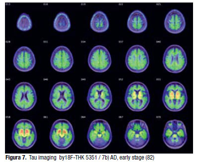
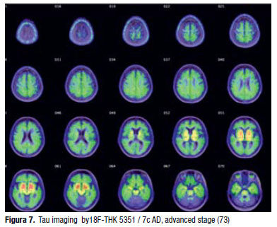
Recent highlight works in PET research are the imaging of amyloidal plaque and active tau protein for Alzheimer's disease (AD). ![]() - or
- or ![]() - labelled thiofavin analogues have been developed as amyloidal plaque marker. High uptake is always observed in AD cases, but sometimes high uptake is also present in the cortex of aged normal subjects. This mismatch makes difficult the confirmatory diagnosis and the evaluation of compounds for the AD treatment or prevention. Active tau protein image by
- labelled thiofavin analogues have been developed as amyloidal plaque marker. High uptake is always observed in AD cases, but sometimes high uptake is also present in the cortex of aged normal subjects. This mismatch makes difficult the confirmatory diagnosis and the evaluation of compounds for the AD treatment or prevention. Active tau protein image by ![]() -THK compounds (quinoline derivative) is a good match to clinical syndrome and to FDG diagnosis in AD [15-17]. The image of active tau is closer related to cell death than amyloidal plaque image (See Figures 7a, 7b, 7c); numbers in parenthesis are the age of patients. The prevention treatment requires more early stage images of brain degeneration.
-THK compounds (quinoline derivative) is a good match to clinical syndrome and to FDG diagnosis in AD [15-17]. The image of active tau is closer related to cell death than amyloidal plaque image (See Figures 7a, 7b, 7c); numbers in parenthesis are the age of patients. The prevention treatment requires more early stage images of brain degeneration.
Another highlight work is the imaging of inflammation. In many cases inflammation is followed by the denature of tissue functions and finally cell death. FDG is a good inflammation biomarker in various organs because of the hyper glucose metabolism that occurs in inflammation. Neuroinflammation images may be important to find tissue denature at early stages in Parkinson Disease (PD), AD and other neurodegenerative diseases. For this purpose, TSPO (translocator protein) ligand will be developed by ![]() or
or ![]() labelling as phenoxyphenyl acetamide and oxopurine derivatives. These works will link direct to preventive therapy of dementia and will lead to a new stage of theragnostic methodology in the near future.
labelling as phenoxyphenyl acetamide and oxopurine derivatives. These works will link direct to preventive therapy of dementia and will lead to a new stage of theragnostic methodology in the near future.
Acknowledgments
Authors appreciate to Ms. Luisa Aniuska Betancourt Hernández (former president of Nuclear Agency) for giving us the opportunity to make this review in the special anniversary edition of “Nucleus” and give many thanks to our research colleagues.
]]>
REFERENCES
[1] IDO T, WON CN, FOWLER JS, WOLF AP. Fluorination with F2. A Convenient Synthesis of 2-Deoxy-2-fluoo-D-glucose. J. Org. Chem. 1977; 42 (13): 2341-2342.
[2] IDO T, WON CN, CASELLA V, et. al. Labeled 2-deoxy-D-glucose analog. 18F-labeled 2-deoxy-2-fuluoro-D-glucose, 2-deoxy-2-fluoo-D-mannose and 14C-2-deoxy-2-fuluoro-D-glucose. J of Labelled Comp. and Radiopharm. 1978; 16(2): 175-183.
[3] REIVICH M, KUHL D, WOLF A, GREENBERG J, PHELPS M, IDO T, et. al. The [18F]Fluorodeoxyglucose method for the measurement of local cerebral glucose utilization in man. Circulation Research. 1979; 44(1): 127-137.
[4] ISHIWATA K, IDO T, KAWAMURA M, et. al. 4-Boron-2-[18F]Fluoro-D,L-phenylalanine as a target compound for boron neutron capture therapy: tumour imaging potential with positron emission tomography. Nucl. Med. Biol. 1991; 18(7): 745-751.
[5] IMAHORI Y, UEDA S, OHMORI Y, KUSAKI T, ONO K, FUJII R, IDO T. Fruorine-18-labeled fluooboronophenylalanine PET in patients with glioma. J. Nucl. Med. 1998; 39(2): 325-333.
[6] TAKAHASHI T, IDO T, IWATA R. Synthesis of 17-[18F]fluoo-5-methylpentadecanoic Acid. Appl. Radiat. Isot. 1992; 43(6): 822-824.
[7] TAKAHASHI T, NISHIMURA S, IDO T, ISHIWATA K, IWATA R. Biological evaluation of 5-Methyl-branched-chain ω-[18F]Fluorofatty acid: a potencial myocardial imaging tracer for positron emission tomography. Nucl. Med. Biol. 1996; 23(3): 303-308. ]]>
[8] HATANO K, ISHIWATA K, KAWASHIMA K, HATAZAWA J, ITOH M, IDO T. D2-Dopamine receptor specificbrain uptake of carbon-11-labeled YM-09151-2. J. NUC. Med. 1989; 30(4): 515-522.
[9] ISHIWATA K, YANAI K, IDO T, MIURA Y, KAWASHMA K. Synthesis and biodistribution of [11C] fludiazepam for imaging benzodiazepine receptors. Nucl. Med. Biol. 1988; 15(4): 365-371.
[10] YANAI K, WATANABE T, YOKOYAMA H, MEGRO K, HATAZAWA J, ITOH M, et.al. Histamine H1 receptors in human brain visualized in vivo by [11C]doxepin and emission tomography. Neuroscience Letters. 1992; 137(2): 145-148.
[11] YANAI K, RYU JH, WATANABE T, IWATA R, IDO T, SAWAI Y, et. al. Histamine H1 receptor occupancy in human brain after single doses of histamine H1 antagonist measured by positron emission tomography. British J Pharmacology. 1995; 116(1): 1649-1655.
[12] HOLLAND JP, SHEH Y, LEWIS JS. Standardized methods for the production of high specific-activity zirconium-89. Nucl. Med. Biol. 2009; 36(7): 729-739.
[13] HOLLAND JP, DIVILOV V, BANDER NH, SMIS-JONES PM, LAR-SO N SM, LEVIS JS. 89Zr-DFO-J591 for immunoPET of prostate-specific membrane antigen expression in vivo. J. Nucl. Med. 2010; 51(8): 1293-1300.
[14] van de WATERRING FCJ, RIJPKEMA M, PERK L, BRINKMANN U, OYEN WJG, BOERMAN OC. Zirconium-89 labeled antibodies: a new tool for molecular imaging in cancer patients (review). BioMed Res Int. 2014; Article ID 203601 http://www.hindawi.com/journals/bmri/2014/203601.
[15] XIA CF, ARTEAGA J, CHEN G, GANGADHARMATH U, GOMEZ LF, et. al. [18F]T807, a novel tau positron emission tomography imaging agent for Alzheimer's disease. Alzheimer’s and Dementia. 2013; 9(6): 666-676.
[16] VILLEMAGNE VL, OKAMURA N. In vivo tau imaging: obstacle and progress. Alzheimer's and Dementia. 2014; 10(3): 254-264.
[17] HARADA R, OKAMURA N, FURUMOTO S, FURUKAWA K, ISHIKI A, TOMITA N, et. al. [18F]THK-5117 PET for assessing neurofibrillary pathology in Alzheimer's disease. Eur. J. Nucl. Med. Mol. Imaging. 2015; 42(7): 1052-1061.
]]>