<?xml version="1.0" encoding="ISO-8859-1"?><article xmlns:mml="http://www.w3.org/1998/Math/MathML" xmlns:xlink="http://www.w3.org/1999/xlink" xmlns:xsi="http://www.w3.org/2001/XMLSchema-instance">
<front>
<journal-meta>
<journal-id>1025-028X</journal-id>
<journal-title><![CDATA[Vaccimonitor]]></journal-title>
<abbrev-journal-title><![CDATA[Vaccimonitor]]></abbrev-journal-title>
<issn>1025-028X</issn>
<publisher>
<publisher-name><![CDATA[Finlay Ediciones]]></publisher-name>
</publisher>
</journal-meta>
<article-meta>
<article-id>S1025-028X2012000200001</article-id>
<title-group>
<article-title xml:lang="en"><![CDATA[In vitro study of protein release from AFCo1 and implications in mucosal immunisation]]></article-title>
<article-title xml:lang="es"><![CDATA[Estudio in vitro de liberación de proteínas de AFCo1 y sus implicaciones en la inmunización mucosal]]></article-title>
</title-group>
<contrib-group>
<contrib contrib-type="author">
<name>
<surname><![CDATA[Acevedo]]></surname>
<given-names><![CDATA[Reinaldo]]></given-names>
</name>
<xref ref-type="aff" rid="A01"/>
</contrib>
<contrib contrib-type="author">
<name>
<surname><![CDATA[Romeu]]></surname>
<given-names><![CDATA[Belkis]]></given-names>
</name>
<xref ref-type="aff" rid="A01"/>
</contrib>
<contrib contrib-type="author">
<name>
<surname><![CDATA[Zayas]]></surname>
<given-names><![CDATA[Caridad]]></given-names>
</name>
<xref ref-type="aff" rid="A01"/>
</contrib>
<contrib contrib-type="author">
<name>
<surname><![CDATA[González]]></surname>
<given-names><![CDATA[Elizabeth]]></given-names>
</name>
<xref ref-type="aff" rid="A01"/>
</contrib>
<contrib contrib-type="author">
<name>
<surname><![CDATA[Lastre]]></surname>
<given-names><![CDATA[Miriam]]></given-names>
</name>
<xref ref-type="aff" rid="A01"/>
</contrib>
<contrib contrib-type="author">
<name>
<surname><![CDATA[del Campo]]></surname>
<given-names><![CDATA[Judith]]></given-names>
</name>
<xref ref-type="aff" rid="A01"/>
</contrib>
<contrib contrib-type="author">
<name>
<surname><![CDATA[Mullen]]></surname>
<given-names><![CDATA[Alex]]></given-names>
</name>
<xref ref-type="aff" rid="A02"/>
</contrib>
<contrib contrib-type="author">
<name>
<surname><![CDATA[Ferro]]></surname>
<given-names><![CDATA[Valerie A]]></given-names>
</name>
<xref ref-type="aff" rid="A02"/>
</contrib>
<contrib contrib-type="author">
<name>
<surname><![CDATA[Pérez]]></surname>
<given-names><![CDATA[Oliver]]></given-names>
</name>
<xref ref-type="aff" rid="A01"/>
</contrib>
</contrib-group>
<aff id="A01">
<institution><![CDATA[,Vicepresidency of Finlay Institute Research and Development ]]></institution>
<addr-line><![CDATA[Havana ]]></addr-line>
<country>Cuba</country>
</aff>
<aff id="A02">
<institution><![CDATA[,University of Strathclyde Strathclyde Institute of Pharmacy and Biomedical Sciences ]]></institution>
<addr-line><![CDATA[Glasgow ]]></addr-line>
</aff>
<pub-date pub-type="pub">
<day>00</day>
<month>08</month>
<year>2012</year>
</pub-date>
<pub-date pub-type="epub">
<day>00</day>
<month>08</month>
<year>2012</year>
</pub-date>
<volume>21</volume>
<numero>2</numero>
<fpage>1</fpage>
<lpage>5</lpage>
<copyright-statement/>
<copyright-year/>
<self-uri xlink:href="http://scielo.sld.cu/scielo.php?script=sci_arttext&amp;pid=S1025-028X2012000200001&amp;lng=en&amp;nrm=iso"></self-uri><self-uri xlink:href="http://scielo.sld.cu/scielo.php?script=sci_abstract&amp;pid=S1025-028X2012000200001&amp;lng=en&amp;nrm=iso"></self-uri><self-uri xlink:href="http://scielo.sld.cu/scielo.php?script=sci_pdf&amp;pid=S1025-028X2012000200001&amp;lng=en&amp;nrm=iso"></self-uri><abstract abstract-type="short" xml:lang="en"><p><![CDATA[Adjuvant Finlay Cochleate 1 (AFCo1) is a Proteoliposome-derived cochleate obtained from Neisseria meningitidis serogroup B. Transformation of proteoliposomes into AFCo1 potentiates the immune response on Neisseria antigens when it is administered by intranasal or intragastric (i.g) routes. However, the i.n route has been demonstrated to be more effective. The aim of this work is to evaluate in vitro the protein release from AFCo1, in simulated gastric fluid (SGF) or simulated nasal fluid (SNF) using a microdissolution test and to provide support for the results found when AFCo1 was administered by i.g or i.n routes in BALB/c mice. Results showed that dilution of AFCo1 in simulated gastric fluid affects the delivery of Neisseria protein antigens because they were released from cochleate structures faster than when simulated nasal fluid was used. In conclusion, conditions simulating gastric environment affect the delivery of protein antigens from AFCo1 and this result could partially explain why i.n administration is more effective in vivo than i.g immunisation.]]></p></abstract>
<abstract abstract-type="short" xml:lang="es"><p><![CDATA[El cocleato adyuvante Finlay (AFCo1) 1 es derivado de un proteopolisoma de Neisseria meningitidis serogrupo B. La transformación de los proteoliposomas en AFCo1 potencia la respuesta inmune de los antígenos de Neisseria cuando se administra por vía intranasal o intragástrica. Se ha demostrado, sin embargo, que la vía intranasal es más efectiva. Los objetivos de este trabajo fueron evaluar in vitro la liberación de proteínas del AFCo1 en líquido nasal o gástrico simulado, usando para ello la prueba de microdisolución y apoyar los resultados obtenidos cuando se administró AFCo1 por vía intranasal o intragástrica en ratones BALB/c. Los resultados demostraron que la dilución de AFCo1 en líquido gástrico o nasal simulado afecta la distribución de los antígenos de proteína de Neisseria , ya que estos se liberaron de las estructuras de cocleatos más rápido cuando se utilizó líquido nasal. Se concluyó que las condiciones que simulan el entorno gástrico afectan la distribución de los antígenos de proteínas de AFCo1 y este resultado puede explicar parcialmente porqué la administración intranasal es más efectiva in vivo que la inmunización intragástrica.]]></p></abstract>
<kwd-group>
<kwd lng="en"><![CDATA[Cochleate]]></kwd>
<kwd lng="en"><![CDATA[adjuvant]]></kwd>
<kwd lng="en"><![CDATA[proteoliposome]]></kwd>
<kwd lng="en"><![CDATA[Neisseria]]></kwd>
<kwd lng="es"><![CDATA[Cocleato]]></kwd>
<kwd lng="es"><![CDATA[adyuvante]]></kwd>
<kwd lng="es"><![CDATA[proteoliposoma]]></kwd>
<kwd lng="es"><![CDATA[Neisseria]]></kwd>
</kwd-group>
</article-meta>
</front><body><![CDATA[ <div align="right">       <p><font face="Verdana" size="2"><b>ART&Iacute;CULO ORIGINAL</b></font></p>       <p>&nbsp;</p>       <p align="justify"> <em><strong><font size="4" face="Verdana">I</font><font face="Verdana"><font size="4">n vitro </font></font></strong></em><font size="4" face="Verdana"><strong> study of protein release from AFCo1 and implications in mucosal immunisation</strong></font>  </p>       <p align="justify">&nbsp;</p>       <p align="justify"> <strong><font size="3" face="Verdana">Estudio <em>in vitro </em> de liberaci&oacute;n de prote&iacute;nas de AFCo1 y sus implicaciones en la inmunizaci&oacute;n mucosal</font></strong></p>       <p align="justify">&nbsp;</p>       <p align="justify">&nbsp;</p>       <p align="justify"><strong><font size="2" face="Verdana">Reinaldo Acevedo<sup>1*</sup>, Belkis Romeu<sup>1</sup>, Caridad Zayas<sup>1</sup>, Elizabeth Gonz&aacute;lez<sup>1</sup>, Miriam Lastre<sup>1</sup>, Judith del Campo<sup>1</sup>, Alex Mullen<sup>2</sup>, Valerie A. Ferro<sup>2</sup>, Oliver P&eacute;rez<sup>1</sup> </font></strong></p>       <p align="justify"><font size="2"><sup><font face="Verdana">1</font></sup><font face="Verdana">Research and Development Vicepresidency of Finlay Institute, Havana, Cuba. Box 16017    ]]></body>
<body><![CDATA[<br>       <sup>2</sup>University of Strathclyde, Strathclyde Institute of Pharmacy and Biomedical Sciences, 161 Cathedral Street, Glasgow, G4 0RE    <br>     email:<a href="mailto:racevedo@finlay.edu.cu">racevedo@finlay.edu.cu</a>     <br>   </font><font size="2" face="Verdana">*Lic en Ciencias Farmac&eacute;uticas. Doctor en Ciencias de la Salud. Subdirector de Investigaciones del Instituto Finlay</font></font></p>       <p align="justify">&nbsp;</p>       <p align="justify">&nbsp;</p>   <hr align="JUSTIFY">       <p align="justify"><font size="2" face="Verdana"><B>ABSTRACT</B></font></p>       <p align="justify"><font size="2" face="Verdana">Adjuvant Finlay Cochleate 1 (AFCo1) is a Proteoliposome-derived cochleate obtained from Neisseria meningitidis serogroup B. Transformation of proteoliposomes into AFCo1 potentiates the immune response on Neisseria antigens when it is administered by intranasal or intragastric (i.g) routes. However, the i.n route has been demonstrated to be more effective. The aim of this work is to evaluate in vitro the protein release from AFCo1, in simulated gastric fluid (SGF) or simulated nasal fluid (SNF) using a microdissolution test and to provide support for the results found when AFCo1 was administered by i.g or i.n routes in BALB/c mice. Results showed that dilution of AFCo1 in simulated gastric fluid affects the delivery of Neisseria protein antigens because they were released from cochleate structures faster than when simulated nasal fluid was used. In conclusion, conditions simulating gastric environment affect the delivery of protein antigens from AFCo1 and this result could partially explain why i.n administration is more effective in vivo than i.g immunisation. </font></p>       <p align="justify"><font size="2" face="Verdana"><strong>Keywords: </strong> Cochleate, adjuvant, proteoliposome, <em>Neisseria</em>.</font></p>   <hr align="JUSTIFY">       <p align="justify"><font face="Verdana"><strong><font size="2">RESUMEN</font> </strong></font></p>       <p align="justify"><font size="2" face="Verdana">El cocleato adyuvante Finlay (AFCo1) 1 es derivado de un proteopolisoma de <em>Neisseria meningitidis </em>serogrupo B. La transformaci&oacute;n de los proteoliposomas en AFCo1 potencia la respuesta inmune de los ant&iacute;genos de <em>Neisseria </em>cuando se administra por v&iacute;a intranasal o intrag&aacute;strica. Se ha demostrado, sin embargo, que la v&iacute;a intranasal es m&aacute;s efectiva. Los objetivos de este trabajo fueron evaluar <em>in vitro </em> la liberaci&oacute;n de prote&iacute;nas del AFCo1 en l&iacute;quido nasal o g&aacute;strico simulado, usando para ello la prueba de microdisoluci&oacute;n y apoyar los resultados obtenidos cuando se administr&oacute; AFCo1 por v&iacute;a intranasal o intrag&aacute;strica en ratones BALB/c. Los resultados demostraron que la diluci&oacute;n de AFCo1 en l&iacute;quido g&aacute;strico o nasal simulado afecta la distribuci&oacute;n de los ant&iacute;genos de prote&iacute;na de <em>Neisseria </em>, ya que estos se liberaron de las estructuras de cocleatos m&aacute;s r&aacute;pido cuando se utiliz&oacute; l&iacute;quido nasal. Se concluy&oacute; que las condiciones que simulan el entorno g&aacute;strico afectan la distribuci&oacute;n de los ant&iacute;genos de prote&iacute;nas de AFCo1 y este resultado puede explicar parcialmente porqu&eacute; la administraci&oacute;n intranasal es m&aacute;s efectiva <em>in vivo </em> que la inmunizaci&oacute;n intrag&aacute;strica. </font></p>       ]]></body>
<body><![CDATA[<p align="justify"><font size="2" face="Verdana"><strong>Palabras clave </strong>: Cocleato, adyuvante, proteoliposoma, Neisseria.</font></p>   <hr align="JUSTIFY">       <p align="justify"><font size="2" face="Verdana"> </font>    <br>   </p>         <p align="justify"><font face="Verdana"><strong><font size="3">INTRODUCTION</font></strong></font></p>       <p align="justify"><font size="2" face="Verdana">Most pathogens either invade      the body or establish infection in mucosal tissues and represent an enormous      challenge for vaccine development by the absence of good mucosal adjuvants.      AFCo1 (Adjuvant Finlay Cochleate 1) is an adjuvant microparticle derived from      <em>Neisseria meningitidis </em> serogroup B proteoliposome (PLn) (1). It      contains several immunopotentiator and antigenic molecules such as lipopolisacharides      (LPS) and outer membrane porins (2). </font></p>       <p align="justify"><font size="2" face="Verdana">Several studies have demonstrated the AFCo1 adjuvant effect on <em>Neisseria </em>, <em>Leishmania </em> and ovalbumin antigens administered either by parenteral or mucosal route (2,3). Recently, del Campo <em>et al </em> (2009) reported that AFCo1 elicited immune responses against <em>Neisseria </em> antigens by intranasal (i.n) or intragastric (i.g) route of immunisation (3) and they also found that i.n was more immunogenic. It was suggested that stomach conditions of low pH and proteases affect delivery or antigen integrity (3). Therefore, the aim of this work was to evaluate the <em>in vitro </em> release of protein antigens from AFCo1 using a microdissolution method with fluids simulating i.g or i.n conditions. </font></p>       <p align="justify"><font size="2" face="Verdana">The suitability of predicting <em>in vivo </em> behavior of drug products using simulated fluid has been demonstrated in several <em>in vitro-in vivo </em> correlation studies (4, 5). However, this correlation may depend on multiple factors such as dosage form, administration route and the test used to evaluate the release     of the encapsulated material (6). The dissolution test is the most used and recognised by pharmacopeia to determine the solubility and <em>in vitro </em> release of active molecules in formulation (6). But this method demands large amounts of drug, and biological active principles are usually only available in low quantities. Therefore, we selected a miniaturized flask method described by Glomme et al (2005) (7), adapted it to our laboratory conditions, referring to it as a microdissolution test.</font></p>       <p align="justify">&nbsp;</p>       <p align="justify"> <font face="Verdana"><strong><font size="3">MATERIALS AND METHODS</font></strong> </font></p>       <p align="justify"><font face="Verdana"><strong><font size="2">Preparation of      AFCo1 structures </font></strong></font></p>       ]]></body>
<body><![CDATA[<p align="justify"><font size="2" face="Verdana">PLn was provided by the production vice-presidency of Finlay Institute. PLn was resuspended and adjusted to 1 mg/mL in a buffer containing 30 mmol/L Tris, 3 mmol/L ethylenediaminetetra-acetic acid and 1.5% (w/v) sodium deoxycholate. AFCo1 formation was performed as previously described by P&eacute;rez O et al (2008) (1). Briefly, detergent was eliminated and calcium (Ca<sup>2+</sup>) was incorporated at the same time using a rotary dialysis method for lab scale. The Ca<sup>2+</sup> formation buffer was prepared with 30 mmol/L Tris, 100 mmol/L NaCl and 5 mmol/L CaCl<sub>2</sub>  at pH 7.4. </font></p>       <p align="justify"><font size="2" face="Verdana">The process was marked by the appearance of a white suspension as cochleates were formed. The efficiency of the incorporation process was estimated by comparing the protein quantities of the precipitate and the supernatant by Lowry. AFCo1 structure was observed by light microscopy using an Opton Standard 25 microscope and the average length size of 200 particles was determined using a gradation scale on an ocular lens. Additionally, AFCo1 was observed by transmission electron microscopy as described in the next section.</font> </p>       <p align="justify"><font size="2" face="Verdana">Negative Staining and Transmission Electron Microscopy (TEM).</font>  <font size="2" face="Verdana">These experiments were carried out as described by Xiaozhong <em>et al </em> (2004)(8). Briefly, Formvar/Carbon-coated 200 mesh copper grids were glow discharged. Cochleate samples were resuspended in 30 mM Tris buffer and dried using filter paper to a thin layer onto the hydrophilic support film. Twenty microlitres of 1% (v/v) aqueous methylamine vanadate stain (Nanovan; Nanoprobes, Stony Brook, NY, USA) was applied and excess moisture removed with filter paper. Dried samples were imaged with a LEO 912 energy filtering transmission electron microscope operating at 120 kV. Contrast enhanced, zero-loss energy filtered digital images were recorded with a 14 bit /2 K Proscan CCD camera. </font></p>       <p align="justify"> <strong><font size="2" face="Verdana">Dissolution test </font></strong></p>       <p align="justify"><font face="Verdana"><strong><font size="2">Media Preparation. </font></strong><font size="2">Simulated fluids (SF) were prepared according to USP 29/NF 24 (9). Briefly, Simulated Gastric Fluid (SGF): 34.2 mM of sodium chloride and 0.09mM of pepsin were dissolved in water with hydrochloric acid 0.2 M to pH 1.2. No references on Simulated Nasal Fluid (SNF) were found in specialized books (USP or Martindale) so we used a formula for Simulated Intestinal Fluid without pancreatin, which is similar to the physiological conditions of nasal mucosa (10). Then, 50.3 mM of potassium dihydrogen phosphate and 22.4 mM of sodium hydroxide were dissolved in water to final pH of 6.8. </font></font></p>       <p align="justify"><font size="2" face="Verdana"><strong>Microdissolution method for release studies. </strong>A variation of the miniaturized shake-flask method (7) was employed to evaluate protein release from AFCo1 when treated with SGF or SNF. Five micro-centrifuge tubes were prepared for each <em>in vitro </em> simulated condition and 2 mL of dissolution sample was added to the simulated fluids and AFCo1 (5:1) v/v. </font></p>       <p align="justify"><font size="2" face="Verdana">The samples were shaken (100 rpm) at 37 &deg;C in an orbital shaker (Heidolph Polymax 1040). At predetermined time intervals (0, 15, 30, 60 and 120 minutes), samples were centrifuged at 10 000 rpm for 5 min. The supernatant was collected and kept at 4 &deg;C until evaluation. The pellet was re-suspended up to the same volume with calcium formation buffer and the protein concentration was evaluated by Lowry assay (1). </font></p>       <p align="justify"><font size="2" face="Verdana"><strong> Immunization and sample conection</strong></font></p>       <p align="justify"><font size="2" face="Verdana"> Groups of five female BALB/c mice were inoculated by i.n or i.g route with AFCo1 (100 &micro;g per dose per mouse). Mice received three inoculations for mucosal routes: 20 &micro;L of AFCo1 (10 &micro;L per nostril) by the i.n route without anesthesia     or 100 &micro;L of AFCo1 by i.g gavage. Placebo groups were immunized with 20 &micro;L or 100 &micro;L of phosphate buffered saline (PBS) by i.n or i.g route respectively. Saliva samples were taken by stimulating salivation using an intraperitoneal injection of 50 &micro;L of 0.5% pilocarpine. This samples were inactivated for 15 min at 56 &ordm;C and centrifuged at 10 000 g for 10 min. For serum collection, animals were bled by retro-orbital puncture 14 days after the last immunisation, and the samples were centrifuged at 5000 g for 10 min. and stored at -20 &ordm;C for subsequent analysis. Animals were housed at the Finlay Institute animal facility and all experiments were performed with approval of the Finlay Institute Ethics Committee. </font></p>       <p align="justify"><font size="2" face="Verdana"> <strong>Enzyme-linked immunosorbent assay (ELISA) for PLn specific IgA and IgG elicited by AFCo1 administration.</strong></font></p>       ]]></body>
<body><![CDATA[<p align="justify"> <font size="2" face="Verdana">Specific IgG anti-PLn antibodies in serum samples and IgA anti-PLn antibodies in saliva were measured by indirect ELISA using polystyrene 96 well plates MaxiSorp coated with PLn (100 &micro;L <em>per </em> well) at 20 &micro;g/mL in PBS buffer (0.15 mol/L, pH 7.3) at 4 &ordm;C overnight, and blocked with 1% (w/v) BSA in PBS (blocking solution) for 1 h at room temperature. </font></p>       <p align="justify"><font size="2" face="Verdana">Serum samples were diluted 1:100 and saliva 1:2 in blocking solution and incubated for 1 h at 37 &ordm;C. Anti-mouse IgG or IgA peroxidase-conjugated antibodies (Sigma) were added (100 &micro;L <em>per </em> well) at 1:2500 dilution in blocking solution and incubated for 1 h at 37 &ordm;C. Bound antibodies were detected with 100 &micro;L <em>per </em> well of the substrate-chromogen mixture (o-phenylenediamine and hydrogen peroxide in citrate-phosphate buffer, pH 5). The reaction was stopped by adding 50 &micro;L of sulphuric acid at 2 mol/L and optical density at 492 nm was measured in a microplate reader (Titertek, Multiskan Plus; Labsystem). </font></p>       <p align="justify"><font size="2" face="Verdana">All incubation steps were followed by three washes with PBS containing 0.05% Tween-20 (v/v). ELISA results were expressed as units per mL, referred to an in-house standard. The means and standard errors of the mean (SEM) of at least three different experiments were analysed. </font></p>       <p align="justify"> <font face="Verdana"><strong><font size="2">Statistical Methods. </font></strong><font size="2"> Data from <em>in vitro </em> experiment was analyzed by a two-way ANOVA followed by Bonferroni post-hoc test. Statistical analysis of <em>in vivo </em> data was carried out with a Student's <em>t </em> test. The samples were first subjected to Kolmogorov-Smirnov normality test with Dallal-Wilkinson-Lillie for p-value. All the statistical analyses were performed using Graph Pad Prism 4 software (CA, USA).</font></font></p>       <p align="justify">&nbsp;</p>       <p align="justify"> <font size="3"><strong><font face="Verdana">RESULTS</font></strong> </font></p>       <p align="justify"><font size="2" face="Verdana"><strong>AFCo1 formation and      characterization</strong> </font></p>       <p align="justify"><font size="2" face="Verdana">PLn was transformed into AFCo1 using the dialysis rotary method. In this process, calcium interacts with phospholipids, proteins and LPS in the PLn (<a href="#f1">Fig. 1A</a>) forming a microtubular     structure as observed in <a href="#f1">Figure 1B</a>. The length of the AFCo1 measured from TEM micrographs was 13.5 &micro;m and the width was 2.6 mm. Similarly, the average length of the cochleates was measured by light microscopy (15.6 &micro;m &plusmn; 4.2 &micro;m). </font></p>       <p align="center"><img src="/img/revistas/vac/v21n2/f0101212.jpg" width="512" height="480"><a name="f1"></a></p>       
<p align="justify"> <strong><font face="Verdana"><em><font size="2">In vitro </font></em><font size="2"> study of protein release from AFCo1 </font></font></strong></p>       ]]></body>
<body><![CDATA[<p align="justify"> <font size="2" face="Verdana">A microdissolution test was used to evaluate protein release from AFCo1 in two different simulated fluids. More than 40% of proteins in AFCo1 were released from the samples incubated in SGF or SNF in the first 15 min of the test, but were faster released from AFCo1 when treated with the SGF (<a href="#f2">Fig. 2</a>). Once the test started, significant differences were observed between treatments at 15, 30, 60 and 120 min (<a href="#f2">Fig. 2</a>). After two hours of treatment with simulated fluids, the AFCo1 in SGF released more than 80% of proteins but more than 40% of protein was still encapsulated in the AFCo1 dissolved in SNF as shown in <a href="#f2">Figure 2</a>.</font></p>       <p align="center"><font size="2" face="Verdana"><img src="/img/revistas/vac/v21n2/f0201212.jpg" width="566" height="480"><a name="f2"></a></font></p>       
<p align="justify"> <strong><font size="2" face="Verdana">Evaluation of immune response induced i.g <em>vs </em> i.n administration of AFCo1</font></strong></p>       <p align="justify"> <font size="2" face="Verdana">
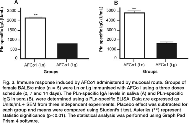
BALB/c mice were immunised by i.n or i.g route with 100 mg of AFCo1 using a three dose schedule (0,7 and 14 days). <a href="/img/revistas/vac/v21n2/f0301212.jpg">Figure 3</a> shows that i.n immunisation elicited higher specific IgA (<a href="/img/revistas/vac/v21n2/f0301212.jpg">Fig. 3A</a>) and IgG (<a href="/img/revistas/vac/v21n2/f0301212.jpg">Fig. 3B</a>) antibodies responses against antigens from <em>N. meningitidis </em> B in the PLn (p&lt;0.01). PLn is the raw material used to obtain the AFCo1 and contains the same <em>Neisseria </em> antigens than AFCo1 </font></p>       
<p align="justify">&nbsp; </p>       <p align="justify"><font face="Verdana"><strong><font size="3">DISCUSSION</font></strong> </font></p>       <p align="justify">&nbsp;</p>       <p align="justify"><font size="2" face="Verdana">  Mucosal vaccine strategies against bacterial infections based on particulate non living delivery systems are an important strategy in the design of adjuvants and vaccine formulations (11), not only because they represent a pain free alternative, but also because they provide the potential for both mucosal and systemic protection. AFCo1 has been used as a mucosal adjuvant with several antigens (2, 3). In particular, i.n immunisation has been more immunogenic than i.g (3), but reasons ruling this behaviour are still unknown. AFCo1 is a complex tubular microparticle formed by the interaction of calcium with negatively charged phospholipids, LPS, and proteins in the PLn structure (1, 2). </font></p>       <p align="justify"><font size="2" face="Verdana">They have a defined multilayered structure consisting of a solid, lipid bilayer sheet rolled up in a spiral like shape as represented in <a href="#f1">Figure 1A</a>. AFCo1 has high amounts of PorA and PorB <em>Neisseria </em>proteins (2). </font></p>       <p align="justify"><font size="2" face="Verdana">These also represent important antigens within PLn which is the core of the Cuban vaccine against <em>N. meningitidis </em> serogroup B and C, VA-MENGOC-BC<sup>&reg;</sup>  . At the same time, PorA and PorB may behave as immunostimulators to activate dendritic cells (DC) through interaction with toll like receptor (TLR)-2 (12), as well as LPS which is considered a potent activator of TLR-4 (12).</font></p>       ]]></body>
<body><![CDATA[<p align="justify"><font size="2" face="Verdana">An important issue for the development of an effective mucosal vaccine is whether DC within mucosal epithelia are involved in priming CD4 and CD8 effector T cells. The efficiency of oral immunisation with protein antigens to prime CD4 and CD8 T cell responses is hampered by antigen dilution/degradation in the gastrointestinal milieu (13). The nasopharingeal mucosa offers several advantages for <em>in vivo </em> stimulation because inductive sites are mostly found in the Waldeyer's ring, a region where lymphoid tissue is less dispersed than limphoid tissue (Peyer's patch) in gastrointestinal compartments (14). </font></p>       <p align="justify"><font size="2" face="Verdana">Therefore we used an <em>in vitro </em> microdissolution test to evaluate if the release of AFCo1 protein antigens/immunostimulators was affected by conditions that simulate i.g environment. Firstly, the amount of protein released over time from AFCo1 was evaluated with different fluids simulating i.n or i.g immunisation. </font></p>       <p align="justify"><font size="2" face="Verdana">We then separated the unbound AFCo1 components by a brief centrifugation step instead of filtration using a Whatman UniPrep filter chamber as recommended by Glomme, et al (2005) (7) because previous experience working with AFCo1 suspensions has demonstrated that the centrifugation step is sufficient to evaluate the incorporation of proteins into AFCo1 (3). </font></p>       <p align="justify"><font size="2" face="Verdana">Results showed that proteins were released faster from samples treated with SGF, although more than 40% of proteins from samples treated with SNG or SGF were released in the first 15 min of dissolution. This result indicates that dissolution may have an important effect at the nasal and gastric compartments <em>in vivo </em>in the early moments of exposure of the cochleates to body fluids. Miclea et al (2007) found similar results when they evaluated <em>in vitro </em> release of recombinant proteins encapsulated in cochleates prepared using commercial phospholipids (15). </font></p>       <p align="justify"><font size="2" face="Verdana">It seems that aqueous media favours unrolling of the AFCo1 structure due to a chemical equilibrium between the calcium forming the structure and that released into the fluid. In addition, the sample treated with SGF showed a higher percentage of protein released than that treated with SNF, probably because the acid environment may also favour the solubilisation of calcium precipitates in organic tissues (16). The i.g environment may also contribute to faster disassembly of AFCo1 and exposure of the antigenic and immunostimulator molecules to degradative effects of enzymes and low pH of the stomach. </font></p>       <p align="justify"><font size="2" face="Verdana">Additionally, the results of mucosal immunisation (<a href="/img/revistas/vac/v21n2/f0301212.jpg">Fig. 3</a>) confirm previous results obtained by del Campo, et al (3) and show significant differences between mucosal and systemic immune responses induced in mice immunised by i.n or i.g routes. However, how much the immunogenic and immunostimulator molecules in AFCo1 have been affected <em>in vivo </em> is a question that needs to be answered. </font></p>       
<p align="justify"> <font size="2" face="Verdana">In general, this study demonstrates that a microdissolution test is an effective way to evaluate the release of proteins from AFCo1. This method may be used to determine the release of molecules incorporated in AFCo1, other than Neisseria proteins and also to evaluate cochleates derived from different bacteria proteoliposomes, such as AFCo2 (17). These results contribute to understand the action mechanism of AFCo1. </font></p>       <p align="justify"> <font face="Verdana"><strong><font size="2">Acknowledgments </font></strong> </font></p>       <p align="justify"><font size="2" face="Verdana"> This research was supported by Finlay Institute and by the Research and Development Fund from Strathclyde University. We would like to thank to Dr. Ram&oacute;n F. Barber&aacute; for kindly providing PLn from production facilities of Finlay Institute. </font></p>       <p align="justify">&nbsp;</p>       ]]></body>
<body><![CDATA[<p align="justify"> <font size="3"><strong><font face="Verdana">REFERENCES</font></strong></font></p>       <p align="justify">&nbsp;</p>       <!-- ref --><p align="justify"><font size="2" face="Verdana">1. P&eacute;rez O, Bracho G, Lastre M, Sierra G, Campa C, Mora N, et al., inventors. Method of obtaining cochlear structures, vaccine compositions, adjuvants and intermediates thereof. WIPO, WO/2004/047805. Granted by Cuban Office for Intellectual Property OCPI23313. 2008.     </font></p>       <!-- ref --><p align="justify"><font size="2" face="Verdana">2. P&eacute;rez O, Bracho G, Lastre M, Mora N, del Campo J, Gil D, et al. Novel adjuvant based on a proteoliposome-derived cochleate structure containing native lipopolysaccharide as a pathogen-associated molecular pattern. Immunol Cell Biol 2004; 82(6):603-10.     </font></p>       <!-- ref --><p align="justify"><font size="2" face="Verdana">3. del Campo J, Zayas C, Romeu B, Acevedo R, Gonz&aacute;lez E, Bracho G, et al. Mucosal immunization using proteoliposome and cochleate structures from <em>Neisseria meningitidis </em> serogroup B induce mucosal and systemic responses. Methods 2009;49:301-8.     </font></p>       <!-- ref --><p align="justify"><font size="2" face="Verdana">4. Sunesen VH, Pedersen BL, Kristensen HG, Mullertz A. <em>In vivo-in vitro </em> correlations for a poorly soluble drug, danazol, using the flow-through dissolution method with biorelevant dissolution media. Eur J Pharm Sci 2005;24:305-13.     </font></p>       ]]></body>
<body><![CDATA[<!-- ref --><p align="justify"><font size="2" face="Verdana">5. Dressman JB, Reppas C. <em>In vitro-in vivo </em> correlations for lipophilic, poorly water-soluble drugs. Eur J Pharm Sci 2000;11:S73-S80.     </font></p>       <!-- ref --><p align="justify"><font size="2" face="Verdana">6. Ranade VV, Hollinger MA. Drug Delivery Systems. 2<sup>nd</sup> ed. Florida, Boca Raton: CRC Press, Inc; 2004.     </font></p>       <!-- ref --><p align="justify"><font size="2" face="Verdana">7. Glomme A, Marz J, Dressman JB. Comparison of a Miniaturized Shake-Flask Solubility Method with Automated Potentiometric Acid/Base Titrations and Calculated Solubilities. J Pharm Sci 2005;94(1)1-16.     </font></p>       <!-- ref --><p align="justify"><font size="2" face="Verdana">8. Wang W, Qu X, Gray AI, Tetley L, Uchegbu IF. Self-Assembly of Cetyl Linear Polyethylenimine To Give Micelles, Vesicles and Dense Nanoparticles. Macromolecules 2004;37:9114-22.     </font></p>       <!-- ref --><p align="justify"><font size="2" face="Verdana">9. USP 29/NF 24, The United State Pharmacopeia/The National Formulary, United States Pharmacopeia Convention Inc., Rockville, USA: USP; 2006.    </font></p>       ]]></body>
<body><![CDATA[<!-- ref --><p align="justify"><font size="2" face="Verdana"> 10. Stippler E, Kopp S, Dressman JB. Comparison of US Pharmacopeia simulated intestinal fluid TS (without pancreatin) and phosphate standard buffer pH 6.8, TS of the International Pharmacopoeia with respect to their use in <em>in vitro </em> dissolution testing. Dissolution Technologies 2004;11(2):6-10.     </font></p>       <!-- ref --><p align="justify"><font size="2" face="Verdana">11. Chadwick S, Kriegel C, Amiji M. Delivery strategies to enhance mucosal vaccination. Expert Opin Biol Ther 2009; 9(4):427-40.     </font></p>       <!-- ref --><p align="justify"><font size="2" face="Verdana">12. Kawai T, Akira S. The roles of TLRs, RLRs and NLRs in pathogen recognition. International Immunology 2009;21(4):317-37.     </font></p>       <!-- ref --><p align="justify"><font size="2" face="Verdana">13. Dieli F. Dendritic cells and the handling of antigen. Clin Exp Immunol 2003;134(2):178-80.     </font></p>       <!-- ref --><p align="justify"><font size="2" face="Verdana">14. Brandtzaeg P. Mucosal Immunity: Induction, Dissemination, and Effector Functions. Scandinavian Journal of Immunology 2009;70:505-15.     </font></p>       ]]></body>
<body><![CDATA[<!-- ref --><p align="justify"><font size="2" face="Verdana">15. Miclea RD, Varma PR, Peng A, Balu-Iyer SV. Development and characterization of lipidic cochleate containing recombinant factor VIII. Biochim Biophys Acta 2007;1768:2890-8.    </font></p>       <!-- ref --><p align="justify"><font size="2" face="Verdana">16. Nurit J, Margerit J, Terol A, Boudeville P. pH-metric study of the setting reaction of monocalcium phosphate monohydrate/calcium oxide-based cements. J Mater Sci Mater Med 2002;13:1007-14.     </font></p>       <!-- ref --><p align="justify"><font size="2" face="Verdana">17. Acevedo R, Callic&oacute; A, del Campo J, Gonz&aacute;lez E, Cedr&eacute; B, Gonz&aacute;lez L, et al. Intranasal administration of proteoliposome-derived cochleates from <em>Vibrio cholerae </em> O1 induce mucosal and systemic immune responses in mice. Methods 2009;49:309-15.     </font></p>       <p align="justify">&nbsp;</p>       <p align="justify">&nbsp;</p>       <p align="justify"> <font size="2" face="Verdana">Recibido: Octubre de 2011     <br>   Aceptado: Diciembre de 2011</font> </p> </div>     ]]></body>
<body><![CDATA[ ]]></body><back>
<ref-list>
<ref id="B1">
<label>1</label><nlm-citation citation-type="">
<person-group person-group-type="author">
<name>
<surname><![CDATA[Pérez]]></surname>
<given-names><![CDATA[O]]></given-names>
</name>
<name>
<surname><![CDATA[Bracho]]></surname>
<given-names><![CDATA[G]]></given-names>
</name>
<name>
<surname><![CDATA[Lastre]]></surname>
<given-names><![CDATA[M]]></given-names>
</name>
<name>
<surname><![CDATA[Sierra]]></surname>
<given-names><![CDATA[G]]></given-names>
</name>
<name>
<surname><![CDATA[Campa]]></surname>
<given-names><![CDATA[C]]></given-names>
</name>
<name>
<surname><![CDATA[Mora]]></surname>
<given-names><![CDATA[N]]></given-names>
</name>
</person-group>
<source><![CDATA[Method of obtaining cochlear structures, vaccine compositions, adjuvants and intermediates thereof]]></source>
<year></year>
</nlm-citation>
</ref>
<ref id="B2">
<label>2</label><nlm-citation citation-type="journal">
<person-group person-group-type="author">
<name>
<surname><![CDATA[Pérez]]></surname>
<given-names><![CDATA[O]]></given-names>
</name>
<name>
<surname><![CDATA[Bracho]]></surname>
<given-names><![CDATA[G]]></given-names>
</name>
<name>
<surname><![CDATA[Lastre]]></surname>
<given-names><![CDATA[M]]></given-names>
</name>
<name>
<surname><![CDATA[Mora]]></surname>
<given-names><![CDATA[N]]></given-names>
</name>
<name>
<surname><![CDATA[del Campo]]></surname>
<given-names><![CDATA[J]]></given-names>
</name>
<name>
<surname><![CDATA[Gil]]></surname>
<given-names><![CDATA[D]]></given-names>
</name>
</person-group>
<article-title xml:lang="en"><![CDATA[Novel adjuvant based on a proteoliposome-derived cochleate structure containing native lipopolysaccharide as a pathogen-associated molecular pattern]]></article-title>
<source><![CDATA[Immunol Cell Biol]]></source>
<year>2004</year>
<volume>82</volume>
<numero>6</numero>
<issue>6</issue>
<page-range>603-10</page-range></nlm-citation>
</ref>
<ref id="B3">
<label>3</label><nlm-citation citation-type="journal">
<person-group person-group-type="author">
<name>
<surname><![CDATA[del Campo]]></surname>
<given-names><![CDATA[J]]></given-names>
</name>
<name>
<surname><![CDATA[Zayas]]></surname>
<given-names><![CDATA[C]]></given-names>
</name>
<name>
<surname><![CDATA[Romeu]]></surname>
<given-names><![CDATA[B]]></given-names>
</name>
<name>
<surname><![CDATA[Acevedo]]></surname>
<given-names><![CDATA[R]]></given-names>
</name>
<name>
<surname><![CDATA[González]]></surname>
<given-names><![CDATA[E]]></given-names>
</name>
<name>
<surname><![CDATA[Bracho]]></surname>
<given-names><![CDATA[G]]></given-names>
</name>
</person-group>
<article-title xml:lang="en"><![CDATA[Mucosal immunization using proteoliposome and cochleate structures from Neisseria meningitidis serogroup B induce mucosal and systemic responses]]></article-title>
<source><![CDATA[Methods]]></source>
<year>2009</year>
<volume>49</volume>
<page-range>301-8</page-range></nlm-citation>
</ref>
<ref id="B4">
<label>4</label><nlm-citation citation-type="journal">
<person-group person-group-type="author">
<name>
<surname><![CDATA[Sunesen]]></surname>
<given-names><![CDATA[VH]]></given-names>
</name>
<name>
<surname><![CDATA[Pedersen]]></surname>
<given-names><![CDATA[BL]]></given-names>
</name>
<name>
<surname><![CDATA[Kristensen]]></surname>
<given-names><![CDATA[HG]]></given-names>
</name>
<name>
<surname><![CDATA[Mullertz]]></surname>
<given-names><![CDATA[A]]></given-names>
</name>
</person-group>
<article-title xml:lang="en"><![CDATA[In vivo-in vitro correlations for a poorly soluble drug, danazol, using the flow-through dissolution method with biorelevant dissolution media]]></article-title>
<source><![CDATA[Eur J Pharm Sci]]></source>
<year>2005</year>
<volume>24</volume>
<page-range>305-13</page-range></nlm-citation>
</ref>
<ref id="B5">
<label>5</label><nlm-citation citation-type="journal">
<person-group person-group-type="author">
<name>
<surname><![CDATA[Dressman]]></surname>
<given-names><![CDATA[JB]]></given-names>
</name>
<name>
<surname><![CDATA[Reppas]]></surname>
<given-names><![CDATA[C]]></given-names>
</name>
</person-group>
<article-title xml:lang="en"><![CDATA[In vitro-in vivo correlations for lipophilic, poorly water-soluble drugs]]></article-title>
<source><![CDATA[Eur J Pharm Sci]]></source>
<year>2000</year>
<volume>11</volume>
<page-range>S73-S80</page-range></nlm-citation>
</ref>
<ref id="B6">
<label>6</label><nlm-citation citation-type="book">
<person-group person-group-type="author">
<name>
<surname><![CDATA[Ranade]]></surname>
<given-names><![CDATA[VV]]></given-names>
</name>
<name>
<surname><![CDATA[Hollinger]]></surname>
<given-names><![CDATA[MA]]></given-names>
</name>
</person-group>
<source><![CDATA[Drug Delivery Systems]]></source>
<year>2004</year>
<edition>2</edition>
<publisher-loc><![CDATA[Boca Raton^eFlorida Florida]]></publisher-loc>
<publisher-name><![CDATA[CRC Press, Inc]]></publisher-name>
</nlm-citation>
</ref>
<ref id="B7">
<label>7</label><nlm-citation citation-type="journal">
<person-group person-group-type="author">
<name>
<surname><![CDATA[Glomme]]></surname>
<given-names><![CDATA[A]]></given-names>
</name>
<name>
<surname><![CDATA[Marz]]></surname>
<given-names><![CDATA[J]]></given-names>
</name>
<name>
<surname><![CDATA[Dressman]]></surname>
<given-names><![CDATA[JB]]></given-names>
</name>
</person-group>
<article-title xml:lang="en"><![CDATA[Comparison of a Miniaturized Shake Flask Solubility Method with Automated Potentiometric AcidBase Titrations and Calculated Solubilities]]></article-title>
<source><![CDATA[J Pharm Sci]]></source>
<year>2005</year>
<volume>94</volume>
<numero>1</numero>
<issue>1</issue>
<page-range>1-16</page-range></nlm-citation>
</ref>
<ref id="B8">
<label>8</label><nlm-citation citation-type="journal">
<person-group person-group-type="author">
<name>
<surname><![CDATA[Wang]]></surname>
<given-names><![CDATA[W]]></given-names>
</name>
<name>
<surname><![CDATA[Qu]]></surname>
<given-names><![CDATA[X]]></given-names>
</name>
<name>
<surname><![CDATA[Gray]]></surname>
<given-names><![CDATA[AI]]></given-names>
</name>
<name>
<surname><![CDATA[Tetley]]></surname>
<given-names><![CDATA[L]]></given-names>
</name>
<name>
<surname><![CDATA[Uchegbu]]></surname>
<given-names><![CDATA[IF]]></given-names>
</name>
</person-group>
<article-title xml:lang="en"><![CDATA[Self-Assembly of Cetyl Linear Polyethylenimine To Give Micelles, Vesicles and Dense Nanoparticles]]></article-title>
<source><![CDATA[Macromolecules]]></source>
<year>2004</year>
<volume>37</volume>
<page-range>9114-22</page-range></nlm-citation>
</ref>
<ref id="B9">
<label>9</label><nlm-citation citation-type="book">
<collab>The United State Pharmacopeia/The National Formulary^dUnited States Pharmacopeia Convention Inc</collab>
<source><![CDATA[]]></source>
<year>2006</year>
<publisher-loc><![CDATA[Rockville ]]></publisher-loc>
<publisher-name><![CDATA[USP]]></publisher-name>
</nlm-citation>
</ref>
<ref id="B10">
<label>10</label><nlm-citation citation-type="journal">
<person-group person-group-type="author">
<name>
<surname><![CDATA[Stippler]]></surname>
<given-names><![CDATA[E]]></given-names>
</name>
<name>
<surname><![CDATA[Kopp]]></surname>
<given-names><![CDATA[S]]></given-names>
</name>
<name>
<surname><![CDATA[Dressman]]></surname>
<given-names><![CDATA[JB]]></given-names>
</name>
</person-group>
<article-title xml:lang="en"><![CDATA[Comparison of US Pharmacopeia simulated intestinal fluid TS (without pancreatin) and phosphate standard buffer pH 6: 8, TS of the International Pharmacopoeia with respect to their use in in vitro dissolution testing]]></article-title>
<source><![CDATA[Dissolution Technologies]]></source>
<year>2004</year>
<volume>11</volume>
<numero>2</numero>
<issue>2</issue>
<page-range>6-10</page-range></nlm-citation>
</ref>
<ref id="B11">
<label>11</label><nlm-citation citation-type="journal">
<person-group person-group-type="author">
<name>
<surname><![CDATA[Chadwick]]></surname>
<given-names><![CDATA[S]]></given-names>
</name>
<name>
<surname><![CDATA[Kriegel]]></surname>
<given-names><![CDATA[C]]></given-names>
</name>
<name>
<surname><![CDATA[Amiji]]></surname>
<given-names><![CDATA[M]]></given-names>
</name>
</person-group>
<article-title xml:lang="en"><![CDATA[Delivery strategies to enhance mucosal vaccination]]></article-title>
<source><![CDATA[Expert Opin Biol Ther]]></source>
<year>2009</year>
<volume>9</volume>
<numero>4</numero>
<issue>4</issue>
<page-range>427-40</page-range></nlm-citation>
</ref>
<ref id="B12">
<label>12</label><nlm-citation citation-type="journal">
<person-group person-group-type="author">
<name>
<surname><![CDATA[Kawai]]></surname>
<given-names><![CDATA[T]]></given-names>
</name>
<name>
<surname><![CDATA[Akira]]></surname>
<given-names><![CDATA[S]]></given-names>
</name>
</person-group>
<article-title xml:lang="en"><![CDATA[The roles of TLRs, RLRs and NLRs in pathogen recognition]]></article-title>
<source><![CDATA[International Immunology]]></source>
<year>2009</year>
<volume>21</volume>
<numero>4</numero>
<issue>4</issue>
<page-range>317-37</page-range></nlm-citation>
</ref>
<ref id="B13">
<label>13</label><nlm-citation citation-type="journal">
<person-group person-group-type="author">
<name>
<surname><![CDATA[Dieli]]></surname>
<given-names><![CDATA[F]]></given-names>
</name>
</person-group>
<article-title xml:lang="en"><![CDATA[Dendritic cells and the handling of antigen]]></article-title>
<source><![CDATA[Clin Exp Immunol]]></source>
<year>2003</year>
<volume>134</volume>
<numero>2</numero>
<issue>2</issue>
<page-range>178-80</page-range></nlm-citation>
</ref>
<ref id="B14">
<label>14</label><nlm-citation citation-type="journal">
<person-group person-group-type="author">
<name>
<surname><![CDATA[Brandtzaeg]]></surname>
<given-names><![CDATA[P]]></given-names>
</name>
</person-group>
<article-title xml:lang="en"><![CDATA[Mucosal Immunity: Induction, Dissemination, and Effector Functions]]></article-title>
<source><![CDATA[Scandinavian Journal of Immunology]]></source>
<year>2009</year>
<volume>70</volume>
<page-range>505-15</page-range></nlm-citation>
</ref>
<ref id="B15">
<label>15</label><nlm-citation citation-type="journal">
<person-group person-group-type="author">
<name>
<surname><![CDATA[Miclea]]></surname>
<given-names><![CDATA[RD]]></given-names>
</name>
<name>
<surname><![CDATA[Varma]]></surname>
<given-names><![CDATA[PR]]></given-names>
</name>
<name>
<surname><![CDATA[Peng]]></surname>
<given-names><![CDATA[A]]></given-names>
</name>
<name>
<surname><![CDATA[Balu-Iyer]]></surname>
<given-names><![CDATA[SV]]></given-names>
</name>
</person-group>
<article-title xml:lang="en"><![CDATA[Development and characterization of lipidic cochleate containing recombinant factor VIII]]></article-title>
<source><![CDATA[Biochim Biophys Acta]]></source>
<year>2007</year>
<volume>1768</volume>
<page-range>2890-8</page-range></nlm-citation>
</ref>
<ref id="B16">
<label>16</label><nlm-citation citation-type="journal">
<person-group person-group-type="author">
<name>
<surname><![CDATA[Nurit]]></surname>
<given-names><![CDATA[J]]></given-names>
</name>
<name>
<surname><![CDATA[Margerit]]></surname>
<given-names><![CDATA[J]]></given-names>
</name>
<name>
<surname><![CDATA[Terol]]></surname>
<given-names><![CDATA[A]]></given-names>
</name>
<name>
<surname><![CDATA[Boudeville]]></surname>
<given-names><![CDATA[P]]></given-names>
</name>
</person-group>
<article-title xml:lang="en"><![CDATA[pH-metric study of the setting reaction of monocalcium phosphate monohydrate/calcium oxide-based cements]]></article-title>
<source><![CDATA[J Mater Sci Mater Med]]></source>
<year>2002</year>
<volume>13</volume>
<page-range>1007-14</page-range></nlm-citation>
</ref>
<ref id="B17">
<label>17</label><nlm-citation citation-type="journal">
<person-group person-group-type="author">
<name>
<surname><![CDATA[Acevedo]]></surname>
<given-names><![CDATA[R]]></given-names>
</name>
<name>
<surname><![CDATA[Callicó]]></surname>
<given-names><![CDATA[A]]></given-names>
</name>
<name>
<surname><![CDATA[del Campo]]></surname>
<given-names><![CDATA[J]]></given-names>
</name>
<name>
<surname><![CDATA[González]]></surname>
<given-names><![CDATA[E]]></given-names>
</name>
<name>
<surname><![CDATA[Cedré]]></surname>
<given-names><![CDATA[B]]></given-names>
</name>
<name>
<surname><![CDATA[González]]></surname>
<given-names><![CDATA[L]]></given-names>
</name>
</person-group>
<article-title xml:lang="en"><![CDATA[Intranasal administration of proteoliposome-derived cochleates from Vibrio cholerae O1 induce mucosal and systemic immune responses in mice]]></article-title>
<source><![CDATA[Methods]]></source>
<year>2009</year>
<volume>49</volume>
<page-range>309-15</page-range></nlm-citation>
</ref>
</ref-list>
</back>
</article>
