


 Custom services
 English (pdf)
 Article in xml format
 Article references
 Send this article by e-mail
 Cited by SciELO
  Similars in
SciELO
Similars in
SciELO 
 Permalink
Introducción
Characterizing brain morphology and its association with age- and sex-related development, function, and neurodegenerative processes in healthy humans, as well as morphological alterations found in psychiatric disorders and neurological diseases is crucial for the development of modern neuroscience.1,2 This fact becomes more relevant if one takes into account that more and more people reach more advanced stages of life where there is an increase in the rate of conditions typical of the elderly, with neurodegenerative diseases standing out.2,3,4
This fact, together with the advancement of computer systems, has influenced Computed Tomography (CT) to become the centerpiece of cranial imaging.5,6 Knowing the normal measurements of the cerebral ventricles in living human beings is of great importance in the diagnosis and monitoring of various pathologies. There is an ongoing debate in the literature about the best method of evaluating the various parts of the ventricular system, and the information known about precise measurements of the cerebral ventricles is limited.6,7
The new criteria suggested for research consider the use of neuroimaging-based biomarkers and the established criteria for diagnosis suggest their use as a clinical tool.6,8,9 Currently, the effort has been focused on the development of segmentation methods for CT images that allow the identification of informative features from a large data set of original features in preclinical stages, before brain damage irreversible occurs.9,10
Current morphometric studies have basically been developed from MRI neuroimaging techniques and other advanced neuroimaging methods, obtained in Caucasian populations from North America and/or Europe, which constitutes an important bias in the standardization of images. On the other hand, there are no reference standards that allow individual differences in neuroimaging metrics to be quantified.5,6
In our environment, although CT is widely used in the clinical setting, segmentation methods are not available to estimate ventricular volume from multislice cranial CT images. Due to the above, the present work is carried out to determine the ventricular volumetry due to its wide use as a marker of cerebral atrophy and to identify the effect of sex on these structures, according to the type of skull and, in correspondence with its statistical behavior, it can be used as a standardized morphometric pattern in a population with normal neurocognitive functions.
Methods
A descriptive observational study of clinical cases was developed. The study population consisted of 30 patients, 15 of them men, grouped in the age ranges 45-54 who presented normal neurocognitive functions, who had previously indicated a head CT, so they attended the imaging service of the Hospital Conrado Benítez, in the period December 2021-May 2022 and whose results were negative. Patients with a confirmed diagnosis of neurological and psychiatric diseases, a history of head trauma, familial neurocognitive disorders, schizophrenic disorders, pregnancy, as well as the presence of cognitive impairment (CD) were excluded. Authorization was requested and approved by the scientific council of the institution and by the ethics committee of each health institution involved in the development of the research. Participation in the study was carried out under the principle of voluntariness. We accepted the ethical principles for research in human beings, this under the Declaration of Helsinki in force in Cuba.
The brain volumetric reconstruction was obtained from a segmentation method based on the analysis of homogeneous textures and the Bicubic interpolation technique, using the gray level co-occurrence matrix (GLCM). mental state (MEEM) standardized and approved for the Cuban population.4 The CT scanner used in this study was SIEMENS, Multislice. Each patient had between 45 to 50 cuts with a thickness of 3 mm in this study. The matrix size of each segment was 512 x 512 pixels and the Hounsfield unit scale was: -1024 to +3071.
The variables studied were sociodemographic (sex), volumetric (volumes of the lateral ventricles, III and IV ventricles) and anatomical (type of skull), which was classified according to the cephalic index (CI) formula proposed by Paul Broca:11
CI= (Maximum transverse diameter /Maximum anteroposterior diameter)*100
Brachycephalic: index of 80 or more.
Mesocephalic: indices between 74.9 to 79.9
Dolichocephalic: index less than 74.9.
In this study, the iMagis technological tool was used, certified for its use by the National Center for the Registration of medical equipment of the Cuban Ministry of Public Health.12 Widely spread and used in radiology services in the country with a more updated version called NeuroiMagis, which allows morphometric calculations and three-dimensional reconstructions to be performed.
For morphometric estimates, the following steps were performed: pre-processing, feature extraction and feature selection.
Pre-processing: The initial stage is the conversion of the image to a grayscale level. In the second step, the existence of noise and artifacts in the image is removed using the anisotropic diffusion filtering technique.8
Feature extraction: Automatic extraction of texture features is performed, based on the Gray Level Co-occurrence Matrix (GLCM), where the image is automatically divided into K clusters by estimating features of homogeneity obtained from a matrix of co-occurrences.9,10
Feature selection: The region of interest is segmented by combining texture information with the region growth approach. Finally, to evaluate the accuracy of the proposed approach, the Dice coefficient was used as a similarity metric and a value of 95% was achieved.13,14,15
Statistic analysis
As part of the exploratory analysis, volumetric measurements were summarized using box plots and descriptive statistics: arithmetic mean, standard error of the mean, standard deviation, first and third quartiles, and median. To identify the possible differences between both sexes, the assumption of normality in the variables that measure volumetry was first verified in order to analyze them as dependent variables using the Shapiro Wilk test.
Then, the possible correlation between them was identified with Pearson's correlation coefficient in the case of those with normal distribution and with Spearman's Rho coefficient for those with non-normal distribution. Once the high correlation was verified, it was decided to use a multivariate linear model that included the factors of sex, skull type, urban/rural origin, schooling, occupation, history of arterial hypertension, and family history of dementia. As obtained by Pillai and Lamda de Wilks trace statistics. A significance level of 5% was considered in all cases of hypothesis tests. Minitab® 19.2 (64-bit) was used as statistical processor.
Results
In relation to the variables studied, the volumetric variables are represented according to their median and interquartile range; mean and outliers were also symbolized. The asymmetry of the ventricular volumes is verified graphically (fig. 1).
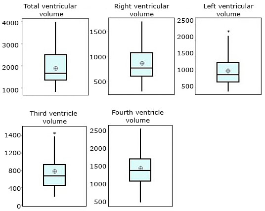
Figure 2 shows that only the volume of the right ventricle (p= 0.193) and that of the fourth ventricle follow a normal distribution (p= 0.782).
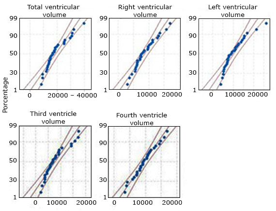
Fig. 2 Probability plots to verify normality in the probability distribution of the volumetric variables.
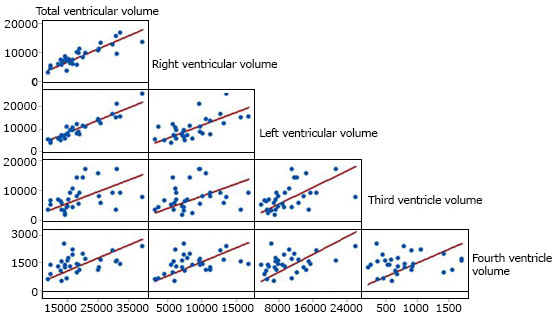
Ventricular volumes showed significant and positive correlation between them, except between the third and fourth ventricles and the third and right ventricular volume. The correlation is of moderate intensity between the right and left volumes and high between these and the total. The volume of the fourth ventricle showed moderate correlation with the rest of the volumes (fig. 3).
The statistics in the multivariate linear model were only significant in terms of sex and skull type. Table 1 shows the point estimate of the summary measurements for the five volumetric variables studied by sex, skull type and in total. In the volume of the fourth ventricle, values close to the mean and median can be seen, while the other ventricular volumes show some asymmetry to the left or lower values.
| Gender | Female | 16644 | 1729 | 6698 | 13625 | 14848 | 18525 | |
| Male | 21034 | 2170 | 8404 | 12781 | 19825 | 25798 | ||
| Cranium | Dolichocephalic | 18250 | 2453 | 8846 | 13315 | 15745 | 21830 | |
| Mesocephalic | 19290 | 1730 | 7135 | 13420 | 17824 | 25403 | ||
| Total | 18839 | 1423 | 7794 | 13470 | 16615 | 25040 | ||
| Gender | Female | 8215 | 1008 | 3903 | 5816 | 7314 | 9774 | |
| Male | 9014 | 767 | 2972 | 6131 | 9601 | 11283 | ||
| Cranium | Dolichocephalic | 8234 | 1058 | 3814 | 5142 | 7341 | 10709 | |
| Mesocephalic | 8905 | 776 | 3200 | 6145 | 7908 | 10944 | ||
| Total | 8614 | 627 | 3433 | 6094 | 7695 | 10798 | ||
| Gender | Female | 8430 | 857 | 3318 | 6311 | 7434 | 9494 | |
| Male | 12020 | 1563 | 6053 | 6801 | 11552 | 14357 | ||
| Cranium | Dolichocephalic | 10016 | 1605 | 5787 | 5899 | 8752 | 12412 | |
| Mesocephalic | 10385 | 1152 | 4750 | 6887 | 10468 | 13461 | ||
| Total | 10225 | 937 | 5132 | 6679 | 9021 | 12886 | ||
| Gender | Female | 518.8 | 62.4 | 241.5 | 360.8 | 449.7 | 688.5 | |
| Male | 1028 | 110 | 425 | 669 | 804 | 1434 | ||
| Cranium | Dolichocephalic | 546.2 | 62.1 | 223.8 | 361.0 | 449.7 | 790.4 | |
| Mesocephalic | 947 | 114 | 468 | 590 | 913 | 1431 | ||
| Total | 773.3 | 78.0 | 427.0 | 447.9 | 678.6 | 928.9 | ||
| Gender | Female | 1414 | 124 | 479 | 1236 | 1365 | 1606 | |
| Male | 1477 | 142 | 552 | 1071 | 1438 | 1989 | ||
| Cranium | Dolichocephalic | 1439 | 164 | 592 | 1052 | 1365 | 1739 | |
| Mesocephalic | 1451 | 110 | 454 | 1088 | 1414 | 1729 | ||
| 1445.6 | 92.9 | 508.6 | 1090.3 | 1389.6 | 1717.5 | |||
SE: Standard error; SD: Standard desviation
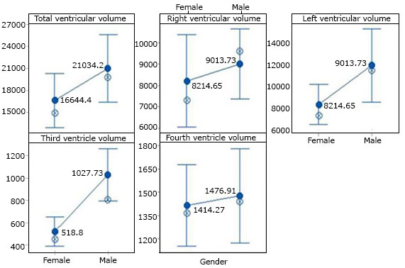
Figure 4 shows the 95% intervals for the mean of each of the volumetric measurements according to sex. The mean value is shown in the bars and the median has been indicated. No significant differences were found regarding sex in any volume except in the third ventricle (p= 0.01). The same occurred by type of skull (p= 0.005) with a graphic behavior very similar to that of sex.
Discussion
In routine clinical practice, it is shown that existing morphometric techniques have not been able to prevail, in part due to the non-accessibility of technical methods despite their diversity, in addition to their high cost.1,6 Due to the absence of morphometric patterns that characterize the study population in the present study, a morphometric quantification method has been developed that includes segmentation of CT images that allow the identification of informative characteristics of a massive set of original characteristics in pre - clinical stages and volumetric calculations, which has allowed using Multislice CT images, it has been possible to quantify these results, obtaining a morphometric pattern that describes the increase in the volume of the ventricles with respect to sex and its possible use as biomarkers of cognitive deterioration.
Consideration of the effects of sex can help to understand a series of brain disorders with different incidence or nature. While there are many similarities in structure, function, and neurotransmission, there are also macroscopic and microscopic sex differences in gray/white matter distribution, cerebral blood flow, neurotransmission profile, neuronal composition, dendritic morphology, and functionality, as well as also in specific brain regions.16,17,18
Most of the authors have studied the morphometrics of the lateral ventricles according to their different parts, not finding robust scientific evidence that shows the morphometric results of the study of these structures as a whole,1 such as that carried out in this study.
Honnegowda et al,7 were studied for the measurements of lateral ventricle, third ventricle and fourth ventricle and it was statistically analyzed. The antero-posterior extent of the body of the lateral ventricles on the right and on the left side were greater in men than in women; Similar behavior was shown by the measurements of the length of the frontal horns, in which men had larger cavities on both sides than women. The width and height of the fourth and third ventricle were increased too in male rather than female.
Polat et al,19) studied the ventricular system in healthy Turkish subjects, reporting statistically significant differences in their frontal horn width, third (3rd) ventricle width, and the maximum transverse inner diameter of the skull values in between sexes. These values were lower in healthy male subjects than females, however; there were not found significantly difference between groups.
Dzefi Tettey,20) when determining the Evans index and the effect of sex on this index in adult Ghanaians, showed that there was no significant difference between both values for men and women.
Sex differences in the brain are demonstrated in the observed prevalence of psychiatric disorders and in some psychological traits. Men have higher raw volumes and white matter areas; women have greater crude cortical thickness and greater complexity of the white matter tract.21 Kijonka et al,22 demonstrated in a Polish population that most of the sex differences in global volumes were revealed to be linked to the difference in head/brain size parameters. They also confirmed age- and sex-related cerebral volumetric loss, the negative age dependence of gray matter volume was more pronounced in men, while in the case of white matter it turned out to be in women.
Namrata Kolsur and coauthors,23 showed that in a sample of the Indian population The ventricular indices showed statistically significant difference between males and females in all the indices except for cella media index. Linear Ventricular Indices, Evans Index, Bifrontal Index, Bicaudate Index, Cella Media Index, Third Ventricular Index, Huckman Index, Ventricular Index.
Buchpiguel et al3 comparing unadjusted brain volumes showed larger volumes of gray and white matter in men. After volumetric correction, these adjusted volumes were higher in women. However, we did not find unified criteria in relation to the sexual dimorphism of the brain.
Jäncke,24 showed that total brain volume, as well as gray and white matter volumes, reveal sex/gender differences with values ranging from 1 to 1.4914. When correcting for brain size, sex/gender differences almost disappear, so these differences are not convincing enough to support the hypothesis of sexual dimorphism in the brain.
Hirnsteina M,25 rejects the concept of "sexual dimorphism" in its extreme binary form, even though they emphasize the importance of small effects that can have behavioral consequences and can play a role in the etiology of many mental disorders. The notion that the structure or function of the human brain may be sexually dimorphic is not entirely new, but it is controversial.26,27,28
Lutz Jäncke reaffirms in his research that there are some relatively strong but also many moderate or even weak sex/gender differences in terms of brain anatomy and brain function. These differences are not large enough to support clear sexual dimorphism.24
Most interestingly, there is currently a lack of a direct and strong correlation between these neuroscientific findings and real-life behavior as well as cognition. However, in the context of modern plasticity research, we need to take much more account of the fact that the brain can adapt and change anatomically and functionally through practice and learning. Therefore, it could be possible that the male and female brains change their structure and functions due to their different experiences and because they are exposed to different social environments. However, it is also possible that genetic, hormonal, and social influences interact in ways that are currently unknown in shaping the brain and behavior. In light of these influences on human brain development, a new area of sex/gender research could be established.24
The present study provides a method for morphometric quantification of the encephalic ventricular system and a normative database, in which it is confirmed that the volume of the lateral ventricles shows a significant effect based on sex and related to normal cognitive functioning associated with aging. . In correspondence with its statistical behavior, it can be used as a standardized morphometric pattern in a population with normal neurocognitive functions and promises to become a sensitive diagnostic tool for individual diagnostic classification. Additional research is required to validate its potential clinical utility.

