


My SciELO
 Custom services
Services on Demand
Article
 Article in xml format
 Article references
 Send this article by e-mail
Indicators
-
 Cited by SciELO
Related links
-
 Similars in
SciELO
Similars in
SciELO
Share

 Permalink
Revista Cubana de Estomatología
On-line version ISSN 1561-297X
Rev Cubana Estomatol vol.52 no.3 Ciudad de La Habana July.-Sept. 2015
ARTÍCULO ORIGINAL
Characterization of late diagnosis of oral cancer in a Brazilian city
Caracterización de diagnóstico tardío de cáncer oral en una ciudad brasileña
Manuela Gouvêa Campêlo dos Santos,I Danúbia Roberta de Medeiros Nóbrega,II Gabriella de Vasconcelos Neves,III Hianne Cristinne de Morais Medeiros,III Jozinete Vieira Pereira,IV Daliana Queiroga de Castro GomesIV
I Department of Dentistry. State University of Paraíba. Araruna, PB, Brazil.
II Faculdades Integradas de Patos. Patos, PB, Brazil.
III State University of Paraíba, Campina Grande, PB, Brazil.
IV Department of Dentistry. State University of Paraíba. Campina Grande, PB, Brazil.
ABSTRACT
Introduction: oral cancer in Brazil still presents high incidence and mortality rates and has different characteristics throughout the national territory. Although in most cases the diagnosis is late, there is a great possibility for cure when patients are treated early.
Objective: to describe the sociodemographic profile of patients with oral squamous cell carcinoma and the possible etiological factors associated.
Methods: this was a descriptive prospective cross-sectional study carried out in Napoleão Laureano Hospital, state of Paraíba, from January 2012 to May 2013. The study included patients with advanced-stage oral squamous cell carcinoma identified during clinical examination and confirmed by histopathology. The following variables were assessed: age, sex, comorbidity, smoking, alcohol use, tumor location, time of development, clinical staging, histopathological grading and proposed treatment.
Results: a total of 15 cases of patients with stage III and IV oral squamous cell carcinoma were found. Of these, 80 % were males with a mean age of 62.59 years and lesions affecting predominantly the mouth floor, followed by the tongue. The most common sign was the presence of tumor greater than 3.0-cm diameter, including ulcerated, leukoplastic and erythroplastic areas, in addition to pain and difficulty in feeding and phonation.
Conclusion: the majority of patients identified, with advanced-stage squamous cell carcinoma showed moderate cellular differentiation between stages III and IV, and was composed by males with smoking and alcohol drinking habits in the seventh decade of life.
Key-words: squamous cell carcinoma, late diagnosis, oral cancer.
Introducción: el cáncer oral en Brasil todavía presenta altos niveles de incidencia y mortalidad y tiene diferentes rasgos en todo el territorio nacional. En la mayoría de los casos el diagnóstico es tardío. Sin embargo, hay una gran posibilidad de cura cuando es tratado desde el principio.
Objetivo: describir el perfil sociodemográfico de los pacientes afectados por carcinoma bucal de células escamosas, así como los posibles factores etiológicos asociados.
Métodos: se realizó un estudio descriptivo prospectivo transversal en el hospital “Dr. Napoleão Laureano”, estado de Paraíba, desde enero de 2012 a mayo de 2013. El estudio incluyó a pacientes con carcinoma oral de células escamosas en estadio avanzado identificado durante el examen clínico y confirmado por histopatología. Se evaluaron las siguientes variables: edad, sexo, comorbilidad, el tabaquismo, el consumo de alcohol, la ubicación del tumor, el tiempo de desarrollo, la estadificación clínica, clasificación histopatológica y el tratamiento propuesto.
Resultados: se encontró un total de 15 casos de pacientes con estadio III y IV del carcinoma oral de células escamosas COCE . De estos, 80 % eran hombres con una edad media de 62,59 años. Las lesiones afectaban predominantemente al piso de la boca, seguido de la lengua. El signo clínico más común fue la presencia de tumor mayor que 3,0 cm de diámetro, incluyendo las zonas ulceradas, áreas con leucoplasia y eritroplasia, además de dolor y dificultad en la alimentación y la fonación.
Conclusión: la mayoría de los pacientes identificados con carcinoma de células escamosas en estadio avanzado mostró moderada diferenciación celular entre los estadios III y IV. Estos eran hombres con hábitos de fumar y beber en la séptima década de la vida.
Palabras clave: carcinoma de células escamosas, diagnóstico tardío, cáncer oral.
INTRODUCTION
Oral cancer can be defined as a neoplasm involving the oral cavity, which begins at the lip and ends at the anterior pillar of the fauces.1 It is a chronic disease that results in high morbidity and mortality, thus being considered a public health issue in Brazil by the National Cancer Institute (INCA).2
A total of about 6.4 million cases of malignant tumors have been diagnosed each year worldwide, of which oral cancer accounts for 10 %,3 causing this type of cancer to be considered a worldwide health issue. In Brazil, the oral squamous cell carcinoma (OSCC) accounts for 95 % of malignant neoplasms of the oral cavity. According to the National Cancer Institute (INCA), the number of registered deaths in 2010 was 4,891. In 2014, 11,280 new cases of oral cancer were diagnosed in men and 4,010 in women.2,4
The causes of oral cancer are mainly related to smoking and alcohol consumption, and it is found to affect in general males above 50 years. Clinically, OSCC appears as white, erythroplastic lesions or mucosal ulcerations, affecting more frequently the lower lip, tongue, mouth floor, soft palate and alveolar ridge.1,5-7
Early detection of oral cancer is the most effective measure to reduce mortality, morbidity and sequelae produced by this disease, whose survival rate at five years is 50 % or less.3 On the other hand, when diagnosed in advanced stages, oral cancer cases are often associated with high mortality rates.4,8,9
Oral cancer is usually disregarded in its initial phase by the common population. However, it can turn to be extremely fatal if left untreated, even in a very early stage of the lesion. Its detection and diagnosis are currently based on clinical examination, histopathological evaluation of biopsy material and molecular methods.10
Given the above, early diagnosis of oral cancer and the immediate referral of the patient to specialized treatment may be important issues to reduce morbidity and mortality caused by the disease. Thus, the aim of this study to observe the sociodemographic profile of patients with oral squamous cell carcinoma (OSCC) and the possible etiological factors associated.
METHODS
A descriptive prospective exploratory quantitative study was carried out including a sample of 15 patients diagnosed with squamous cell carcinoma from January 2012 to May 2013 at the state hospital Dr. Napoleão Laureano, which is considered a referral hospital for cancer treatment located in the state of Paraíba, Brazil.
The universe was composed of all patients with clinical signs and symptoms of oral squamous cell carcinoma confirmed by histopathological examination.
Data were collected during clinical examination and those patients with advanced-stage oral cancer, confirmed by histopathological examination, were included in our study. A data collection instrument was developed by the authors, containing information on age; sex; comorbidity; smoking; alcohol use; tumor location; time of development; clinical staging; histopathological grading and proposed treatment.
Non-smoker patients were considered as such if they had reported never having smoked or having quit smoking for at least 10 years.11 Likewise, non-alcoholic patients were considered as such if they had reported never having drunk or having stopped such habit for at least one year.12
This study was conducted in accordance with the ethical principles of the Declaration of Helsinki and was approved by the Institutional Review Board of the State University of Paraíba (UEPB) under protocol number 0006.0.133.000-12. Before enrolment, patients consented their participation by signing an informed consent form.
This study included patients with moderate- or advanced-stage (III or IV stage) OSCC confirmed by histopathological diagnosis after incisional biopsy. The histopathological classification of malignancy adopted herein was proposed by the World Health Organization (WHO),13 which is based on the degree of cell differentiation. Accordingly, malignancies are classified into three categories: poorly differentiated, moderately differentiated and well differentiated. Late diagnosis was assessed in months from the date that the patient reported the onset of signs or symptoms until diagnosis in a specialized facility. The tumor stage at diagnosis was also considered in the assessment. The data were collected, grouped in tables and analyzed using descriptive statistics.
RESULTS
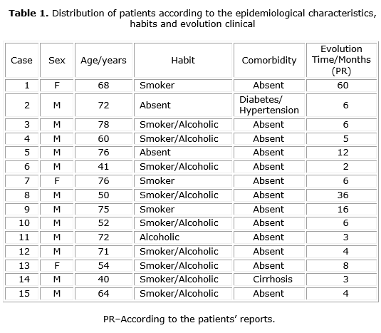
The mean time of lesion development was 11, 8 months. The clinical features of some lesions and important information regarding the sample profile are shown in table 1. Of the 15 patients assessed, 80 % (n= 12) were males, with a mean age of 62.59 years, and 20 % (n= 3) were females, with a mean age of 66 years. A total of 80 % of patients (n= 12) were found to be smokers and 66.66 % (n= 10) alcoholics and the mean time of lesion development was 11, 8 months.
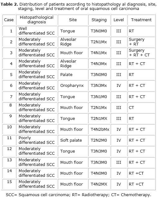
The distribution of patients according to histopathological data oral squamous cell carcinoma is reported in table 2. The majority of cases showed moderately differentiated histopathological diagnosis (n= 13) in stage III (n= 8).The most common anatomical location was the mouth floor. The most frequent treatment was combination chemotherapy and radiotherapy (n= 9).
DISCUSSION
The oral squamous cell carcinoma is a malignant neoplasm originating from the lining epithelium that accounts for about 95 % of the malignant lesions in the oral region.14,15 With regard to sex, most of the sample of this study corresponded to males, which corroborates the findings by Neville et al.,1 Almeida et al.,16 Santos et al.17 and Brasil.2 The mean age of the patients was 63.26 years, in agreement with the studies by Alvarenga et al.,15 Almeida et al.,16 Deng et al.,18 in which patients with advanced diagnosis of oral cancer had an average age around 64 years.
As to the anatomical locations of the tumor, the mouth floor followed by the tongue were the most affected sites. These data are in agreement with those of several studies.1,3,15,16,18-21 Contrarily, Gervásio et al.22 conducted a study with 740 OSCC cases and found that the tongue was the most common site, accounting for 44 % of the cases, followed by the mouth floor with 16 %.
Current and previous history of tobacco and alcohol consumption are considered the most important risk factors for OSCC.7,23 The present results showed that both deleterious habits were frequently reported by OSCC patients. Despite the fact that oral cancer has multifactorial causes, the habits of smoking and consuming alcohol have often been documented as the main risk factors associated with the development of this disease. Tobacco contains more than 60 potentially carcinogen substances that may be related to the onset of oral cancer. These products release reactive oxygen species that can damage cell proteins, carbohydrates and DNA.1,24 Acetaldehyde, a product of the metabolism of ethanol, is considered a carcinogen due to its mutagenic effects on DNA.23 These agents act synergistically, given that alcohol increases the permeability of oral mucosal cells to tobacco carcinogens.1,14,25
As to therapeutic approaches, there was a combination of radiotherapy and chemotherapy in most cases presented, corroborating the data reported by Ruback et al.26 In advanced stages, treatment modalities have been shown to promote increased control of locoregional tumor. Nevertheless, the approaches vary according to the location of the lesion, histological type, clinical staging and patients’ physical condition.26
In this study, patients looked for medical care from 02 to 60 months after the onset of the first signs and symptoms, with a mean evolution time of 11.8 months. This variation in time was higher in our study than in others reported in the literature, which identified a range from 03 to 18 months.8,27 However, we observed that 10 patients (2/3) of our sample sought treatment within six months after the initial complaint of the first signs and/or symptoms. This result is similar to that pointed out by Pires et al.,7 who reinforce the importance of considering the possible diagnosis of OSCC in cases of leukoplakia and erythroplakia lesions. The authors also highlight the need to obtain biopsy samples from all lesions in this population of patients, so that to increase their chances of having a favorable prognosis and longer survival.
The delay in diagnosis may be caused by several factors, namely: failure of professionals to recognize that the lesion is malignant; delay of patients to look for medical care; neglecting the first cancer signs and/or symptoms; or even persistence of the neoplasm resulting from an incomplete removal of the primary tumor.4,8,16,28 A study by Peacock et al.29 found that the delay in diagnosis was due to the delay in scheduling the appointments in the centers of primary health care.
The diagnostic delay period cancer oral in Brazil and the world is still very long,30 whereas it was longer than 100 days in the United States.29 It is worth noting that the chief determinant of a poor prognosis is the stage of disease at diagnosis. By analyzing the data related to staging in oral cancer patients, Gervásio et al.22 found that around 80 % of patients were found to be at an advanced stage in the very first appointment in specialized services. In this study, all patients were diagnosed at advanced stages of the disease according to Barnes,13 with cellular differentiation between moderately and well-differentiated patterns. Advanced stages of oral cancer refer to tumors invading adjacent structures, for instance, cortical bone, deep muscle of tongue, maxillary sinus and skin.9,29
The awareness of patients and health professionals for the early detection of lesions with malignant potential and referral for immediate care would certainly contribute to a more favorable prognosis and, consequently, a better quality of life for the patient.
CONCLUSION
The majority of patients identified, with advanced-stage squamous cell carcinoma showed moderate cellular differentiation between stages III and IV, and was composed by males with smoking and alcohol drinking habits in the seventh decade of life.
BIBLIOGRAPHIC REFERENCES
1. Neville BW, Allen CM, Damm DD. Patologia oral e maxilofacial. 2da.ed. Rio de Janeiro: Guanabara Koogan; 2009.
2. Brasil, Ministério da Saúde. Estimativas de incidência e mortalidade por câncer no Brasil [acesso em 2015 jan 7]. Disponível em: http://www.inca.gov.br/estimativa/2014/estimativa-24042014.pdf
3. Leite IC, Nunes LC, Moreira RC, Couto CA, Teixeira MT. Mortalidade por Câncer de Boca e Faringe em Cidade de Médio Porte na Região Sudeste do Brasil, 1980-2005. Rev Bras Cancerol. 2010;56(1):17-23.
4. Kujan O, Sloan P. Dilemmas of Oral Cancer Screening: An Update. Asian Pac J Cancer Prev. 2013;14(5):3369-73.
5. Chhabra N, Chhabra S, Sapra N. Diagnostic modalities for squamous cell carcinoma: an extensive review of literature-considering toluidine blue as a useful adjunct. J Maxillofac Oral Surg. 2015;14(2):188-200.
6. Van Zyl A, Bunn BK. Clinical features of oral cancer. SADJ. 2012;67(10):566-9.
7. Pires FR, Ramos AB, Oliveira JB, Tavares AS, Luz PS, Santos TC. Oral squamous cell carcinoma: clinicopathological features from 346 cases from a single oral pathology service during an 8-year period. J Appl Oral Sci. 2013;21(5):460-7.
8. Van der Waal I, de Bree R, Brakenhoff R, Coebergh JW. Early diagnosis in primary oral cancer: is it possible? Med Oral Patol Oral Cir Bucal. 2011;16(3):300-5.
9. Seoane-Romero JM, Vázquez-Mahía I, Seoane J, Varela-Centelles P, Tomas I, López-Cedrún JL. Factors related to late stage diagnosis of oral squamous cell carcinoma. Med Oral Patol Oral Cir Oral. 2012;17(1):35-40.
10. Masthan KM, Babu NA, Dash KC, Elumalai M. Advanced diagnostic aids in oral cancer. Asian Pac J Cancer Prev. 2012;13(8):3573-6.
11. Takács D, Koppány F, Mihályi S, Suba Z. Decreased oral cancer risk by moderate alcohol consumption in non-smoker postmenopausal women. Oral Oncol. 2011;47(6):537-40.
12. Balaram P, Sridhar H, Rajkumar T, Vaccarella S, Herrero R, Nandakumar A, et al. Oral cancer in southern India: the influence of smoking, drinking, paan-chewing and oral hygiene. Int J Cancer. 2002;98(3):440-5.
13. Barnes L, Eveson, JW, Reichart P, Sidransky D. World Health Organization Classification of Tumours. Pathology and Genetics of Head and Neck Tumours. Lyon: IARC Press; 2005.
14. Brener S, Jeunon FA, Barbosa AA, Grandinetti HAM. Carcinoma de células escamosas bucal: uma revisão de literatura entre o perfil do paciente, estadiamento clínico e tratamento proposto. Rev Bras Cancerol. 2007;53(1):63-9.
15. Alvarenga LM, Ruiz MT, Pavarino-Bertelli EC, Ruback MJ, Maniglia JV, Goloni-Bertollo EM. Avaliação epidemiológica de pacientes com câncer de cabeça e pescoço em um hospital universitário do noroeste do estado de São Paulo. Rev Bras Otorrinolaringol. 2008;74(1):68-73.
16. Almeida PAS, Catão MFM, Costa LJ. Fatores relacionados ao diagnóstico tardio do câncer de boca no estado da Paraíba – Brasil: relatos de pacientes portadores. Braz Dent Sci. 2009;12(4):18-24.
17. Santos LC, Batista OM, Cangussu MC. Characterization of oral cancer diagnostic delay in the state of Alagoas. Braz J Otorhinolaryngol. 2010;76(4):416-22.
18. Deng, Z, Kiyuna A, Hasegawa M, Nakasone I, Hosokawa A, Suzuki M. Oral candidiasis in patients receiving radiation therapy for head and neck cancer. Otolaryngol Head Neck Surg. 2010;143(2):242-7.
19. Dedivitis RA, França CM, Mafra ACB, Guimarães FT, Guimarães AV. Características clínico-epidemiológicas no carcinoma espinocelular de boca e orofaringe. Rev Bras Otorrinolaringol. 2004;70(1):35-40.
20. Silva PSL, Leao VML, Scarpel RD. Caracterização da população portadora de câncer de boca e orofaringe atendida no setor de cabeça e pescoço em hospital de referência na cidade de Salvador–BA. Rev CEFAC. 2009;11(3):441-7.
21. Melo LCM, Bernardo JMP, Marques EB, Leite ICG. Perfil epidemiológico de casos incidentes de câncer de boca e faringe. Rev Gauch Odontol, 2010;58(3):351-5.
22. Gervásio OLAS, Dutra RA, Tartaglia SMA, Vasconcelos WA, Barbosa AA, Aguiar MCF. Oral squamous cell carcinoma: A retrospective study of 740 cases in Brazilian population. Braz Dent J. 2001;12(1):57-61.
23. Johnson NW, Jayasekara P, Amarasinghe AA. Squamous cell carcinoma and precursor lesions of the oral cavity: epidemiology and aetiology. Periodontol 2000. 2011;57(1):19-37.
24. Frydrych AM , Slack-Smith LM, Parsons R, Threlfall T. Oral cavity squamous cell carcinoma -characteristics and survival in aboriginal and non- aboriginal Western australians. Open Dent J. 2014;29(8):168-74.
25. Johnson NW, Warnakulasuriya S, Gupta PC, Dimba E, Chindia M, Otoh EC, et al. Global oral health inequalities in incidence and outcomes for oral cancer: causes and solutions. Adv Dent Res. 2011;23(2):237-46.
26. Ruback MJ, Galbiatti AL, Arantes LM, Marucci GH, Russo A, Ruiz-Cintra MT, et al. Clinical and epidemiological characteristics of patients in the head and neck surgery department of a university hospital. Sao Paulo Med J. 2012;130(5):307-13.
27. Brouha X, Tromp D, Hordijk GJ, Winnubst J, DE Leeuw R. Role of alcohol and smoking in diagnostic delay of head and neck cancer patients. Acta Otolaryngol. 2005;125(5):552-6.
28. Costa EG, Migliorati CA. Câncer bucal: avaliação do tempo decorrente entre a detecção da lesão e o início do tratamento. Rev Bras Cancerol. 2001;47(3):283-9.
29. Peacock ZS, Pogrel MA, Schmidt BL. Exploring the reasons for delay in treatment of oral cancer. J Am Dent Assoc. 2008;139(10):1346-52.
30. Veloso DJ, Ribeiro CF, Albuquerque Júnior RL, Ramalho LM, Gueiros LA, Melo AU. Retardo no Diagnóstico do Câncer Bucal: Entendendo os Fatores Relacionados. R Bras Ci Saúde. 2012;16(4):579-84.
31. Ministério da Saúde (BR). Secretaria de Atenção à Saúde. Instituto Nacional de Câncer. TNM: classificação de tumores malignos. Rio de Janeiro: Instituto Nacional do Cancer; 2004.
Recibido: 14 de enero de 2015.
Aprobado: 13 de abril de 2015.
Manuela Gouvêa Campêlo dos Santos. Department of Dentistry. State University of Paraíba (UEPB). Araruna, Paraíba, Brazil.
Correo electrónico: manuelagouvea@hotmail.com

